DOI 10.1007/s00221-012-3392-1 RESEARCH ARTICLE
Chronic homocysteine exposure causes changes in the intrinsic
electrophysiological properties of cultured hippocampal neurons
Christina Schaub · Mischa Uebachs · Heinz Beck ·Michael Linnebank
Received: 8 June 2012 / Accepted: 20 December 2012 / Published online: 10 January 2013 © Springer-Verlag Berlin Heidelberg 2013
This spike broadening was unaltered by acute application of homocysteine. However, it was significantly diminished when incubation with homocysteine was extended to 24 h prior to recording. Furthermore, the number of action poten-tials elicited by low current injections was reduced after long-term incubation with homocysteine, but not by the acute application. After 24 h of homocysteine incubation, the input resistance was reduced which might have contrib-uted to the observed alterations in membrane excitability. We conclude that homocysteine exposure causes changes in the intrinsic electrophysiological properties of cultured hippocampal neurons as a mechanism of neurological symptoms of hyperhomocysteinemia.
Keywords Homocysteine · Excitability of neurons ·
Epilepsy
Introduction
Homocysteine is a sulfur containing, reactive, non- proteinogenic amino acid occurring as ubiquitous inter-mediate of methionine metabolism. Depending on the cell type, homocysteine can be metabolized within or released from the cell (Mudd et al. 2001). Several enzymes, trans-porter proteins and transmembrane carriers are involved in methionine–homocysteine metabolism, in which folate, riboflavin, vitamin B6 and vitamin B12 act as co-factors. Several hereditary enzyme defects and vitamin deficien-cies can cause severe hyperhomocysteinemia, in which disulfides of homocysteine form homocystine. Because homocystine is renally excreted, this metabolic condi-tion is denominated homocystinuria (Mudd et al. 2001). Neurological symptoms associated with untreated homo-cystinuria include psychomotor retardation, disturbances
Abstract Homocystinuria is an inborn error of
metabo-lism characterized by plasma homocysteine levels up to 500 μM, premature vascular events and mental retarda-tion. Mild elevations of homocysteine plasma levels up to 25 μM, which are common in the general population, are associated with vascular disease, cognitive impairment and neurodegeneration. Several mechanisms of homocysteine neurotoxicity have been investigated. However, informa-tion on putative effects of hyperhomocysteinemia on the electrophysiology of neurons is limited. To screen for such effects, we examined primary cultures of mouse hippocam-pal neurons with the whole-cell patch-clamp technique. Homocysteine was applied intracellularly (100 μM), or cell cultures were incubated with 100 μM homocysteine for 24 h. Membrane voltage was measured in current-clamp mode, and action potential firing was induced with short and prolonged current injections. Single action potentials induced by short current injections (5 ms) were not altered by acute application or incubation of homocysteine. When we elicited trains of action potentials with prolonged cur-rent injections (200 ms), a broadening of action potentials during repetitive firing was observed in control neurons.
C. Schaub · M. Uebachs · M. Linnebank
Department of Neurology, University of Bonn, Bonn, Germany C. Schaub · M. Uebachs · H. Beck
Department of Epileptology, University of Bonn, Bonn, Germany M. Linnebank (*)
Department of Neurology, University Hospital Zürich, Frauenklinikstrasse 26, 8091 Zurich, Switzerland e-mail: michael.linnebank@usz.ch
H. Beck
Deutsches Zentrum für Neurodegenerative Erkrankungen e.V. (DZNE), Bonn, Germany
of central nervous system myelination and epilepsy (Mudd et al. 1985). Whereas homocysteine plasma levels in homocystinuria patients can reach 500 μM, mild hyperho-mocysteinemia is defined by homocysteine plasma levels above the upper reference range of 10–15 μM up to 25 μM (Stanger et al. 2009). Polymorphic variants of proteins involved in homocysteine metabolism, vitamin deficien-cies and renal dysfunction are common reasons of mild hyperhomocysteinemia (Mudd et al. 2001). Numerous studies investigated the relevance of mild hyperhomo-cysteinemia for central nervous system disorders. Mild hyperhomocysteinemia is associated not only with cerebro-vascular disease, but also with mild cognitive impairment, Alzheimer’s disease and epilepsy (Stanger et al. 2009). Moreover, the recent VITACOG trial showed a signifi-cant reduction in brain atrophy as effect of homocysteine- lowering vitamins in individuals with mild cognitive impairment (Smith et al. 2010). Thus, a causal relation-ship between homocysteine and neuronal damage may be assumed, but the underlying mechanisms remain specu-lative. Homocysteine acts as a partial antagonist of the glycine site of the N-methyl-d-aspartate (NMDA) subtype of glutamate receptors, but is also an agonist at the glu-tamate binding site of this receptor. As NMDA receptors are up-regulated in alcohol-overconsuming individuals, because alcohol acts as an antagonist at NMDA receptors, elevated homocysteine plasma levels in actively drink-ing patients may contribute to seizures in alcohol with-drawal and explain the correlation between homocysteine plasma levels and the risk of withdrawal seizures (Bleich et al. 2006). Further, neuronal calcium influx mediated by effects of glutamatergic hyperexcitation of NMDA recep-tors is a pathomechanism of Alzheimer’s disease and can induce cell death of retinal ganglion cells (Ganapathy et al. 2011). Thus, the rationale of this study is the assump-tion that homocysteine impacts the electrophysiology of neurons as one underlying mechanism of the association of hyperhomocysteinemia with mild cognitive impairment and dementia (Chan et al. 2008; Stanger et al. 2009; Smith et al. 2010). In the present study, we aimed at elucidating acute and chronic effects of homocysteine on the excitabil-ity of primary mouse hippocampal neurons.
Materials and methods
Patch-clamp recordings
Primary cultured neurons were superfused with an extra-cellular solution containing (in mM) 125 NaCl, 3.5 KCl, 1.25 NaH2PO4, 2 MgSO4, 2 CaCl2, 26 NaHCO3, and 15
d-glucose, osmolality 315 mOsm. Patch pipettes
(resist-ance 3–4 MΩ) were filled with an intracellular solution
containing (in mM) 130 K-gluconate, 20 KCl, 10 HEPES, 0.16 ethylene glycol-bis (2-aminoethyl ether)-N,N,N
′,N′-tetraacetic acid (EGTA), 2 Mg-adenosine 5′-triphosphate
(ATP) and 2 Na2-ATP; pH was titrated to 7.25 with KOH; osmolality was adjusted to 295 mOsm using sucrose.
A liquid junction potential of −15 mV was measured between the intra- and extracellular solutions and corrected so that the data points were shifted in a hyperpolarizing direction relative to the voltage axis. We obtained tight-seal whole-cell recordings with an EPC9 amplifier (HEKA, Lamprecht, Germany). Voltage signals were filtered at 10 kHz (3 dB, four-pole low-pass Bessel filter) and sampled at 20 kHz or more by an interface (ITC16, Instrutech). All experiments were done at room temperature (21–23 °C). Neuronal primary cell culture
Primary hippocampal neurons were prepared from Wistar rats as described previously (Schoch et al. 2001). Pregnant rats were killed, and the uterus containing the embryos (embryonic days 17–18) was removed. The hippocampus was isolated, and the tissue was washed and then digested with trypsin (0.025 g/ml for 10 min at 37 °C). After several washing steps, the tissue was dissociated by trituration using a plastic tip. The solution was centrifuged at 1,000 rpm for 10 min, and medium was added. After counting, the cells were plated at a density of 40,000/24 well on glass cover-slips coated with poly-l-lysine (Sigma-Aldrich). Neurons were cultured at 37 °C in a humidified incubator with 95 % air/5 %CO2 in basal medium eagle (BME) supplemented
with 0.5 % glucose, 10 % fetal calf serum (FCS), 2 % B-27 and 0.5 mM l-glutamine (all Invitrogen, Karlsruhe, Germany) for 9–14 days before use.
Dissociated hippocampal neurons were incubated with 100 μM homocysteine (DL-homocysteine, Sigma-Aldrich) for 24 h or were treated with acute intracellular applica-tion of 100 μM homocysteine via inclusion into the patch pipette. This concentration is ten times higher than physio-logical homocysteine plasma concentrations, but commonly observed in inborn errors of homocysteine metabolism, for example, due to deficiency of cystathionine beta-synthase or methylenetetrahydrofolate reductase. Also, severe vita-min B12 deficiency can lead to such plasma concentrations (Stanger et al. 2009).
All neurons used in our experiments had a pyramidal neuron-like morphology with one or two major dendrites emanating from the cell body.
Analysis and statistics
The analysis of the recorded traces was performed by an automated algorithm using the software Igor (Wavemetrics). The activation threshold of action potentials was determined
as the point with the maximal increase in the slope. Param-eters extracted from the traces were averaged, and unpaired Student’s t tests were performed for statistical comparison (Excel, Microsoft). The analysis was performed automated and blinded to avoid a bias. All data are presented as mean values, error bars represent the SEM. A significance level <0.05 is marked with asterisks throughout all figures. The number of recorded cells ranged from 10 to 20 (see figure legends for details).
Results
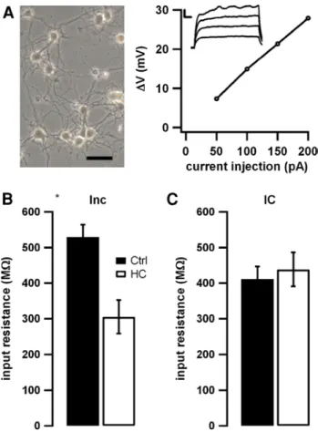
We obtained patch-clamp recordings from primary cultures of hippocampal neurons and first assessed the passive mem-brane properties of these neurons. No significant differences between the resting membrane potential of the cells could be observed when comparing cells cultured under control conditions, following incubation with homocysteine or dur-ing acute application of homocysteine (data not shown). We injected small subthreshold depolarizing currents to exam-ine input resistance (Fig. 1a, inset). Fitting the subthresh-old current–voltage relation allowed us to determine the input resistance (Fig. 1a). We found that preincubation with homocysteine (24 h, see “Methods”) caused a pronounced reduction in the input resistance by 42 % (Fig. 1b, from 529 to 305 MΩ, n = 10 and 10, p < 0.05). In contrast, acute intracellular application of 100 μM homocysteine via inclu-sion into the patch pipette did not affect input resistance (Fig. 1c, n = 17 and n = 16 for control and homocysteine). The apparent differences between the two control groups are due to variability between batches of cell cultures and consistently turned out to be not significant, whereas the comparison between control and application/incubation was always performed within one batch.
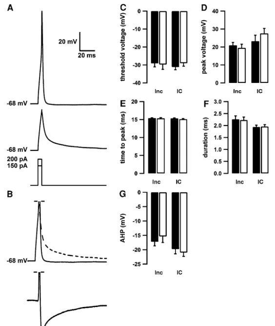
We next studied if the properties of action potentials are altered by homocysteine preincubation (Inc) or intracel-lular application of homocysteine (IC). Individual action potentials were elicited by brief current injections (5 ms, lowermost trace in Fig. 2a) elicited firing of single action potentials (uppermost trace in Fig. 2a). The threshold for generation of an action potential was unaffected by homo-cysteine, regardless of its mode of application (Fig. 2c), as was the action potential peak voltage (Fig. 2d), the time until the action potential peak was attained (Fig. 2e) and the dura-tion of the acdura-tion potential at threshold level (Fig. 2f). To examine spike afterpotentials, the depolarizing effects of the current injection were subtracted. To achieve this, we sub-tracted the voltage trace elicited by just subthreshold current injection (middle trace in Fig. 2a) scaled to the size of the initial depolarization preceding the action potential (dashed line in Fig. 2b). This revealed an after spike hyperpolariza-tion (lowermost trace in Fig. 2b), which was also not altered
by homocysteine preincubation or intracellular application (Fig. 2g). Thus, it appears that neither application of homo-cysteine via the patch pipette nor 24-h preincubation with homocysteine modifies the properties of individual action potentials.
We next studied the input–output relation of hippocam-pal neurons, and if this can be affected by homocysteine. The input–output relation was assessed by injecting current pulses of various amplitudes (200 ms, Fig. 3a, b, lowermost traces) and counting the number of resultant action poten-tials. We hypothesized that the decrease in input resistance seen in Fig. 1 would render homocysteine preincubated neurons less excitable and reduce action potential out-put. Indeed, homocysteine preincubated cells generated significantly less action potential output for small current injections (100 and 150 pA, see Fig. 3c; asterisks indicate
Fig. 1 Homocysteine effects on passive properties of
hippocam-pal neurons. a Photomicrograph of a representative neuronal culture on the left. Right panel shows input resistance calculated according to Ohm’s law from the current–voltage relation of passive voltage deflections (see inset, scale bars indicate 5 mV and 20 ms) elicited by small subthreshold current injections. b, c Preincubation with homo-cysteine (Inc, open bars in b, n = 10), but not acute application of homocysteine (IC, open bars in c, n = 16), resulted in a decreased input resistance compared to control cells (filled bars in b, n = 10 and c, n = 17)
p < 0.05, n = 10 for homocysteine and control groups). Conversely, the application of homocysteine via the patch pipette did not affect the input–output relation (n = 17 and
n = 16 for control and homocysteine, data not shown).
To characterize the shape of the action potentials and their activity-dependent change during action potential trains, we analyzed the first voltage trace that displayed five action potentials. The action potential firing displayed
Fig. 2 Homocysteine does not affect the action potential shape. a Representative voltage trace of an action potential (upper trace)
and a passive depolarization (middle trace, see lowermost part for current injections). Neither the action potential threshold (c), the minimum current injection needed to elicit an action potential (d), the peak voltage (d), the time to peak (e) nor the width of the action potential at threshold level (f) is altered following acute intracellular application (open bars, IC, n = 15) or 24-h incubation (open bars, Inc, n = 14) with 100 μM homocysteine compared to control cells (filled bars n = 16 (IC group) and 13 (Inc group)). b To analyze the afterpotentials of the action potential, the voltage trace elicited
with a subthreshold current injection (a middle trace) was scaled to fit the passive phase of the action potential trace (a upper trace). The up-scaled subthreshold trace (dashed line) was subtracted from the action potential trace (full line) to correct the voltage trace for the artificial voltage deflection due to the experimental setup. The resulting trace (lowermost part) represents the active voltage changes during an action potential and reveals a pronounced after spike hyperpolarization. g The AHP was quantified in control cells (filled bars) and following intracellular application (IC) or 24-h incubation (Inc) with homocysteine (open bars) and was unaffected by homocysteine
a frequency adaptation during the train in control conditions (see Fig. 4a). This adaptation turned out to be significant when quantified as increasing interspike intervals for all recorded control cells (as indicates by the arrow in Fig. 4a, see filled circles and asterisks in Fig. 4b, c). Following 24-h preincubation with homocysteine, this frequency adaptation was markedly reduced and no longer significant (open cir-cles in Fig. 4b). This was not the case when homocysteine was introduced via the patch (open circles in Fig. 4c). Nei-ther preincubation nor acute application of homocysteine affected the initial spike interval.
Action potentials physiologically broaden during a train of action potentials, reflecting the progressive inac-tivation of voltage-gated ion channels (Fig. 5a). Spike broadening was quantified at the level of the half Ap amplitude (see dashed lines in Fig. 5b1, b2). The spike broadening during trains was significantly reduced in cells preincubated 24 h in homocysteine (open circles in Fig. 5c). In contrast to the homocysteine effects on input resistance and frequency adaptation, this loss of spike broadening was also exerted by acute application of homocysteine (open circles in Fig. 5d). These results show that long-term exposure to homocysteine strongly affects neuronal excitability, whereas short-term expo-sure has only discrete effects.
Discussion
The present study aimed at analyzing electrophysiologi-cal effects of homocysteine as a possible mechanism of the association between hyperhomocysteinemia and neurologi-cal diseases.
We tested the effects of intra- and extracellular elevations of homocysteine concentrations on primary mouse neurons and observed that the input resistance decreases follow-ing long-term homocysteine exposure. This phenomenon results in a decrease in the neuronal input–output relation, rendering the neurons less excitable to lower- and mid-range current injections. Further, we found a significant reduction in both spike broadening and frequency adaptation during repetitive firing following 24-h homocysteine incubation. While a reduction in input resistance may be interpreted as a reduction in excitability, this latter effect is consistent with an increased excitability, as spike accommodation limits the capability of cells to fire high-frequency discharges (Zhou and Hablitz 1996). These results indicate that the effects of homocysteine on neurons elude a simple classification into pro- and anti-excitatory effects.
Fig. 3 Reduced repetitive firing following homocysteine
incuba-tion. a Prolonged current injections (200 ms) of varying magnitude (50–400 pA) were applied to elicit repetitive action potential firing.
b Representative voltage traces recorded in control cells (b1) and
fol-lowing incubation with homocysteine (b2). c The action potential
fre-quency for all cells was depicted versus the magnitude of the current injection. In control cells (filled circles, n = 10), the action potential firing rate increased initially with larger current injections (see also lower traces in b1). When further increasing the magnitude of the
cur-rent injection, the repolarization became insufficient, and the action potential firing was blocked (see uppermost traces in b1) resulting
in reduced firing rates. In homocysteine-treated cells (open circles,
n = 10), the firing rate following small current injections was
signifi-cantly smaller (see asterisks, p < 0.05). With larger current injections, the firing rate increased continuously without block due to insuffi-cient repolarization (see also upper traces in b2)
Homocysteine is associated with vascular and neurode-generative diseases, mechanisms are only poorly understood. This study suggests that interaction of homocysteine with electrophysiological properties of cells may be a relevant mechanism. It remains unclear whether interaction with ion channels or ions themselves is the reason. We have shown that homocysteine and copper interact probably by binding of copper by homocysteine (Linnebank et al. 2006). Thus, for the future, the channels and ions homocysteine is inter-fering with, should be explored. As limitation, we used an artificial patch-clamp approach with experimental 24-h incubation or intracellular application of homocysteine, whereas toxicity of mild or severe hyperhomocysteinemia occurs over years and decades.
Alterations of biophysical properties of cells may arise from unspecific toxicity or apoptosis, and homocysteine has been reported to induced apoptosis of neurons even at low concentrations (50–250 μM, Ganapathy et al. 2011; Kruman et al. 2000). No degradation of cells was seen, and we selected cells in healthy conditions by visual inspection for our recordings, but we did not quantitatively evaluate the neuronal cultures for cell loss or changes in neuronal morphology. However, the functional changes were quite selective, with the resting membrane potential and most action potential properties being unaffected. We believe this argues against, but does not completely exclude, large toxic effects of homocysteine in our cultures.
As we aimed at analyzing toxicity of hyperhomocysteine-mia as a chronic condition in humans, we did not analyze per-acute effects of homocysteine incubation, but incubated cells for 24 h before testing as a compromise. Homocysteine is not taken up by cells, but can be excreted to the extracel-lular space (Fowler 2005). Thus, concerning intracellular application, we made analyses immediately, as intracellu-lar concentrations after application were unpredictable and could not be monitored.
In patients, available evidence suggests that hyper-homocysteinemia is associated with increased CNS excitability. For instance, hyperhomocysteinemia is a risk factor for epileptic seizures in alcohol-dependent patients under conditions of alcohol withdrawal (Bleich et al.
2006). One explanation for this hyperexcitable phenotype in vivo may be that in intact neuronal networks, the pro-excitatory effects of homocysteine dominate. Alternately, synaptic effects of homocysteine may become relevant. In the case of withdrawal seizures, NMDA receptors, which are inhibited by alcohol, are up-regulated in actively drinking patients. Homocysteine is both an ago-nist at the glutamate binding site of the NMDA receptor complex and an antagonist at the glycine co-agonist site. This raises the possibility that homocysteine may exert NMDA agonistic actions, in particular, if glycine lev-els in the CNS are increased (Lipton et al. 1997; Bleich et al. 2006).
Fig. 4 Homocysteine
abol-ishes the frequency adaptation of repetitive firing. a The first action potential train with at least 5 action potentials was analyzed to address the frequency adaptation during the train. Interspike intervals as indicated by the arrow were quantified. During the recorded period, a slight but signifi-cant increase in the interspike interval was observed (filled
circles in b, n = 10 and c, n = 17, p < 0.05, see asterisks).
Following 24-h preincubation (Inc) with homocysteine, this frequency adaptation was abol-ished (open circles in b, n = 10, not significant), but it was not affected by acute intracellular application (IC) of homocyst-eine (c open circles, n = 16)
Epilepsy is one of the most common neurological dis-orders. In most cases, a lifetime therapy with antiepilep-tic drugs (AED) is necessary. Despite development of a significant number of new AEDs, about 30 % of patients with epilepsy remain refractory to drug treatment (Regesta and Tanganelli 1999). Even though drug resistance is a significant clinical problem, the molecular and cellu-lar mechanisms underlying the clinical phenomenon of drug resistance remain incompletely understood. Most AED are associated with decreased folate or vitamin B12 plasma levels and thus dispose to hyperhomocysteinemia (Linnebank et al. 2011). Hyperhomocysteinemia due to AED long-term treatment may contribute to secondary pharmacoresistance observed in several patients with initial
AED response. In addition, inhibitory and excitatory effects of hyperhomocysteinemia may contribute to long-term AED side effects on the central nervous system like fatigue and cognitive impairment in addition to the increased risk for ischemic heart disease (+34 %) and fatal cardiovascu-lar disease (+68 %) in individuals with epilepsy (Gaitatzis et al. 2004).
In conclusion, our data describe heterogeneous electro-physiological effects of homocysteine on neurons, some of which may contribute to neurological impairment and sei-zures associated with hyperhomocysteinemia.
Acknowledgments This work was supported by the BONFOR
program of the University of Bonn Medical Center (ML).
Fig. 5 Spike broadening is
reduced following incuba-tion with homocysteine. a To address the spike broadening during the train, the same traces as in Fig. 3 were analyzed.
b Action potential width was
measured at half width level (dashed horizontal line) as depicted for the first (b1 as
indicated in a) and last action potential (b2 as indicated in a).
Width of all action potentials was normalized to the width of the first action potential, averaged for all cells. In control cells, the spikes broadened to the ~1.5 width of the first action potential (filled circles in c,
n = 10 and d, n = 17). This
broadening was significantly reduced following 24-h incuba-tion (Inc) with homocysteine (c open circles, n = 10) or acute application (IC) of homocyst-eine (d open circles, n = 16)
References
Bleich S, Bayerlein K, Hillemacher T, Degner D, Kornhuber J, Frieling H (2006) An assessment of the potential value of elevated homocysteine in predicting alcohol-withdrawal seizures. Epilep-sia 47:934–938
Chan AY, Alsaraby A, Shea TB (2008) Folate deprivation increases tau phosphorylation by homocysteine-induced calcium influx and by inhibition of phosphatase activity: alleviation by S-adenosyl methionine. Brain Res 1199:133–137
Fowler B (2005) Homocysteine: overview of biochemistry, molecular biology, and role in disease processes. Semin Vasc Med 5:77–86 Gaitatzis A, Johnson AL, Chadwick DW, Shorvon SD, Sander JW
(2004) Life expectancy in people with newly diagnosed epilepsy. Brain 127:2427–2432
Ganapathy PS, White RE, Ha Y, Bozard BR, McNeil PL, Caldwell RW, Kumar S, Black SM, Smith SB (2011) The role of
N-methyl-d-aspartate receptor activation in homocysteine-induced death of
retinal ganglion cells. Invest Ophthalmol Vis Sci 52(8):5515–5524 Kruman II, Culmsee C, Chan SL, Kruman Y, Guo Z, Penix L, Mattson
MP (2000) Homocysteine elicits a DNA damage response in neu-rons that promotes apoptosis and hypersensitivity to excitotoxic-ity. J Neurosci 20(18):6920–6926
Linnebank M, Lutz H, Jarre E, Vielhaber S, Noelker C, Struys E, Jakobs C, Klockgether T, Evert BO, Kunz WS, Wullner U (2006) Binding of copper is a mechanism of homocysteine toxicity lead-ing to COX deficiency and apoptosis in primary neurons, PC12 and SHSY-5Y cells. Neurobiol Dis 23:725–730
Linnebank M, Moskau S, Semmler A, Widman G, Stoffel-Wagner B, Weller M, Elger CE (2011) Antiepileptic drugs interact with folate and vitamin B12 serum levels. Ann Neurol 69:352–359
Lipton SA, Kim WK, Choi YB, Kumar S, D’Emilia DM, Rayudu PV, Arnelle DR, Stamler JS (1997) Neurotoxicity associated with dual actions of homocysteine at the N-methyl-d-aspartate receptor. Proc Natl Acad Sci USA 94:5923–5928
Mudd SH, Skovby F, Levy HL, Pettigrew KD, Wilcken B, Pyeritz RE, Andria G, Boers GH, Bromberg IL, Cerone R et al (1985) The nat-ural history of homocystinuria due to cystathionine beta-synthase deficiency. Am J Hum Genet 37:1–31
Mudd SH, Levy HL, Kraus JP (2001) Disorders of transsulfuration. In: Scriver CR, Beaudet AL, Sly WS, Valle D, Childs B, Kinzler K, Vogelstein B (eds) The metabolic and molecular bases of inherited disease. Mc Graw-Hill, New York, pp 2007–2056
Regesta G, Tanganelli P (1999) Clinical aspects and biological bases of drug-resistant epilepsies. Epilepsy Res 34:109–122
Schoch S, Deák F, Königstorfer A, Mozhayeva M, Sara Y, Südhof TC, Kavalali ET (2001) SNARE function analyzed in synaptobrevin/ VAMP knockout mice. Science 294(5544):1117–1122
Smith AD, Smith SM, de Jager CA, Whitbread P, Johnston C, Agacin-ski G, Oulhaj A, Bradley KM, Jacoby R, Refsum H (2010) Homo-cysteine-lowering by B vitamins slows the rate of accelerated brain atrophy in mild cognitive impairment: a randomized con-trolled trial. PLoS ONE 5:e12244
Stanger O, Fowler B, Piertzik K, Huemer M, Haschke-Becher E, Semmler A, Lorenzl S, Linnebank M (2009) Homocysteine, folate and vitamin B12 in neuropsychiatric diseases: review and treat-ment recommendations. Expert Rev Neurother 9:1393–1412 Zhou FM, Hablitz JJ (1996) Layer I neurons of rat neocortex. I.
Action potential and repetitive firing properties. J Neurophysiol 76:651–667