HAL Id: hal-02530660
https://hal.sorbonne-universite.fr/hal-02530660
Submitted on 3 Apr 2020
HAL is a multi-disciplinary open access
archive for the deposit and dissemination of sci-entific research documents, whether they are pub-lished or not. The documents may come from teaching and research institutions in France or abroad, or from public or private research centers.
L’archive ouverte pluridisciplinaire HAL, est destinée au dépôt et à la diffusion de documents scientifiques de niveau recherche, publiés ou non, émanant des établissements d’enseignement et de recherche français ou étrangers, des laboratoires publics ou privés.
portable device: cross-validation against a standard
isokinetic dynamometer
Jean-Yves Hogrel, Olivier Benveniste, Damien Bachasson
To cite this version:
Jean-Yves Hogrel, Olivier Benveniste, Damien Bachasson. Routine monitoring of isometric knee ex-tension strength in patients with muscle impairments using a new portable device: cross-validation against a standard isokinetic dynamometer. Physiological Measurement, IOP Publishing, 2020, 41 (1), pp.015003. �10.1088/1361-6579/ab6b49�. �hal-02530660�
Routine monitoring of isometric knee extension strength in patients with muscle
1
impairments using a new portable device: cross-validation against a standard
2
isokinetic dynamometer
3 4
Jean-Yves Hogrel1, Olivier Benveniste2, Damien Bachasson1
5
1Institute of Myology, Neuromuscular Investigation Center, Pitié-Salpêtrière University
6
Hospital, Paris, France 7
2Department of Internal Medicine and Clinical Immunology and
Inflammation-8
Immunopathology-Biotherapy Department (I2B), Pitié-Salpêtrière University Hospital, 9
Assistance Publique-Hôpitaux de Paris, East Paris Neuromuscular Diseases Reference Center, 10
Inserm U974, Sorbonne Université, Paris 6, Paris, France 11 12 Corresponding author 13 Jean-Yves Hogrel 14 Institut de Myologie 15
Hôpital Universitaire Pitié-Salpêtrière 16 75651 Paris Cedex 13 17 France 18 Phone: +33 1 42 16 58 80 19 Fax: +33 1 42 16 58 81 20 E-mail: jy.hogrel@institut-myologie.org 21 22 3 4 5 6 7 8 9 10 11 12 13 14 15 16 17 18 19 20 21 22 23 24 25 26 27 28 29 30 31 32 33 34 35 36 37 38 39 40 41 42 43 44 45 46 47 48 49 50 51 52 53 54 55 56 57 58 59 60
Abstract
23
Objective: Muscle strength is a critical clinical hallmark in both health and disease. The 24
current study introduces a novel portable device prototype (MyoQuad) for assessing and 25
monitoring maximal voluntary isometric knee extension torque (MVIT). Approach: Fifty-six 26
patients with inclusion body myositis were studied. Knee extension weakness is a key feature 27
in this inflammatory muscle disease. Cross-validation with an isokinetic dynamometer 28
(Biodex System 3 Pro) was performed. Between-day reproducibility and ability to monitor 29
changes in muscle strength over time compared to the gold standard method as a reference, 30
were also investigated. Main results: The measurement was feasible even in the weakest 31
patients. Agreement between methods was excellent (standard error of measurement (SEM) 32
was 3.8 Nm and intra-class correlation coefficient (ICC) was 0.973). Least significant 33
difference (LSD) was 4.9 and 5.3 Nm for the MyoQuad and the Biodex, respectively 34
Measurements using the MyoQuad exhibited excellent between-day reproducibility (SEM 35
was 2.4 Nm and ICC was 0.989 versus 2.6 Nm and 0.988 using the Biodex). Changes in 36
MVIT at 6 and 12 months were similar between methods (timepoint × method interaction was 37
not significant; all p > 0.19); strength changes classified according to LSD at 6 and 12 months 38
were consistent between methods (>70% consistent classification)). Significance: The 39
measurement of maximal voluntary isometric knee extension torque using the MyoQuad 40
offers a cost-effective, portable and immediate alternative for the routine measurement of 41
maximal voluntary isometric strength of the quadriceps. The MyoQuad offers a comfort and 42
stability that cannot be provided by standard hand-held dynamometers. These results support 43
quantitative muscle strength assessment using fixed yet flexible dynamometry within clinical 44
routine and multicenter trials. 45 46 3 4 5 6 7 8 9 10 11 12 13 14 15 16 17 18 19 20 21 22 23 24 25 26 27 28 29 30 31 32 33 34 35 36 37 38 39 40 41 42 43 44 45 46 47 48 49 50 51 52 53 54 55 56 57 58 59 60
Keywords: device; muscle strength dynamometer; muscle weakness; myositis; outcome 47 measures 48 49 List of abbreviations 50
MMT, manual muscle testing; HHD, hand-held dynamometry; MVIT, maximal isometric 51
knee extension voluntary torque; SD, standard deviation; CIM, change in mean; ICC, intra-52
class correlation coefficient; SEM, standard error of measurement; LSD, least significant 53 change 54 55 3 4 5 6 7 8 9 10 11 12 13 14 15 16 17 18 19 20 21 22 23 24 25 26 27 28 29 30 31 32 33 34 35 36 37 38 39 40 41 42 43 44 45 46 47 48 49 50 51 52 53 54 55 56 57 58 59 60
Introduction
56Muscle weakness has been demonstrated to be an independent predictor of all-cause mortality 57
in an apparently healthy population (Garcia-Hermoso et al., 2018). Knee extension has been 58
identified as a preferential target for detecting muscle weakness in various chronic disorders 59
and other conditions such as aging or immobilization. Availability of a simple portable setup 60
and methodology for assessing knee extensor strength is critical to generalize the 61
measurement of this important clinical hallmark. 62
Strength assessment within clinical settings is most frequently performed using manual 63
muscle testing (MMT) (Hogrel et al., 2006). As a semi-quantitative and operator-dependent 64
method, MMT is poorly responsive (Bohannon, 2005). MMT may be used for rough detection 65
of muscle weakness but not to finely quantify its severity and its evolution over time. 66
Quantitative measurement of muscle strength allows precise temporal monitoring of muscle 67
strength and enables the use of Z-scores or percentage of predicted values computed from 68
datasets and predictive equations (Hogrel et al., 2007; Harbo et al., 2012; Seymour et al., 69
2010; McKay et al., 2016). Portable dynamometers have been demonstrated to be particularly 70
relevant for quantifying muscle strength at low-cost and ease of use within daily clinical 71
practice and multicenter research trials. Good agreement has been reported between isometric 72
strength measurement using hand-held dynamometry (HHD) and the “gold standard” 73
isokinetic dynamometer (Stark et al., 2011). However, evaluator strength limits the magnitude 74
of isometric force that can be measured using HHD (Deones et al., 1994). For powerful 75
muscle groups such as the quadriceps, belt-stabilization may be used to improve the reliability 76
and the range of measurable muscle strength using HHD (Bohannon et al., 2011; Bohannon et 77
al., 2012; Bachasson et al., 2014). However stabilized HHD is mostly achieved using home-78
made methods that may be imperfectly adapted, may lead to discomfort and are unlikely to be 79
standardized for repeated testing (Hansen et al., 2015). Some approaches may also fail to 80 3 4 5 6 7 8 9 10 11 12 13 14 15 16 17 18 19 20 21 22 23 24 25 26 27 28 29 30 31 32 33 34 35 36 37 38 39 40 41 42 43 44 45 46 47 48 49 50 51 52 53 54 55 56 57 58 59 60
provide accurate assessment of muscle strength in the weakest patients, partly due to design 81
flaws and metrological limitations inherent in the hardware used, as typically observed in 82
patients with muscle dystrophy for instance (Servais et al., 2013). 83
The current manuscript introduces a novel portable device prototype (namely, the MyoQuad) 84
for the assessment and monitoring of isometric knee extension strength that may be used in 85
most clinical setups. This article is organized as follows: description of the device, evaluation 86
of the device in patients including cross-validation with the gold standard, between-day 87
reproducibility of measurements and ability to monitor changes in muscle strength over time 88
with the gold standard method as a reference. 89 90
Methods
91 Participants 92The device was tested in patients with inclusion body myositis enrolled in a natural history 93
study (NCT00898989) and in a pharmacological trial (NCT02481453). The patients were 94
included according to the criteria defined by Benveniste and Hilton-Jones (2010). These 95
studies conformed to the Declaration of Helsinki and were approved by the local ethics 96
committee (CPP-Ile de France VI). All participants gave written informed consent. All tests 97
were performed between April 2013 and April 2017. 98
Description of the device for the assessment of isometric knee extension strength
99
The device used in the present study was a first-generation prototype. The MyoQuad was 100
specifically designed for the assessment of maximal isometric strength, even in very weak 101
individuals. It embeds a high precision load cell (Interface SML-300, Scottsdale, Arizona, 102
USA) and electronic board dedicated to signal acquisition and processing, wireless signal 103
transmission (Bluetooth), and operating-energy controls. The current prototype has a 104
measurement range from 0 to 136 kg with 10 g resolution and 50 g accuracy over the whole 105 3 4 5 6 7 8 9 10 11 12 13 14 15 16 17 18 19 20 21 22 23 24 25 26 27 28 29 30 31 32 33 34 35 36 37 38 39 40 41 42 43 44 45 46 47 48 49 50 51 52 53 54 55 56 57 58 59 60
nominal range. The load cell and the board are included in a 3D-printed case, which can be 106
firmly and securely attached to the structure of any examination bed/table using a clamp (see 107
Figure 1A). A dedicated software was developed allowing the visualization and analysis of 108
the acquired strength signal. The software can be run from a phone/tablet or laptop. One 109
extremity of the load cell is equipped with a hook on which a strap can be attached (see 110
Figure 1B). Other interfaces may also be screwed directly onto the load cell for other 111
applications in traction and compression. The MyoQuad device was checked for calibration 112
using strict standardized operating procedures. A set of M3 class masses from 0.2 to 50 kg 113
was used for calibration. The calibration was checked every week, then every month after 6 114
months, then every 2 months after 12 months. The metrological properties of the device were 115
always within the requirements (50-g accuracy over the whole range of measurement). A 116
typical calibration curve is presented in Figure 2. 117
Measurement protocol
118
Subjects were seated on a standard examination table with the hip and knee flexed to 90°. A 119
small dense foam pillow was placed below the tested thigh (see Figure 1.A. and Figure 1.B.), 120
so that the leg was vertical in order to avoid any effects of gravity effect, which can be 121
detrimental for the assessment of very weak patients. The desired strap location (lower part of 122
the strap above the medial malleolus) and the medial tibiofemoral joint space were marked on 123
the skin. The distance between the two marks i.e. the lever arm, was carefully measured to the 124
nearest half-centimeter using a measuring tape (Figure 2) to compute the torque as force × 125
lever arm, allowing direct comparison between methods (MyoQuad vs. isokinetic 126
dynamometer). The ankle strap was then fixed and attached to the dynamometer. The height 127
of the table was adjusted to ensure the strap was horizontal. Participants were asked to keep 128
their hands on their thighs. The evaluator secured the patient at the thigh and shoulder levels 129
on the tested side in order to limit compensatory movements. 130 3 4 5 6 7 8 9 10 11 12 13 14 15 16 17 18 19 20 21 22 23 24 25 26 27 28 29 30 31 32 33 34 35 36 37 38 39 40 41 42 43 44 45 46 47 48 49 50 51 52 53 54 55 56 57 58 59 60
Isokinetic dynamometer
131
Participants sat (85° hip flexion) on an ergometer (Biodex System 3 Pro, Biodex Medical, 132
Shirley, NY, USA). The upper body was stabilized with straps across the thorax and the 133
abdomen. Knee joint axis of rotation was aligned with the measurement axis of the system. 134
The thigh was strapped around the mid-thigh of the leg to be tested. The knee angle was 135
placed at 90° to cancel the effect of gravity. All measurements were performed in an isometric 136
mode. 137
Assessment of maximal voluntary isometric knee extension voluntary torque (MVIT)
138
Patients were instructed to perform maximal effort during a static knee extension and to limit 139
countermovement. Three maximal voluntary contractions were recorded for each 140
dynamometer. A fourth and possibly fifth trial were performed if the value reached during the 141
last trial was higher than the preceding ones, or if the difference between trials was greater 142
than 10%. Strong verbal encouragements were provided to the subjects. For MyoQuad 143
measurements, the patient was stabilized using one hand on the thigh and the other on the 144
shoulder on the measured side. Measurements were performed on both sides. The maximal 145
value from all trials was used for analyses. 146
Data analysis
147
Torque were expressed as absolute values and as percentage of predictive values using 148
previous published equations (Hogrel et al., 2007). 149
Statistics
150
Data within text and tables are shown as mean ± standard deviation (SD) or mean [lower 95% 151
confidence interval, upper 95% confidence interval]. The assumptions of normality and 152
sphericity were confirmed using the D’Agostino K-squared and Mauchly tests, respectively. 153
For cross-validation and between-day reproducibility, change in mean (CIM) and paired t-154
tests were used for detection of systematic bias. Standard error of measurement (SEM) was 155 3 4 5 6 7 8 9 10 11 12 13 14 15 16 17 18 19 20 21 22 23 24 25 26 27 28 29 30 31 32 33 34 35 36 37 38 39 40 41 42 43 44 45 46 47 48 49 50 51 52 53 54 55 56 57 58 59 60
used to study absolute reliability. Relative reliability was assessed using intra-class correlation 156
coefficients (ICC2,1). Regression analysis and Bland–Altman plots were also performed.
157
Individual coefficients of correlation were computed for between-day measurements and a 158
paired sample t-test was used to compare individual coefficients of correlation between 159
methods. The same approach was used to compare change between methods at follow-up. In 160
addition, a two-way ANOVA (timepoint × method) was used to compare methods. Tukey’s 161
honest significant difference post-hoc tests were conducted when a significant main and/or 162
interaction effect was found. Least significant difference was defined as SEM × 2 and 163
outcome at follow-up was defined as impaired, unchanged, and improved. A Fisher exact 164
probability test was used to compare contingency tables of outcomes at follow-up. All 165
analyses were performed in the computing environment R Version 3.2.3. Statistical 166
significance was set at p < 0.05 for all tests. 167
168
Results
169
A total of 56 patients (age = 67 ± 9 years) with inclusion body myositis were included. Thirty-170
three patients were assessed for between-day reproducibility. Thirty-two of which were 171
reassessed after 6 and 12 months. Amongst them, 29 were tested using both methods. The 172
reason for the smaller number of patients at follow-up was time constraints or unavailability 173
of the MyoQuad. Ultimately, a total of 287 observations using both methods were gathered. 174
Agreement of methods are summarized in Table 1 and Figure 3. The MVIT measured using 175
the MyoQuad was significantly lower than the MVIT measured using the Biodex. Between-176
day reproducibility results for both methods are displayed in Table 2 and Figure 4. No 177
systematic bias was detected and least significant difference was 4.9 and 5.3 Nm for the 178
MyoQuad and the Biodex, respectively. Individual coefficients of variation for between-day 179
measurements were 5.4 ± 7.4 % and 6.7 ± 6.6 % using MyoQuad and the Biodex with no 180 3 4 5 6 7 8 9 10 11 12 13 14 15 16 17 18 19 20 21 22 23 24 25 26 27 28 29 30 31 32 33 34 35 36 37 38 39 40 41 42 43 44 45 46 47 48 49 50 51 52 53 54 55 56 57 58 59 60
significant difference between methods (mean of the differences = -1.4 Nm; 95%CI: [-3.7, 181
0.9] Nm; p = 0.26). 182
Change in MVIT at 6 and 12 months using both methods is shown in Figure 5. When 183
performed using absolute values (at baseline, 6, and 12 months), ANOVA showed significant 184
main effects of method and time (both p < 0.01) and no significant timepoint × method 185
interaction (p = 0.19) (Figure 5.A). When performed using absolute changes at 6 and 12 186
months, ANOVA showed significant main effects of method and time (both p < 0.05) and no 187
significant timepoint × method interaction (p = 0.46) (Figure 5.B). Variability in changes 188
between methods at follow-up is displayed in Table 3. A contingency table of outcomes at 189
follow-up is shown in Table 4. 190
191
Discussion
192
The aim of this study was to introduce a novel device for the assessment of isometric strength 193
of knee extensors, for use in most clinical environments. Agreement with the gold standard, 194
between-day reliability, and the ability of the device to monitor changes in muscle strength 195
over time compared to the gold standard method as a reference were investigated. Main 196
results are as follows: i) the device demonstrates excellent metrological accuracy, ii) 197
measurements obtained using the device exhibit excellent agreement with the gold standard, 198
iii) reliability of measurements obtained using the device was excellent and comparable to 199
results obtained using the gold standard, iii) change in strength over time was similar using 200
the device and the gold standard. 201
The MyoQuad device was developed in order to conveniently assess quadriceps strength 202
within routine clinical practice, including in very weak patients. This may be commonly 203
performed using HHD, which, however, presents several limitations. In very strong patients, 204
the evaluator may have difficulties in maintaining the dynamometer in a steady hold, whereas 205 3 4 5 6 7 8 9 10 11 12 13 14 15 16 17 18 19 20 21 22 23 24 25 26 27 28 29 30 31 32 33 34 35 36 37 38 39 40 41 42 43 44 45 46 47 48 49 50 51 52 53 54 55 56 57 58 59 60
the influence of the evaluator may be too important in the weakest patients leading to possible 206
large relative errors. Fixed dynamometers are attractive to tackle these limitations (Mentiplay 207
et al., 2015). However, muscle strength may be improperly or even impossible to assess if the 208
device is improperly designed. For instance, within a recent therapeutic trial in IBM, the 209
system was designed in a way that patients had to carry the weight of the strain gauge before 210
strength produced could be actually measured. As a result, knee extensor strength could not 211
be measured in 3 out of 12 (25%) patients and the consistency of the measurements in other 212
patients was largely flawed (unpublished results). The design of the proposed device tackles 213
these issues as the MyoQuad is directly secured on a fixed frame. HHD have been repeatedly 214
observed to underestimate knee extension strength compared to isometric measurements using 215
an isokinetic dynamometer (Stark et al., 2011; Martin et al., 2006). This is particularly true in 216
stronger individuals where the evaluator may have difficulties stabilizing the measurement 217
chain (Martin et al., 2006; Bohannon et al., 2012). The high agreement between 218
measurements performed using the MyoQuad and the Biodex support that fixed dynamometry 219
improves the robustness of assessments through improved comfort and stability. These data 220
are in line with previous work reporting higher consistency of measurements using a modified 221
procedure of stabilized HHD as compared to standard HHD (Kim et al., 2014; Hansen et al., 222
2015). 223
Our data shows a low SEM < 8.5 % Nm and high ICC > 0.98 for between-day reproducibility 224
when using the MyoQuad. This was similar to that observed using the Biodex (SEM was < 225
10.0% and ICC was > 0.98). We also reported similar individual coefficients of variation for 226
between-day measurement using the MyoQuad and the Biodex. This high reproducibility 227
yielded similar least significant change using both methods. These data are in line with 228
previous reports that have investigated between-day reliability using gold standard methods 229
(Ruschel et al., 2015; Kean et al., 2010). Importantly, our data showed no significant 230 3 4 5 6 7 8 9 10 11 12 13 14 15 16 17 18 19 20 21 22 23 24 25 26 27 28 29 30 31 32 33 34 35 36 37 38 39 40 41 42 43 44 45 46 47 48 49 50 51 52 53 54 55 56 57 58 59 60
difference between methods for monitoring change in MVIT over time (Figure 5) and good 231
agreement when comparing changes at follow-up (Table 3). This was confirmed by consistent 232
classification of strength changes using both methods at follow-up (Table 4). To the best of 233
our knowledge, this is the first report that compares temporal changes in knee extensors using 234
fixed dynamometry and standard isokinetic dynamometry. 235
As this study was only performed in patients with inclusion body myositis, generalizability of 236
findings to other disorders remains to be demonstrated. However, similar findings regarding 237
the reliability of MVIT have been previously reported in various clinical fields (Nuzzo et al., 238
2019). Volitional maneuvers such as maximal voluntary contraction embraces both peripheral 239
and central factors, which render its estimation variable (Millet et al., 2012). Therefore, 240
observed differences between Biodex and MyoQuad also reflect this variability. Another 241
potential source of error using the MyoQuad is the measurement of the lever arm to compute 242
torque that is circumvented when using torque meter as in standard isokinetic dynamometers 243
like the Biodex (Ruschel et al., 2015). 244
245
Conclusions
246
Measurement of maximal voluntary isometric knee extension torque using the MyoQuad 247
offers a cost-effective, portable and immediate alternative for the routine measurement of 248
maximal voluntary isometric contraction of the quadriceps by offering comfort and stability 249
that cannot be provided using hand-held dynamometry. It may be used for both baseline and 250
follow-up assessments of muscle strength and may also be used to assess other muscle groups 251
(e.g. knee flexion, shoulder abduction) using proper patient positioning and adapted 252
interfaces. Altogether, our results support the adoption of quantitative muscle strength 253
assessment using fixed yet flexible dynamometry within routine clinical practice and 254 multicenter trials. 255 3 4 5 6 7 8 9 10 11 12 13 14 15 16 17 18 19 20 21 22 23 24 25 26 27 28 29 30 31 32 33 34 35 36 37 38 39 40 41 42 43 44 45 46 47 48 49 50 51 52 53 54 55 56 57 58 59 60
256
Acknowledgments
257
We gratefully thank all the patients who participated in this study. This study was supported by 258
the Association Française contre les Myopathies (AFM). The authors are grateful to Simone 259
Birnbaum for English editing. 260
261
ORCID iDs
262
Jean-Yves Hogrel: https://orcid.org/0000-0003-0045-7505 263
Olivier Benveniste: https://orcid.org/0000-0002-1167-5797 264
Damien Bachasson: https://orcid.org/0000-0001-6335-9916
265 266
References
267
Bachasson D, Villiot-Danger E, Verges S, Hayot M, Perez T, Chambellan A and Wuyam B 268
2014 [Maximal isometric voluntary quadriceps strength assessment in COPD] Revue 269
des Maladies Respiratoires 31 765-70 270
Benveniste O and Hilton-Jones D 2010 International Workshop on Inclusion Body Myositis 271
held at the Institute of Myology, Paris, on 29 May 2009 Neuromuscul Disord 20 414-272
21 273
Bohannon R W 2005 Manual muscle testing: does it meet the standards of an adequate 274
screening test? Clinical Rehabilitation 19 662-7 275
Bohannon R W, Bubela D J, Wang Y C, Magasi S R and Gershon R C 2011 Adequacy of belt-276
stabilized testing of knee extension strength Journal of Strength and Conditioning 277 Research 25 1963-7 278 3 4 5 6 7 8 9 10 11 12 13 14 15 16 17 18 19 20 21 22 23 24 25 26 27 28 29 30 31 32 33 34 35 36 37 38 39 40 41 42 43 44 45 46 47 48 49 50 51 52 53 54 55 56 57 58 59 60
Bohannon R W, Kindig J, Sabo G, Duni A E and Cram P 2012 Isometric knee extension force 279
measured using a handheld dynamometer with and without belt-stabilization Physiother 280
Theory Pract 28 562-8 281
Deones V L, Wiley S C and Worrell T 1994 Assessment of quadriceps muscle performance by 282
a hand-held dynamometer and an isokinetic dynamometer Journal of Orthopaedic & 283
Sports Physical Therapy 20 296-301 284
Garcia-Hermoso A, Cavero-Redondo I, Ramirez-Velez R, Ruiz J R, Ortega F B, Lee D-C and 285
Martinez-Vizcaino V 2018 Muscular Strength as a Predictor of All-Cause Mortality in 286
an Apparently Healthy Population: A Systematic Review and Meta-Analysis of Data 287
From Approximately 2 Million Men and Women Arch Phys Med Rehabil 99 2100-13.e5 288
Hansen E M, McCartney C N, Sweeney R S, Palimenio M R and Grindstaff T L 2015 Hand-289
held Dynamometer Positioning Impacts Discomfort During Quadriceps Strength 290
Testing: A Validity and Reliability Study Int J Sports Phys Ther 10 62-8 291
Harbo T, Brincks J and Andersen H 2012 Maximal isokinetic and isometric muscle strength of 292
major muscle groups related to age, body mass, height, and sex in 178 healthy subjects 293
European Journal of Applied Physiology 112 267-75 294
Hogrel J Y, Ollivier G and Desnuelle C 2006 [Manual and quantitative muscle testing in 295
neuromuscular disorders. How to assess the consistency of strength measurements in 296
clinical trials?] Rev Neurol (Paris) 162 427-36 297
Hogrel J Y, Payan C A, Ollivier G, Tanant V, Attarian S, Couillandre A, Dupeyron A, 298
Lacomblez L, Doppler V, Meininger V, Tranchant C, Pouget J and Desnuelle C 2007 299
Development of a French isometric strength normative database for adults using 300
quantitative muscle testing Archives of Physical Medicine and Rehabilitation 88 1289-301 97 302 3 4 5 6 7 8 9 10 11 12 13 14 15 16 17 18 19 20 21 22 23 24 25 26 27 28 29 30 31 32 33 34 35 36 37 38 39 40 41 42 43 44 45 46 47 48 49 50 51 52 53 54 55 56 57 58 59 60
Kean C O, Birmingham T B, Garland S J, Bryant D M and Giffin J R 2010 Minimal detectable 303
change in quadriceps strength and voluntary muscle activation in patients with knee 304
osteoarthritis Archives of Physical Medicine and Rehabilitation 91 1447-51 305
Kim W K, Kim D K, Seo K M and Kang S H 2014 Reliability and validity of isometric knee 306
extensor strength test with hand-held dynamometer depending on its fixation: a pilot 307
study Ann Rehabil Med 38 84-93 308
Martin H J, Yule V, Syddall H E, Dennison E M, Cooper C and Aihie Sayer A 2006 Is hand-309
held dynamometry useful for the measurement of quadriceps strength in older people? 310
A comparison with the gold standard Bodex dynamometry Gerontology 52 154-9 311
McKay M J, Baldwin J N, Ferreira P, Simic M, Vanicek N, Hiller C E, Nightingale E J, 312
Moloney N A, Quinlan K G, Pourkazemi F, Sman A D, Nicholson L L, Mousavi S J, 313
Rose K, Raymond J, Mackey M G, Chard A, Hubscher M, Wegener C, Fong Yan A, 314
Refshauge K M, Burns J and Norms Project C 2016 1000 Norms Project: protocol of a 315
cross-sectional study cataloging human variation Physiotherapy 102 50-6 316
Mentiplay B F, Perraton L G, Bower K J, Adair B, Pua Y H, Williams G P, McGaw R and 317
Clark R A 2015 Assessment of Lower Limb Muscle Strength and Power Using Hand-318
Held and Fixed Dynamometry: A Reliability and Validity Study PLoS One 10 e0140822 319
Millet G Y, Bachasson D, Temesi J, Wuyam B, Feasson L, Verges S and Levy P 2012 Potential 320
interests and limits of magnetic and electrical stimulation techniques to assess 321
neuromuscular fatigue Neuromuscul Disord 22 Suppl 3 S181-6 322
Nuzzo J L, Taylor J L and Gandevia S C 2019 CORP: Measurement of upper and lower limb 323
muscle strength and voluntary activation J Appl Physiol (1985) 126 513-43 324
Ruschel C, Haupenthal A, Jacomel G F, Fontana Hde B, Santos D P, Scoz R D and Roesler H 325
2015 Validity and reliability of an instrumented leg-extension machine for measuring 326
isometric muscle strength of the knee extensors Journal of Sport Rehabilitation 24 327 3 4 5 6 7 8 9 10 11 12 13 14 15 16 17 18 19 20 21 22 23 24 25 26 27 28 29 30 31 32 33 34 35 36 37 38 39 40 41 42 43 44 45 46 47 48 49 50 51 52 53 54 55 56 57 58 59 60
Servais L, Deconinck N, Moraux A, Benali M, Canal A, Van Parys F, Vereecke W, 328
Wittevrongel S, Mayer M, Desguerre I, Maincent K, Themar-Noel C, Quijano-Roy S, 329
Serari N, Voit T and Hogrel J Y 2013 Innovative methods to assess upper limb strength 330
and function in non-ambulant Duchenne patients Neuromuscul Disord 23 139-48 331
Seymour J M, Spruit M A, Hopkinson N S, Natanek S A, Man W D, Jackson A, Gosker H R, 332
Schols A M, Moxham J, Polkey M I and Wouters E F 2010 The prevalence of 333
quadriceps weakness in COPD and the relationship with disease severity Eur Respir J 334
36 81-8
335
Stark T, Walker B, Phillips J K, Fejer R and Beck R 2011 Hand-held dynamometry correlation 336
with the gold standard isokinetic dynamometry: a systematic review PM R 3 472-9 337 338 3 4 5 6 7 8 9 10 11 12 13 14 15 16 17 18 19 20 21 22 23 24 25 26 27 28 29 30 31 32 33 34 35 36 37 38 39 40 41 42 43 44 45 46 47 48 49 50 51 52 53 54 55 56 57 58 59 60
Tables
339
Table 1. Agreement of measurements obtained using the MyoQuad and the Biodex (n =
340
52 participants with a total of 283 measurements).
341
MyoQuad (Nm) Biodex (Nm)
CIM (Nm) [95% CI]
P value ICC [95% CI] SEM (Nm) [95% CI]
29.46 ± 23.12 31.14 ± 24.68 1.68 [1.06, 2.30] < 0.001 0.973 [0.966, 0.979] 3.76 [3.47, 4.10] CIM = change in mean; SEM = standard error of measurement; ICC = intra-class correlation 342 coefficient. 343 344 345 3 4 5 6 7 8 9 10 11 12 13 14 15 16 17 18 19 20 21 22 23 24 25 26 27 28 29 30 31 32 33 34 35 36 37 38 39 40 41 42 43 44 45 46 47 48 49 50 51 52 53 54 55 56 57 58 59 60
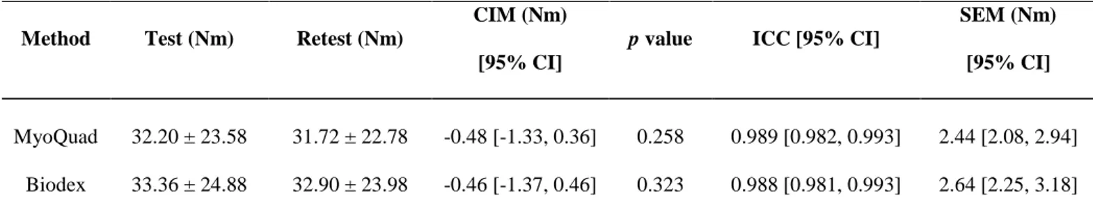
Table 2. Test-retest reliability of maximal isometric knee extension voluntary torque using
346
the MyoQuad and the Biodex (n = 33 participants with a total of 66 measurements).
347
Method Test (Nm) Retest (Nm)
CIM (Nm) [95% CI]
p value ICC [95% CI]
SEM (Nm) [95% CI]
MyoQuad 32.20 ± 23.58 31.72 ± 22.78 -0.48 [-1.33, 0.36] 0.258 0.989 [0.982, 0.993] 2.44 [2.08, 2.94] Biodex 33.36 ± 24.88 32.90 ± 23.98 -0.46 [-1.37, 0.46] 0.323 0.988 [0.981, 0.993] 2.64 [2.25, 3.18]
CIM = change in mean; SEM = standard error of measurement; ICC = intra-class correlation 348 coefficient. 349 350 351 3 4 5 6 7 8 9 10 11 12 13 14 15 16 17 18 19 20 21 22 23 24 25 26 27 28 29 30 31 32 33 34 35 36 37 38 39 40 41 42 43 44 45 46 47 48 49 50 51 52 53 54 55 56 57 58 59 60
Table 3. Variability of changes in maximal isometric knee extension voluntary torque at
352
follow-up using the MyoQuad and the Biodex (n = 44 participants with a total of 88
353 measurements). 354 Biodex (Nm) MyoQuad (Nm) CIM (Nm) [95% CI]
p value ICC [95% CI]
SEM (Nm) [95% CI] -3.15 ± 9.01 -3.17 ± 7.57 -0.02 [-1.29, 1.24] 0.971 0.756 [0.648, 0.836] 4.22 [3.68,4.96]
CIM = change in mean; ICC = intra-class correlation coefficient; SEM = standard error of 355 measurement. 356 357 358 359 3 4 5 6 7 8 9 10 11 12 13 14 15 16 17 18 19 20 21 22 23 24 25 26 27 28 29 30 31 32 33 34 35 36 37 38 39 40 41 42 43 44 45 46 47 48 49 50 51 52 53 54 55 56 57 58 59 60
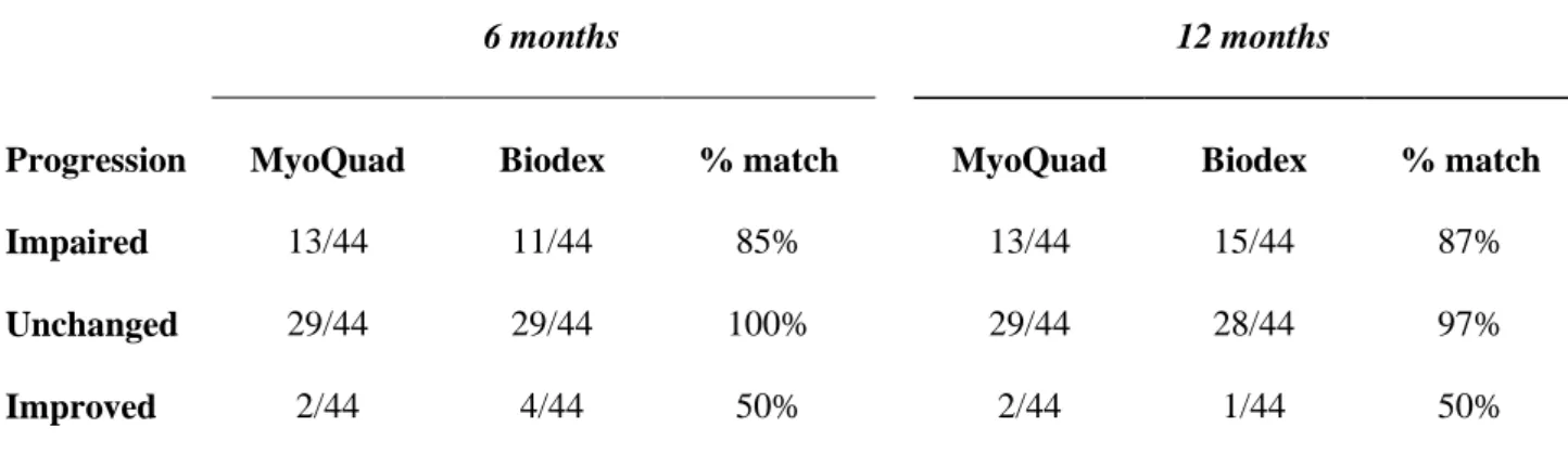
Table 4. Contingency table illustrating classified changes in strength using the MyoQuad
360
and the Biodex.
361
6 months 12 months
Progression MyoQuad Biodex % match MyoQuad Biodex % match
Impaired 13/44 11/44 85% 13/44 15/44 87%
Unchanged 29/44 29/44 100% 29/44 28/44 97%
Improved 2/44 4/44 50% 2/44 1/44 50%
Least significant change determined from between-day reliability was used to classify changes 362
in strength after 6 and 12 months. 363 364 365 3 4 5 6 7 8 9 10 11 12 13 14 15 16 17 18 19 20 21 22 23 24 25 26 27 28 29 30 31 32 33 34 35 36 37 38 39 40 41 42 43 44 45 46 47 48 49 50 51 52 53 54 55 56 57 58 59 60
Figure legends
366
Figure 1. Patient installation and device. Close-up view of the device paired with tablet
367
computer (A); Close-up view of the device attached to table legs with round (B) or 368
rectangular section (C); Overview of the setup with one participant and stabilization by one 369
evaluator (D). A small pillow is inserted under the distal part of the thigh to ensure the thigh is 370
horizontal. 371
372
Figure 2. Typical calibration curve. The dashed line represents the identity line, and the
373
solid line indicates the linear regression line. 374
375
Figure 3. Agreement of measurements obtained using the MyoQuad versus the Biodex.
376
Bland–Altman plots (A) and regression analysis (B) of measurements obtained using the
377
MyoQuad and the Biodex. In A, the solid line indicates the mean difference between the
378
measurements and dashed lines the limits of agreement. In B, the dashed line represents the 379
identity line, and the solid line indicates the linear regression line. 380
381
Figure 4. Between-day reproducibility of measurements obtained using the MyoQuad
382
and the Biodex. Bland–Altman plots (A, C) and regression analysis (B, D) for
between-383
day reliability of measurements obtained using the MyoQuad and the Biodex,
384
respectively. In A and C, the solid line indicates the mean difference between the
385
measurements and the dashed line indicates the limit of agreements. In B and D, the dashed 386
line represents the identity line, and the solid line indicates the linear regression line. 387
Logarithmic scales are used for better data visualization. 388 389 3 4 5 6 7 8 9 10 11 12 13 14 15 16 17 18 19 20 21 22 23 24 25 26 27 28 29 30 31 32 33 34 35 36 37 38 39 40 41 42 43 44 45 46 47 48 49 50 51 52 53 54 55 56 57 58 59 60
Figure 5. Change in torque over time using the MyoQuad and the Biodex. Absolute (A)
390
values and change (B) in knee extensor strength over time. 391 392 3 4 5 6 7 8 9 10 11 12 13 14 15 16 17 18 19 20 21 22 23 24 25 26 27 28 29 30 31 32 33 34 35 36 37 38 39 40 41 42 43 44 45 46 47 48 49 50 51 52 53 54 55 56 57 58 59 60
Figure 1 393 394 395 3 4 5 6 7 8 9 10 11 12 13 14 15 16 17 18 19 20 21 22 23 24 25 26 27 28 29 30 31 32 33 34 35 36 37 38 39 40 41 42 43 44 45 46 47 48 49 50 51 52 53 54 55 56 57 58 59 60
Figure 2 396 397 398 3 4 5 6 7 8 9 10 11 12 13 14 15 16 17 18 19 20 21 22 23 24 25 26 27 28 29 30 31 32 33 34 35 36 37 38 39 40 41 42 43 44 45 46 47 48 49 50 51 52 53 54 55 56 57 58 59 60
Figure 3 399 400 401 3 4 5 6 7 8 9 10 11 12 13 14 15 16 17 18 19 20 21 22 23 24 25 26 27 28 29 30 31 32 33 34 35 36 37 38 39 40 41 42 43 44 45 46 47 48 49 50 51 52 53 54 55 56 57 58 59 60
Figure 4 402 403 404 3 4 5 6 7 8 9 10 11 12 13 14 15 16 17 18 19 20 21 22 23 24 25 26 27 28 29 30 31 32 33 34 35 36 37 38 39 40 41 42 43 44 45 46 47 48 49 50 51 52 53 54 55 56 57 58 59 60
Figure 5 405 406 3 4 5 6 7 8 9 10 11 12 13 14 15 16 17 18 19 20 21 22 23 24 25 26 27 28 29 30 31 32 33 34 35 36 37 38 39 40 41 42 43 44 45 46 47 48 49 50 51 52 53 54 55 56 57 58 59 60