Serogroups and genotypes of Leptospira spp. strains from bovine aborted fetuses 1
Delooz Laurent1,2, Czaplicki Guy1, Gregoire Fabien1, Dal Pozzo Fabiana2, Pez Floriane3, Kodjo 2
Angeli4, Saegerman Claude2 3
1 Regional Association for Animal Registration and Health (ARSIA) asbl, 5590 Ciney, Belgium 4
2 Research Unit of Epidemiology and Risk Analysis applied to veterinary science (UREAR-5
ULg), Fundamental and Applied Research for Animals & Health (FARAH) Center, Faculty of 6
Veterinary Medicine, University of Liege, 4000 Liege, Belgium 7
3 BioSellal, 69007 Lyon, France 8
4 Laboratoire des Leptospires, VetAgro Sup, Campus Vétérinaire de Lyon, 69280 Marcy-9 l’Etoile, France 10 11 ABSTRACT 12
Leptospirosis is a global disease of animals, with potential major economic impact on livestock 13
industry and important zoonotic capacities. The disease represents a major challenge in the 14
developing countries as humans and animals frequently live in close association. The serovar 15
Hardjo of Leptospira whose primary host is cattle has been studied extensively, but few data 16
exist on other current circulating or emerging serotypes. To better understand the disease in 17
cattle and how to prevent and/or control it, it is necessary to identify the genotype and the 18
serotype of circulating Leptospira. This paper presents results of several investigations 19
performed on a historical Belgian collection of congenital jaundice in bovine aborted fetuses 20
coming from the leptospirosis emerging episode of 2014 (Delooz et al., 2015). The results 21
revealed that L. Grippotyphosa and L. Australis were the most prevalent serogroups with 22
respectively 17/42 and 13/42 positive MAT during this emerging event associated with the 23
same clinical pattern. The study also confirms that congenital jaundice is associated with L. 24
kirscheneri and L. interrogans and provides the genotyping of DNA obtained from these two
25
species. 26
Keywords: Abortion, Cattle, Icteric, Jaundice, Genotyping, Leptospira, Leptospirosis, 27 Belgium 28 29 INTRODUCTION 30
In Belgium, a national surveillance program based on the compulsory reporting of 31
abortions and subsequent analyses on their products reached several objectives including 32
official surveillance of bovine brucellosis but also the monitoring of other bovine abortive 33
diseases. Some emerging or re-emerging pathogens could be identified, including Bluetongue 34
virus serotype 8, Brucella abortus and more recently Schmallenberg virus. 35
Since July 2014, the Belgian Walloon region faced an unexpected situation with a 36
drastic increase of congenital jaundice in bovine aborted fetuses (Delooz et al., 2015). During 37
the last six years, abortions associated with jaundice had been notified but the monthly 38
incidence of cases remained stable. From July to December 2014, an increase of bovine aborted 39
fetuses with jaundice was reported by ARSIA pathologists, with a significantly higher incidence 40
than previous months and years. The standardized panel of analyses designed to extend the 41
diagnosis did not allow identifying the etiology. After additional analyzes, high levels of 42
antibodies against Leptospira serogroups Australis and Grippotyphosa were found in cows after 43
abortion of icteric fetuses. Serology performed during the emergence identified serogroups 44
without providing information on the genotype of the involved pathogenic Leptospira strains. 45
A leptospiral infection was consequently hypothezised (Delooz et al., 2015). 46
Leptospirosis is a transmissible disease of animals and humans caused by the spirochete 47
Leptospira. All the pathogenic leptospires were formerly classified as members of the species
48
Leptospira interrogans. However, the genus has been reorganised and pathogenic Leptospira
49
are now classified in 23 species (Adler, 2010; Levett, 2001; Morey et al., 2006) from which 50
more than 300 distinct serovars included within 24 serogroups are distinguished (Levett, 2015). 51
Laboratory diagnosis of leptospirosis can be complex and involves tests designed to detect 52
specific antibodies against Leptospira, as well as for direct isolation of leptospires, antigens 53
detection and detection of Leptospira nucleic acid in animal tissues or body fluids (OIE, 2014). 54
In cattle, the seroprevalence of the serogroup Sejroe is one of the most studied and varies 55
widely from one country to another with 33% in France (Ayral et al., 2014), 30% in the 56
Netherlands (Hartman et al., 1989) and up to 83.59% in Ireland (Ryan et al., 2012). In addition, 57
antibodies against Leptospira serovar Hardjo were found in tank milk of 9.2% bovine dairy 58
herds, with a higher incidence in the southern part of the country (Dom et al., 1991). In France, 59
microscopic agglutination test (MAT) performed on samples collected in 394 cattle herds 60
allowed to determine the distribution of the following three predominant circulating serogroups 61
(Australis, Sejroe, and Grippotyphosa) (Ayral et al., 2014). In this part of Europe, leptospirosis 62
is prevalent and may be responsible for pathological events in human (Mori et al., 2014) and 63
animal health (Delooz et al., 2015). 64
Leptospirosis is a known cause of abortions and infection can be accompanied by a wide 65
variety of clinical signs. However, fetal jaundice was not observed during experimental 66
infections (Ellis and Michma, 1977; Ellis et al., 1986; Smith et al., 1997). On the contrary based 67
on the recent field observations, the hypothesis of the association of bovine fetal jaundice with 68
leptospirosis was formulated (Delooz et al., 2015). Currently, little epidemiological information 69
exists concerning the different serovars of Leptospira circulating among cattle in Belgium. 70
While the manifestations of the disease can be very different depending on the strain and the 71
animal species, it is important to identify the pathogenic strain in order to tackle the best 72
prevention and control measures and thereby, prevent potential transmission to humans 73
(Evangelista and Coburn, 2010). 74
The aim of this work was to characterize the Leptospira infection detected in bovine 75
abortion cases associated with fetal jaundice which occurred in Southern Belgium in 2014. 76
77
MATERIAL AND METHODS 78
Study desing 79
In the context of the Belgian passive surveillance program for bovine brucellosis, a total 80
of 42 bovine abortion cases collected from October 2011 to December 2014 were included in 81
this study. They originated from 39 cattle farms distributed among the 5 Walloon provinces. 82
They were included in the study according to the diagnosis of congenital jaundice (N = 41) or 83
the PCR positivity against pathogenic Leptospira (N = 1). 84
Information issued from the anamnesis, such as sampling date, herd identification 85
number, cattle breed, month of pregnancy and number of parity were encoded in the laboratory 86
information management system (LIMS) or in an Access database. None of these herds 87
applied vaccination against Leptospira species and, moreover, no Leptospira vaccine has a 88
marketing authorization in Belgium. The geographical localization of each case of abortion was 89
possible using the Lambert coordinates and the Belgian cattle identification and movement 90
traceability system (SANITRACE). 91
92
Laboratory analyses 93
A standardized panel of analyses was first applied to perform the laboratory diagnosis 94
of bovine abortion on submitted fetuses. Direct and/or indirect detection of pathogens was 95
performed, including bacteria (Brucella spp., Campylobacter spp., Coxiella burnetii, 96
Leptospira borgpetersenii and interrogans serovar Hardjo, Listeria monocytogenes, Neospora
97
caninum, Salmonella Dublin), viruses (bluetongue virus serotype 8 (BTV-8), bovine
98
herpesvirus 1 (BoHV-1), bovine herpesvirus 4 (BoHV-4), bovine viral diarrhea virus (BVDV), 99
Schmallenberg virus), several mycotic agents, and many other opportunistic bacteria (Table I). 100
101
Microscopic agglutination tests 102
MAT was performed on the 42 maternal sera sampled on the aborted cows at the time of 103
abortion using twenty-four serovars representing 14 serogroups of pathogenic Leptospira 104
species: Icterohaemorrhagiae, Copenhageni, Australis, Bratislava, Munchen, Autumnalis, Bim, 105
Castellonis, Bataviae, Canicola, Hebdomadis, Panama, Mangus, Pomona, Mozdok, Pyrogenes, 106
Sejroe, Saxkoebing, Hardjo, Wolffi, Tarassovi, Cynopteri, Vanderhoedoni, and Grippotyphosa 107
(Table II). According to observations recorded by Chappel and collaborators in 2004, titers of 108
160 or higher were defined as positive agglutination reactions for ruminants. The end-point was 109
the highest dilution of serum in which 50% agglutination still occured. 110
111
Pathogenic Leptospira DNA detection (real-time PCR). 112
During the necropsy, a spleen, kidney, liver and placenta fragment were sampled on 113
each abortion cases, pooled and stored at -20°C. PCR analysis was performed on 26 pools of 114
organs sampled from icteric fetuses, retrieved in the historical abortion samples collection 115
described in the study design (not available for 16 other fetuses). DNA extraction was 116
performed using KingFisher TM Flex 96 Magnetic Particle Processors (Thermo Scientific TM, 117
UK) and LSI MagVet TM Universal Isolation Kit (Life Technologies, UK) and was followed by 118
pathogenic Leptospira DNA detection using a commercial PCR test (TaqVet TM PathoLept TM, 119
Thermofisher, France) on organ pool according to the manufacturer's instructions (Levett et al., 120
2005). 121
The PCR reactions were performed with a Stratagene Mx3500P (Agilent Technologies, 122
USA). According to the manufacturer's instructions, a sample was considered positive with a 123
threshold cycle (= Ct) value lower than 46. 124
125
Leptospira genotyping method by multilocus sequence typing (MLST) by Next-Generation 126
Sequencing (NGS) 127
Among 10 randomly selected samples (i.e. PCR real-time positive group), genotyping was 128
realized in the Biosellal laboratory of Lyon (France) according to the consensus MLST scheme 129
developed by Boonsilp and collaborators (2013). Obtained PCR amplicons were purified using 130
1.8X Agencourt AMPure XP beads (Beckman Coulter). Qubit fluorometer 2.0 (Life 131
technologies) was used to quantitate and normalize amplicon concentrations, accounting for the 132
different amplicon fragment lengths; this was followed by equimolar pooling of all MLST loci 133
for each sample. 134
Preparation of Illumina libraries was performed with 1 ng of pooled amplicons according to the 135
Nextera XT protocol (Version January 2015) and libraries were sequenced using the MiSeq 136
Personal Sequencer (Illumina). Bio-Informatic analyses were performed using CLC Genomics 137
Workbench (CLC Bio, Qiagen, Aarhus, Denmark). 138
The combination of the sequences of these seven loci was compared with a public database, the 139
Leptospira spp. MLST Databases. This publication made use of the Leptospira MLST website
140
(http://pubmlst.org/leptospira/) developed by Keith Jolley and sited at the University of Oxford 141
(Jolley and Maiden, 2010). This is located at the Imperial College of London and was funded 142
by the Wellcome Trust using Boonsilp and collaborators (2013) protocols (MLST scheme 1) to 143
determine the species of Leptospira and serogroup. Briefly, each sequenced MLST locus for 144
one sample was assigned to allele numbers and combined into an allelic profile which is then 145
assigned to a unique sequence type (ST). 146
147
RESULTS 148
Among the 42 abortion cases, using the standardized panel of analyses (Table I), it was 149
possible to identify one pathogen in 11 cases, Anaplasma phagocytophilum (1), Coxiella 150
burnetii (2), Neospora caninum (1) including 7 opportunistic bacteria such as Escherichia coli
151
(5), Hafnia alvei (1), and Lactococcus lactis (1). Only one maternal serum was seropositive for 152
the Leptospira serovar Hardjo Elisa but the MAT revealed negative results for all the tested 153
serogroups including for serovar Hardjo with low titers of 40. 154
155
Microscopic agglutination tests 156
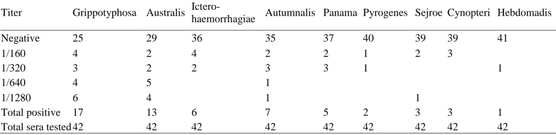
Among the 42 samples of maternal serum, 29 had a positive result with respect to at 157
least one serogroup (Table III). Among the positive samples, agglutination was observed 158
against an average of 2 and a maximum of 5 serogroups per sample. A titer of ≥ 160 was used 159
to define a seropositivity reaction, no positive results have been observed for the following 5 160
serogroups as Ballum, Bataviae, Canicola, Pomona and Tarassovi. The results revealed that 161
Leptospira serogroups Grippotyphosa and Australis were the most prevalent with respectivly
162
17/42 and 13/42 positive MAT (Table III). 163
164
Leptospira interrogans spp. (RT-PCR) 165
Of the 26 organ samples analysed, DNA of pathogenic Leptospira was detected in 21 166
cases. Among the 21 PCR positive cases, 5 sera were negative by MAT against the 24 serovars 167
(14 serogroups) tested. 168
Concerning the 5 PCR negative cases, a positive MAT was observed in 4 samples. For 169
only one sample, the PCR and the MAT were negative. The results of serology (MAT) and 170
molecular detection (real-time PCR) tests are summarized in Table III. 171
172
Leptospira MLST genotyping 173
Among the 10 samples, 2 were successfully amplified and sequenced for all the 7 loci. 174
For these two samples, different sequence types (ST) were obtained, the ST number 110 profile 175
for sample CI-14-061536-002 and the ST number 24 profile for sample CI-12-000889-002 176
(Table III). In the Leptospira MLST database, ST number 110 profile corresponds to 177
Leptospira kirschneri species and Grippotyphosa serogroup (Table III), whereas the ST
178
number 24 profile corresponds to Leptospira interrogans species and Australis serogroup 179 (Table III). 180 181 DISCUSSION 182
MAT and PCR results support the hypothesis that the jaundice observed in fetuses was 183
due to leptospiral infection. Furthermore, no other cause of abortion was identified despite the 184
wide range of analyzes. 185
The MAT is the serological reference test, particularly appropriate for carrying out 186
epidemiological studies, since it can be applied to sera from any animal species, and because 187
the range of antigens utilized can be expanded or decreased as required (Levett, 2004). Most 188
cases of leptospirosis are diagnosed by serology and antibodies are detectable in the blood 189
approximately 5 to 7 days after the onset of clinical signs. In our conditions, the sampling was 190
performed at the time of abortion but the presence of jaundice indicated an earlier infection that 191
could explain the relative high titer in MAT observed. 192
Australis and Grippotyphosa are identified as the two predominant serogroups in this 193
study. These results are consistent with the findings of two other recent studies, in Germany 194
concerning dogs (Mayer-Scholl et al., 2013) and in France concerning dogs and cattle (Ayral 195
et al., 2014). In France, the two predominant infecting serogroups involved in clinical bovine
and canine leptospirosis from 2008 to 2011 are also Australis and Grippotyphosa for the two 197
species. 198
On average, sera show a seropositive reaction to two serogroups with a maximum of 199
five. Indeed, MAT is a complex test to control, perform, and interpret. It is a serogroup-specific 200
assay but interpretation of the MAT is complicated by the high degree of cross-reaction that 201
occurs between different serogroups, especially in acute-phase samples. This “paradoxical” 202
reaction, in which the highest titers are detected to a serogroup unrelated to the infecting one, 203
are also common and studied (Blanco et al., 2016; Lin et al., 1997). The broad cross-reactivity 204
in the acute phase is followed by relative serogroup specificity in convalescent-phase samples 205
(Levett, 2001). Than, an average of two serogroups per sample seems relatively high compared 206
to other studies where only one serovar is highlighted. But this observation argues for 207
paradoxical reaction due to IgM during acute infection. Unfortunatly, paired sera are not 208
available to confirm the infected serogroup with certainty. Moreover, a positive result does not 209
identify with certainty the cause of abortions and it is impossible to date the infection, the 210
reaction kinetics with respect to different serovars may vary (Levett, 2001). 211
In order to ensure the involvement of bacteria in the abortive process, PCR were 212
performed and bacterial DNA was detected in the great majority of cases. Following these PCR 213
analyzes, different profiles combining PCR and MAT are observed. These profiles may depend 214
on the delay between the infection and the abortion, the immunity of the infected animals, the 215
type of serogroups concerned or the laboratory assays. 216
In total, in the great majority of cases, both analyzes provide a positive response and support 217
the diagnosis of leptospirosis. 218
Because of the difficulties associated with serological identification of leptospiral 219
isolates, there has been great interest in molecular methods for identification and subtyping 220
(Terpstra, 1992; Herrmann, 1993). The reclassification of leptospires on genotypic grounds is 221
taxonomically correct and provides a strong foundation for future classifications. Genotyping 222
of two species of Leptospira is a key point that allows knowing with certainty the infecting 223
species. Leptospira interrogans and Leptospira kirschneri were genotyped and have a positive 224
response to serogroup Australis and Grippotyphosa respectively. These results are consistent 225
with Leptospira MLST database. 226
During this episode in 2014, many questions arise about the almost simultaneous 227
distribution on a broad territory of a disease that does not have the dissemination power of an 228
arbovirus. The disease is maintained in nature by chronic infection of reservoir hosts. The most 229
important reservoir hosts are small mammals, which may transfer infection to domestic farm 230
animals, dogs, and humans. Different rodent species may act as reservoir of the serogroups 231
highlighted in this study (Levett, 2001). Distinct variations in reservoir hosts and the serovars 232
they carry occur throughout the world (Hartskeerl and Terpstra, 1996). Currently, the source of 233
infection remains unknown and therefore complicates the usefulness of implementation of 234
preventive measures. From the affected farms, only one case of bovine aborted fetus was 235
identified in 95% of the farms with a maximum of 3 cases in one farm (Delooz et al., 2015). 236
That allows hypothesizing that infection does not spread to the entire herd by other cattle that 237
therefore do not appear to be potential maintenance hosts. 238
The spectrum of clinical signs is extremely broad. Formerly it was considered that 239
distinct clinical syndromes were associated with specific serogroups. However, this hypothesis 240
was questioned by some authors and following studies refuted this hypothesis (Levett, 2001). 241
Grippotyphosa and Australis are the two main serogroups revealed during this emerging event 242
associated with the same clinical pattern, which joins the idea that clinical syndromes were not 243
associated with specific serogroups. However, congenital jaundice from aborted fetuses coming 244
from clinically healthy cows is a new clinical sign to add to those caused by pathogenic 245
Leptospira.
The results of this study indicated that Sejroe serogroup was rarely diagnosed and that 247
methods of the diagnosis of leptospirosis must be adapted for a better surveillance and control. 248
This finding suggests that the available bovine vaccine targeting this serogroup is capable of 249
preventing a minority of the clinical cases. Nevertheless, additional serogroups, such as 250
Grippotyphosa and Australis should be included in the vaccine to eliminate most Leptospira-251
related diseases in cattle. 252
253
CONCLUSION 254
255
Jaundice was a known clinical sign of leptospirosis but, to our kowledge, had never been 256
diagnosed in bovine aborted fetuses coming from clinically healthy cows in the literature. This 257
work allows the association of two pathogenic Leptospira species (L. interrogans or L. 258
kirschneri) to congenital jaundice in bovine aborted fetuses. This new clinical sign should be
259
added to the clinical picture of bovine leptospirosal abortion. 260
Leptospira are often difficult to isolate from infected cattle and therefore diagnosis
261
usually depends on the detection of specific antibodies. This work showed a feasible method of 262
direct diagnostic approach under field conditions where the veterinary practitioner performs 263
samples in cattle farms. 264
Finally, despite that the sources of infection during the emergence remain unknow, this 265
study provided useful information in the knowledge of bovine leptospirosis in south part of 266
Belgium. It seems necessary to be prepared to tackle appropriate prevent and control measures 267
and to further explores the epidemiology of this disease in this region, especially in wild life. 268
269
AKNOWLEDGEMENT 270
We thank Magnée D. (Thermofisher, UK) to provide PCR kits. We thank the Federal Agency 271
for the Safety of the Food Chain (FAFSC) to support the costs of the basic protocol 272
corresponding to the standardized panel of analyses. The authors gratefully acknowledge their 273
veterinary colleagues from ARSIA, i.e. Christian Quinet (serology), Marc Saulmont and 274
Thierry Petitjean (autopsy and microbiology) for their technical assistance. 275
276
REFERENCES 277
Adler, B., 2010: Leptospira and Leptospirosis. Current Topics in Microbiology and 278
Immunology 387: 1–293.
279
Ayral, F. C., D. J. Bicout, H. Pereira, M. Artois, and A. Kodjo, 2014: Distribution of Leptospira 280
serogroups in cattle herds and dogs in France. The American journal of tropical 281
medicine and hygiene 91, 756-759.
282
Blanco, R. M., L. F. dos Santos, R. L. Galloway, and E. C. Romero, 2016: Is the 283
microagglutination test (MAT) good for predicting the infecting serogroup for 284
leptospirosis in Brazil? Comparative immunology, microbiology and infectious diseases 285
44, 34-36. 286
Boonsilp, S., Thaipadungpanit, J., Amornchai, P., Wuthiekanun, V., Bailey, M.S., Holden, 287
M.T., Zhang, C., Jiang, X., Koizumi, N., Taylor, K., Galloway, R., Hoffmaster, A.R., 288
Craig, S., Smythe, L.D., Hartskeerl, R.A., Day, N.P., Chantratita, N., Feil, E.J., 289
Aanensen, D.M., Spratt, B.G., and S.J. Peacock, 2013: A single multilocus sequence 290
typing (MLST) scheme for seven pathogenic Leptospira species. PLoS Negl Trop 291
Dis. 7(1), e1954. 292
Chappel, R. J., M. Goris, M. F. Palmer, and R. A. Hartskeerl, 2004: Impact of proficiency 293
testing on results of the microscopic agglutination test for diagnosis of leptospirosis. 294
Journal of clinical microbiology 42, 5484-5488.
Delooz, L., M. Mori, T. Petitjean, J. Evrard, G. Czaplicki, and C. Saegerman, 2015: Congenital 296
jaundice in bovine aborted foetuses: an emerging syndrome in southern Belgium. 297
Transboundary and emerging diseases 62, 124-126.
298
Dom, P. P., F. Haesebrouck, R. Vandermeersch, J. Descamps, and K. Van Ommeslaeghe, 1991: 299
Prevalence of Leptospira interrogans serovar hardjo antibodies in milk in Belgian dairy 300
herds. The Veterinary quarterly 13, 118-120. 301
Ellis, W. A., and S. W. Michna, 1977: Bovine leptospirosis: experimental infection of pregnant 302
heifers with a strain belonging to the Hebdomadis serogroup. Research in veterinary 303
science 22, 229-236.
304
Ellis, W. A., J. J. O'Brien, S. D. Neill, and D. G. Bryson, 1986: Bovine leptospirosis: 305
experimental serovar hardjo infection. Veterinary microbiology 11, 293-299. 306
Evangelista, K. V., and J. Coburn, 2010: Leptospira as an emerging pathogen: a review of its 307
biology, pathogenesis and host immune responses. Future microbiology 5, 1413-1425. 308
Guerra, M. A., 2009: Leptospirosis. Journal of the American Veterinary Medical Association 309
234, 472-478, 430. 310
Hartman, E. G., P. Franken, B. A. Bokhout, and D. J. Peterse, 1989: [Leptospirosis in cattle; 311
milker's fever in cattle farmers]. Tijdschrift voor diergeneeskunde 114, 131-135. 312
Hartskeerl, R. A., and W. J. Terpstra, 1996: Leptospirosis in wild animals. The Veterinary 313
quarterly 18 Suppl 3, S149-150.
314
Herrmann, J. L., 1993: Genomic techniques for identification of Leptospira strains. Pathologie-315
biologie 41, 943-950.
316
Jolley, K. A., and M. C. Maiden, 2010: BIGSdb: Scalable analysis of bacterial genome variation 317
at the population level. BMC bioinformatics 11, 595. 318
Levett, P. N., 2001: Leptospirosis. Clinical microbiology reviews 14, 296-326. 319
Levett, P. N., 2004: Leptospirosis: a forgotten zoonosis? Clinical and Applied Immunology 320
Reviews 4, 435–448.
321
Levett, P. N., R. E. Morey, R. L. Galloway, D. E. Turner, A. G. Steigerwalt , and L. W. Mayer, 322
2005: Detection of pathogenic leptospires by real-time quantitative PCR. Journal of 323
Medical Microbiology 54(1): 45-49.
324
Levett, P. N., 2015: Systematics of Leptospiraceae p. 11-20. In Ben Adler. Leptospira and 325
Leptospirosis, Current Topics in Microbiology and Immunology. Springer Berlin 326
Heidelberg. 327
Lin, M., O. Surujballi, K. Nielsen, S. Nadin-Davis, and G. Randall, 1997: Identification of a 328
35-kilodalton serovar-cross-reactive flagellar protein, FlaB, from Leptospira 329
interrogans by N-terminal sequencing, gene cloning, and sequence analysis. Infection
330
and immunity 65, 4355-4359.
331
Mayer-Scholl, A., E. Luge, A. Draeger, K. Nockler, and B. Kohn, 2013: Distribution of 332
Leptospira serogroups in dogs from Berlin, Germany. Vector borne and zoonotic
333
diseases 13, 200-202.
334
Morey, R. E., R. L. Galloway, S. L. Bragg, A. G. Steigerwalt, L. W. Mayer, and P. N. Levett, 335
2006: Species-specific identification of Leptospiraceae by 16S rRNA gene sequencing. 336
Journal of clinical microbiology 44, 3510-3516.
337
Mori, M., M. Esbroeck, S. Depoorter, W. Decaluwe, S. J. Vandecasteele, D. Fretin, and M. 338
Reynders, 2015: Outbreak of leptospirosis during a scout camp in the Luxembourg 339
Belgian province, Belgium, summer 2012. Epidemiology and infection 143, 1761-1766. 340
OIE Terrestrial Manual, 2014: Leptospirosis. Available atw: 341
http://www.oie.int/fileadmin/Home/fr/Health_standards/tahm/2.01.09_LEPTO.pdf 342
(accessed 19 November 2015). 343
Ryan, E. G., N. Leonard, L. O'Grady, M. L. Doherty, and S. J. More, 2012: Herd-level risk 344
factors associated with Leptospira Hardjo seroprevalence in Beef/Suckler herds in the 345
Republic of Ireland. Irish veterinary journal 65, 6. 346
Smith, C. R., M. R. McGowan, C. S. McClintock, B. G. Corney, P. J. Ketterer, L. Smythe, and 347
W. Ward, 1997: Experimental Leptospira borgpetersenii serovar hardjo infection of 348
pregnant cattle. Australian veterinary journal 75, 822-826. 349
Terpstra, W. J., 1992: Typing Leptospira from the perspective of a reference laboratory. Acta 350
Leidensia 60, 79-87.
351
Tables and figures caption 352
Table I. List of pathogens included and diagnostic methods applied in the standardized panel 353
of analyses 354
Pathogens
Fetus Foetal serum Maternal serum
Samples Methods Methods Methods
Brucella spp. Abomasal fluid Culture SAW /ELISA Ab
Campylobacter foetus spp. Abomasal fluid Culture
Coxiella burnetii Abomasal fluid PCR** ELISA Ab
Listeria monocytogenes Abomasal fluid Culture Mycotic agents Abomasal fluid Culture Opportunistic bacteria Abomasal fluid Culture $
Salmonella spp. Abomasal fluid Culture
BTV-8 Brain PCR*
Neospora caninum Brain PCR ELISA Ab ELISA Ab
Schmallenberg virus Brain PCR*
BoHV-4 Spleen PCR ELISA Ab
Leptospira serovar Hardjo ELISA Ab 355
Legend: PCR, Polymerase chain reaction; Ab, Antibody; Ag, Antigen; SAW, Sero-356
agglutination of Wright; $, Only the presence of a pure culture on blood agar is indicative of 357
opportunistic bacteria; *, Applied only if suspected case (congenital abnormalities); BVDV, 358
Bovine Viral Diarrhoea Virus; BTV-8, Bluetongue virus serotype 8; BoHV-4, Bovine 359
herpesvirus 4. 360
Table II. Distribution of MAT results among the tested cow sera according to different leptospiral serogroup (only serogroups with positive 362
results are listed) 363
364
Titer Grippotyphosa Australis Ictero-
haemorrhagiae Autumnalis Panama Pyrogenes Sejroe Cynopteri Hebdomadis
Negative 25 29 36 35 37 40 39 39 41 1/160 4 2 4 2 2 1 2 3 1/320 3 2 2 3 3 1 1 1/640 4 5 1 1/1280 6 4 1 1 Total positive 17 13 6 7 5 2 3 3 1
Total sera tested 42 42 42 42 42 42 42 42 42
Table III. Results of serological and antigenical analysis according to Leptospira serogroup 366
(only serogroups with positive results are listed, the highest titer of each serogroup is presented) 367 Leptospiral serogroup ID Pathogenic Leptospira strains (PCR) Genotyping
(MLST) AUS AUT CYN GRI HEB ICT PAN PYR SEJ
CI-11-023436 - - - -
CI-11-029715 - - - -
CI-12-000889 Pos L. interrogans 640 - - - - - 640 320 -
CI-12-001123 - - - - CI-13-006899 - - - - CI-13-011101 - - - 160 CI-13-018383 - - - - CI-13-019237 - - - - CI-13-022971 - - - - CI-13-032292 - - - 320 - - - - - CI-13-032707 160 - - 640 - - - - - CI-14-034435 Pos - - - - CI-14-034966 Pos - - - 160 - - - - - CI-14-037270 Neg - - 160 1280 - - - - - CI-14-038297 1280 160 - - - 160 - - - CI-14-038323 320 - 160 - - - - CI-14-038596 Pos 1280 1280 - - - 160 640 - - CI-14-040708 Neg 160 - - - - CI-14-042496 640 - - 320 - - - - - CI-14-042765 - - - 160 - - - 160 - CI-14-044328 Neg - - - 640 - - - - - CI-14-044569 Pos - 160 - - 320 - - - 1280 CI-14-044707 Pos - - - 640 - - - - - CI-14-046867 - - 160 1280 - - - - - CI-14-047394 Pos - - - 1280 - - - - - CI-14-047531 Pos 1280 320 - - - - 160 - - CI-14-047968 Pos 1280 320 - - - 320 - - - CI-14-048785 Pos 640 - - - 160 - - CI-14-049712 Pos 640 640 - - - 320 640 - 160 CI-14-050512 Pos - - - - CI-14-050870 Neg - - - - CI-14-057066 Pos - - - - CI-14-057564 Pos * - - - 640 - - - - - CI-14-057925 Pos - - - - CI-14-058607 - - - 1280 - - - - - CI-14-058713 Pos - - - 1280 - - - - -
CI-14-061536 Pos L. kirschneri 320 - - 1280 - - - - -
CI-14-062221 Pos * - - - -
CI-14-067483 Pos 640 320 - - - -
CI-14-069253 Neg - - - 320 - - - - -
368
Legend: 369
* Unsuccessful amplification and sequencing; AUS, Australis ; AUT, Autumnalis ; CYN, 370
Cynopteri ; GRI, Grippotyphosa; HEB, Hebdomadis; ICT, Icterohaemorrhagiae ; PAN, Panama 371
; PYR, Pyrogenes ; SEJ, Sejroe. 372
Figure captions 374
Figure 1. Geographical distribution of icteric abortion's case, years 2008-2014 (N=152) 375
Legend: White dots correspond to icteric cases where complementary analysis (MAT and/or 376
RT-PCR) are performed; black dots corresponds to icteric cases without complementary 377
analysis. 378
379
Figure 2. Trends of icteric bovine aborted fetuses rate and the absolute number of notified 380
abortions 381
Fig. 1 383
384 385
Fig. 2 386
387 388 389