THE IMMUNE RESPONSE TO LIVE VACCINES
AGAINST BOVINE ANAPLASMOSIS
Thèse présentée à la faculté des Sciences Institut de Biologie
Université de Neuchâtel
Pour l’obtention du grade de docteur ès Sciences (Ph.D.) Par
Rachel Kenneil
Jury:
Prof. Dr. Kurt Pfister, Directeur de thèse (Université de Neuchâtel, Switzerland) Prof. Dr. Lise Gern (Université de Neuchâtel, Switzerland)
Prof. Dr. Patrick Guerin (Université de Neuchâtel, Switzerland) Prof. hon. Bruno Betschart (Université de Neuchâtel, Switzerland)
Prof. Dr. Steffen Rehbein (Merial GmbH, Germany)
Supervisor: Prof. Dr. Lygia Passos (Ludwig Maximilian University, Germany)
Université de Neuchâtel 2015
ABSTRACT
Anaplasma marginale causes bovine anaplasmosis, a hemolytic disease which is a serious problem for the cattle industry in tropical and sub-tropical regions worldwide. A. centrale is a less pathogenic species which is closely related to A. marginale and used as a live vaccine against anaplasmosis. Unfortunately the A. centrale vaccine can only be produced from the blood of infected cattle, risking contamination with other pathogens. This project compared the A. centrale live vaccine with A. marginale UFMG1, a low pathogenicity Brazilian strain which has been proposed as a potentially safer live vaccine derived from cell culture. Calves were infected with UFMG1 or A. centrale, and then challenged with the pathogenic Israeli A. marginale Gonen strain.
Previous infection with UFMG1 did not significantly reduce the severity of disease caused by challenge with the Gonen strain, whereas A. centrale infection did provide cross-protection against Gonen. In comparison to the antibody response to UFMG1 infection, the response to A. centrale infection had higher overall levels of IgG and showed higher cross-reactivity to Gonen strain antigen. The antibody response to A. centrale also had higher levels of IgG2, and showed more opsonophagocytic activity. All of these characteristics correlated significantly with protection from disease upon challenge. Understanding how A. centrale infection stimulates this effective immune response would be a valuable direction for future vaccine research.
Keywords: Anaplasma marginale, bovine anaplasmosis, immune response, opsonophagocytosis,
RÉSUMÉ
Le rickettsies Anaplasma marginale provoque l'anaplasmose bovine, une maladie hémolytique qui est un grave problème pour l'industrie bovine dans les régions tropicales et subtropicales du monde entier. L’A. centrale est une espèce moins pathogène qui est étroitement liée à l’A. marginale et elle est utilisée comme vaccin vivant contre l'anaplasmose. Malheureusement, le vaccin A. centrale ne peut être produit qu’à partir du sang de bovins infectés, qui risquent d’être contaminés par d'autres agents pathogènes. Ce projet a comparé le vaccin vivant A. centrale avec A. marginale UFMG1, une souche brésilienne faiblement pathogène qui a été proposée comme un vaccin vivant issu de culture cellulaire et potentiellement plus sûr. Les veaux ont été infectés par UFMG1 ou A. centrale, puis inoculés avec la souche israélienne pathogène A. marginale Gonen.
L’infection précédente avec UFMG1 n'a pas significativement réduit la sévérité de la maladie causée par la souche inoculée avec le Gonen, alors que l'infection par A. centrale a fournit une protection croisée contre le Gonen. Par rapport à la réaction des anticorps à l’infection UFMG1, la réaction à l'infection par A. centrale avait des niveaux globaux plus élevés d'IgG et a montré une réactivité croisée ultérieure à l'antigène de la souche Gonen. La réaction des anticorps à A. centrale avait également des niveaux plus élevés d'IgG2, et a montré une activité plus opsonophagocytaire. Toutes ces caractéristiques sont en corrélation significative avec la protection contre la maladie lors de l'épreuve. Comprendre comment l'infection par A. centrale stimule cette réponse immunitaire efficace serait une piste de recherche vaccinale prometteuse.
Mots-clés : Anaplasma marginale, anaplasmose bovine, réponse immunitaire, opsonophagocytose,
CONTENTS
1: General Introduction
1.1. What is A. marginale?...…... 1 1.1.1. Classification 1 1.1.2. Transmission 2 1.1.3. Disease 3 1.1.4. Economic Impact 3 1.2. Immunology...…... 4 1.2.1. Serological Response 51.2.2. Cell-mediated immune response 7
1.3. Control Measures...…... 9
1.3.1. Chemotherapy 9
1.3.2. Vaccination 9
1.4. Thesis Structure...…... 15 1.4. References...…... 16
2:Clinical results of the live vaccine trial of A. marginale UFMG1 and A. centrale
2.1. Introduction...…... 27 2.1.1. A. centrale as a live vaccine against A. marginale 27 2.1.2. Cell culture-derived A. marginale UFMG1 as a potential vaccine 27 2.1.3. The problem of cross-protection between strains 28 2.1.4. Protection with or without infection-exclusion 29
2.1.5. Aims 30
2.2. Material and Methods....…... 31
2.2.1. A. marginale and A. centrale strains 31
2.2.2. Trial design 32
2.2.3. Statistical Analysis 33
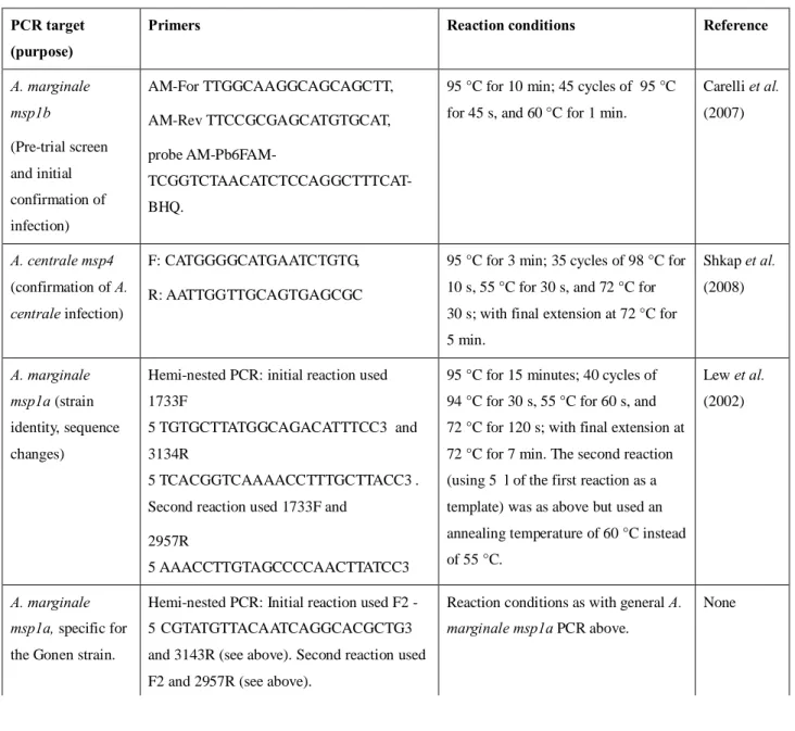
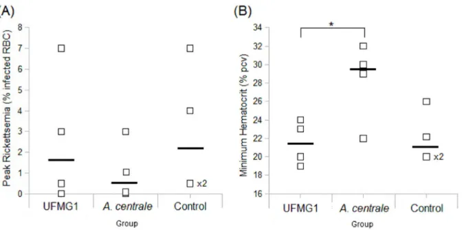
2.2.4. Confirmation of infection and genotype analysis 34 2.3. Results...…... 36 2.3.1. Clinical response to infection with UFMG1 and A. centrale 36 2.3.2. Clinical response after challenge with Gonen 37
2.3.3. Establishment and persistence of infection 39
2.4. Discussion...…... 41 2.4.1. Response to initial infection with UFMG1 vs. A. centrale 41
2.4.2. Protection from challenge 42
2.4.3. Variation in response to infection 43
2.4.4. MSP1a tracking 44
2.4.5. Conclusions 45
3: Serological response to infection with A. marginale or A. centrale
3.1. Introduction...…... 51 3.1.1. Importance of the antibody response against bovine anaplasmosis 51 3.1.2. Characteristics of a protective antibody response 51
3.1.3. Aims 52
3.2. Material and Methods..…... 52
3.2.1. Sample collection 52
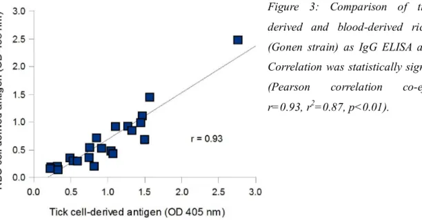
3.2.2. Source of A. marginale and A. centrale antigenic material 52
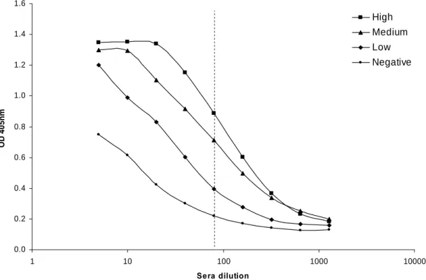
3.2.3. Measurement of seroconversion 53
3.2.4. IgG ELISAs 53
3.2.5. Data analysis 54
3.3. Results...…... 55
3.3.1. Seroconversion 55
3.3.2. Total IgG ELISA development 56
3.3.3. IgG response to the homologous strain 57
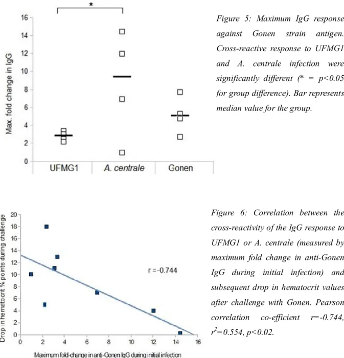
3.3.4. IgG response to the challenge strain 58
3.3.5. Cross-reactivity of the UFMG1 IgG response against Brazilian and 59 Israeli strains
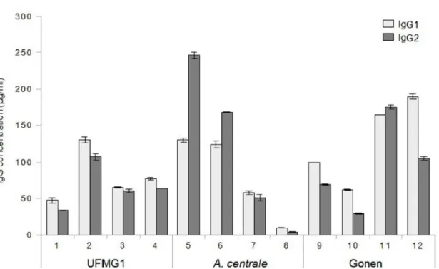
3.3.6. IgG subclass of the antibody response 59
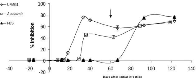
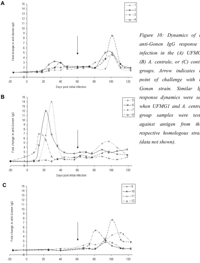
3.3.7. Dynamics of the IgG response to infection 61
3.4. Discussion...…... 63
3.4.1. Seroconversion 63
3.4.2. Cross-reactivity between A. marginale strains and A. centrale 63
3.4.3. Subclass of the IgG response 65
3.4.4. Overall antibody levels and possible link to T-cell function and 66 regulation
3.4.5. Variation in response to infection 66
3.4.6. Dynamics of the IgG response to initial infection and challenge 67
3.4.7. Conclusions 68
3.5. References...…... 68
4: Cell-mediated immune response to infection with A. marginale or A. centrale
4.1. Introduction...…... 73 4.1.1. Cell-mediated immunity also plays a role in protection from 73 anaplasmosis
4.1.2. Cell-mediated immune response to live vaccines 74
4.3. Results...…... 77 4.3.1. Optimization of freezing procedure and PBMC proliferation assay 77
4.3.2. Proliferation of fresh PBMCs 78
4.3.3. Proliferation of cryopreserved PBMCs 79
4.3.4. IFNγ production in vitro and in vivo 83
4.4. Discussion...…... 88
4.4.1. PBMC proliferation 88
4.4.2. IFNγ production by PBMCs 90
4.4.3. In vivo IFNγ levels during A. marginale and A. centrale infection 90
4.4.4. Conclusions 92
4.5. References...…... 93
5: Opsonophagocytic activity of immune sera against A. marginale
5.1. Introduction...…... 97 5.1.1. The value of measuring antibody function as a correlate of protection 97 5.1.2. Opsonophagocytosis as a correlate of protection for bovine 97 anaplasmosis
5.1.3. Measuring opsonophagocytosis 98
5.1.4. Aims 100
5.2. Material and Methods...…... 100
5.2.1. Sera 100 5.2.2. Bacteria 100 5.2.3. Cell preparation 101 5.2.4. Assay set-up 102 5.2.5. Flow cytometry 102 5.2.6. Statistical analysis 103 5.3. Results...…... 103 5.3.1. Using bacterial staining to measure opsonophagocytosis 103 5.3.2. Using the oxidative burst to measure opsonophagocytosis 105 5.3.3. Defining and analyzing the in vitro opsonophagocytosis assay 106 5.3.4. Optimization of the opsonophagocytosis assay 109 5.3.5. Testing serum samples from the live vaccine trial against 112 A. marginale Gonen
5.3.6. Comparing opsonophagocytic activity (OPA) levels to clinical 113 symptoms, IgG2, and IFNγ
5.4. Discussion...…... 115 5.4.1. Measuring phagocytosis through the uptake of 115 fluorochrome-stained bacteria
5.4.2. Measuring phagocytosis through the oxidative burst 115 5.4.3. OPA response to A. marginale and A. centrale infection 117 5.4.3. The relationship of OPA to other immune parameters 118
5.4.5. Conclusions and future research 118
6: General Conclusion
... 1236.1. Rationale for the project 123 6.2. Main findings of the project 123 6.3. Future Research 125
6.4. References 125
7. Appendix: Summary of clinical and immunological data
7.1 Summary of clinical data
129
7.2 Summary of immunological data
130
8. Abbreviations
... 1319. List of Figures and Tables
... 13310. Curriculum Vitae
…... 137CHAPTER 1: GENERAL INTRODUCTION
1.1. WHAT IS A. MARGINALE?1.1.1. Classification
Anaplasma marginale Theiler 1910 is a gram negative rickettsia and the causative agent of bovine anaplasmosis - a tick-transmitted hemolytic disease which causes serious economic problems to the cattle industry in tropical and sub-tropical areas worldwide (Kocan et al., 2010). At present, there is no safe and globally effective vaccine against bovine anaplasmosis, and vaccine development remains an area of active research.
A. marginale belongs to the order Rickettsiales and the family Anaplasmataceae. This family consists of small, obligate intracellular bacteria, which are found within membrane-bound vacuoles in the cytoplasm of host cells (Dumler et al., 2001). Within the Anaplasmataceae family, the genus Anaplasma includes several species of particular economic interest: A. marginale, the type species for the genus; A. centrale, sometimes described as a sub-species of A. marginale (Shkap et al., 2008); and A. phagocytophilum, which was formerly known by several different names - Ehrlichia phagocytophila, E. equi, or the causative agent of Human Granulocytic Ehrlichiosis (Dumler et al., 2001).
A. marginale was first isolated from cattle by Theiler (1910), who inaugurated the genus Anaplasma. He named the type species A. marginale after the 'marginal points' that the rickettsia formed in the infected erythrocytes. A. marginale infection has since been found in a wide range of wild and domestic ruminants, including cattle, bison, buffalo, wildebeest, deer and elk. However, reports of severe disease have exclusively been confined to cattle - experimental infection of other ruminant species produced only mild symptoms of anaplasmosis (Kuttler, 1984).
Within the ruminant host, A. marginale primarily infects erythrocytes. Endothelial cell infection has also been detected in vivo (Carreño et al., 2007), but this does not appear to play an important role in the A. marginale life cycle (Wamsley et al., 2011).
1.1.2. Transmission
A. marginale can be transmitted biologically by ticks, mechanically by biting flies or blood-contaminated fomites, and transplacentally (Kocan et al., 2010). Around 20 different species of ticks have been implicated as vectors for biological transmission, with the most important genera being Dermacentor and Rhipicephalus. Across the majority of the tropical and sub-tropical range of A. marginale, Rhipicephalus (Boophilus) spp. are the most critical tick vectors, in particular R. (B). microplus and R. (B). annulatus. Since these species were eradicated from the United States in the first half of the 20th century, Dermacentor spp. particularly D. andersonii and D. variabilis, and D. albipictus, have taken over as the predominant biological vectors for A. marginale in the US. In Europe, multiple tick species have been proposed to play a role in anaplasmosis transmission (De la Fuente et al., 2005). Of the species suggested, D. reticulatus has the strongest evidence of being an important A. marginale vector in Europe: it is capable of experimental transmission of A.marginale (Zivkovic et al., 2007), A. marginale DNA has been isolated from D.reticulatus ticks during an anaplasmosis outbreak in Hungary (Hornok et al., 2012), and its range runs from France, through Eastern Europe to Central Asia, covering many of the areas in which A. marginale has been found (Karbowiak, 2014).
Tick transmission can be transstadial or intrastadial; transovarial transmission has not been reported (Stich et al., 1989). When the tick feeds on infected cattle, the rickettsia are taken up as part of the blood meal, infect the gut cells of the tick, and then spread to other tissues. These tissues include the salivary glands, from where A. marginale can then be transmitted to new hosts during tick feeding (Ge et al., 1996).
On a global scale, biological transmission by ticks is considered the most common route of A. marginale infection. But not all strains of A. marginale are tick-transmissable, and in some regions no tick vector species occur. Therefore, although it has lower transmission efficacy, mechanical transmission can also play an important role in the spread of A. marginale (Scoles et al., 2005). Mechanical transmission can be through various genera of biting flies (Tabanus, Stomoxys) or mosquitoes (Culex and Aedes), or through blood-contaminated fomites such as de-horning saws, ear-tagging devices, or castration instruments.
1.1.3. Disease
In the vertebrate host, A. marginale primarily infects erythrocytes. As part of the host response, these infected cells - along with considerable numbers of uninfected erythrocytes - are then destroyed by the reticuloendothelial system. Baker et al. (1961) measured up to a ten-fold increase in the rate of erythrocyte phagocytosis during acute A. marginale infection. This considerable loss of erythrocytes leads to anaemia and icterus, without hemoglobinemia or hemoglobinuria (Richey, 1981). Other symptoms of bovine anaplasmosis include fever, lethargy, weight loss, lowered milk production, and abortion. It is often fatal in older cattle if they are not treated early (Kocan et al., 2010). The severity of bovine anaplasmosis increases with age: calves under six months of age rarely become ill; between 6-12 months they usually develop mild disease, which is more acute at 1-2 years old; finally, adults over two years old suffer acute and often fatal disease (Roby et al., 1961; Aubry and Geale, 2011). The reason behind this increasing severity with age is as yet unknown.
If cattle recover from acute disease, they usually remain persistently infected, often at microscopically undetectable levels (<107 rickettsia/ml). Throughout persistent infection, cattle show cyclical rises in rickettsemia (Eriks et al., 1993). These peaks in rickettsemia represent the generation of antigenic variants, which escape immune control and multiply rapidly, before they stimulate a variant-specific immune response that brings the infection under control again (French et al., 1999). Due to this consistent stimulation, persistently infected cattle maintain a strong immune response against A. marginale, and are protected from disease when subsequently challenged with the homologous A. marginale strain. Unfortunately they also act as reservoirs of infection for naïve cattle (Palmer et al., 1999).
1.1.4. Economic Impact in Endemic Areas
Bovine anaplasmosis has a serious economic impact on the cattle industry of endemic areas. Its effect is most serious in areas of endemic instability, where the transmission rate is too low to ensure that all cattle become infected as calves Aubrey and Geale, 2011). When cattle are first exposed to A. marginale as adults, they are much more likely to develop serious disease (Roby et al., 1961).
Anaplasmosis causes direct losses through mortality, abortion, lowered weight of beef cattle and lowered milk production from dairy cattle. In addition, anaplasmosis frequently limits breed improvement (Lombardo, 1976; Ocampo Espinoza et al., 2006). In Mexico, a highly endemic area,
anaplasmosis is estimated to cause up to a quarter of total deaths in national cattle improvement programmes (Rodriguez-Camarillo et al., 1999). When pedigree Bos taurus cattle are imported from temperate areas to endemic tropical or sub-tropical areas, the new cattle are likely to be extremely susceptible to A. marginale infection. This is due to both a lack of previous exposure, and to the greater susceptibility of B. taurus breeds (e.g. Holstein, Hereford) to tick-borne diseases in comparison to B. indicus cattle (Kocan et al., 2003).
There have been relatively few studies quantifying the economic impact of bovine anaplasmosis. In the 1970s, McCallon (1973) estimated that the disease caused annual losses of over 300 million US dollars (USD) to the American cattle industry. More recently, Tanzania was estimated to lose 47.3 million USD solely due to the direct costs of bovine anaplasmosis (Kivaria, 2006).
A. marginale is often considered in conjunction with the protozoan parasites Babesia bovis and B. bigemina, as all three are tick-borne pathogens which cause serious disease to cattle in tropical and sub-tropical regions of the world, and may often co-exist in the same animal. For example in Brazil, the three pathogens are referred to as one complex of disease, 'Tristeza Parasitária Bovina' (Gonçalves, 2000). This complex was estimated to cause a loss of 875 million USD to the cattle industry in South American countries (Brown, 1997).
Global climate change is likely to increase the range of tick vector species, and so increase the regions affected by the pathogens they can transmit (Howden et al., 2010). As such, an ever-increasing number of countries are working to develop a deeper understanding of tick-borne diseases such as anaplasmosis.
1.2. IMMUNOLOGY
Multiple parts of the immune system appear to play a role in the immune response to A. marginale. The serological response has been identified as an important part of protection after vaccination (reviewed by Palmer et al., 1999), and yet immune serum alone is not protective (Gale et al., 1992). Cell-mediated responses have also been shown to play a role in protection (Brown et al., 1998), but the T-cell response is inhibited by the pathogen to facilitate persistent infection (Han et al., 2010).
1.2.1. Serological Response
High antibody levels, particularly of the subclass IgG2 (Brown et al., 1998; Barigye et al., 2004; Vega et al., 2007) or against major surface proteins (MSPs), (Tebele et al., 1991) have frequently been shown to correlate with protection. Antibodies can act through several routes: neutralization, opsonization, and complement-mediated bactericidal activity (Siegrist, 2012).
Palmer and McGuire (1984) demonstrated that polyclonal immune sera could neutralize the infectivity of A. marginale for calves, and Palmer et al. (1987) showed the same neutralization effect with anti-MSP1a monoclonal antibody.
Opsonophagocytosis, i.e. antibody-enhanced phagocytosis, has been proposed as an important mechanism to clear A. marginale infection (Palmer et al., 1999). An opsonin, which literally means 'a flavoring', is any substance which makes whatever it binds to more likely to be 'eaten' by phagocytic cells (Murphy et al., 2012). The antibody classes IgM and IgG are the most effective opsonins. Within IgG, different IgG subclasses have different levels of efficacy in opsonization; in cattle, the IgG2 subclass is the best opsonin (McGuire and Musoke, 1981).
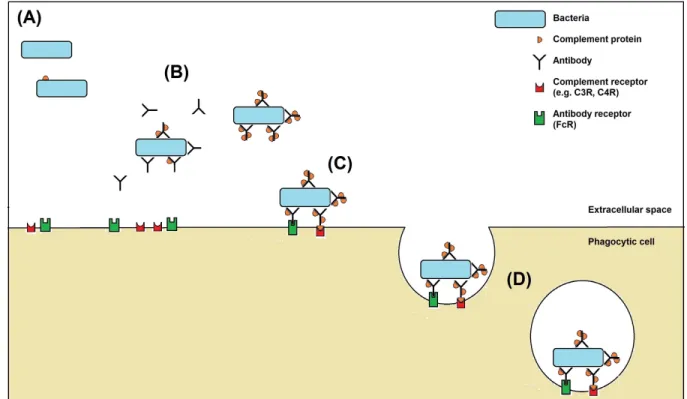
In the presence of immune serum, antigen-specific antibodies coat the surface of bacteria, allowing the C1 complement protein to bind to the antibody-antigen complex. Once bound, C1 is activated and triggers the complement cascade, binding and activating a sequence of complement proteins including the opsonins C3 and C4. Phagocytic cells will then bind greatly enhanced numbers of these opsonized or 'flavored' bacteria, through their receptors for C3 and C4 complement proteins, and for the Fc portion of antibody (Murphy et al., 2012). In the absence of any antibody, bacteria can still be recognized by phagocytic cells via pathogen recognition receptors (PRRs) which bind to common pathogen components such as lipopolysaccharide (LPS). The complement protein C3 also binds at low levels directly to the bacterial surface, leading to its activation and recognition by the C3 receptor on the phagocyte (Murphy et al., 2012). An overview of the opsonophagocytosis of bacteria is shown in Figure 1.
Figure 1: An overview of opsonophagocytosis of bacteria by immune sera.
(A) Without immune sera, only a small amount of complement protein will bind to bacteria.
(B) When immune serum is added, antibodies bind to epitopes on the surface of the bacteria. High amounts of complement proteins then bind to the antibody-antigen complex that has formed on the bacteria.
(C) Phagocytic cells recognize opsonized bacteria through the cell's receptors for antibody (FcR) and for complement proteins (C3R, C4R).
(D) The binding of opsonized bacteria to these receptors triggers the cell to internalize the bacteria in a membrane vesicle (the phagosome). The phagosome then fuses with other vesicles called lysosomes, which contain a range of antimicrobial components, and the bacteria are broken down by toxic reactive oxygen species and nitrogen oxides, acidification, proteolytic enzymes, and antimicrobial peptides.
Cantor et al. (1993) demonstrated that sera from MSP1-immunized cattle could significantly enhance in vitro opsonization of A. marginale. Melendez (2005) showed that immune sera from
Serum bactericidal activity against A. marginale has not yet been assessed.
Despite the demonstrated activity of antibodies against A. marginale, immune serum alone is not sufficient to prevent disease. In a classic passive protection experiment, Gale et al. (1992) showed that transfer of serum from hyper-immune to naïve calves was insufficient to protect the latter from disease. In addition, immune suppression by splenectomy or drugs leads persistently infected asymptomatic cattle to suffer a relapse in symptoms within 1-2 weeks – this relapse occurs before circulating IgG levels show a significant fall (Jones et al., 1968; Kuttler and Adams, 1977).
1.2.2. Cell-mediated Immune Response
There is ample evidence that cell-mediated immunity also plays a role in the host response to A. marginale. In early experiments, protection against bovine anaplasmosis correlated with inhibition of leukocyte migration (Buening, 1976) and development of cutaneous hypersensitivity (Carson et al., 1976), indicating an association with T-cell responsiveness and macrophage activation, respectively. Brown et al. (1998) demonstrated that after cattle were vaccinated with outer membrane proteins, their protection from challenge correlated with their level of CD4+ T-cell proliferation, and with functions associated with CD4+ T-cells, namely the production of IFNγ and IgG2.
These findings led to the current paradigm for a protective immune response to vaccination, proposed by Palmer et al. (1999). This model centers around antigen-specific CD4+ T-cells, and their production of IFNγ, which activates macrophages and stimulates B-cells to produce more IgG2. Antigen-specific IgG2 opsonises the rickettsia, increasing their uptake by the activated macrophages and leading to control of the infection.
This model has been challenged by more recent research, which suggests that CD4+ T-cells do not appear to be essential to protection from anaplasmosis. When calves were thymectomized (removing their ability to develop new T-cells), and then had their existing CD4+ T-cells depleted, they were still able to control A. marginale infection. They showed no significant difference in disease severity compared to untreated calves (Valdez et al., 2002). There is a caveat to this study in that CD4+ T-cell depletion was not absolute (as this is very difficult to achieve).
Interestingly, a natural depletion of T-cells during infection appears to be a persistence strategy of A. marginale (Han et al, 2010). Vaccination with A. marginale surface proteins MSP2 (Abbott et al.,
2005) or MSP1a (Han et al., 2008) induced high levels of antigen-specific CD4+ T-cells; these high levels rapidly fell after the cattle were infected with A. marginale. Later in infection only low and sporadic T-cell responses were seen. This phenomenon was not the general immunosuppression seen after A. phagocytophilum infection. The T-cell reduction was confined to those cells which responded to A. marginale antigen, with the level of CD4+ T-cells specific for a control antigen remaining high after A. marginale infection (Han et al., 2010).
The inhibition of a T-cell response to A. marginale is very likely to contribute to its long-term persistence in infected cattle. However, the inhibition may also benefit the host. Continuous CD4+ T-cell activity in response to the cyclically high bacterial loads seen in persistent A. marginale infection would likely lead to systemic inflammation, damaging the host (Han et al., 2010). In fact, down-regulation of T-cell responses is frequently seen in long-term persistent infections where antigen loads remain high (Jenson et al., 2002; Xu et al., 2002).
The majority of studies on the T-cell response to A. marginale have focused on alpha-beta T-cells. These are the most abundant T-cell type, encompassing CD4+ and CD8+ T-cells, and a range of regulatory T-cells. Another type of T-cell which may also play a role in the response to A. marginale infection is the gamma-delta T-cell. Gamma-delta T-cells are particularly abundant in ruminants, and are at their highest levels in younger animals – the age at which anaplasmosis causes the mildest symptoms (Hein and MacKay, 1991; Roby et al., 1961). Gamma-delta T-cell clones specific for A. marginale MSP2 have been shown to respond to antigen by proliferating, producing IFNγ, and expressing a range of chemokines which recruit inflammatory cells (Lahmers et al., 2005; 2006). However, the dynamics of gamma-delta T-cells during infection with A. marginale have not yet been reported.
Much of the nature of the T-cell response to A. marginale remains to be clarified - in particular, whether the effects of T-cell deletion during A. marginale infection prevent the T-cell response from having any protective effect against bovine anaplasmosis.
1.3. CONTROL MEASURES AGAINST A. MARGINALE
Control measures vary in different areas, according to their expense, practicality, and the preferences of the region. Controlling arthropods with acaricide treatment is useful for limiting the spread of various vector-borne diseases, including anaplasmosis. But as A. marginale can also be transmitted mechanically and transplacentally, acaracides cannot totally eliminate the spread of this rickettsia. In addition, regular use of acaricides is expensive and can hasten the development of resistance (Kocan et al., 2010).
1.3.1. Chemotherapy
Tetracycline antibiotics are by far the predominant treatment for bovine anaplasmosis. Trials of imidocarb (Roby et al., 1972) and enrofloxacin (Facury-Filho et al., 2012) have also shown good results, but they are not commonly used (Coetzee et al., 2006). Repeated doses of tetracycline can eliminate persistent A. marginale infections, although complete elimination is not always achieved (Swift and Thomas, 1983).
Tetracycline is most effective in the earlier stages of the disease (Kuttler et al., 1980), so it can be difficult to catch the disease early enough in range cattle (Kocan et al., 2010). Therefore it is sometimes used prophylactically, particularly in the US (Kocan et al., 2010). Such frequent use of antibiotics has the potential to cause selection of resistant strains, but to date this has not been reported as a problem. The withholding period in antibiotic-treated cattle before they can be used for meat or milk can be an issue for farmers, particularly with long-lasting oxytetracycline preparations (Lew-Taylor, 2012).
In some regions, tetracyclines are used for the 'infection-treatment' control method: cattle are inoculated with A. marginale-infected erythrocytes, and then treated early in the patent phase of disease by low doses of tetracycline drugs. The aim is the development of persistent infection without acute disease. As the animals have the opportunity to develop an immune response, they will subsequently be immune to challenge with homologous strains. However, the infection-treatment method requires careful monitoring to ensure acute disease does not develop, and so can be unsuitable for large herds of cattle (Kocan et al. 2003).
1.3.2. Vaccination
Despite the effectiveness of antibiotic treatment, demand for a vaccine is still high (Spath et al., 1990; Kocan et al., 2010). Existing vaccines can be effective control methods for bovine
anaplasmosis, but developing improved vaccines which are safer and globally effective has long been an objective of the cattle industry (Palmer et al., 1989).
No vaccines tested to date reliably induce sterile immunity. Instead, when vaccinated cattle are challenged, they develop persistent infection without acute disease, and so can still act as reservoirs of infection for naïve cattle (Bock and deVos, 2001). Various types of vaccine against bovine anaplasmosis have been studied. Killed and live vaccines have been frequently used in the field; recombinant protein, outer membrane, and DNA vaccines have so far been confined to small research trials (Kocan et al., 2010).
1.3.2.1 Killed vaccines
The commercial vaccine 'Anaplaz' consisted of killed A. marginale derived from the blood of infected cattle (Fort Dodge Laboratories, Fort Dodge, Iowa). 'Anaplaz' was marketed for several decades in the US before being withdrawn due to company restructuring (Kocan et al., 2003). Killed vaccines have several advantages over live vaccines: less chance of contamination with other pathogens, greater ease of storage, and low post-inoculation reactions. However, they offer limited cross-protection against strains from different regions (Kuttler et al., 1984), and require yearly boosters since they only induce short-term immunity (Brock et al., 1965). Moreover, expensive purification procedures are needed to remove erythrocyte debris (McCorkle-Shirley et al., 1985). If the rickettsia are insufficiently purified from erythrocyte stroma, repeated vaccination may stimulate isoantibodies against erythrocyte proteins. Calves ingesting colostrum from cows with high isoantibody titres may then develop hemolytic anemia (Dennis et al., 1970). The occurrence of this problem was reduced by improved purification protocols (Hart et al., 1990), and the introduction of guidelines not to vaccinate cows in the later stages of pregnancy (Luther et al., 1989).
1.3.2.2. Outer Membranes, Recombinant Proteins, and DNA Vaccines
Outer membrane proteins (OMPs) are common vaccine targets as they will be exposed to the host's immune system during infection (Grandi, 2010). The A. marginale proteins that have been considered as vaccine candidates are therefore OMPs, with the majority of research being focused
MSP1a and MSP1b form the MSP1 complex, which is involved in cell entry. MSP1a is necessary and sufficient for attachment to both erythrocytes and tick cells (de la Fuente et al., 2003), while MSP1b only plays a role in attachment to erythrocytes (de la Fuente et al., 2001). MSP2 and MSP3 are both transcribed from large multi-gene families. They vary considerably between isolates (Alleman et al., 1997; Palmer et al., 1994), and even express variant sequences over the course of persistent infection (Brayton et al., 2003; Barbet et al., 2001; French et al., 1999). MSP4 and MSP5 are both encoded by single-copy genes, and are highly conserved. As such they are useful for phylogenetic analysis (de la Fuente et al., 2002b) and diagnostics, with MSP5 used for a commercial diagnostic ELISA (de Eschaide et al., 1998).
Outer membrane protein vaccines
Outer membrane protein (OMP) vaccines are a complex mix of proteins of varying immunogenicity. At least 25 different antigenic OMPs have been identified (Lopez et al., 2005; Noh et al., 2008). Several studies have demonstrated that OMP-based vaccines can induce protection against homologous challenge (Tebele et al., 1991; Brown et al., 1998; Noh et al., 2008), but Palmer et al. (1994b) found they did not significantly reduce disease symptoms after heterologous challenge. In addition, OMP vaccines are expensive to prepare and difficult to standardize (Noh et al., 2013).
Recombinant protein vaccines
Recombinant protein vaccines are often less protective than OMP vaccines. This is perhaps unsurprising, as they usually consist of a single protein rather than the complex mix of immunogens seen in OMP vaccines (Palmer and McElwain, 1995).
The majority of recombinant protein vaccines have used MSPs. Despite good results with native protein (Palmer et al., 1989), recombinant MSP1 (in the form of the MSP1a/MSP1b complex) has had mixed results, with some studies finding protection against homologous strains (Camacho-Nuez et al., 2000), and others not (Palmer and McElwain, 1995). Although MSP2 and MSP3 are highly immunogenic (Palmer et al., 1999), their high variability makes them unsuitable targets for a widely protective vaccine (Albarrak et al., 2012). There has been very limited study of recombinant MSP4 and MSP5; the former induced protection against homologous challenge, the latter not (A.F. Barbet, unpublished data, cited in Palmer and McElwain, 1995).
some groups to focus on sub-dominant OMP antigens. Candidate sub-dominant antigens are those which are highly conserved between strains and over the course of infection, and are still immunogenic despite being less abundant. Multiple antigens have been identified which fit these requirements (Lopez et al., 2005; Noh et al., 2008; Sutten et al., 2010; Agnes et al., 2011), but so far only one, AM779, has been tested as a recombinant protein vaccine. It was immunogenic but not protective, raising the possibility that multiple sub-dominant antigens may have to be combined create an effective recombinant protein vaccine (Albarrak et al., 2012).
DNA vaccines
DNA vaccines have been tested in mice (Kano et al., 2008) and cattle (Arulkanthan et al., 1999; Mwangi et al., 2007). Interestingly, Arulkanthan et al. (1999) found that the bovine antibody response to a MSP1a DNA vaccine was largely restricted to IgG1, which is not associated with protection (Brown et al., 1998). However, an MSP1b-based DNA vaccine has been shown to induce partial protection from disease (de Andrade et al., 2006).
1.3.2.3. Live vaccines
Live vaccines have been used against bovine anaplasmosis for over a century, beginning shortly after A. marginale was first identified (Theiler, 1912). Today live vaccines are perhaps still the most widely used type (Kocan et al., 2003). As live vaccines lead to a persistent infection in vaccinated cattle, they provide them with lifelong protection against severe disease. They produce a stronger immune response than killed vaccines, with the generation of antigenic variants during persistent infection leading to a broader immune response (Palmer et al. 1999).
The practise of deliberately establishing a persistent infection with a less pathogenic strain in order to create immunity against a more severe infection is known as premunition or premunisation (Kuttler et al., 1984b). Vaccines for premunisation are customarily produced from the blood of splenectomized calves, which develop high levels of rickettsemia after infection (Kocan et al., 2003). The infected blood must then be kept chilled (for use within a week) or cryopreserved (for longer term storage, and quality control) (McElwain, 2008). Natural variation can be a problem with live vaccines: it can lead to variants which either cause unexpectedly serious disease, or do not
use as a live vaccine: passage through sheep (Ristic et al., 1968), passage through deer (Kuttler and Zaugg, 1988), or 60Co irradiation (Sharma and Bansal, 1986). The most successful attenuated vaccine was produced in sheep by Ristic et al. (1968). It induced protection with limited side effects in several trials (Welter and Ristic, 1969; Osorno et al., 1975; Ristic and Carson, 1977). However, other investigators found severe post-vaccination reactions (Anziani et al., 1981; Henry et al., 1983).
Naturally low pathogenic A. marginale strains
Low pathogenic strains of A. marginale cause mild or no disease without needing deliberate attenuation. Several low pathogenic strains have been tested as live vaccines: the Dawn strain from Australia (Bock et al. 2003; Carter et al., 2006); the Yucatan strain from Mexico (Rodriguez-Camarillo et al., 2008); and the UFMG1 strain from Brazil (Bastos et al., 2010). A low pathogenicity field isolate from Colombia has also been tested as part of a combination babesiosis-anaplasmosis live vaccine (Benavides et al., 2000).
All these strains, with the exception of the combination vaccine, induced good protection against homologous strains, and against heterologous A. marginale strains from the same geographic area. However, none have yet successfully progressed into wider use outside their country of origin.
Anaplasma centrale
Anaplasma centrale is a low pathogenic species closely related to A. marginale, which was first identified in South Africa by Sir Arnold Theiler, shortly after he isolated A. marginale (Theiler, 1912). As only a few, quite variable, A. centrale isolates have been sequenced, it is difficult to determine whether A.centrale should be identified as a variant of A.marginale, a sub-species, or a separate species. 16S rRNA analysis based on a Japanese A. centrale isolate classified it as a distinct species (Inokuma et al., 2001), but analysis of two South African A.centrale (including the vaccine strain, A. centrale Theiler, 1911) by 16S rRNA (Dumler et al., 2001) and GroEL sequences (Lew et al., 2003) classified A. centrale as an A. marginale sub-species.
As the name suggests, A. centrale inclusions are generally seen in the centre of erythrocytes, as opposed to the 'marginal points' of A. marginale (Theiler, 1912). Theiler determined that animals infected with A. centrale only showed mild disease or were asymptomatic, and were subsequently protected from challenge with pathogenic A. marginale. In the decades after this discovery, A. centrale was exported to various countries to be used as a live vaccine against bovine anaplasmosis
– not only to other countries in Africa, but further afield to Israel, Australia, and parts of South America (Shkap et al., 2008; Bock and de Vos, 2001; De Wall, 2000; Melendez et al., 2003).
In all these regions, A.centrale is generally very effective at protecting cattle against bovine anaplasmosis (Bock and de Vos, 2001). Nevertheless there have been some reports of vaccine failure, particularly against highly pathogenic A. marginale strains (Wilson et al., 1980; Payne et al., 1990; Turton et al., 1998; Brizuela et al., 1998; Bock and de Vos, 2001), and A. centrale itself has been reported to cause disease if administered to adult cattle (Pipano, 1976; Potgieter 1979; Bigalke 1980; Pipano et al., 1985).
As with all the live vaccines that have been used in the field so far, A. centrale is produced from the blood of infected splenectomized calves (Pipano, 1995). Blood-derived vaccines risk accidental transmission of other blood-borne pathogens. For example when one calf used for vaccine production became accidentally infected with bovine leucosis virus, over 10,000 A. centrale vaccines doses were contaminated (Rogers et al., 1988).
1.3.2.4. Tick-cell culture as a source of vaccines
Tick cell culture has been proposed as a safer and more easily standardized source of live vaccines. A. centrale has never successfully been cultivated in vitro, but multiple A. marginale strains have been propagated in tick cell lines (Passos, 2012). In vitro-derived A. marginale remain infective for cattle and ticks (Munderloh et al., 1996), and keep the same antigenic composition through repeated passages in tick cell culture (Barbet et al., 1999). Tick cell-derived A. marginale have been shown to be immunogenic when used as a killed (Kocan et al., 2001; de la Fuente et al., 2002) or live inoculum (Bastos et al., 2010; Hammac et al., 2013). When compared with blood-derived A. marginale of the same strain, they induced similar levels of protection (de la Fuente et al., 2002; Bastos et al., 2010).
blood-1.4. THESIS STRUCTURE
The aim of this project was to investigate the immune response to live vaccines against bovine anaplasmosis. A. centrale, the current 'gold-standard' live vaccine, was compared with a naturally low pathogenic Brazilian A. marginale strain (UFMG1), which has been successfully established in cell culture and so has the potential of being a cell culture-derived live vaccine.
Chapter 2 describes a trial comparing the effectiveness of A. centrale and UFMG1 in protecting calves against challenge with a pathogenic Israeli strain, A. marginale Gonen.
Chapter 3 investigates the serological response induced by A. centrale and A. marginale UFMG1.
Chapter 4 investigates the cell-mediated response to A. centrale and A. marginale UFMG1 – namely PBMC proliferation and IFNγ production.
Chapter 5 describes the development of an in vitro assay for serum opsonophagocytosis activity (OPA) against A. marginale, based on flow cytometric measurement of the oxidative burst response to phagocytosis. OPA has been suggested as a correlate of protection for bovine anaplasmosis, but this has never been experimentally confirmed.
Chapters 2 and 3 are partially based on:
R.Kenneil, V. Shkap, B.Leibovich, E. Zweygarth, K. Pfister, M.F. Ribeiro, L.M.Passos (2013). Cross-protection between geographically distinct Anaplasma marginale isolates appears to be constrained by limited antibody responses. Journal of Transboundary and Emerging Diseases 60 Suppl.2: 97-104.
1.5. REFERENCES
Abbott JR, Palmer GH, Kegerreis KA, Hetrick PF, Howard CJ, Hope JC, Brown WC (2005). Rapid and long-term disappearance of CD4-T lymphocyte responses specific for Anaplasma marginale major surface protein-2 (MSP2) in MSP2 vaccinates following challenge with live A. marginale. J. Immunol. 174: 6702–6715.
Agnes JT, Brayton KA, LaFollett M, Norimine J, Brown WC, Palmer GH (2011). Identification of Anaplasma marginale outer membrane protein antigens conserved between A. marginale sensu stricto strains and the live A. marginale subsp. centrale vaccine. Infect. Immun. 79: 1311-1318. Albarrak SM, Brown WC, Noh SM, Reif KE, Scoles GA, Turse JE, Norimine J, Ueti MW, Palmer GH (2012). Subdominant antigens in bacterial vaccines: AM779 is subdominant in the Anaplasma marginale outer membrane vaccine but does not associate with protective immunity. PloS One 7:e46372. doi: 10.1371/journal.pone.0046372.
Alleman AR, Palmer GH, McGuire TC, McElwain TF, Perryman LE, Barbet AF (1997). Anaplasma marginale major surface protein 3 is encoded by a polymorphic, multigene family. Infect. Immun. 65: 156–163.
Anziani OS, Hadani A, Ford CA, Guglielmone AA, Bermudez AC, Mangold AJ, Suarez CM, Tarabla DH (1981). Observaciones de campo y laboratorio sobre la inoculaciòn de bovinos Holando Argentino con una cepa de Anaplasma marginale. Gac. Vet. 43: 962–974.
Arulkanthan A, Brown WC, McGuire TC, Knowles DP (1999). Biased immunoglobulin G1 isotype responses induced in cattle with DNA expressing msp1a of Anaplasma marginale. Infect. Immun. 67: 3481-3487.
Aubry P and Geale DW (2010). A review of bovine anaplasmosis. Transbound. Emerg. Dis. 58: 1-30.
Baker NF, Osebold JW, Christensen JF (1961). Erythrocyte survival in experimental anaplasmosis. Am. J. Vet. Res. 22: 590-596.
Barbet AF, Blentlinger R, Yi J, Lundgren AM, Blouin EF, Kocan KM (1999). Comparison of surface proteins of Anaplasma marginale grown in tick cell culture, tick salivary glands, and cattle. Infect Immun. 67: 102-107.
Barbet AF, Yi J, Lundgren A, McEwen BR, Blouin EF, Kocan KM (2001). Antigenic variation of Anaplasma marginale: major surface protein 2 diversity during cyclic transmission between ticks and cattle. Infect. Immun. 69: 3057–3066.
Barigye R, García-Ortiz MA, Rojas Ramírez EE, Rodríguez SD (2004). Identification of IgG2-specific antigens in Mexican Anaplasma marginale strains. Ann. N. Y. Acad. Sci. 1026: 84-94.
Benavides E, Vizcaino O, Britto CM, Romero A, Rubio A (2000). Attenuated trivalent vaccine against babesiosis and anaplasmosis in Colombia. Ann. N. Y. Acad. Sci. 916: 613–616.
Bigalke RD (1980). The control of ticks and tick-borne diseases of cattle in South Africa. Zim. Vet. J. 11: 20–21.
Bock RE, de Vos AJ (2001). Immunity following use of Australian tick fever vaccine: a review of the evidence. Aust. Vet. J. 79: 832-839
Bock RE, deVos AJ, Kingston TG, Carter PD (2003). Assessment of a low virulence Australian isolate of Anaplasma marginale for pathogenicity, immunogenicity and transmissibility by Boophilus microplus. Vet. Parasitol.118: 121-131.
Brayton KA, Meeus PF, Barbet AF, Palmer GH (2003). Simultaneous variation of the immunodominant outer membrane proteins, MSP2 and MSP3, during Anaplasma marginale persistence in vivo. Infect Immun. 71: 6627-6632.
Brizuela CM, Ortellado CA, Sanabria E, Torres O, Ortigosa D (1998). The safety and efficacy of Australian tick-borne disease vaccine strains in cattle in Paraguay. Vet Parasitol. 76: 27-41. Brock WE, Kliewer IO, Pearson CC (1965). A vaccine for anaplasmosis. J. Am. Vet. Med. Assoc. 147: 948–951.
Brown DCG (1997). Dynamic and impact of tick-borne diseases of cattle. Tropical Animal Health and Production 29:1S–3S.
Brown WC, Shkap V, Zhu D, McGuire TC, Tuo W, McElwain TF, Palmer GH (1998). CD4(+) T-lymphocyte and immunoglobulin G2 responses in calves immunized with Anaplasma marginale outer membranes and protected against homologous challenge. Infect. Immun. 66: 5406-5413. Buening GM (1976). Cell mediated immune response in anaplasmosis as measured by a micro cell-mediated cytotoxicity assay and leukocyte migration inhibition test. Am. J. Vet. Res. 34: 1215–1218 Camacho-Nuez M, de Lourdes Muñoz M, Suarez CE, McGuire TC, Brown WC, Palmer GH (2000). Expression of polymorphic msp1beta genes during acute Anaplasma marginale rickettsemia. Infect Immun. 68: 1946-1952.
Cantor GH, Pontzer CH, Palmer GH (1993). Opsonization of Anaplasma marginale mediated by bovine antibody against surface protein MSP-1. Vet. Immunol. Immunopathol. 37: 343–350
Carreño AD, Alleman AR, Barbet AF, Palmer GH, Noh SM, Johnson CM (2007). In vivo Endothelial Cell Infection by Anaplasma marginale. Vet. Pathol. 44: 116-118
Carson CA, Sells DM, Ristic M (1976). Cell mediated immunity in bovine anaplasmosis and correlation with protection induced by vaccination. Vet. Parasitol. 2: 75–81
Carter PD, Bock RE, deVos AJ (2006) Benefits of Improved Vaccine for Anaplasmosis in Australia. Proceedings of the 11th International Symposium on Veterinary Epidemiology and Economics. Available from www.sciquest.org.nz [Accessed 24th July 2013].
Coetzee JF, Apley MD, Kocan KM (2006). Comparison of the efficacy of enrofloxacin, imidocarb, and oxytetracycline for clearance of persistent Anaplasma marginale infections in cattle. Vet Ther. 7: 347-360.
de Andrade GM, Machado RZ, Vidotto MC, Vidotto O (2004). Immunization of bovines using a DNA vaccine (pcDNA3.1/MSP1b) prepared from the Jaboticabal strain of Anaplasma marginale. Ann. N. Y. Acad. Sci. 1026: 257-266.
de Eschaide ST, Knowles DP, McGuire TC, Palmer GH, Suarez CE, McElwain TF (1998).
Detection of cattle naturally infected with Anaplasma marginale in a region of endemicity by nested PCR and a competitive enzyme-lined immunosorbent assay using recombinant major surface protein 5. J. Clin. Microbiol. 36: 777–782.
de la Fuente J, Garcia-Garcia JC, Blouin EF, Kocan KM (2001). Differential adhesion of major surface proteins 1a and 1b of the ehrlichial cattle pathogen Anaplasma marginale to bovine erythrocytes and tick cells. Int. J. Parasitol. 31: 145–153.
de la Fuente J, Kocan KM, Garcia-Garcia JC, Blouin EF, Claypool PL, Saliki JT (2002). Vaccination of cattle with Anaplasma marginale derived from tick cell culture and bovine erythrocytes followed by challenge-exposure by infected ticks. Vet. Microbiol. 89: 239–251. de la Fuente J, Van Den Bussche RA, Garcia-Garcia JC, Rodriquez SD, Garcia MA, Guglielmone AA, Mangold AJ, Passos LMF, Ribeiro MFB, Blouin EF, Kocan KM (2002). Phylogeography of New World isolates of Anaplasma marginale (Rickettsiaceae: Anaplasmataceae) based on major surface protein sequences. Vet. Microbiol. 88: 275–285.
de la Fuente J, Garcia-Garcia JC, Blouin EF, Kocan KM (2003). Characterization of the functional domain of major surface protein 1a involved in adhesion of the rickettsia Anaplasma marginale to host cells. Vet. Microbiol. 91: 265–283.
De La Fuente J, Naranjo V, Ruiz-Fons F, Höfle U, Fernández De Mera IG, Villanúa D, Almazán C, Torina A, Caracappa S, Kocan KM, Gortázar C (2005). Potential vertebrate reservoir hosts and invertebrate vectors of Anaplasma marginale and A. phagocytophilum in central Spain. Vector Borne Zoonotic Diseases 54:390-401.
Dennis RA, O’Hara PJ, Young MF, Dorris KD (1970). Neonatal immunohemolytic anemia and icterus of calves. J. Am. Vet. Med. Assoc. 156: 1861–1869.
De Wall DT (2000). Anaplasmosis control and diagnosis in South Africa. Ann. N. Y. Acad. Sci. 916: 474-483.
Dumler JS, Barbet AF, Bekker CP, Dasch GA, Palmer GH, Ray SC, Rikihisa Y, Rurangirwa FR (2001). Reorganization of genera in the families Rickettsiaceae and Anaplasmataceae in the order Rickettsiales: unification of some species of Ehrlichia with Anaplasma, Cowdria with Ehrlichia
Facury-Filho EJ, de Carvalho AÚ, Ferreira PM, Moura MF, Apolinário BC, Santos LDPH, Ribeiro MF (2012). Effectiveness of enrofloxacin for the treatment of experimentally-induced bovine anaplasmosis. Rev. Bras. Parasitol. Vet. 21: 32-36.
French DM, Brown WC, Palmer GH (1999). Emergence of Anaplasma marginale antigenic variants during persistent rickettsemia. Infect.Immun. 67: 5834–5840.
Gale KR, Leatch G, Gartside M, Dimmock CM (1992). Anaplasma marginale: failure of sera from immune cattle to confer protection in passive-transfer experiments. Parasitol Res. 78: 410-415. Ge NL, Kocan KM, Blouin EF, Murphy GL (1996). Developmental studies of Anaplasma marginale (Rickettsiales: Anaplasmataceae) in male Dermacentor andersoni (Acari: Ixodidae) infected as adult using nonradioactive in situ hybridization. J. Med. Entomol. 33: 911–920. Gonçalves PM (2000). Epidemiology and control of bovine babesiosis and anaplasmosis in Southeast region of Brazil. Ciência Rural 30: 187-194.
Grandi G (2010). Bacterial surface proteins and vaccines. F1000 Biology Reports 2:36.
Hammac GK, Ku PS, Galletti MF, Noh SM, Scoles GA, Palmer GH, Brayton KA (2013). Protective immunity induced by immunization with a live, cultured Anaplasma marginale strain. Vaccine 31: 3617-3622.
Han S, Norimine J, Palmer GH, Mwangi W, Lahmers KK, Brown WC (2008). Rapid deletion of antigen-specific CD4+ T cells following infection represents a strategy of immune evasion and persistence for Anaplasma marginale. J. Immunol. 181: 7759-7769.
Han S, Norimine J, Brayton KA, Palmer GH, Scoles GA, Brown WC (2010).Anaplasma marginale infection with persistent high-load bacteremia induces a dysfunctional memory CD4+ T lymphocyte response but sustained high IgG titers. Clin. Vaccine Immunol. 17: 1881-1890.
Howden KJ, Geale DW, Paré J, Golsteyn-Thomas EJ, Gajadhar AA (2010). An update on bovine anaplasmosis (Anaplasma marginale) in Canada. Can. Vet. J. 51: 837-840.
Hein WR and Mackay CR (1991). Prominence of γδ T cells in the ruminant immune system. Immunol. Today 12: 30-34.
Hart LT, Todd WJ, Luther DG (1990). Anaplasma marginale antigen, antigen composition, vaccine and process for production of said antigen, antigen composition and vaccine. US Patent 4,956,278. Henry ET, Norman BB, Fly DE, Wichmann RW, York SM (1983). Effects and use of modified live Anaplasma marginale vaccine in beef heifers in California. J. Am. Vet. Med. Assoc. 183: 66–69. Hornok S, Micsutka A, Fernández de Mera IG, Meli ML, Gönczi E, Tánczos B, Mangold AJ, Farkas R, Lutz H, Hofmann-Lehmann R, de la Fuente J (2012). Fatal bovine anaplasmosis in a herd with new genotypes of Anaplasma marginale, Anaplasma ovis and concurrent haemoplasmosis. Research in Veterinary Science 2: 30-35.
apoptosis of CD4 T lymphocytes: a mechanism of immune unresponsiveness in filariasis. Eur. J. Immunol. 32: 858–867.
Jones EW, Norman BB, Kliewer IO, Brock WE (1968). Anaplasma marginale infection in splenectomized calves. Am J Vet Res. 29: 523-533.
Kano FS, Tamekuni K, Coelho AL, Garcia JL, Vidotto O, Itano EN, Vidotto MC (2008). Induced -immune response of DNA vaccine encoding an association MSP1a, MSP1b, and MSP5 antigens of Anaplasma marginale. Vaccine. 26: 3522-3527.
Karbowiak G (2014).The occurrence of the Dermacentor reticulatus tick - its expansion to new areas and possible causes. Annals in Parasitology 60:37-47.
Kivaria FM (2007). The control of East Coast Fever in Africa: a constant battle for impoverished dairy farmers. Vet. J. 174: 221–222.
Kocan KM, Halbur T, Blouin EF, Onet V, de la Fuente J, Garcia-Garcia JC, Saliki JT (2001). Immunization of cattle with Anaplasma marginale derived from tick cell culture. Vet. Parasitol. 102: 151–161.
Kocan, K. M., J. de la Fuente, A. A. Guglielmone, and R. D. Melendez (2003). Antigens and alternatives for control of Anaplasma marginale infection in cattle. Clin.Microbiol.Rev. 16: 698-712.
Kocan KM, de la Fuente J, Blouin ED, Coetzee JF, Ewing SA (2010). The natural history of Anaplasma marginale. Vet. Parasitol. 167: 95-107.
Kuttler KL, Adams LG (1977). Influence of dexamethasone on the recrudescence of Anaplasma marginale in splenectomized calves. Am. J. Vet. Res. 38: 1327-1330.
Kuttler KL, Johnson LW, Simpson JE (1980). Chemotherapy to eliminate Anaplasma marginale under field and laboratory conditions. Proc. Annu. Meet. U.S. Anim. Health. Assoc. 84: 73-82. Kuttler KL (1984). Anaplasma infections in wild and domestic ruminants: a review. J. Wildl. Dis. 20: 12-20.
Kuttler KL, Zaugg JL, Johnson LW (1984). Serologic and clinical responses of premunized, vaccinated, and previously infected cattle to challenge exposure by two different Anaplasma marginale isolates. Am. J. Vet. Res. 45: 2223-2226.
Kuttler KL, Zaugg JL (1988). Characteristics of an attenuated Anaplasma marginale of deer origin as an anaplasmosis vaccine. Trop. Anim. Health. Prod. 20: 85-91.
associated genes by WC1+ gamma-delta T cells. J. Leukoc. Biol. 80: 939-952.
Lew AE, Gale KR, Minchin CM, Shkap V, de Waal DT (2003). Phylogenetic analysis of the erythrocytic Anaplasma species based on 16S rDNA and GroEL (HSP60) sequences of A. marginale, A. centrale, and A. ovis and the specific detection of A. centrale vaccine strain. Veterinary Microbiology 92: 145–160.
Lew-Taylor AE (2012). Anaplasmosis. In: The Merck Veterinary Manual (Eds. Aiello SE and Moses MA). Available from http://www.merckmanuals.com/vet/index.html. [Accessed 29th January 2014]. Lombardo RA (1976). Socioeconomic importance of the tick problem in the Americas. PAHO Scientific Publication 326: 79-89.
Lopez JE, Siems WF, Palmer GH, Brayton KA, McGuire TC, Norimine J, Brown WC (2005). Identification of novel antigenic proteins in a complex Anaplasma marginale outer membrane immunogen by mass spectrometry and genomic mapping. Infect. Immun. 73: 8109-8118. Luther DG, Hart LT, Todd WJ, Morris NG, Taylor ND, McRae J (1989). Field study of an
experimental anaplasmosis vaccine on pregnant cows and neonatal isoerythrolisis. In: Proceedings of the 8th National Veterinary Hemoparasite Disease Conference (pp. 559–562).
Ocampo-Espinoza V, Vázquez JE, Aguilar MD, Ortiz MA, Alarcón GJ, Rodríguez SD (2006). Anaplasma marginale: lack of cross-protection between strains that share MSP1a variable region and MSP4. Vet. Microbiol. 114: 34–40.
McCallon, B.R. 1973. Prevalence and economic aspects of anaplasmosis. Proceedings of the 6th National Anaplasmosis Conference: 1–3.
McCorkle-Shirley S, Hart LT, Larson AD, Todd WJ, Myhand JD. (1985). High-yield preparation of purified Anaplasma marginale from infected bovine red blood cells. Am. J. Vet. Res. 46: 1745– 1747.
McElwain TF (2008). Bovine Anaplasmosis. In: OIE Manual of Diagnostic Tests and Vaccines for Terrestrial Animals (pp.599-610). Paris: World Organisation for Animal Health.
McGuire TC, Musoke AJ (1981). Biologic activities of bovine IgG subclasses. Adv. Exp. Med. Biol. 137: 359-366.
Meléndez RD, Toro Benítez M, Niccita G, Moreno J, Puzzar S, Morales J (2003). Humoral immune response and hematologic evaluation of pregnant Jersey cows after vaccination with Anaplasma centrale. Vet. Microbiol. 94: 335-339.
Meléndez RD (2005). Phagocytosis of Anaplasma marginale infected and uninfected erythrocytes by bovine peripheral blood leukocytes. Revista Científica 15: 305-309.
Munderloh UG, Blouin EF, Kocan KM, Ge NL, Edwards WL, Kurtti TJ (1996). Establishment of the tick (Acari:Ixodidae)-borne cattle pathogen Anaplasma marginale
(Rickettsiales:Anaplasmataceae) in tick cell culture. J. Med. Entomol. 33: 656-664.
York: Garland Science.
Mwangi W, Brown WC, Splitter GA, Davies CJ, Howard CJ, Hope JC, Aida Y, Zhuang Y, Hunter BJ, Palmer GH (2007). DNA vaccine construct incorporating intercellular trafficking and
intracellular targeting motifs effectively primes and induces memory B- and T-cell responses in outbred animals. Clin. Vaccine Immunol. 14: 304-311.
Noh SM, Brayton KA, Brown WC, Norimine J, Munske GR, Davitt CM, Palmer GH (2008). Composition of the surface proteome of Anaplasma marginale and its role in protective immunity induced by outer membrane immunization. Infect. Immun. 76: 2219-2226.
Noh SM, Turse JE, Brown WC, Norimine J, Palmer GH (2013). Linkage between Anaplasma marginale outer membrane proteins enhances immunogenicity but is not required for protection from challenge. Clin. Vaccine Immunol. 20: 651-656.
Osorno MB, Solana PM, Perez JM, Lopez TR (1975). Study of an attenuated Anaplasma marginale vaccine in Mexico: Natural challenge of immunity in an enzootic area. Am. J. Vet. Res. 36: 631– 633.
Palmer GH, McGuire TC (1984). Immune serum against Anaplasma marginale initial bodies neutralizes infectivity for cattle. J. Immunol. 133: 1010-1015.
Palmer GH, Waghela SD, Barbet AF, Davis WC, McGuire TC (1987). Characterization of a neutralization-sensitive epitope on the Am 105 surface protein of Anaplasma marginale. Int. J. Parasitol. 17: 1279-1285
Palmer GH, McElwain TF (1995). Molecular basis for vaccine development against anaplasmosis and babesiosis. Vet. Parasitol. 57: 233-253.
Palmer GH, Barbet AF, Cantor GH, McGuire TC (1989). Immunization of cattle with the MSP-1 surface protein complex induces protection against a structurally variant Anaplasma marginale isolate. Infect. Immun. 57: 3666–3669.
Palmer GH, Eid G, Barbet AF, McGuire TC, McElwain TF (1994). The immunoprotective Anaplasma marginale major surface protein 2 is encoded by a polymorphic multigene family. Infect. Immun. 62: 3808–3816.
Palmer GH, Munodzana D, Tebele N, Ushe T, McElwain TF (1994b). Heterologous strain challenge of cattle immunized with Anaplasma marginale outer membranes. Vet. Immunol. Immunopathol. 42: 265-273.
Palmer GH, Rurangirwa FR, Kocan KM, Brown WC (1999). Molecular basis for vaccine
Pipano E (1976). Control of bovine theileriosis and anaplasmosis in Israel. Bull. Off. Int. Epizootiol. 86: 55-59.
Pipano E, Mayer E, Frank M (1985) Comparative response of Friesian milking cows and calves to Anaplasma centrale vaccine. Br. Vet. J. 141: 174-8.
Pipano E (1995). Live vaccines against hemoparasitic diseases in livestock. Vet. Parasitol. 57: 213-31.
Potgieter FT (1979). Epizootiology and control of anaplasmosis in South Africa. J. S. Afr. Vet. Assoc. 50: 367-372.
Potgieter FT, van Rensburg L (1987). The persistence of colostral Anaplasma antibodies and incidence of in utero transmission of Anaplasma infections in calves under laboratory conditions. Onderstepoort J. Vet. Res. 54: 557-560.
Richey EJ (1981). Bovine anaplasmosis. In: Howard RJ (Ed.) Current Veterinary Therapy Food Animal Practice (pp. 767–772). Philadelphia: The W.B. Saunders Co.
Ristic M, Sibinovic S, Welter CJ (1968). An attenuated Anaplasma marginale vaccine. Proc. Ann. Meet. U. S. Anim. Health. Assoc. 72:56-69.
Ristic M, Carson CA (1977). Methods of immunoprophylaxis against bovine anaplasmosis with emphasis on use of the attenuated Anaplasma marginale vaccine. Adv. Exp. Med. Biol. 93: 151-188.
Roby TO, Gates DW, Mott LO (1961). The comparative susceptibility of calves and adult cattle to bovine anaplasmosis. Am. J. Vet. Res.22: 982–985.
Roby TO (1972). The inhibitory effect of imidocarb on experimental anaplasmosis in splenectomized calves. Res. Vet. Sci. 13: 519-522.
Rodriguez-Camarillo SD, Garcia-Ortiz MA, Canto-Alarcon GJ, Hernandez-Salgado G, Santos-Cerda N, Abortes-Torres R (1999). Ensayo de un inmunogeno experimental inactivado contra Anaplasma marginale. Tec. Pecu. Mex. 37: 1–12.
Rodríguez-Camarillo SD, García-Ortiz MA, Rojas-Ramírez EE, Cantó-Alarcón GJ, Preciado de la Torre JF, Rosario-Cruz R, Ramos-Aragón JA, Aboytes-Torres R (2008). Anaplasma marginale Yucatan (Mexico) Strain. Assessment of low virulence and potential use as a live vaccine. Ann. N. Y. Acad. Sci. 1149: 98-102.
Rogers RJ, Dimmock CK, de Vos AJ, Rodwell BJ (1988). Bovine leucosis virus contamination of a vaccine produced in vivo against bovine babesiosis and anaplasmosis. Aust. Vet. J. 65: 285-287. Scoles GA, Broce AB, Lysyk TJ, Palmer GH (2005). Relative efficiency of biological transmission of Anaplasma marginale (Rickettsiales: Anaplasmataceae) by Dermacentor andersoni (Acari: Ixodidae) compared with mechanical transmission by Stomoxys calcitrans (Diptera: Muscidae). J. Med. Entomol. 42: 668-675.
Anaplasma marginale. Indian J. Anim. Sci. 56: 490–493.
Shkap V, Leibovitz B, Krigel Y, Molad T, Fish L, Mazuz M, Fleiderovitz L, Savitsky I (2008). Concomitant infection of cattle with the vaccine strain Anaplasma marginale ss centrale and field strains of A. marginale. Vet Microbiol. 130: 277-284.
Siegrist C (2012). Vaccine immunology. In: Plotkin SA, Orenstein WA, Offit PA (Eds), Vaccines 6th Edition (pp. 14-32). Philadelphia: W.B. Saunders Company.
Spath EJ, Mangold AJ, Guglielmone AA (1990). Estimation of the potential demand for a bovine babesiosis and anaplasmosis vaccine in Argentina. Vet. Parasitol. 36: 131 – 140.
Stich RW, Kocan KM, Palmer GH, Ewing SA, Hair JA, Barron SJ (1989). Transstadial and attempted transovarial transmission of Anaplasma marginale by Dermacentor variabilis. Am. J. Vet. Res. 50: 1377-80.
Sutten EL, Norimine J, Beare PA, Heinzen RA, Lopez JE, Morse K, Brayton KA, Gillespie JJ, Brown WC (2010). Anaplasma marginale type IV secretion system proteins VirB2, VirB7, VirB11, and VirD4 are immunogenic components of a protective bacterial membrane vaccine. Infect. Immun. 78: 1314-1325.
Swift BL, Thomas GM (1983). Bovine anaplasmosis: elimination of the carrier state with injectable long-acting oxytetracycline. J. Am. Vet. Med. Assoc. 183: 63-65.
Tebele N, McGuire TC, Palmer GH (1991). Induction of protective immunity by using Anaplasma marginale initial body membranes. Infect Immun. 59: 3199-3204.
Theiler A (1910). Anaplasma marginale (gen. spec. nov.). The marginal points in the blood of cattle suffering from a specific disease. Report of the government veterinary bacteriologist,
1908–1909. Transvaal, South Africa.
Theiler A (1912). Gallsickness of imported cattle and the protective inoculation against this disease. Agric. J. Union South Africa 3: 7–46.
Turton, JA, Katsande TC, Matingo MB, Jorgensen WK, Ushewokunze-Obatolu U, Dalgliesh RJ (1998). Observations on the use of Anaplasma centrale for immunization of cattle against anaplasmosis in Zimbabwe. Onderstepoort J. Vet. Res. 65: 81-86.
Valdez RA, McGuire TC, Brown WC, Davis WC, Jordan JM, Knowles DP (2002). Selective in vivo depletion of CD4(+) T lymphocytes with anti-CD4 monoclonal antibody during acute infection of calves with Anaplasma marginale. Clin. Diagn. Lab. Immunol. 9: 417-424.
Vega LE, Rodríguez SD, Alarcón GJ, Flores RL, Ocampo RJ, Ortiz MA, de la Torre JF, Ramírez EE (2007). Anaplasma marginale field challenge: protection by an inactivated immunogen that shares
vaccine. Proc. Annu. Meet. U. S. Anim. Health Assoc. 73: 122-130.
Wilson AJ, Parker R, Trueman KF (1980). Experimental immunization of calves against Anaplasma marginale infection: observations on the use of living A. centrale and A. marginale. Vet Parasitol 7: 305-311.
Xu H, Wipsa J, Yan H, Zeng M, Makobongo MO, Finkelman FD, Kelso A, Good MF (2002). The mechanism and significance of deletion of parasite-specific CD4 T cells in malaria infection. J. Exp.Med. 195: 881–892.
Zivkovic Z, Nijhof AM, de la Fuente J, Kocan KM, Jongejan F (2007). Experimental transmission of Anaplasma marginale by male Dermacentor reticulatus. BMC Veterinary Research 3:32.