(Diptera: Culicidae)
HANS BRIEGEL AND EMANUEL HORLER
Institute of Zoology, University of Zurich, CH-8057 Zurich, Switzerland
J. Med. Entomol. 30(6): 975-985 (1993)
ABSTRACT Multiple blood meals within one gonotrophic cycle were taken readily at 6-24-hr intervals by nulliparous female Anopheles albimanus Wiedemann. Up to five blood meals were ingested and with each blood meal, more primary follicles matured as well as some secondary ones. This produced an irregular oviposition pattern that ques-tioned the concept of gonotrophic concordance. The first blood meal initiated limited vitellogenesis and the maturation of few follicles. Instead, protein was diverted to the synthesis of maternal, extraovarian lipid and protein deposits; fecundity increased with successive blood meals. This pattern of protein and energy utilization may be explained in terms of the low caloric lipid and protein content of nulliparous females before the first blood meal. In An. albimanus, a critical female body size, =0.25 cal of lipid per female, was necessary for the initiation of oogenesis; below this threshold at least two blood meals were required for follicle maturation. Less than 10% of the caloric input from a blood meal was utilized in the synthesis of ovarian protein and lipids, whereas a similar percentage was transferred to maternal deposits of protein and lipid. In nonoogenic females, a replete blood meal increased total body protein and lipid by 17 and 113%, respectively. Alto-gether, the efficiency of blood protein utilization was rather low, as indicated by losses of excretory nitrogen that regularly exceeded 75% of the input. Anopheles gambiae Giles and An. stephensi Liston also fed multiply, but fecundity was less affected. Instead, maternal deposits were synthesized from the blood meal in substantial amounts. In general, gonotrophic discordance also was found routinely in these anopheline species. The ratio of yolk protein to lipids varied inter- and intra-specifically, as well as among consecutive blood meals, indicating a considerable plasticity in the caloric distribution of these two yolk components.
KEY WORDS Anopheles spp., reproduction, multiple feeding
IN MOSQUITOES, reproduction is usually cyclic 1964, Boreham & Garrett-Jones 1973, Burkot et and initiated by a blood meal. Oviposition marks al. 1988). Reisen & Aslamkhan (1976) presented the end of this cycle, when the female is ready evidence for three blood meals from eclosion to for another blood meal. For anopheline mosqui- first oviposition in Anopheles culicifacies Giles, toes particularly, this cyclic mode of reproduc- For Anopheles gambiae Giles and Anopheles tion was called gonotrophic concordance by funestus Giles, Hocking & Maclnnes (1948) Swellengrebel (1929). Variations in this relation- suggested that at least two blood meals were ship were first observed by Swellengrebel (1929) necessary for each gonotrophic cycle. For these in Anopheles labranchiae atroparvus Van Thiel, two species, Gillies (1954, 1955) reported where females did not mature eggs after blood gonotrophic discordance in —20% of the popula-meals were ingested in the autumn, and, by de tion. According to Gillies, the ovaries reached Buck & Swellengrebel (1934) in Anopheles ma- only the late resting stage with the first blood
culipennis messeae Falleroni, where females re- meal, and a second meal was required for
com-quired more than one blood meal to complete a plete maturation. A similar situation appears to single gonotrophic cycle. The terms gonotrophic occur in An. stephensi Liston and An. culici-dissociation and gonotrophic discordance were fades (Reisen et al. 1986). Altogether, multiple coined by Swellengrebel (1929) for these situa- blood meals appear to be a wide-spread require-tions, respectively. In the field, blood-fed mos- ment for completing the gonotrophic cycle of quitoes have been collected attempting to feed anophelines.
on humans (e.g., Gillies & De Meillon 1968, An earlier report (Briegel & Rezzonico 1985) Magnarelli 1977). The frequency of multiple described an experiment with Anopheles albi-blood feeding in field-collected females has manus Wiedemann, in which fecundity in-been found to vary from 5 to 50% in a number of creased from 52 to 185 eggs per female when
Anopheles species (Senior White 1952, Davidson nullipars were fed three consecutive blood
1954, Smith & Weitz 1959, Edman & Downe meals during a single oviposition cycle. This
suit was indicative of gonotrophic discordance. Garrett-Jones (1970) clearly stated that blood-feeding activity "may or may not correspond to the gonotrophic cycle" and "close correspon-dence is often assumed as the cycle itself in the absence of sufficient evidence."
Multiple blood meals also have been reported for culicine mosquitoes. In Culex tarsalis Co-quillett, 43% of a population refed according to Bang & Reeves (1942). Edman & Downe (1964) found mixed blood meals in all of 14 species tested. More recently, in Aedes aegypti L., dou-ble blood meals were detected by Trpis & Hausermann (1986) and multiple meals by Scott et al. (1993). Previously, gonotrophic discor-dance seemed to be associated primarily with hibernation (Roubaud 1929, Mitchell & Briegel 1989), but the report by Scott et al. (1993) indi-cated frequent occurrence in tropical conditions and species more often than previously assumed. Multiple blood feedings might be the result of a first partial or interrupted blood meal. Indeed, Klowden & Lea (1979b) demonstrated for Ae.
aegypti that females could refeed within a
lim-ited time period after a small initial blood meal. Obviously, neural and humoral mechanisms have evolved, in most cases, preventing repeated blood meals within one gonotrophic cycle (Gwadz 1969; Klowden & Lea 1979a, b). As a further safety mechanism against overloading, the oostatic hormone prevents additional oogen-esis (Meola & Lea 1972, Else & Judson 1972).
The reports of reproductive aberrations in anophelines are descriptive in nature and gath-ered under field conditions in connection with malaria surveys or other epidemiological studies. Multiple meals have been attributed to inter-rupted feeding as a consequence of host defen-sive behavior. In the present report, we provide laboratory evidence that the nutritive state of anophelines rather than interrupted blood meals force repeated blood feeding. Low energy re-serves at the time of eclosion (Briegel 1990b) form the metabolic basis for this multiple blood-meal requirement. Female Anopheles appear to rely on a massive protein input from the blood meal for reproduction and for the acquisition of additional lipid and protein reserves.
Materials and Methods
The Anopheles used in this study were An.
(Nyssorhynchus) albimanus Wiedemann, strain
"San Diego del Norte," An. (Cellia) stephensi Liston, strain of Indian origin (obtained from NIH), and An. (Cellia) gambiae Giles, strain 16c55. The taxonomic status of An. gambiae s. str. was confirmed by W. Maier (Bonn, Germany).
All larvae were reared at 27°C under 14:10 (L:D) h long-day conditions and were fed a high-protein diet comprising equal weights of lactal-bumin, rat chow, and brewers yeast (Briegel
1990a, b). Imagoes were maintained in large cages (30 by 40 by 60 cm) at 27°C and 85% RH with access to sucrose ad lib under the same long-day conditions that included a 20-min sim-ulated sunrise and sunset. Stock colonies were fed weekly on 2 consecutive d for 20 min on a restrained guinea pig.
For experiments, mated females (1 or 2 d old) were fed on one of us (E. H.) who was not aller-gic to anopheline bites. Females were fed in groups of 20—30 until all stopped feeding as in-dicated by voluntary withdrawal of the probos-cis. Subsequently, the females were checked in-dividually for the presence of blood, then kept in beakers in groups of 20—30. At a given interval, they were refed on the same subject for 10—20 min until all stopped feeding again. Each female then was inspected for the presence of fresh blood in the midgut by trans-illuminating her abdomen with a fiber optic halogen lamp. Even traces of fresh blood could be recognized around or above the dark bolus of the previous blood meal. The few females that did not refeed were kept as controls for single blood meals. All the females that refed were transferred individually to a reacting tube (14 by 100 mm) to save their feces; a small cotton ball containing a drop of distilled water maintained humidity. After an-other 24 h, the females were pooled in groups of 20 and were reexposed to the same host. Having fed to repletion, as judged again by withdrawal of the proboscis, they were examined a second time for the presence of fresh blood. The few that refused to feed were discarded. Fed females were transferred individually into new glass tubes to catch their feces during the next 24 h. In some experiments, this procedure was repeated until five blood meals were taken. Females in these experiments were never offered sugar. Af-ter their last blood meals, females were kept in-dividually in tubes for 48—60 h until defecation was complete and no black haematin remained in their midguts. Females then were anesthe-tized, their wing lengths measured as an esti-mate of body size (cubic value of wing length), the ovaries excised with fine needles, and the mature oocytes counted within a drop of 100% ethanol. Subsequently, these oocytes were col-lected with watchmaker's forceps and trans-ferred to a test tube (10 by 70 mm), fixed with ethanol at 90°C for 5 min, then stored at room temperature until they were analyzed. Alterna-tively, females were allowed to oviposit over-night, the eggs were counted the next morning and fixed and analyzed in the same way. No differences in number or caloric content were detected between mature oocytes collected by ovarian dissection or oviposition. While dissect-ing the ovaries, the intestinal tract was routinely checked for traces of haematin; if present, the female was discarded because of incomplete or delayed defecation. Thus, the number of eggs
matured, their total protein or lipid content, and the total excretory haematin value were deter-mined for each female.
The biochemical analyses were performed as described previously (Briegel 1990a, b). Total pro-teins were estimated from total nitrogen through Kjeldahl digestion and subsequent Nessleriza-tion (Minari & Zilversmit 1963). Total lipids were determined according to the methods of Van Handel (1985b). Total carbohydrates of whole mosquitoes were measured with the hot anthrone reaction (Van Handel 1985a), which could not distinguish between glycogen and oli-gosaccharides. Carbohydrates never contributed more than a small percentage of the total caloric content; they were not given further consider-ation in this study.
Fecal material was eluted in 1% lithium car-bonate to quantify haematin output (Briegel 1980). Based on the haematin readings per indi-vidual female, blood consumption could be de-termined retrospectively and the caloric input to the mosquito calculated (Briegel 1986). With this method, ingested protein (hemoglobin) actually digested and metabolized was measured; this eliminated hemoglobin losses as a result of bloodmeal concentration during feeding (Briegel & Rezzonico 1985). The hemoglobin titer of the blood donor was monitored with Drabkin's solu-tion (Briegel et al. 1979). His total blood protein was recorded simultaneously by the Kjeldahl procedure to convert the hemoglobin readings to total protein input for each blood meal.
In most cases, sufficient oocytes were present in single females for the analysis of the yolk protein or lipid. If <30 eggs were found, ovaries from two such females were pooled. Females were placed in small single cages (20 by 30 mm) with a water-soaked pad of cellucotton for ovipo-sition. These females subsequently were fixed with ethanol to analyze their protein or lipid reserves in comparison with their prereproduc-tive conditions. Extraovarian nitrogen and lipid deposits were estimated either by subtracting ovarian values from the total female contents ob-tained shortly before oviposition, or by compar-ing caloric contents before blood feedcompar-ing and shortly after oviposition. Analyses of carcasses would have falsified such determinations through leakage of haemolymph or inevitable losses of fat body tissue. All protein and lipid values were expressed in calories per female with 0.004 and 0.009 cal//xg as respective conver-sion factors. For single oocytes, the unit of mil-licalories (1 meal = 0.001 cal) was used. Means were compared by t test statistics.
Results
Multiple blood meals in An. albimanus. When
An. albimanus were fed to repletion on a human
within 1-2 d of eclosion, they frequently refed .Ot Q L±J
5
O UJ(2
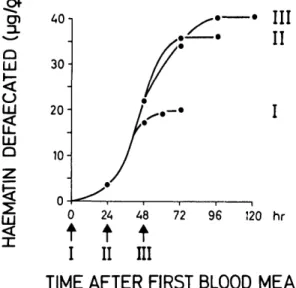
LU Q LU < 40 30- 20-10-III
II
24 48 72 96 120 hrt t t
I II
III
TIME AFTER FIRST BLOOD MEAL
Fig. 1. Haematin defecation by female An. albi-manus fed three blood meals to repletion on a human host at daily intervals (arrows). Each point represents the measurement of one pool of excreta from 10-24 females.three to five times. These females were insemi-nated and were not offered sugar during the ex-periment. To follow the digestion of repeated blood meals, haematin defecation was recorded during 120 h until oviposition had occurred (Fig. 1). The haematin readings could be assigned re-liably to the actual number of blood meals given (I—III). The average volume of blood consumed by the females was estimated to be 3.3 fi\ for the first blood meal, 2.6 /xl for the second meal, and «=0.8 ix\ for the third meal; total blood-meal vol-ume was 6.7 fd (5 cal of blood protein).
To describe the effect of multiple feedings on oocyte maturation, some blood-fed females were dissected to measure the length of the yolk mass in the maturing follicles (and some were sacri-ficed for measuring midgut enzymes) (unpub-lished data). After the first blood meal (n = 306), the females were divided in two groups for re-cording oogenesis. In a group that imbibed only one blood meal (n = 108), 88 females initiated oogenesis, and yolk was deposited into the folli-cles as shown in Fig. 2A. The maximal length of the yolk mass, 420 /xm (100%), was observed at =»40 h in an average of 50 follicles per female. The remaining group of females received a sec-ond blood meal (n = 198). During the next 24-h interval, 37 females were dissected, of which 23 continued to deposit yolk. The remaining 14 fe-males started oogenesis after this second meal and followed the same biphasic line but with a considerably steeper slope that attained the final yolk length within 24 h (Fig. 2B). Thus, matura-tion was accelerated by a factor of 1.7 (24 versus 40 h).
X < LL
o
LUo
a:
LUa.
01 too
L Lo
I
LU 120 hr 0 24 48 72 96 120 hr 24 48 72 96 120 hrTIME AFTER FIRST BLOOD MEAL
Fig. 2. Oogenesis in An. albimanus traced by mea-suring the length of the yolk mass at various intervals after blood meals. Three blood meals were given at daily intervals to 1.5-d-old females from the same hu-man host (arrows). Oocyte measurements for follicles stimulated by the first (A), the second (B), or the third (C) blood meals within the same cohort of females. Each point is the mean (±SE) of two to seven follicles.
Of the group with three blood meals (n = 104), nearly half of the females (n = 48) started to deposit yolk into their penultimate follicles at the normal rate (Fig. 2C). Fig. 3 illustrates an ovary of a female 70-80 h after the first blood meal with a mature follicle, recognized by the chorionic features such as floats. Furthermore, this figure shows penultimate follicles in matu-ration with 325 /Am of yolk (77% of the maxi-mum). In the latter follicles, the chorionic struc-tures were not yet differentiated because only 30-40 h had passed since their stimulation by the third blood meal. The number of mature
Fig. 3. Oogenesis in An. albimanus with three blood meals. Ovary dissected 72 h after the first blood meal (i.e., 48 h after the second and 24 h after the third). Ovariole at far left is still intact, showing a ma-ture primary follicle (floats visible, 405 /im of yolk length, n = 61), and its secondary follicle in the process of maturation (311 fim of yolk length, n = 54). In the secondary follicles (three more are visible), the exo-chorionic structures have not yet differentiated. The germarium is recognized in all of them. The bar on the Nomarski-DIC photograph equals 100 /tun.
oocytes per female was doubled from 76 ± 20 (n = 155) with two blood meals to 153 ± 23 (n = 41) with a third blood meal. The rate of yolk deposition always followed a biphasic pattern: a slower and a faster part with the transition period between yolk lengths of 130-170 fxm (30-40% of the maximum), irrespective of the feeding his-tory.
To further investigate the frequency of multi-ple blood meals, female An. albimanus were ex-posed to the human host at shorter intervals. Even 6, 8, or 12 h after a replete blood meal, all females refed. Even with these shorter intervals, it was still possible to distinguish the most recent blood by transillumination. The bright red color of the recent meal was readily distinguished from the previous blood (characterized by a faint darkening of its color). This distinction was reli-able only when screened within minutes after termination of feeding, because mixing of these blood meals started soon afterward because of an incomplete or weak peritrophic membrane for-mation at this time.
The metabolic fate of multiple blood meals was investigated by offering two blood meals within a 12-h interval. All females obtained the first blood meal at 24-26 h after eclosion and before they had access to either 10% sucrose or water. Afterward, they were kept with access to water for 2 d until the mature oocytes were counted and related to body size (Fig. 4). The control group was treated in the same way with only one blood meal. Even with sugar available,
100- 80-LU AO-L AO-L
cr
LU 0 L LUo
o
o
LU 60-20" 0J 100 80-60 A0- 20-0J 2.8D
3.0 3.2 3.4 3.6 3.0 3.2 3.4 3.6 2.8 3.0 3.2 3.4 3.6WING LENGTH (mm)
Fig. 4. Effect of body size (wing length) and prefeeding conditions on the fecundity of female An. albimanus after their first (A, C) or second (B, D) blood meal. Females were fed sucrose ad lib (A, B) or water (C, D) from eclosion until blood meal. The first blood meal was given 24-26 h after eclosion and the second 12 h later. Mature oocytes were counted by dissecting the ovaries =48 h after the blood meal. The dotted vertical lines indicate the threshold in body size required for oogenesis.
81% of the females remained nonoogenic after their first blood meal; the remaining 19% pro-duced 63 ± 17 eggs per female (n = 10) and had wing lengths ^3.2 mm (Fig. 4A). With the sec-ond blood meal, fecundity did not increase sig-nificantly (69 ± 19 eggs per female; n = 48; t = 0.987, df = 56, P > 0.3), but 66% of these females initiated vitellogenesis, irrespective of their body size (Fig. 4B). In contrast, with preblood-meal access only to water, none of the females entered oogenesis with one blood meal (Fig.
4C). With two blood meals, 33% matured an av-erage of 52 ± 12 eggs per female (n = 11, Fig. 4D), irrespective of body size. When these fe-males were analyzed for their prefeeding lipid reserves, females with access to only water had significantly fewer lipid reserves (0.16 ± 0.03 cal per female; n = 50; t = 7.993, df = 108, P < 0.001) than those with access to sugar (0.23 ± 0.06 cal per female, n = 60).
When individual prefeeding lipid reserves were regressed on wing length (not shown),
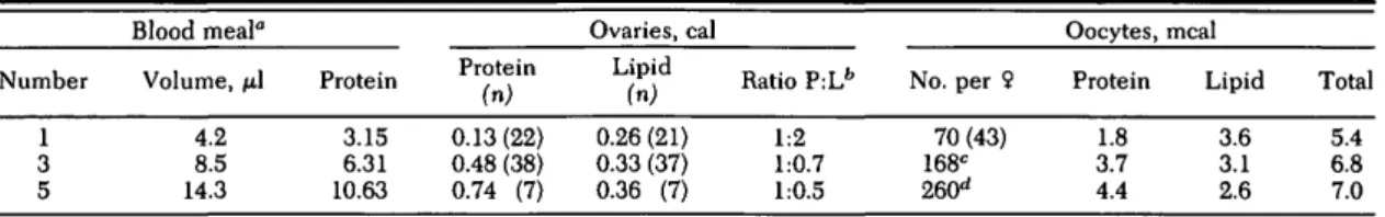
fe-Table 1. Caloric content of An. albimanus fed five consecutive blood meals at 24-h intervals on the same human host Number 1 3 5 Blood meal" Volume, /J.1 4.2 8.5 14.3 Protein 3.15 6.31 10.63 Protein (n) 0.13(22) 0.48 (38) 0.74 (7) Ovaries, cal Lipid (n) 0.26 (21) 0.33 (37) 0.36 (7) Ratio P:Lfc 1:2 1:0.7 1:0.5 No. per 9 70 (43) 168C 260d Oocytes, Protein 1.8 3.7 4.4 meal Lipid 3.6 3.1 2.6 Total 5.4 6.8 7.0 ° Bloodmeal data show protein values accumulated through blood meals 1 to 5 (cal per female).
b
L, lipid; P, protein.
c
Eight pools of 5-30 females each.
d
Two pools of 6 and 34 females.
males with wings longer than 3.2 mm and access to sugar had lipid contents of ^0.27 cal. In con-trast, the lipid content of females with access to water ranged from 0.11 to 0.25 cal. There ap-peared to be a threshold lipid level between 0.25 and 0.27 cal per female, above which oogenesis was initiated with one blood meal. Below that level, however, an additional blood meal was required before oogenesis was started. With this supplementary blood meal, even the smallest fe-males were able to initiate oogenesis (Fig. 4 B and D) and in the larger females, fecundity rose to 100 eggs.
Caloric Allocation and Utilization of Blood-meal Protein in Female An. albimanus. Because
multiple blood meals improved fecundity and their effect appeared to depend on female lipid levels, we carried out a detailed quantitative analysis of the role of additional protein in yolk deposition and maternal metabolism. Over 300 females were fed five blood meals at 24-h inter-vals. The total female protein and lipid was com-pared before the blood meal, before oviposition, and after oviposition. In addition, mature folli-cles were removed, counted, then analyzed for protein and lipids to estimate the caloric content of mature oocytes.
As shown in Table 1, oogenic females pro-duced 70 ± 22 eggs (n = 43) with one blood meal. In the mature ovaries, as well as in single oocytes, the caloric ratio of protein to lipid was 1:2. After three blood meals, fecundity increased to 168 ± 22 (n = 8), and the caloric ratio of ovarian protein to lipid now was 1:0.7. Protein per oocyte doubled (3.7 meal; t = 9.06, df = 63, P < 0.001), whereas lipid per oocyte (3.1 meal) had decreased significantly when compared with oocytes matured with only one blood meal (3.6 meal; t = 4.99, df = 62, P < 0.001).
Data in Table 1 also indicated that fecundity increased with five blood meals, attaining an av-erage of 260 eggs per female. The caloric propor-tion of total yolk protein to lipid was 1:0.5. This shift was caused by a nearly 6-fold increase in yolk protein plus a 1.5-fold increase in yolk lipid in mature ovaries compared with one blood meal. However, protein per single oocyte more than doubled (from 1.8 to 4.4 meal), whereas the lipid per oocyte was reduced by roughly
one-quarter (from 3.6 to 2.6 meal; t = 5.10, df = 26, P < 0.001) (Table 1). Thus, with subsequent blood meals, the total caloric content of mature oocytes (protein plus lipid) increased from 5.4 (first blood meal) to 6.8 (third), and finally to 7.0 meal per oocyte (fifth). Thus, total yolk per oo-cyte was enhanced with each additional blood meal because of considerable flexibility in the ratios of protein and lipids within the maturing oocytes. However, the total yolk mass synthe-sized per female never exceeded 10—13% of the caloric blood protein input, indicating that there were limitations in the efficiency of utilizing the blood meal for vitellogenesis.
To test for alternative metabolic routes in the utilization of host blood protein, we compared female protein and lipid before the blood meal and after oviposition with one and three blood meals (Table 2). After the first blood meal, half of the females matured an average of 46 ± 12 (n = 20) eggs, utilizing 9.7% of bloodmeal protein for the synthesis of yolk protein plus lipid. Oogene-sis was not detected in the remaining females (n = 23). Instead, they showed a net gain of 0.10 cal of protein and 0.41 cal of lipid, comprising 15.9% of the caloric value of the blood meal. With three blood meals, the females matured an average of 78 ± 21 (n = 40) eggs, utilizing 0.45 cal for the synthesis of yolk protein plus lipid (6.8% of the caloric protein input). In addition, they synthesized 0.16 cal of extraovarian protein and 0.29 cal of extraovarian lipid deposits, rep-resenting a net gain of 6.7% in maternal deposits. To demonstrate the priority in synthesis of ma-ternal deposits, sugar-fed female An. albimanus were given enemas that contained protein insuf-ficient for oogenesis, because by this route the mechanism of protein concentration was abol-ished (Briegel & Rezzonico 1985). With 1 fx\ of human blood (0.91 cal protein), none of the fe-males initiated oogenesis but within 48 h, their total body protein had increased significantly
(t = 3.643, df = 34, P < 0.001) from 0.54 to 0.67
cal per female (Table 3), utilizing 14% of the protein input. Total lipid insignificantly in-creased from 0.79 to 0.87 cal per female (t = 0.985, df = 33, P > 0.3), representing 9% of the bloodmeal input. Therefore, a total of 23% of the bloodmeal protein was retained by nonoogenic
Table 2. Gain of protein and lipid reserves in female An. albimanus fed one to three blood meals at 24~h intervals on the same human host
Parameter Preblood meal One blood meal
Ovaries (46 ± 12 eggs) Nonoogenic 9
% Gain per nonoogenic 9a Three blood meals
Ovaries (78 ± 21 eggs) Postoviposition 9 % Gain per 9° Protein (n) 0.55 3.20 0.12 0.65 0.10 6.58 0.18 0.71 0.16 ± 0.08 (37) ± 0.05 (10) ± 0.06 (12) = 17% ± 0.13 (20) ± 0.09 (24) = 28% 0.37 0.19 0.78 0.41 0.27 0.65 0.28 Lipid (n) ± 0.09 (38) ± 0.07 (10) ± 0.23 (11) « 113% ± 0.08 (20) ± 0.18 (32) = 78% Total 0.92 — 0.31 1.43 0.51 0.45 1.36 0.44 % Caloric bloodmeal input 100 9.7 15.9 100 6.8 — 6.7 Values are mean calories per female ± SE.
° Percentage of prebloodmeal values.
females and was used to augment maternal pro-tein and lipid.
With 2-JU.I enemas (1.83 cal protein), a third of the females (n = 5) showed limited oogenesis. The remaining two-thirds (n = 11) now had a total body protein content of 0.84 cal per female (16% of the blood meal input; t = 10.207, df = 39, P < 0.001) and of 1.25 cal per female of lipid (25% of the blood meal input; t = 4.658, df = 28, P < 0.001). Therefore, a total of 41% of the blood-meal protein was retained by the nonoogenic females, improving their content of protein and lipids by 55 and 58%, respectively. Conse-quently, bloodmeal protein was utilized predom-inantly for the synthesis of maternal deposits, and the improvement of fecundity was post-poned.
To resolve the metabolic fate of sequential blood meals, five blood meals were given to fe-males at 24-h intervals and oviposition was re-corded in 12-h intervals during 144 h. Feeding success was consistently >90%. Immediately af-ter the third blood meal, 65% of the individually blood-fed females oviposited 61 ± 13 eggs (n = 97). After blood meal 4, an average of 55 ± 19 eggs (n = 56) was deposited by 67% of the sur-viving females during the next day. After blood
meal 5, another 69 ± 34 eggs (n = 23) were laid by 32% of the fed females within 24 h, and dur-ing the last collectdur-ing interval, 90 ± 27 eggs (n = 48) were laid by 68% of the females. Total fecun-dity amounted to an average of 275 eggs per female (range, 107-480 eggs). Finally, 168 h af-ter the first blood meal, these females had empty ovaries, and the female protein and lipids were 0.81 ± 0.10 cal (n = 35) and 0.82 ± 0.18 cal (n = 33), respectively. The corresponding values for teneral females of the same body size was 0.80 cal of protein and 0.22 cal of lipid. Despite the processing of five blood meals and production of several hundred eggs per female, total lipids had more than tripled (from 0.22 to 0.82 cal).
In conclusion, this experiment indicated that oviposition regularly took place within 48-60 h of a blood meal, each of which appeared to ini-tiate maturation of a new set of follicles. Each "wave" of oviposition comprised between 50 and 100 eggs per female. Consequently, batches of >150 eggs, as observed above and reported earlier (Briegel & Rezzonico 1985), appeared to be an artifact because of oviposition deprivation. Therefore, the double follicles shown in Fig. 3 might be the consequence of the lack of ovipo-sition sites or ovipoovipo-sition stimuli. Furthermore,
Table 3. Gain of protein and lipid reserves in female An. albimanus with blood meals given by enema Parameter Preblood meal Human blood, 1 /xl Nonoogenic 9 % Gain per 9 2/xl Nonoogenic 9 % Gain per 9 Preblood meal Rodent blood, 1 pi Nonoogenic 9 % Gain per 9 Protein (n) 0.54 ± 0.09 (25) 0.91 ± 0.11 (5) 0.67 ± 0.11 (11) 0.13 = 24%fc (= 14%)c 1.83 0.84 ± 0.08 (16) 0.30 = 55%b (= 16%)c 0.53 ± 0.08 (11) 0.79 ± 0.01 (7) 0.81 ± 0.13 (4) 0.27 = 51%b (= 34%)c Lipid (n) 0.79 ± 0.2° (25) 0.87 ± 0.23 (10) 0.08 = 10%b (= 9%)c 1.25 ± 0.15 (5) 0.46 = 58%fc (= 25%c) 0.51 ± 0.09 (10)c 0.66 ± 0.19 (4) 0.15 = 30%b (1) (= 19%)c Total 1.33 1.54 0.21 2.09 0.76 1.04 1.46 0.43 Values are mean calories per female ± SE. Volume and source of blood was varied.
° Gain in percentage of preblood meal female content ().
b
Gain in percentage of blood meal content.
c
The preblood meal lipid content of 0.79 cal was higher than in Table 2 because the females had access to sugar in this experiment.
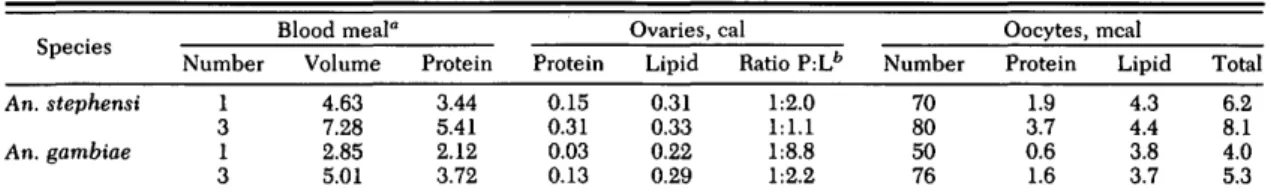
Table 4. Caloric content of An. stephensi and An. gambiae fed three consecutive blood meals at 24-h intervals on the same human host
An. stephensi An. gambiae Number 1 3 1 3 Blood meal" Volume 4.63 7.28 2.85 5.01 Protein 3.44 5.41 2.12 3.72 Protein 0.15 0.31 0.03 0.13 Ovaries, Lipid 0.31 0.33 0.22 0.29 cal Ratio P:Lb 1:2.0 1:1.1 1:8.8 1:2.2 Number 70 80 50 76 Oocytes, Protein 1.9 3.7 0.6 1.6 meal Lipid 4.3 4.4 3.8 3.7 Total 6.2 8.1 4.0 5.3 Values are mean calories or millicalories per female ± SE.
° Bloodmeal data show protein values accumulated through blood meals 1 to 3 (calories per female).
b
L, lipid; P, protein.
protein reserves are acquired with the first few blood meals (Tables 2 and 3) and lipid reserves are conserved at a fairly high level.
Multiple Blood Meals in An. stephensi. To test whether observations on An. albimanus could be generalized to other anophelines, An. stephensi was offered three consecutive blood meals at 24-h intervals. The females refed at these inter-vals, and blood feeding easily could have been continued at this pace. With their first blood meal, 70 ± 16 (n = 44) eggs matured. Before the blood meal, the females were equicaloric in pro-tein (0.64 ±0.11 cal per female, n = 38) and lipid (0.64 ± 0.13 cal per female, n = 39). After depos-iting the first batch of eggs, their protein re-mained the same (0.68 ± 0.08 cal per female, n = 7), but their lipid content was reduced by 40% to 0.38 ±0.11 cal per female (n = 8; t = 5.355, df = 45, P < 0.001). Surprisingly, after three blood meals, fecundity rose to only 80 ± 13 (n = 50) eggs per female, a small but significant increase
(t = 3.342, df = 92, P < 0.01). In these females,
the total ovarian lipid was nearly the same as in females fed only once (0.31 versus 0.33 cal; Ta-ble 4), but the ovarian protein had douTa-bled (0.15 versus 0.31 cal; Table 4). These changes also became evident when comparing the protein and lipid per oocyte. The oocyte lipid was the same whether females were fed one or three blood meals, but oocyte protein had doubled (Table 4). Although multiple blood meals were evident in this species, hardly any new oocytes were stim-ulated to undergo oogenesis. Subsequent blood meals were used to supplement the protein con-tent of oocytes already in maturation. In addi-tion, females with three blood meals showed an increase of their total nonovarian protein content from 0.64 to 0.84 cal per female, representing an increase of 31% compared with prefeeding con-ditions (t = 7.025, df = 63, P < 0.001).
Protein acquisition from the blood meals was accompanied by a significant lipid reduction of 33% from 0.64 to 0.43 cal per female (t = 6.366, df = 63, P < 0.001). Taken together, the total caloric content (protein plus lipid) of each female after metabolizing three blood meals was the same after oviposition as it was before feeding: 1.27 and 1.28 cal, respectively. This might imply some sort of reallocation of reserve components;
i.e., the inevitable loss of 33% of lipids through oogeneses and oviposition was compensated by a net gain of 31% of protein compared with prebloodmeal conditions. Therefore, in An.
stephensi, multiple blood meals appear to
im-prove the yolk and maternal protein from the first blood meal as a possible compensation for the concomitant loss of lipids.
Multiple Blood Meals in An. gambiae. Fe-males were kept on sucrose ad lib from eclosion for —12 h, after which they were fed on a human host. These females (n = 28) digested and ex-creted their blood meal but did not mature any eggs. A similar result was observed when they fed 24 h after eclosion. At the age of 48 h, 40% of the females matured 46 ± 19 eggs (n = 7) and at 72 h, this percentage rose to 73% with 47 ± 15 eggs (n = 13) per female. Females that had fed but failed to initiate oogenesis had synthesized maternal, extraovarian deposits of protein and lipid to the extent of 7 and 13-17% of the caloric bloodmeal input, respectively. In this way the prefeeding protein content was markedly im-proved. Structural changes in preresting stage oocytes were not followed. Although biting, feeding, and digestion were possible at 12 h, female metabolism was not competent yet to al-low oogenesis. Synthesis of yolk components de-veloped only after 24 h of imaginal life, indicat-ing once more the priority of reserve acquisition over vitellogenesis in teneral females.
All female An. gambiae avidly refed when of-fered the same host within 12 or 24 h. At 6 h after a blood meal, the females were reluctant to re-feed, and of the 10% that did, the second blood meal could not be recognized reliably by our transillumination test because the first blood meal was still bright red in color. With three blood meals, fecundity rose to 76 ± 21 eggs per female (n = 75, t = 6.588, df = 115, P < 0.001), which is 50% more than with the first blood meal (50 ± 17 eggs, n = 42). The dominance of lipid over protein in the yolk resulting from one blood meal was reduced with three blood meals (Table 4). This change occurred exclusively on the level of yolk protein, because yolk lipid remained con-stant at 3.7-3.8 meal per oocyte. With three blood meals, female metabolism had increased extra-ovarian protein values by 25%. Concomitantly,
however, lipids were reduced by 24%, indicating a redistribution of maternal reserves when en-hancing fecundity through repetitive blood meals, similar to An. stephensi.
Discussion
Three Anopheles vectors of malaria took mul-tiple blood meals on a human host, irrespective of their nutritional or gonotrophic status. In our experiments, where host-seeking may have been bypassed by close contact with the host, three to five blood meals were taken readily at intervals of 6, 12, or 24 h. This result might indicate the absence of neural as well as hormonal biting inhibitions at least from 6 h onward, contrary to sugar-fed Ae. aegypti (Klowden & Lea 1979b). Rapid refeeding led us to postulate a strategy of blood meal acquisition and utilization in anophelines different from that known for the culicines, which are characterized by the evolu-tion of clear-cut gonotrophic cycles. In general, the neuro-endocrine system (Clements 1963; Meola & Lea 1972; Lea 1975; Klowden & Lea 1979a, b) enforces gonotrophic concordance. In contrast, anophelines having fed "to repletion" avidly refeed when exposed to the same host at almost any intervals. Recently, the validity of gonotrophic concordance was questioned by Ed-man et al. (1992), even in Ae. aegypti in view of their blood-feeding pattern. In Anopheles, mul-tiple feeding is facilitated by an efficient mech-anism of blood protein concentration during feeding (Briegel & Rezzonico 1985) which is fol-lowed later by diuresis. The first blood meal was used primarily for the synthesis of maternal re-serves, whereas sequential blood meals were uti-lized for improving fecundity by synthesizing additional yolk protein and yolk lipid. Thus, full fecundity in An. albimanus and An. gambiae was reached only through continued yolk production by repeated blood feeding.
Initiation of yolk synthesis after the first blood meal depended on female body size. Teneral anophelines were characterized by limited re-serves per unit body size (Briegel 1990a, b). In
An. albimanus, females with wing lengths of
<3.2 mm did not start oogenesis with one blood meal but required additional protein input. Ten-eral An. albimanus females with wing lengths of 3.2 mm contained 0.25 cal of lipids. (Briegel 1990b). All the smaller females with <0.25 cal of lipids required a second blood meal for the stim-ulation of oogenesis. This critical level is crucial for the initiation of oogenesis following a blood meal, confirming the threshold size described previously (Briegel 1990b).
Hocking & Maclnnes (1948), working on An.
gambiae and An. funestus, reported that
"fe-males . . . sometimes feed daily"; this was re-lated to frequent flights of females of various gonoactive status in and out of huts to rest
in-doors during daytime (Gillies & De Meillon 1968). Gillies (1955) already found the "pre-gravid phase of ovarian development" in An.
fu-nestus to require multiple feedings in the field.
All these behavioral adaptations of anophelines appear to compensate the low lipid contents per female. In the case of sufficient lipid reserves through carbohydrate feeding and a large enough blood meal (>0.5 cal of protein), vitello-genesis is possible for a batch of 40—50 eggs. But with an opportunity to feed on a host, additional blood meals readily are ingested to improve both reserves and fecundity. Therefore, the complex and refined endocrine regulations of gonotrophic concordance would be a hindrance. In Ae.
ae-gypti, there also was a minimal energetic status
required for oogenesis (Briegel 1985). Unless a minimal energy requirement of 1 cal per female was attained by preblood meal caloric content plus caloric protein input from blood feeding, oogenesis was not initiated (H. B., unpublished data).
In recent field studies, Edman et al. (1992) and Scott et al. (1993) reported that multiple blood meals occurred regularly in Ae. aegypti under natural conditions in preference to sugar meals. Based on earlier results (Briegel 1990a), and not having access to thorough investigations on the various physiological parameters, we believe that this multiple blood feeding represents an-other mechanism contrasting with that described herein for the anophelines. In Ae. aegypti, the subsequent meals appear to improve (in a nutri-tive sense) a gonotrophic cycle already under-way along the line reported by Lea et al. (1978), rather than to stimulate additional follicles for maturation, as in Anopheles. The less efficient lipogenic potential of Anopheles indeed sharply contrasts with Aedes or Culex (Van Handel 1965, 1984; Mitchell & Briegel 1989; Briegel 1990a).
In Ae. aegypti, all reproductive processes typ-ically ensue on a higher level in absolute caloric terms, which in turn may require strict regula-tion to prevent females from temporarily over-loading while feeding on protein from a host. In contrast to the energetic dumping characterizing culicine reproduction, anophelines appear to op-erate permanently at their energetic minimum, which leads to behavioral and nutritional con-straints for the acquisition of additional protein as provided by multiple blood meals.
In spite of all these adaptations, the compara-tively low efficiency of the utilization of these blood meals remains an enigma. Even after con-secutive blood meals with a 3-fold increase in fecundity, there was always an excretory nitro-gen loss of up to 75% of the input. Further ex-periments are required to relate the oogenic ef-ficiencies to the enormous protein catabolism.
The dynamics of yolk deposition was interest-ing to follow for two reasons. First, it was bipha-sic under all circumstances. There was a slower
rate of yolk deposition until 30—40% of the max-imal yolk length was reached, followed by a clearly steeper and rapid segment. Rates in-creased by =S550%, and durations were about half the first segment. Similar biphasic yolk curves have been observed for various species (H. B., unpublished data). More interesting, however, was the absolute time requirement for the matu-ration of the oocytes. In An. albimanus after the first blood meal, maximal yolk length was reached within 40 h. With a second blood meal, additional follicles started to deposit yolk, but these reached maturity within half the time. With a third blood meal, still another set of oocytes entered vitellogenesis, again requiring «=40 h to reach maximal yolk length. Conse-quently, when proper ovipositional stimuli were provided, almost continuous oviposition was ob-served.
Depending on their dietary history, a high flexibility in the yolk composition of Anopheles eggs was observed. In An. albimanus given one, three, or five blood meals, yolk protein increased by a factor of nearly 2.5 (1.8, 3.9, 4.4 meal, re-spectively), whereas yolk lipids decreased by one-third (3.6, 3.1, 2.6 meal, respectively). In An.
gambiae and An. stephensi, oocyte values were
similar. With three blood meals, their protein per oocyte increased ==2.5 times, whereas the lipid per oocyte of these species was not affected by the additional protein input. As far as yolk lipids are concerned, An. gambiae and An. stephensi were different from An. albimanus. This also was true for the number of maturing oocytes, because in the former two species this figure remained almost constant despite additional blood meals, but in An. albimanus it clearly had an enhancing effect. Under such complex metabolic condi-tions, recording the frequencies of blood meals and counting eggs may be misleading for rec-ognizing gonotrophic cycles. It appears that gonotrophic cycles are doubtful, for certain
Anopheles at least, as clearly stated by
Garrett-Jones (1970). Accordingly, we are left with the impression that gonotrophic discordance pre-vails in many anopheline species. Thus in
Anopheles, mixed meals do not necessarily
rep-resent interrupted feedings. Instead, they may reflect a well-adapted strategy, primarily to ame-liorate female reserves and improve the fecun-dity from subsequent blood meals. In contrast, in the culicines, mixed blood meals are thought to indicate interrupted feedings, because host-seeking behavior was found clearly to operate under narrowly defined conditions (Klowden 1990). In this context, the infrequent double feedings reported for culicines are exceptional, whereas multiple feedings appear to be adaptive for anophelines. Although gathered under trolled or manipulated (or both) laboratory con-ditions, our results demonstrate the physiologi-cal and metabolic potential of the species
investigated and they may elucidate the concept of gonotrophic cycles in anopheline vector spe-cies.
Note Added in Proof
To refute the argument of multiple blood meals as an artifact of forced-feeding under laboratory conditions we have carried out the following experiment. An. albimanus (n = 197) were bloodfed and exposed 12 h later to one of us at night while sleeping under a bednet of =2 m3 volume. After a 2-h exposure the females were collected and analyzed in comparison to controls (n = 71) having had only one blood meal: 81% of the females refed a second time and significant increases (P < 0.001) were observed for mean hematin excretion (+ 44%) and fecun-dity (+ 32%).
Acknowledgments
We thank S. Zaba and I. Fliickiger for dependable rearing of the mosquito colonies, and R. Haigis for excellent technical assistance. We appreciate the crit-ical reading of the manuscript and the help given by R. Graf (all from the University of Zurich). Substantial improvements suggested by referees and the editor are kindly acknowledged. Financial support was received from the Swiss National Science Foundation.
References Cited
Bang, F. B. & W. C. Reeves. 1942. Mosquitoes and encephalitis in the Yakima Valley, Washington. HI. Feeding habits of Culex tarsalis Coq., a mosquito host of the viruses of Western equine and St. Louis encephalitis. J. Infect. Dis. 70: 273-274.
Boreham, P.F.L. & C. Garrett-Jones. 1973. Preva-lence of mixed blood meals and double feeding in a malaria vector (Anopheles sacharovi Favre). Bull. W.H.O. 48: 605-614.
Briegel, H. 1980. Determination of uric acid and he-matin in a single sample of excreta from blood-fed insects. Experientia 36: 1428.
1985. Mosquito reproduction: incomplete utiliza-tion of the blood meal protein for oogenesis. J. In-sect Physiol. 31: 15-21.
1986. Protein catabolism and nitrogen partitioning during oogenesis in the mosquito Aedes aegypti. J. Insect Physiol. 32: 455-462.
1990a. Metabolic relationship between female body size, reserves, and fecundity of Aedes aegypti. J. Insect Physiol. 36: 165-172.
1990b. Fecundity, metabolism, and body size in Anopheles (Diptera: Culicidae), vectors of malaria. J. Med. Entomol. 27: 839-850.
Briegel, H. & L. Rezzonico. 1985. Concentration of host blood protein during feeding by anopheline mosquitoes (Diptera: Culicidae). J. Med. Entomol. 22: 612-618.
Briegel, H., A. O. Lea & M. J. Klowden. 1979. He-moglobinometry as a method for measuring blood meal sizes in mosquitoes. J. Med. Entomol. 15: 235-239.
Burkot, T. R., P. M. Graves, R. Paru & M. Lagog. 1988. Mixed blood feeding by the malaria vectors in the Anopheles punctulatus complex (Diptera: Culicidae). J. Med. Entomol. 25: 205-213. Clements, A. N. 1963. The physiology of
mosqui-toes. Pergamon, London.
Davidson, G. 1954. Estimation of the survival-rate of anopheline mosquitoes in nature. Nature 174: 792-793.
De Buck, A. & N. H. Swellengrebel. 1934. Behav-iour of Dutch Anopheles atroparvus and messeae in winter under artificial conditions. Riv. Malariol. 13: 404-416 (in Clements 1963).
Edman, J. D. & A.E.R. Downe. 1964. Host-blood sources and multiple-feeding habits of mosquitoes in Kansas. Mosq. News 24: 154-160.
Edman, J. D., D. Strickman, P. Kittayapong & T. W. Scott. 1992. Female Aedes aegypti (Diptera: Culicidae) in Thailand rarely feed on sugar. J. Med. Entomol. 29: 1035-1038.
Else, J. G. & C. L. Judson. 1972. Enforced egg-retention and its effects on vitellogenesis in the mosquito Aedes aegypti. J. Med. Entomol. 9: 527-530.
Garrett-Jones, C. 1970. Problems of epidemiologi-cal entomology as applied to malariology, pp. 168-180. In Miscellaneous Publications 7, Entomologi-cal Society of America, College Park, MD. Gillies, M. T. 1954. The recognition of age-groups
within populations of Anopheles gambiae by the pre-gravid rate and the sporozoite rate. Ann. Trop. Med. Parasitol. 48: 58-74.
1955. The pre-gravid phase of ovarian development in Anopheles funestus. Ann. Trop. Med. Parasitol. 49: 320-325.
Gillies, M. T. & B. De Meillon. 1968. The Anophelinae of Africa South of the Sahara (Ethio-pian zoogeographical region). Publ. South African Inst. Med. Res. 54, 343 pp. Johannesburg. Gwadz, R. W. 1969. Regulation of blood meal size
in the mosquito. J. Insect Physiol. 15: 2039-2044. Hocking, K. S. & D. G. Maclnnes. 1948. Notes on
the bionomics of Anopheles gambiae and A. funes-tus in East Africa. Bull. Entomol. Res. 39: 453-465. Klowden, M. J. 1990. The endogenous regulation of mosquito reproductive behavior. Experientia 46: 660-670.
Klowden, M. J. & A. O. Lea. 1979a. Humoral inhi-bition of host-seeking in Aedes aegypti during oocyte maturation. J. Insect Physiol. 25: 231-235. 1979b. Abdominal distention terminates
subse-quent host-seeking behaviour of Aedes aegypti fol-lowing a blood meal. J. Insect Physiol. 25: 583-585. Lea, A. O. 1975. The control of reproduction by a blood meal: the mosquito as model for vector endo-crinology. Acta Trop. 32: 112-115.
Lea, A. O., H. Briegel & H. M. Lea. 1978. Arrest, resorption, or maturation of oocytes in Aedes ae-gypti: dependence on the quantity of blood and the interval between blood meals. Physiol. Entomol. 3: 309-316.
Magnarelli, L. A. 1977. Physiological age of mos-quitoes (Diptera: Culicidae) and observations on
partial blood-feeding. J. Med. Entomol. 13: 445-450.
Meola, R. & A. O. Lea. 1972. Humoral inhibition of egg development in mosquitoes. J. Med. Entomol. 9: 99-103.
Minari, O. & D. B. Zilversmit. 1963. Use of KCN for stabilization of color in direct Nesslerization of Kjeldahl digests. Anal. Biochem. 6: 320-327. Mitchell, C. J. & H. Briegel. 1989. Inability of
dia-pausing Culex pipiens (Diptera: Culicidae) to use blood for producing lipid reserves for overwinter survival. J. Med. Entomol. 26: 318-326.
Reisen, W. K. & M. Aslamkhan. 1976. Observations on the swarming and mating behaviour of Anophe-les culicifacies GiAnophe-les in nature. Bull. W.H.O. 54: 155-158.
Reisen, W. K., F. Mahmood, S. Niaz, K. Azra, T. Parveen, R. Mukhtar, Y. Aslam & T. F. Siddiqui. 1986. Population dynamics of some Pakistan mos-quitoes: temporal changes in reproductive status, age structure and survivorship of Anopheles culici-facies, An. stephensi and Culex taeniorhynchus.
Ann. Trop. Med. Parasitol. 80: 77-95.
Roubaud, E. 1929. Cycle autogene d'attente et ge-nerations hivernales suractives inapparentes chez le moustique commun, Culex pipiens. C. R. Acad. Sci. (Paris) 188: 735-738.
Scott, T. W., G. G. Clark, L. H. Lorenz, P. H. Amera-singhe, P. Reiter & J. D. Edman. 1993. Detec-tion of multiple blood feeding in Aedes aegypti (Diptera: Culicidae) during a single gonotrophic cy-cle using a histologic technique. J. Med. Entomol. 30: 94-99.
Senior White, R. A. 1952. Studies on the bionomics of Anopheles aquasalis Curry 1932. Ind. J. Malariol. 6: 29-72.
Smith, A. & B. Weitz. 1959. The feeding habits of Anopheles gambiae, with particular reference to subsidiary hosts. Ann. Trop. Med. Parasitol. 53: 414-415.
Swellengrebel, N. H. 1929. La dissociation des fonctions sexuelles et nutritives (dissociation gonotrophique) o\'Anopheles maculipennis comme cause du paludisme dans les pays-bas et ses rap-ports avec 'Tinfection domicilaire". Ann. Inst. Past. (Paris). 43: 1370-1389 (in Clements 1963). Trpis, M. & W. Hausermann. 1986. Dispersal and
other population parameters of Aedes aegypti in an African village and their possible significance in epidemiology of vector-borne diseases. Am. J. Trop. Med. Hyg. 35: 1263-1279.
Van Handel, E. 1965. The obese mosquito. J. Phys-iol. 181: 478-486.
1984. Metabolism of nutrients in the adult mos-quito. Mosq. News 44: 573-579.
1985a. Rapid determination of glycogen and sugars in mosquitoes. J. Am. Mosq. Control Assoc. 1: 299-301.
1985b. Rapid determination of total lipids in mos-quitoes. J. Am. Mosq. Control Assoc. 1: 302-304.
Received for publication 11 November 1992; ac-cepted 10 May 1993.