Glycomimicry: display of fucosylation on the lipo-oligosaccharide
of recombinant Escherichia coli K12
Elif Yavuz&Carola Maffioli&Karin Ilg&Markus Aebi& Bernard Priem
Received: 9 November 2010 / Revised: 6 December 2010 / Accepted: 21 December 2010 / Published online: 1 February 2011 # Springer Science+Business Media, LLC 2011
Abstract We recently described the design of Escherichia coli K12 and Salmonella enterica sv Typhimurium to display the gangliomannoside 3 (GM3) antigen on the cell surface [1]. We report here the fucosylation of modified lipooligosaccharide in a recombinant E.coli strain with a truncated lipid A core due to deletion of the core glycosyltransferases genes waaO and waaB. This trunca-ted structure was used as a scaffold to assemble the Lewis Y motif by consequent action of the heterologously expressed β-1,4 galactosyltransferase LgtE (Neisseria gonorrheae), the β-1,3 N-acetylglucosaminyltransferase LgtA and the β-1,3 galactosyltransferase LgtB from Neisseria meningitidis, as well as the α-1,2 and α-1,3 fucosyltransferases FutC and FutA from Helicobacter pylori. We show the display of the Lewis Y structure by immunological and chemical analysis.
Keywords Bacterial glycosylation . Glycoengineering . Fucosylation . Lewis Y. Chimeric LOS
Abbreviations
LPS Lipopolysaccharide LOS Lipooligosaccharide
Introduction
Carbohydrates are common molecular tools to mediate interactions between cells. Eukaryotes as well as bacteria and archaea synthesise numerous oligo- and polysaccha-ride structures, many of which are located on the cell surface. In bacteria, surface carbohydrates are important structural motives involved in many biological processes including virulence, phage protection, and host immunity. The main classes of carbohydrate-containing extracellular compounds are capsular polysaccharides, lipo-oligo/poly-saccharides, and to a certain extent, outer membrane glycoproteins [2, 3]. The unravelling of the respective biosynthetic pathways has opened new strategies to engineer bacterial surfaces. One aspect of this field of research is the engineering of non-pathogenic bacterial strains to display pathogen-related motifs. These newly generated strains can then be used to initiate and study an immune response against carbohydrate structures without any additional harmful effects of the pathogenic strain. Additionally, bacteria can be engineered to express and display carbohydrate motives of a mammalian host for subsequent use in pathogen neutralisation after infection of the host [4, 5]. The engineering of heterologous carbohydrate structures on bacterial cell surfaces in vivo requires several steps: The provision of a donor substrate for the glycosyltransferase(s), the availability of an acceptor structure and the possibility to export the in vivo created foreign carbohydrate structure to the cell surface. The last aspect often prevents the display of engineered oligosaccharides on the cell surface. For example, although the biosynthesis of the lipo-oligosaccharide core of Escherichia coli is well known, the details of the exportation pathway from the cytoplasm to the outer-membrane are not completely elucidated. Recently, it
E. Yavuz
:
B. Priem (*) CERMAV-CNRS, Grenoble, FranceE. Yavuz
:
C. Maffioli:
K. Ilg:
M. Aebi (*) Institute of Microbiology, ETH,Zurich, Switzerland
e-mail: aebi@micro.biol.ethz.ch B. Priem
could be demonstrated that LPS transport involves the periplasmic protein LptA, the cytosolic protein LptB and the inner membrane proteins LptC, LptF and LptG, which constitute an ABC transporter with accessory proteins [6]. The nascent LPS molecule is then targeted to the outer leaflet of the outer membrane by the outer membrane proteins LptD/LptE [7–9].
To create an acceptor substrate for heterologously expressed glycosyltransferases, bacteria with a truncated LOS structure can be obtained by deletion of genes encoding glycosyltransferases involved in the first steps of the lipid A core synthesis. The heterologous expres-sion of glycosyltransferases able to act on such truncated LOS of Escherichia coli R1 allowed surface display of globotriaose (Gb3), lac-N-neotetraose (LnNT), and gan-gliosides GM1 and GM3 [4]. The resulting strains were used as neutralizing bio-agents of the Shiga toxin (Stx) and enterotoxic E. coli [5]. Another possibility is to build the heterologous carbohydrate structure on the lipid linker undecaprenylphosphate, with a subsequent transfer of this structure to a full lipid A core by the O-antigen ligase WaaL. For example, the overexpression of Haemophilus influenzae genes encoding glycosyltransferases in E. coli resulted in the endogenous synthesis of a recombinant LOS carrying Haemophilus LPS motifs [10]. Altogether, these data show that the lipid A export system of E. coli has a certain flexibility towards its substrate and is able to transport foreign structures to the outer membrane. However, none of the chimeric LOS structures reported above contain fucose, an important monosaccharide involved in cell recognition. On the host side, fucose forms part of a target for a number of pathogens such as Pseudomonas aeruginosa, or Campylobacter jeujuni [11,
12]. On the bacterial side, fucose is for example present on the gastric pathogen Helicobacter pylori.
Our recent work focused on the glyco-engineering of E. coli to produce recombinant oligosaccharides [11]. The technology is based on the internalisation ofβ-galactosides such as lactose followed by endogenous glycosylation when glycosyltransferases from other bacteria were expressed recombinantly. With this system, we successively achieved the synthesis of fucosylated oligosaccharides including the human Lewis X and H antigens [13–16]. Additionally, the co-expression ofα1,2- and α1,3- fucosyl-transferases of H. pylori led to the production of oligosac-charides carrying the Lewis Y antigen [17], which is a structural motif of the lipooligosaccharide of H. pylori [18], and a tumor-associated antigen in humans [19].
In the present report, we addressed the fucosylation of the LOS of Escherichia coli K12 withα1,2- and α1,3- fucosyl-transferases of H. pylori. A double mutant over-expressing a β-galactosyltransferase of Neisseria gonorrhoea was designed in order to exhibit a terminal lactosyl motif at the distal moiety
of lipid A. The recombinant enzymes previously used in our lab to produce fucosylated oligosaccharides from lactose were over-expressed in that strain in order to glycosylate the lactosyl-lipidA core. Chemical analyses and immunological assays were performed and attested for synthesis and exportation of the Lewis Y epitope Fucα-2Galβ-4[Fucα-3] GlcNAcβ-3Gal (Fuc = fucose, Gal = galactose, GlcNAc = N-acetylglucosamine) amongst other glycoforms.
Materials and methods
Bacterial strains and growth conditions
A summary of bacterial strains used in this study can be found in Table 1. Bacteria were grown in Luria-Bertani (LB) medium (10 g/l Bacto tryptone, 5 g/l Bacto yeast extracts, 5 g/l NaCl). LB agar plates were supplemented with 1.5% (w/v) agar. Antibiotics were used at the following final concentrations: Tetracyclin 10μg/ml, ampicillin 100 μg/ml, kanamycin 50μg/ml, chloramphenicol 25 μg/ml.
Construction of the E. coli K-12ΔwcaJΔwaaOΔwaaB strain LPS1
To inactivate wcaJ, primers (a+b) (Table1) were used for PCR amplification of 0.64 kb of DNA flanking the 5′ end of wcaJ from JM107, whereas primers (c+d) were used for PCR amplification of 1.27 kb of DNA flanking the 3′ end of wcaJ. The reverse sequence of primer (c) was contained in primer (b), allowing a fusion of both DNA fragments by PCR amplification with primers (a+d). Fused DNA of 1.91 kb containing a truncated wcaJ was cloned into BamHI site of the suicide plasmid pKO3. The resulting recombinant suicide plasmid pKO3-J containing a trun-cated wcaJ sequence and flanking DNA to guaranty recombination was transformed into TA1 cells, mutants were obtained according to author’s instructions [20]. Positive clones were screened by PCR with primers (a) and (c). The inactivation of waaO and waaB is described elsewhere [1].
Construction of plasmids pSUlgtA and pSU-lgtAfutC
Plasmid pLNT1T [16] was digested with NdeI to remove lgtB, thus ligated, providing pBBR-lgtA. LgtA was then obtained by KpnI and XbaI digestion for the cloning into pSU2718 to give pSU-lgtA. FutC was obtained by KpnI and SalI digestion of plasmid pEXT20futC [14] and sub-cloned into pBluescript KS, thus giving pBS-futC. This plasmid was then digested with XbaI to obtain futC flanked by two XbaI sites, allowing a cloning into XbaI site of pSU-A to give pSU-lgtAfutC.
Cell-based ELISA
Bacterial cultures that had been induced overnight with 0.6 mM IPTG were equalized to 2 OD600/ml with PBS.
Flat-bottom ELISA plates were coated with 50 μL of cells and fixed by addition of 20μL of formaldehyde for 30 min at room temperature. Afterwards, the formalde-hyde was removed and unspecific binding was blocked by addition of 1% (w/v) BSA in PBS for 1 h at room temperature. The blocking agent was then removed and the cells were incubated for 1 h at 4°C with anti-Lewis Y antibody (Abcam) in 0.1% (w/v) BSA in PBS. After four washing steps with 0.05% Tween-20/PBS, the bound anti-Lewis-Y antibody was detected by incubation of the cells with an anti-mouse-IgM-HRP conjugate (Santa Cruz) for 45 min at room temperature. Unbound HRP-conjugate was washed away with 0.05% Tween-20/PBS in four washing steps before the cells were resuspended in 200μl ABTS solution (2,2′-azino-bis(3-ethylbenzthia-zoline-6-sulphonic acid, final concentration 1 mM in 70 mM phosphocitrate buffer pH 4.2 with addition of 1% H2O2). Vmax was determined by recording the color
development at 405 nm for 5 min in a SpectraMaxPlus (Molecular Devices).
Electrophoretic analysis of chimeric LOS by SDS-PAGE and immunological detection
The equivalent of 1 OD600 of exponentially growing
induced cultures with 0.6 mM IPTG was pelleted and resuspended in 500μl 0.065 M Tris–HCl pH 6.8, 2% SDS (w/v), 5% β- Mercaptoethanol (v/v), 10% Glycerin (v/v), 0.05% Bromophenol blue (w/v) and lysed for 5 min at 95°C. After cooling to room temperature, proteins were digested by addition of proteinase K (Roche, final concen-tration 0.4 mg/ml) for 1 h at 60°C. Equal volumes of each sample were then separated on a 17% Tris-Tricine-PAGE and stained with silver as described [26].
To detect LewisY structures in the chimeric LOS after gel electrophoresis, a monoclonal anti-LewisY antibody (Abcam) was used. Bound anti-LewisY was visualised with a goat-anti-mouse-IgM-HRP conjugate (Santa Cruz) and ECL (Amersham) as recommended by the manufacturer.
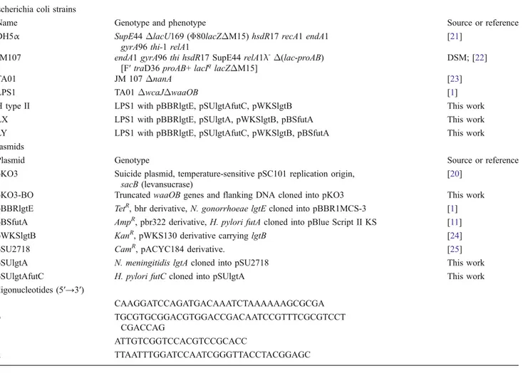
Table 1 Bacterial strains, plasmids and oligonucleotides used in this study Escherichia coli strains
Name Genotype and phenotype Source or reference
DH5α SupE44ΔlacU169 (Φ80lacZΔM15) hsdR17 recA1 endA1
gyrA96 thi-1 relA1
[21] JM107 endA1 gyrA96 thi hsdR17 SupE44 relA1λ-Δ(lac-proAB)
[F′ traD36 proAB+ lacIqlacZΔM15]
DSM; [22]
TA01 JM 107ΔnanA [23]
LPS1 TA01ΔwcaJΔwaaOB [1]
H type II LPS1 with pBBRlgtE, pSUlgtAfutC, pWKSlgtB This work
LX LPS1 with pBBRlgtE, pSUlgtA, pWKSlgtB, pBSfutA This work
LY LPS1 with pBBRlgtE, pSUlgtAfutC, pWKSlgtB, pBSfutA This work
Plasmids
Plasmid Genotype Source or reference
pKO3 Suicide plasmid, temperature-sensitive pSC101 replication origin, sacB (levansucrase)
[20]
pKO3-BO Truncated waaOB genes and flanking DNA cloned into pKO3 This work
pBBRlgtE TetR, bhr derivative, N. gonorrhoeae lgtE cloned into pBBR1MCS-3 [1] pBSfutA AmpR, pbr322 derivative, H. pylori futA cloned into pBlue Script II KS [11]
pWKSlgtB KanR, pWKS130 derivative carrying lgtB [24]
pSU2718 CamR, pACYC184 derivative. [25]
pSUlgtA N. meningitidis lgtA cloned into pSU2718 This work
pSUlgtAfutC H. pylori futC cloned into pSUlgtA This work
Oligonucleotides (5′→3′) a CAAGGATCCAGATGACAAATCTAAAAAAGCGCGA b TGCGTGCGGACGTGGACCGACAATCCGTTTCGCGTCCT CGACCAG c ATTGTCGGTCCACGTCCGCACC d TTAATTTGGATCCAATCGGGTTACCTACGGAGC
Isolation of lipooligosaccharides
Isolation of LOS was carried out by phenol/chloroform/ petrol ether as reported [27], except that it was recovered from the phenol phase by precipitation with 6 volumes of diethyl ether/acetone (1:5 v/v) as described [28].
Preparation of de-O-acylated LPS
De-O-acylation was obtained by conventional mild hydrazinolysis except that hydrazine-hydrate was used instead of anhydrous hydrazine. The solution (20 mg LOS/ml) was stirred at 37°C for 2 h. De-O-acylated LOS was recovered from the hydrazine phase by precipitation with 15 volumes of ice-cold acetone. The pellet was recovered by centrifugation, washed with acetone and air-dried.
Mass spectrometry analysis
MALDI-MS spectra of the sample were obtained with an Autoflex Bruker MALDI-TOF spectrometer. DHB (2, 5-Dihydroxybenzoic acid, SIGMA) was used as a matrix. The De-O-acylated LOS sample was dissolved in water at a concentration of 0.4 mg/mL, DHB at 50 mg/ml in methanol. One μl of each solution was loaded and mixed directly on the target by the dried droplet method. The analysis was performed in the linear negative ion mode with an accelerating voltage of 20 kV and an extraction delay of 90-80 nsec. Spectra were calibrated externally using a peptides mix standard from Bruker Daltonics.
Methylation linkage analysis
The LOSs (about 70 mg) were degraded with 0.1 M sodium acetate buffer, pH 4.2, at 100°C for 4 h to cleave the acid-labile ketosidic linkage between the core OS and lipid A. The water-insoluble lipid A was removed by centrifugation at 20,000 g for 20 min. The oligosaccharide fraction was precipitated from the soluble phase by adding 4 vol of−20°C acetone [29]. Samples were centrifuged at 13,000 g at 4°C for 20 min, and the salt-containing 80% acetone supernatant was discarded. The pellet was dried, solubilised in dime-thylsulfoxide, and methylated with methyl iodine [30]. Permethylated oligosaccharides were hydrolysed in 3 M trifluoroacetic acid for 3 h at 100°C. Methylated sugars were analysed after reduction using NaBD4 and acetylation with
an equal mixture of acetic anhydride and pyridine. Partially methylated alditol acetates (PMAA) were analysed by GC-MS using an agilent chromatograph 6850 equipped with a 5975C MS detector. The neutral sugars and the glucos-amine derivatives were separated on a SP2380 column and a HP5-MS column, respectively.
Results
Engineered E. coli overexpressing fucosyltransferase-genes from H. pylori produced the Lewis Y epitope and presented it on the cell surface
In our previous work, a mutant of E. coli K12 synthesizing a glucose-terminal truncated lipid A core was prepared by inactivation of glycosyltransferases genes waaO and waaB [1]. To construct strains displaying fucose-containing structures, we additionally deleted wcaJ involved in the assembly of colanic acid synthesis, thus providing GDP-fucose availability under over-expression of the positive regulator of the CPS operon (capsular polysaccharide) named Rcsa [16]. The resulting strain was termed LPS1. Five glycosyltransferase genes necessary for the display of the Lewis Y antigen from different bacterial sources were expressed heterologously: lgtE, encoding a β1,4-galacto-syltransferase of Neisseria gonorrhoea reported to transfer to glucose in the LOS structure of N. gonorrhoea [31]; lgtA from Neisseria meningitidis encoding a β1,3-N-acetylglu-cosaminyltranferase with galactose as an acceptor, and lgtB, encoding a β1,3-galactosyltranferase with N-acetylglucos-amine as an acceptor, also from N. meningitidis [32]. In addition, futA and futC, encoding α1,3- and
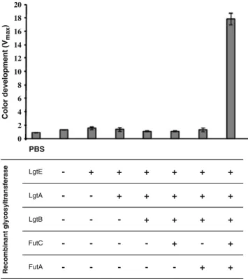
α1,2-FutA FutC LgtB LgtA LgtE -+ + + + + -+ + + + + + + + + + + -+ -+ + -Recombinant glycosyltransferase PBS Color development (V max ) 20 18 16 14 12 10 8 6 4 2 0
Fig. 1 Cell based ELISA for the quantification of Lewis Y display on LeY cells using a monoclonal anti-Lewis Y antibody. From left to right: 1, no cells; 2, LPS1 cells; 3, LPS1 + pBBRlgtE; 4, LPS1 + pBBRlgtE + pSUlgtA; 5, LPS1 + pBBRlgtE + pSUlgtA + pWKSlgtB; 6, H type II cells; 7, LX cells; 8, LY cells
fucosyltransferases of Helicobacter pylori, respectively, were expressed [14–16]. The transformation of the E. coli mutant strain LPS1 with compatible plasmids carrying the five glycosyltransferase genes should result in the produc-tion of a modified LPS that terminates in the Lewis Y motif Fucα-2Galβ-4[Fucα-3]GlcNAcβ-3Gal. To test this hy-pothesis, a cell-based enzyme immunosorbent assay was carried out with a Lewis Y specific monoclonal antibody. Microtiter plate wells were coated with E. coli cells LPS1, H type II, LX and LY as listed in Table 1, expressing intermediate glycan structures between the truncated LOS and the Lewis Y motif. Only the LY cells expressing all necessary enzymes for Lewis Y synthesis led to a significant signal (Fig.1). These data indicated the display
of the Lewis Y motif at the surface of the recombinant strain LY.
In order to visualize glycosylation of the truncated LOS acceptor by apparent molecular weight increase of the LOS in gel electrophoresis, a time-course experiment was carried out with the recombinant strain expressing the glycosyl-transferases necessary for display of the Lewis Y motif. Cell lysates of these cells were then prepared and analysed by Tris-Tricine polyacrylamide gel electrophoresis followed by silver stain (Fig. 2a) and immunoblot (Fig. 2b). The addition of monosaccharides on the LOS acceptor structure was clearly visible by the appearance of bands with higher apparent molecular weight over time as can be seen in the silver nitrate stained gel which correlated with recognition by the anti-Lewis Y monoclonal antibody in the immuno-blot analysis. Several bands of an apparent molecular weight intermediate of the truncated lipid A core and the Lewis Y carrying lipid A core could be observed in the silver nitrate staining (Fig. 2a), which suggested some heterogeneity in the glycosylation profile, although a single band was detected by the antibody (Fig.2b).
Chemical analyses revealed presence but low abundance of Lewis Y motif due to the broad specificity
of the α3-fucosyltransferase of H. pylori
In order to correlate the immunological description of the strains generated to a chemical characterization of recom-binant LOSs, MALDI-TOF mass spectrometry analysis was conducted with de-O-acylated LOSs of the different E. coli strains. This technique was proven to be suitable to
Table 2 Methylation linkage analysis of the core oligosaccharides released by mild hydrolysis from recombinant lipo-oligosaccharides
Derivativea Assignment Relative detector responseb(%)
H type II LX LY
1,5-Ac2-2,3,4-Me3-fucitol terminal Fuc 7.9 11.4 10.0
1,5-Ac2-2,3,4,6-Me4-glucitol terminal Glc 8.7 26.0 6.0
1,5-Ac2-2,3,4,6-Me4-galactitol terminal Gal 14.8 13.6 23.0
1,3,5-Ac3-2,4,6-Me3-galactitol 3-linked Galc 17.6 38.5 23.6
1,2,5-Ac3-3,4,6-Me3-galactitol 2-linked Galc 3.6 0 3.3
1,4,5-Ac3-2,3,6-Me3-glucitol 4-linked Glc 17.3 2.3 11.7
1,3,5-Ac3-2,4,6,7–Me4-heptitol 3-linked Hepd 11.6 2.6 1.0
1,3,4,5-Ac3-2,6-Me2-glucitol 3,4-linked Glcd 0 3.0 8.5
1,4,5-Ac3-2,3,6-Me3-glucosaminitol 4-linked GlcNAc 7.6 2.0 12.5
1,3,4,5-Ac4-2,6-Me2-glucosaminitol 3,4-linked GlcNAc 0 0.4 0.6
aAccording to fragmentation profiles and laboratory standards, at the exception of 3-linked Hep and 3,4-linked Glc, identified on the base of their mass spectrum and according to the total sugar composition.
bTotal ion count. Cell-specific derivatives are bolded. cSeparated one from the other with the SP2380 column only d
Separated one from the other with the HP5-MS column only.
Fig. 2 Silver-staining (A) and immunoblot analysis with a monoclo-nal antibody against Lewis Y (B) of proteinase K-treated whole cell extracts from the LY E. coli strain during a time-course experiment
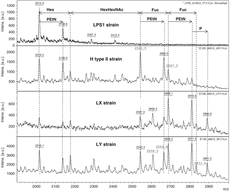
determine the relative abundance of bacterial LOS-glycoforms [33]. In parallel, methylation linkage analyses were carried out with the de-O-acylated LOSs. Results are summarized in Table2and Fig.3.
The de-O-acylated LOS sample of the LPS1 strain contained a main species at 2015.9 Da, fitting to the molecular composition P3-Glc-Hep2-Kdo2-lipid A as
previ-ously reported for this strain [1]. An additional species corresponding to P3-Glc-Hep2-Kdo2-PEtN-lipid A
(+123 Da) was also observed (Fig.3, uppermost panel). The spectra of the three recombinant strains H type II, LX, and LY showed progressively larger molecular weight LOS as a result of the overexpression of recombinant glycosyltransferase genes. All three strains overexpressed the genes encoding for LgtE, LgtA and LgtB, which are responsible for the synthesis of Gal
β-3GlcNAcβ-3Galβ-4-Glc-P3-Glc-Hep2-Kdo2-lipid A by glycosylation of the
truncated core present in the LPS1 strain. The MALDI-TOF spectra of the three strains (H type II, LX, LY) showed a common peak arising at 2542 Da (Fig. 3) from the
addition of two hexoses (Hex) and one
N-acetylhexosamine (HexNAc) to the truncated Glc-LOS of the LPS1 strain, compatible with an additional [Gal-GlcNAc-Gal] motif. An associated Galβ-3GlcNAcβ-3Galβ-4Glc-P3-Glc-Hep2-Kdo2-PEtN-lipid A species was
also observed (2665 Da, Fig.3).
The chemical analysis of the H type II strain LOS sample. Which specifically expressed the α1,2-fucosyltrans-ferase FutC in addition to LgtE, LgtA and LgtB contained a peak at 2687.5 Da corresponding to an additional fucose residue present (Fig.3, second panel from top), matching to the carbohydrate backbone [Fuc-Gal-GlcNAc-Gal-P3
-Glc-2015.9
2138.6
2287.3 2410.4
* LPSI_d140\ 0_I7\1\1Lin, Smoothed
0 200 400 600 800 1000 1200 Intens. [a.u.] 2665.0 2016.2 2138.9 * E129_d40\ 0_J8\1\1Lin 500 1000 1500 2000 Intens. [a.u.] 2688.1 2810.6 2606.4 2541.3 2890.8 * E128_d40\ 0_J7\1\1Lin 200 400 600 Intens. [a.u.] 2811.0 2688.2 2016.1 2542.4 2891.0 E130_d40\ 0_I9\1\1Lin 1000 2000 3000 4000 Intens. [a.u.] 2000 2100 2200 2300 2400 2500 2600 2700 2800 2900 m/z PEtN
Hex HexHexNAc Fuc Fuc
PEtN PEtN P 2542.5 H type II strain LPS1 strain LX strain LY strain 2833.4 2687.5 2608.0 2664.8
Hep2-Kdo2-lipidA]. The methylation analysis (Table 2)
confirmed the presence of a terminal fucose residue as well as 2-linked galactose, characterizing the motif Fucα-2Gal and therefore glycoform I (Fig.4). This is in agreement with the specificity of FutC onβ-galactosides.
When the de-O-acylated LOS sample of LX strain, which specifically expressed the α1,3-fucosyltransferase FutA in addition to LgtE, LgtA and LgtB, was analysed by MALDI-TOF it resulted in a spectrum very similar to the one of the H type II strain, although the proportion of the mono-fucosylated species [Fuc-Gal-GlcNAc-Gal-P3
-Glc-Hep2-Kdo2-lipidA] (and the corresponding PEtN-lipidA
glycoform) was much higher (Fig. 3, second panel from bottom). The methylation analysis revealed the presence of a high amount of 3,4-linked glucose and a low amount of 3,4-linked N-acetylglucosamine. This indicated a fucosyla-tion of the C3-OH of GlcNAc (in the lactosaminyl motif) but on the other hand of the Glc residue (in the lactosyl motif) as well, thereby demonstrating the presence of glycoforms II and III (see Fig. 4). These data fit to the known specificity of the fucosyltransferase FutA used in this study, which was reported to have a good activity on lactosyl as well as N-acetyllactosaminyl motifs of H type II acceptors [34]. A preferential fucosylation on glucose was also obtained when the same co-expressed enzymes acted
on free lactose as an acceptor [16]. A small glycoform at m/ z 2838 Da (Fig.3, second panel from bottom) corresponded to a difucosylated carbohydrate backbone [Fuc2
-Gal-GlcNAc-Gal-P3-Glc-Hep2-Kdo2-lipidA], which meant that
both the Glc and the GlcNAc residue of the Gal-GlcNAc-Gal-P3-Glc-Hep2-Kdo2-lipidA] backbone were fucosylated,
leading to glycoform IV (Fig.4).
The LY strain used in our study combined the two α1,2-and α1,3- fucosyltransferases FutC and FutA, which had been expressed separately in the strains LX and H type II. When the de-O-acylated LOS sample of the LY strain was analysed, the MALDI-TOF spectrum obtained was compa-rable with the spectrum of the LX strain, but showed a slight increase of difucosylated forms (Fig.3, lowest panel). The methylation analysis revealed the combined derivative products of the α-1,2-fucosyltransferase action (2-linked Gal) and of theα-1,3-fucosyltransferase action (3,4-linked Glc/GlcNAc), thus corresponding to glycoforms V and VI, the last one bearing the Lewis Y antigen (Table 2 and Fig. 4).
Discussion
Our data show that fucosylation of the lipid A core of Escherichia coli K12 can be obtained by design of a suitable LOS mutant co-expressing various glycosyltrans-ferases. This is the first report describing the endogenous fucosylation of the lipid A core of E coli. The human cell surface antigen Lewis Y was displayed on the bacterial cell surface of the LY strain as visible in a cell-based immunoassay using a monoclonal antibody raised against Lewis Y (Fig. 1). The chemical analysis of de-O-acylated LOS showed that the Lewis Y structure was not predom-inant, since a major form carrying a fucose on glucose was also observed. These data can be explained by the reduced specificity of theα1,3-fucosyltransferase used in this study. A possibility to increase the expression of the Lewis Y epitope could include the use of a more specific fucosyl-transferase. In this respect our bacterial system displaying fucosylated structures on the cell surface would enable researchers to sort clones (e.g. by fluorescence-activated cell sorting) exhibiting an improved Lewis Y expression after the molecular evolution of theα1,3-fucosyltransferase used. It is interesting to note that, although the α1,3-fucosyltransferase employed in this study shows relaxed specificity towards the acceptor substrate, we can assume that the configuration of the oligosaccharide product created in the E. coli cells by heterologous glycosyltrans-ferase expression is corresponding to the configuration seen in the Lewis Y epitope e.g. on human blood cells as it is unlikely that recognition by the monoclonal antibody against the Lewis Y epitope would not be hindered by a
Fucαα 2Galβ 4GlcNAcβ 3Galβ 4Glcα
3HepII-Galβ 4GlcNAcβ 3Galβ 4Glcα
3HepII-Fucα
3
Fucα 2Galβ 4GlcNAcβ 3Galβ 4Glcα
3HepII-Fucα
3
Galβ 4GlcNAcβ 3Galβ 4Glcα 3HepII-Fucα
3
Fucα 2Galβ 4GlcNAcβ 3Galβ 4Glcα
3HepII-Fucα
3
Strain Glycoform Structure
I II III IV V VI
Galβ 4GlcNAcβ 3Galβ 4Glcα
3HepII-Fucα 3 Fucα 3 LX LY H type II
Fig. 4 Proposed chemical structures of fucosylated LOS of recombi-nant E. coli strains
change configuration of the monosaccharides involved. Earlier studies in our lab also showed that the oligosaccha-ride product created by heterologous expression of the α1,3-fucosyltransferase as well as the other glycosyltrans-ferases except theα1,2-fucosyltransferase could be shown to contain the wild type monosaccharide configuration by NMR when produced in a soluble form [15].
The strains H type II and LX synthesized the group H-type 2 and the Lewis X oligosaccharide motifs, respec-tively, as judged by the chemical analysis of their respective LOS. The fucosylation on LOS was detected by lectin-based cell detection assays with the fucose-binding lectin of Ralstonia solanacearum [35], but our attempt to make a specific detection with a Lewis X antibody was not successful.
The MALDI-TOF analysis suggests that all recombinant strains possess PEtN-modified lipid A core species. The PEtN substitution is normally not important in Escherichia coli K12, but has been associated with the outer membrane adaptation to environmental stress induced by divalent cations and some antibiotics [36, 37]. A possible explana-tion for the PEtN substituexplana-tions visible in our strains is that the chimeric glycosylation of the outer core affects the outer membrane stability, resulting in an induction of PEtN substitution of the lipid A core.
This work is a step forward in the glyco-mimicry engineering of bacteria. Lewis antigens are common host targets for pathogens to bind to, such as pathogenic C. jejuni, and P. aeruginosa [11,12]. Competition experiments with our fucose-displaying non-pathogenic strains therefore might be a prelude to the design of new probiotic-based therapies. Furthermore, these strains present the researcher with the possibility to change the specificity of glycosyl-transferases by molecular evolution and subsequent testing with lectins and cell sorting. In addition, our strains could in the future be used to study the interaction between host receptors with the mentioned carbohydrate structures on non-pathogenic bacteria, thereby enabling us to dissect the effects mediated by the carbohydrate structures from the ones mediated by other bacterial virulence factors.
Acknowledgements We thank Bernard Brasme and Stéphanie Beffy for performing the mass analyses. This work was supported by a SNF grant (31003A_127098/1) to MA and a Marie Curie Early Stage Research Training Fellowship of the European Community’s Sixth Framework Programme (MEST-CT-2004-5033) to EY.
References
1. Ilg, K., Yavuz, Y., Maffioli, C., Priem, B., Aebi, M.: Glycomimi-cry: display of the GM3 sugar epitope on Escherichia coli and Salmonella enterica sv Typhimurium. Glycobiology 20, 1289– 1297 (2010)
2. Raetz, C.R., Whitfield, C.: Lipopolysaccharide endotoxins. Annu. Rev. Biochem. 71, 635–700 (2002)
3. Valvano, M.A.: Export of O-specific lipopolysaccharide. Front. Biosci. 8, s452–471 (2003)
4. Paton, A.W., Morona, R., Paton, J.C.: A new biological agent for treatment of Shiga toxigenic Escherichia coli infections and dysentery in humans. Nat. Med. 6, 265–270 (2000)
5. Paton, A.W., Jennings, M.P., Morona, R., Wang, H., Focareta, A., Roddam, L.F., Paton, J.C.: Recombinant probiotics for treatment and prevention of enterotoxigenic Escherichia coli diarrhea. Gastroenterology 128, 1219–1228 (2005)
6. Morona, J.K., Paton, J.C., Miller, D.C., Morona, R.: Tyrosine phosphorylation of CpsD negatively regulates capsular polysaccha-ride biosynthesis in streptococcus pneumoniae. Mol. Microbiol. 35, 1431–1442 (2000)
7. Bos, M.P., Tefsen, B., Geurtsen, J., Tommassen, J.: Identification of an outer membrane protein required for the transport of lipopolysaccharide to the bacterial cell surface. Proc. Natl Acad. Sci. USA 101, 9417–9422 (2004)
8. Sperandeo, P., Cescutti, R., Villa, R., Di Benedetto, C., Candia, D., Dehò, G., Polissi, A.: Characterization of lptA and lptB, two essential genes implicated in lipopolysaccharide transport to the outer membrane of Escherichia coli. J. Bacteriol. 189, 244–53 (2007)
9. Sperandeo, P., Lau, F.K., Carpentieri, A., De Castro, C., Molinaro, A., Dehò, G., Silhavy, T.J., Polissi, A.: Functional analysis of the protein machinery required for transport of lipopolysaccharide to the outer membrane of Escherichia coli. J. Bacteriol. 190, 4460– 4469 (2008)
10. Phillips, N.J., Miller, T.J., Engstrom, J.J., Melaugh, W., McLaughlin, R., Apicella, M.A., Gibson, B.W.: Characterization of chimeric lipopolysaccharides from Escherichia coli strain JM109 transformed with lipooligosaccharide synthesis genes (lsg) from Haemophilus influenzae. J. Biol. Chem. 275, 4747–4758 (2000)
11. Ruiz-Palacios, G.M., Cervantes, L.E., Ramos, P., Chavez-Munguia, B., Newburg, D.S.: Campylobacter jejuni binds intestinal H(O) antigen (Fuc alpha 1, 2Gal beta 1, 4GlcNAc), and fucosyloligosaccharides of human milk inhibit its binding and infection. J. Biol. Chem. 278, 14112–20 (2003)
12. Wu, A.M., Wu, J.H., Singh, T., Liu, J.H., Tsai, M.S., Gilboa-Garber, N.: Interactions of the fucose-specific Pseudomonas aeruginosa lectin, PA-IIL, with mammalian glycoconjugates bearing polyvalent Lewis(a) and ABH blood group glycotopes. Biochimie 88, 1479–92 (2006)
13. Priem, B., Gilbert, M., Wakarchuk, W.W., Heyraud, A., Samain, E.: A new fermentation process allows large-scale production of human milk oligosaccharides by metabolically engineered bacte-ria. Glycobiology 12, 235–240 (2002)
14. Drouillard, S., Driguez, H., Samain, E.: Large-scale synthesis of H-antigen oligosaccharides by expressing Helicobacter pylori alpha1, 2-fucosyltransferase in metabolically engineered Escherichia coli cells. Angew. Chem. Int. Ed Engl. 45, 1778–1780 (2006)
15. Dumon, C., Bosso, C., Utille, J.P., Heyraud, A., Samain, E.: Production of Lewis x tetrasaccharides by metabolically engineered Escherichia coli. Chembiochem 7, 359–365 (2006)
16. Dumon, C., Priem, B., Martin, S.L., Heyraud, A., Bosso, C., Samain, E.: In vivo fucosylation of N-neotetraose and lacto-N-neohexaose by heterologous expression of Helicobacter pylori alpha-1, 3 fucosyltransferase in engineered Escherichia coli. Glycoconj. J. 18, 465–474 (2001)
17. Randriantsoa, M. Synthèse microbiologique des antigènes gluci-diques des groupes sanguins. 2008. PhD thesis, University of Grenoble, France
18. Wang, G., Ge, Z., Rasko, D.A., Taylor, D.E.: Lewis antigens in Helicobacter pylori: biosynthesis and phase variation. Mol. Microbiol. 36, 1187–1196 (2000)
19. Boghaert, E.R., Sridharan, L., Armellino, D.C., Khandke, K.M., DiJoseph, J.F., Kunz, A., Dougher, M.M., Jiang, F., Kalyandrug, L.B., Hamann, P.R., Frost, P., Damle, N.K.: Antibody-targeted chemotherapy with the calicheamicin conjugate hu3S193-N-acetyl gamma calicheamicin dimethyl hydrazide targets Lewis y and eliminates Lewis y-positive human carcinoma cells and xeno-grafts. Clin. Cancer Res. 10, 4538–4549 (2004)
20. Link, A.J., Phillips, D., Church, G.M.: Methods for generating precise deletions and insertions in the genome of wild-type Escherichia coli: application to open reading frame characteriza-tion. J. Bacteriol. 179, 6228–6237 (1997)
21. Hanahan, D.: Studies on transformation of Escherichia coli with plasmids. J. Mol. Biol. 166, 557–580 (1993)
22. Yanisch-Perron, C., Vieira, J., Messing, J.: Improved M13 phage cloning vectors and host strains: nucleotide sequences of the M13mp18 and pUC19 vectors. Gene 33, 103–119 (1985) 23. Antoine, T., Priem, B., Heyraud, A., Greffe, L., Gilbert, M.,
Wakarchuk, W.W., Lam, J.S., Samain, E.: Large-scale in vivo synthesis of the carbohydrate moieties of gangliosides GM1 and GM2 by metabolically engineered Escherichia coli. Chembiochem 4, 406–412 (2003)
24. Cottaz, S., Samain, E.: Genetic engineering of Escherichia coli for the production of NI, NII-diacetylchitobiose (chitinbiose) and its utilization as a primer for the synthesis of complex carbohydrates. Metab. Eng. 7, 311–317 (2005)
25. Martinez, E., Bartolome, B., de la Cruz, F.: pACYC184-derived cloning vectors containing the multiple cloning site and lacZ alpha reporter gene of pUC8/9 and pUC18/19 plasmids. Gene 68, 159– 162 (1988)
26. Tsai, C.M., Frasch, C.E.: A sensitive silver stain for detecting lipopolysaccharides in polyacrylamide gels. Anal. Biochem. 119, 115–119 (1982)
27. Galanos, C., Luderitz, O., Westphal, O.: A new method for the extraction of R lipopolysaccharides. Eur. J. Biochem. 9, 245–249 (1969) 28. Qureshi, N., Takayama, K., Ribi, E.: Purification and structural
determination of nontoxic lipid A obtained from the lipopolysac-charide of Salmonella typhimurium. J. Biol. Chem. 257, 11808– 11815 (1982)
29. Verostek, M.F., Lubowski, C., Trimble, R.B.: Selective organic precipitation/extraction of released N-glycans following large-scale enzymatic deglycosylation of glycoproteins. Anal. Biochem. 278, 111–122 (2000)
30. Hakomori, S.: A rapid permethylation of glycolipid, and polysac-charide catalyzed by methylsulfinyl carbanion in dimethyl sulfoxide. J. Biochem. 55, 205–208 (1964)
31. Piekarowicz, A., Stein, D.C.: Biochemical properties of Neisseria gonorrhoeae LgtE. J. Bacteriol. 184, 6410–6406 (2002) 32. Wakarchuk, W., Martin, A., Jennings, M.P., Moxon, E.R.,
Richards, J.C.: Functional relationships of the genetic locus encoding the glycosyltransferase enzymes involved in expression of the lacto-N-neotetraose terminal lipopolysaccharide structure in Neisseria meningitidis. J. Biol. Chem. 271, 19166–73 (1996) 33. Gibson, B.W., Engstrom, J.J., John, C.M., Hines, J.W., Falick, W.
H.: Characterization of bacterial lipooligosaccharides by delayed extraction matrix-assisted laser desorption ionization time-of-flight mass spectrometry. J. Am. Soc. Mass Spec. 8, 645–658 (1997) 34. de Vries, T., Srnka, C.A., Palcic, M.M., Swiedler, S.J., van den
Eijnden, D.H., Macher, B.A.: Acceptor specificity of different length constructs of human recombinant alpha 1, 3/4-fucosyltransferases. Replacement of the stem region and the transmembrane domain of fucosyltransferase V by protein A results in an enzyme with GDP-fucose hydrolyzing activity. J. Biol. Chem. 270, 8712–8722 (1995) 35. Sudakevitz, D., Imberty, A., Gilboa-Garber, N.: Production, proper-ties and specificity of a new bacterial L-fucose- and D-arabinose-binding lectin of the plant aggressive pathogen Ralstonia solanacearum, and its comparison to related plant and microbial lectins. J. Biochem. 132, 353–358 (2002)
36. Kanipes, M.I., Lin, S., Cotter, R.J., Raetz, C.R.: Ca2+-induced phosphoethanolamine transfer to the outer 3-deoxy-D-mannooctulo-sonic acid moiety of Escherichia coli lipopolysaccharide. A novel membrane enzyme dependent upon phosphatidylethanolamine. J. Biol. Chem. 276, 1156–1163 (2001)
37. Kim, S.H., Jia, W., Parreira, V.R., Bishop, R.E., Gyles, C.L.: Phosphoethanolamine substitution in the lipid A of Escherichia coli O157: H7 and its association with PmrC. Microbiology 152, 657–666 (2006)