DOI 10.1007/s10067-005-1146-1
Madeleine M e l i • Gabriela G i t z e i m a n n
Renate Koppensteiner - B e a t r i c e R. Amann-Vesti
Predictive value of naUfold capiUaroscopy in patients
with Raynaud's phenomenon
Received: 16 November 2004/ Revised: 23 March 2005/Accepted: 23 March 2005/Published online: 11 June 2005 © Clinical Rheumatology 2005
Abstract The objective of this study was to evaluate the long-term follow-up of patients with Raynaud's phenomenon (RP) and pathological nailfold capilla- roscopy (NC) in order to analyse the predictive value of specific features of capillaroscopy for the develop- ment of a connective tissue disease (CTD). From 1992 to 2002, NC alone or combined with fluorescence vid- eomicroscopy with sodium fluorescein (NaF) was per- formed in 1024 consecutive patients because of RP. We analysed the follow-up and pathological features of NC in all patients who had neither clinical nor serological signs of a CTD at the time of NC. Of 308 patients with neither serological findings nor clinical signs of CTD but with RP and pathological features in NC suspi- cious for CTD, follow-up data were available for 133 patients. An additional N a F test had been performed in 51 (38.4%) patients. After a mean follow-up o f 6.5 years (range: 1-15 years), 109 patients had devel- oped a CTD and 24 patients did not show any clinical signs or serological markers for a CTD after a mean follow-up of 8.5 years (range: 2-15 years). There were no differences in age, duration of RP or of follow-up in patients who developed a C T D compared to patients who did not. Significantly more giant capillaries (p = 0.0001), avascular fields (p=0.02) and irregular architecture (/9=0.0001) had been observed in patients who had developed a C T D during the follow-up o f 6.5 years. The presence o f giant capillaries, avascular fields and irregular architecture of nailfold capillaries is predictive for the development of a CTD in patients with RP.
M. Meli • G. Gitzelmann
R. Koppensteiner • B. R. Amann-Vesti (5:~)
Division of Angiology, Department of Internal Medicine, University Hospital, Ramistrasse 100,
8091 Zurich, Switzerland E-mail: beatrice.amann @ usz.ch Tel.: +41-1-2551111
Fax: +41-1-2554510
K e y w o r d s Connective tissue disease - Nailfold capillaroscopy • Raynaud's phenomenon - Systemic sclerosis
Introduction
R a y n a u d ' s phenomenon (RP) is classified as primary when no underlying cause can be identified and as sec- ondary when its presence is explained by an associated condition such as systemic lupus erythematosus (SLE) and systemic sclerosis. Estimates of the prevalence of RP in the general population range from 5 to 20% [1], and 3-5% have been reported to develop a connective tissue disease (CTD) within 3-6 years [2]. In scleroderma, RP has been reported to be the presenting sign in 50-70% of patients and in 15% of patients with SLE [3-5]. How- ever, RP may precede the development of the disease by m a n y years. In several small studies it has been reported that the presence of autoantibodies may predict the presence or future development of systemic disease [6-8]. Nailfold capillaroscopy (NC) allows the in vivo assessment of morphology and of some functional as- pects of cutaneous capillaries and has been accepted as a diagnostic tool for evaluating microvascular involvement in systemic sclerosis, dermatomyositis, overlap syn- dromes and SLE [9-12]. Only little is known about the role of NC in identifying patients with RP who are at risk of developing a CTD [13-15]. Furthermore, by fluores- cence videomicroscopy with sodium fluorescein (NaF) patients with a CTD might exhibit halo enlargement with a typical " d w a r f h a t " formation as a sign of disturbed barrier function of the vessel wall and structural changes of the pericapillary space [16]. However, it has been shown in one study that of 69 patients with CTD 16% had normal findings in conventional N C but pathologi- cal halo formation after N a F injection [17]. The predic- tive value of combined N C with N a F has not been studied yet. In the present report, we describe the follow- up of 133 patients with pathological findings in NC with
154
or w i t h o u t N a F w h o were referred to o u r clinic because o f RP. A t the time o f the study, all patients h a d negative a u t o a n t i b o d i e s a n d no o t h e r clinical signs o f a C T D .
Patients and methods
In the p e r i o d f r o m 1992 to 2002, 1024 patients were re- ferred to o u r institution for vascular assessment includ- ing N C because o f a history o f R P o f the h a n d s and fingers. In all patients N C was p e r f o r m e d ; fluorescence v i d e o m i c r o s c o p y with N a F was only p e r f o r m e d in part o f the patients. Exclusion criteria for N a F were drug or f o o d allergies, history o f renal disease, p a t h o l o g i c a l cre- atinine clearance, a n d p r e g n a n c y . In some cases the N a F test was n o t p e r f o r m e d because o f difficulties in punc- turing a vein or no specific r e a s o n was given. Fluores- cence v i d e o m i c r o s c o p y with N a F was p e r f o r m e d as previously described [18]. N C was p e r f o r m e d with a fluorescence v i d e o m i c r o s c o p y system consisting o f an incident light fluorescence m i c r o s c o p e (Leica, Heerb- rugg, Switzerland), a 3 - C C D video c a m e r a (model D X C - 930P, Sony, T o k y o , J a p a n ) with a c a m e r a a d a p t e r and sensitivity set o n a u t o m a t i c c o n t r o l ( C M A - D 2 , Sony), a video timer ( V T G - 2 2 ) and scale m a r k e r (IV-600, both f r o m F o r - A - C o m p a n y , T o k y o , Japan), a video m o n i t o r (Picture M o n i t o r m o d e l P M 171T, Ikegami Tsushinki, T o k y o , J a p a n ) a n d a video tape r e c o r d e r (S-VHS, A G - 7350, P a n a s o n i c , Osaka, Japan). T h e m i c r o s c o p e is equipped with 1.0/0.04, 2.5/0.08, 6.3/0.20 a n d 10/0.25 p l a n a r objectives (Leica, H e e r b r u g g , Switzerland), which allow a m a g n i f i c a t i o n o f 24, 62, 165 a n d 240 times, respectively, o n the m o n i t o r . T h e fluorescence excitation filter w o r k s at 450-490 n m a n d the b a r r i e r filter at 515 nm. T h e N a F was injected into the a n t e c u b i t a l vein as a bolus; the d o s a g e was adjusted a c c o r d i n g to the estimated b l o o d volume. A t o u r institution 0.2~).3 ml N a F 2 0 % per liter estimated b l o o d v o l u m e is used and is a d e q u a t e f o r visualization o f skin capillaries.
T h e following p a r a m e t e r s o f N C were e v a l u a t e d from the v i d e o t a p e [19]:
- Irregular a r c h i t e c t u r e (capillaries n o t in one r o w as normal, small areas ( < 500 ~tm) with missing capil- laries next to areas with clusters o f capillaries) - Avascular fields (loss o f capillaries in a field o f at least
500 ~tm)
- D i l a t a t i o n o f capillaries (arterial side > 15 ~m, venous side > 20 I~m)
- G i a n t capillaries (apical d i a m e t e r > 50 I~m, Fig. 1) - N a F images were evaluated for apical halo e n l a r g e m e n t
or " d w a r f h a t " f o r m a t i o n , respectively (Fig. 2) [16].
Fig. 1 Typical giant capillary of the nailfold in a female patient with RP who developed CREST 10 years after NC (×63).
R h e u m a t i s m A s s o c i a t i o n ( A R A ) [20]. T h e diagnosis o f C R E S T was m a d e w h e n the following criteria were fulfilled: calcinosis, R a y n a u d ' s p h e n o m e n o n , o e s o p h a - geal dysmotility, sclerodactyly, and telangiectasia in the
Diagnostic criteria
The diagnosis o f systemic sclerosis (Scl) was m a d e according to the p r e l i m i n a r y criteria o f the A m e r i c a n
Fig. 2 a After the intravenous injection of NaF apical halo enlargement or "dwarf hat" formation might be seen in patients with CTD. b In healthy subjects the cells are surrounded by the fluorescent plasma layer and NaF does not pass the narrow pericapillary border (×63).
presence o f anticentromere antibodies. Mixed connec- tive tissue disease ( M C T D ) was diagnosed in the pres- ence o f symptoms as described by Alarcon-Segovia and Cardiel [21]. The diagnoses of SLE and rheumatoid arthritis (RA) were based on the diagnostic criteria of the ARA. Dermatomyositis (DM) was considered the diagnosis when the criteria o f Bohan and Peter [22] were present, Sj6gren's syndrome was diagnosed when the diagnostic criteria from the A m e r i c a n - E u r o p e a n Con- sensus G r o u p were present, including the presence o f autoantibodies (anti-Ro/SSA, anti-La/SSB or both) [23].
Statistical analysis
Analyses were performed with the statistical software package Stat View 5.0. Continuous variables are reported as means and categorical variables as percentages. Com- parison between groups o f patients was done by means o f unpaired Student's t-test and for categorical variables by using Fisher's exact test. A test for logistic regression was performed as a means o f studying the diagnostic value o f the different features o f N C to predict development o f CTD. Significance was defined as p < 0.05.
Results
O f a total o f 1024 patients seen with RP between 1992 and 2002, the results o f 968 patients were evaluated for the present study; 19 patients with Osler's disease and 37 patients for whom some data were missing had been excluded. In 693 patients, the N a F test was performed in addition to conventional NC. In 201 (155 females and 46 males) patients referred to us because o f RP the diag- nosis o f a C T D had already been made at the time of N C due to serological features and the above-mentioned criteria. The diagnosis and results o f N C from these 201 patients are shown in Table 1. Microangiopathy was defined as being present if at least one o f the above- mentioned criteria was found. The diagnosis o f primary RP was made in 459 patients because the following criteria were fulfilled: normal findings in NC, negative autoantibodies, no signs o f a C T D according to the
A R A criteria, no calcinosis, no oesophageal motility disturbances, no sclerodactyly, no telangiectasia and no xerostomia. In 308 patients with normal serological findings and no clinical signs o f a C T D despite RP, we found pathological features in NC suspicious for CTD. Of these 308 patients with RP and microangiopathy in NC, follow-up data were available for 133 patients: 107 females (mean age: 50.0 years, range: 10-89 years) and 26 males (mean age: 55.1 years, range: 26-82 years). The N a F had been administered in 51 (38.4%) patients. After a mean follow-up o f 6.5 years (range: 1-15 years), 109 patients (82%, group 1) had developed a C T D according to the above-described criteria, The patient characteristics, diagnoses and findings o f N C in these patients are shown in Table 2,
The remaining 24 patients (group 2, 19 females, 5 males, mean age: 45.6 years, range: 21-70) had not developed any clinical signs o f a C T D and serological tests were still negative after a mean follow-up of 8.5 years (range: 2-15 years). Patient characteristics and features o f N C are shown in Table 3. There were no statistically significant differences in age, duration o f RP or duration o f follow-up in patients who developed a C T D (group 1) compared to patients who had not developed a C T D (group 2) during the follow-up. F o r comparison between the two groups, only patients who had not developed a C T D after a follow-up o f at least 6 years had been included: four patients with a follow- up o f 4 years (two patients), one patient with a follow- up of 5 years and one patient with a follow-up o f 2 years had been excluded from the analysis [2]. The mean fol- low-up o f these 20 patients was 9.3 years (range: 6 - 15 years). The features and comparison o f N C in the two groups are shown in Fig. 3. In group 1 significantly more giant capillaries (p=0.0001), avascular fields (p=0.02) and irregular architecture (p=0.0001) had been observed at the initial investigation. Including all 24 patients the results were almost identical; the same features (giant capillaries, avascular fields and irregular architecture) had been identified as significantly different in the two groups (p < 0.05).
F u r t h e r m o r e , the presence o f giant capillaries or an irregular architecture was predictive for the development o f a C T D (p < 0.005). Only two patients developed SLE
Table 1 Age, sex and presence
or absence of pathological Patients
features in nailfold (n/%)
capillaroscopy (NC) of 201 patients with RP, in whom an underlying disease had been already diagnosed at the time of
NC. Scl systemic sclerosis, Scl 93/46.3
C R E S T calcinosis, Raynaud's CREST 8/4.0
phenomenon, oesophageal SLE 29/14.4
dysfunction, sclerodactyly, MCTD 29/14.4 telangiecstasia, S L E systemic DM 14/7.0 lupus erythematosus, M C T D Sj6gren's 9/4.5 mixed connective tissue disease, syndrome
D M dermatomyositis, RA RA 19/9.4
rheumatoid arthritis Total 201
Diagnosis Age ( m e a n , Female/male Presence of
range, years) (%) microangiopathy
(%) Yes No 52.4 (13-78) 72.0/28.0 83.9 16.1 62.4 (36-76) 87.5/12.5 87.5 12.5 39.1 (12~53) 82.8/17.2 37.9 62.1 50.7 (27-83) 86.2/13.8 65.5 34.5 52.6 (17-70) 57.1/42.9 57.1 42.9 52.6 (17~59) 88.9/11. l 44.5 55.5 50,3 (22-73) 84.2/15.8 31.6 68.4 51.5 (12-83) 77.1/22.9 60.7 39.3
156
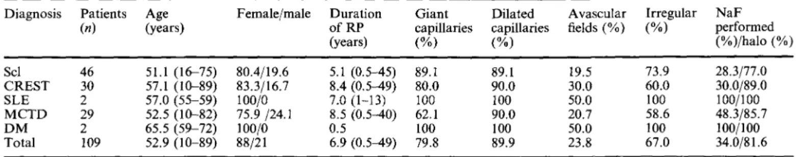
Table 2 Characteristics of 109 patients with RP and pathological features in the nailfold capillaroscopy and their diagnosis after a mean follow-up of 6.5 years (1-15 years). Scl systemic sclerosis,
C R E S T calcinosis, Raynaud's phenomenon, oesophageal dys-
function, sclerodactyly, telangiecstasia, S L E systemic lupus eryth- ematosus, M C T D mixed connective tissue disease, D M
dermatomyositis
Diagnosis Patients Age Female/male Duration Giant Dilated Avascular Irregular NaF
(n) (years) of RP capillaries capillaries fields (%) (%) performed
(years) (%) (%) (%)/halo (%) Scl 46 51.1 (16-75) 80.4/19.6 5.1 (0.5-45) 89.1 89.1 19.5 7 3 . 9 28.3/77.0 CREST 30 57.1 (10-89) 83.3/16.7 8.4 (0.5-49) 80.0 90.0 30.0 60.0 30.0/89.0 SLE 2 57.0 (55-59) 100/0 7.0 (1-13) 100 100 50.0 100 100/100 MCTD 29 52.5 (10-82) 75.9/24.1 8.5 (0.5-40) 62.1 90.0 20.7 58.6 48.3/85.7 DM 2 65.5 (59-72) 100/0 0.5 100 100 50.0 100 100/100 Total 109 52.9 (10-89) 88/21 6.9 (0.5-49) 79.8 89.9 23.8 67.0 34.0/81.6
Table 3 Characteristics of 24 patients with RP and pathological features in the nailfold capillaroscopy who did not develop CTD after a mean follow-up of 8.5 years (2-15 years)
Patients Age Female/male Duration Giant Dilated Avascular Irregular NaF performed
(n) (years) (%) of RP (years) capillaries capillaries fields (%) (%) (%)/halo (%)
(%) (%)
24 45.6 (21-70) 79.2/20.8 6.5 (1-20) 41.7 83.3 4.1 29.2 58.3/85.7
a n d D M , respectively; t h e r e f o r e n o c o n c l u s i o n s f o r these diseases c a n be d r a w n f r o m o u r d a t a .
Discussion
N C h a s been a c c e p t e d as o n e o f the m o s t valuable d i a g n o s t i c tools for the early d e t e c t i o n o f C T D , in par- ticular f o r Scl a n d C R E S T [24-26]. I n the p r e s e n t study, we a n a l y s e d the features o f N C in p a t i e n t s with R P in o r d e r to e v a l u a t e the predictive value o f p a t h o l o g i c a l findings. W e f o u n d t h a t d u r i n g the m e a n f o l l o w - u p o f 100 80 60 40 20 0 % p=O.O001 giant capillaries dilated capillaries p=O.02
_J
avascular fieldsp]00001
irregular halo architecture enlargementFig. 3 Comparison of features of nailfold capillaroscopy in patients with RP. In patients who developed a connective tissue disease (CTD) during follow-up significantly more giant capillaries (p = 0.0001), avascular fields (p= 0.02) and irregular architecture (p=0.0001) have been found than in the 20 patients without any laboratory or clinical signs of a CTD after a follow-up of at least 6years (mean follow-up: 9.3 years, range: 6-15years). The enlargement of the apical halo after injection of NaF was not different in the two groups.
6.5 years 8 2 % o f p a t i e n t s with R P a n d p a t h o l o g i c a l findings in N C d e v e l o p e d a C T D , m a i n l y Scl, C R E S T a n d M C T D . T h e s e p a t i e n t s h a d s h o w n significantly m o r e giant capillaries, i r r e g u l a r a r c h i t e c t u r e a n d avas- cular fields in the initial N C t h a n the g r o u p w h o did n o t d e v e l o p a C T D . T h e p r e s e n c e o f giant capillaries a n d irregular architecture in N C were b o t h predictive for the d e v e l o p m e n t o f a C T D in o u r p a t i e n t p o p u l a t i o n .
I t has been s h o w n t h a t p a t i e n t s w i t h C T D , especially with Scl a n d C R E S T , a typical " c a p " f o r m a t i o n or " l a k e - l i k e " a r e a s c a n be seen at the a p e x o f the capillary after injection o f N a F as a sign o f d i s t u r b e d barriers for diffusion o f the dye at the c a p i l l a r y wall a n d at the o u t e r b o r d e r o f the skin p a p i l l a (halo) [17, 27]. I n 38.4% o f o u r patients with R P , the N a F test w a s p e r f o r m e d . We f o u n d a p a t h o l o g i c a l apical diffusion in 81.6% o f pa- tients w h o d e v e l o p e d a C T D d u r i n g the follow-up; h o w e v e r , 85.7% o f p a t i e n t s w i t h o u t a n y signs o f a C T D after a m e a n f o l l o w - u p o f 8.5 y e a r s also s h o w e d the s a m e h a l o p a t t e r n . F r o m o u r d a t a we c a n conclude t h a t the a d d i t i o n a l use o f N a F d o e s n o t facilitate early detection o f a C T D . H o w e v e r , even w h e n we analyse only p a t i e n t s with a f o l l o w - u p o f a t least 6 years the presence o f p a t h o l o g i c a l h a l o f o r m a t i o n was n o t pre- dictive f o r C T D .
D i l a t e d capillaries were a very c o m m o n b u t unspecific finding in o u r p a t i e n t p o p u l a t i o n . T h e y were present in o v e r 8 0 % a n d their p r e s e n c e w a s n o t predictive f o r the d e v e l o p m e n t o f a C T D . H o w e v e r , they h a v e b e e n de- scribed in C T D , in a c r o c y a n o s i s a n d less p r o n o u n c e d in p r i m a r y R P [28, 29]. I n C T D dilated capillaries are usually n o t the o n l y p a t h o l o g i c a l f e a t u r e o f N C , whereas in p r i m a r y R P a n d p r i m a r y a c r o c y a n o s i s a d d i t i o n a l p a t h o l o g i c a l features such as g i a n t capillaries o r avas- cular fields a r e missing.
It is k n o w n t h a t a C T D might develop m a n y years after the onset o f RP; therefore, the Allen and B r o w n criteria requiring at least a 2-year history o f R P for the diagnosis o f a p r i m a r y R P have been revised [30]. In o u r analysis the m e a n d u r a t i o n o f R P at the time o f N C was 6.9 years in the g r o u p w h o developed a C T D and 6.5 years in the g r o u p w h o did not. In patients w h o developed a C T D d u r i n g the follow-up, d u r a t i o n o f R P up to 49 years has been reported. O u r data suggest that the d u r a t i o n o f R P is a v e r y p o o r criterion for identi- fying patients at risk for the d e v e l o p m e n t o f s e c o n d a r y RP.
The m e a n age was n o t different in the two patient groups, a l t h o u g h children and very y o u n g adults (age between 10 and 21) were only seen in the g r o u p w h o developed a C T D d u r i n g the follow-up. T h e n u m b e r might be t o o low for a conclusion, but it suggests that in children and y o u n g adults with R P and pathological capillaries the risk for a n underlying disease is higher.
In 201 patients (77.1% female) the diagnosis o f an underlying disease h a d a l r e a d y been made at the time o f NC. A b o u t h a l f o f the patients had been diagnosed with a f o r m o f Scl; in these patients a m i c r o a n g i o p a t h y in N C was present in o v e r 80%. In contrast, only in 37.9% o f patients with S L E was m i c r o a n g i o p a t h y present. P a t h - ological capillary m o r p h o l o g y has been r e p o r t e d in 2 - 90% o f S L E p a t i e n t s [10, 31]. This variation in published n u m b e r s m i g h t be due, at least in part, to the definition o f m i c r o a n g i o p a t h y used b y the authors. F u r t h e r m o r e , it has been speculated t h a t the presence o f m i c r o a n g i o p - a t h y might be associated with clinical features, such as R P or specific antibodies. F u r t a d o et al. [32] f o u n d a significant association b e t w e e n m i c r o a n g i o p a t h y in S L E and RP, a n d similar findings were reported by C a s p a r y et al. [33]; h o w e v e r , in a study o f 51 patients with S L E Bongard et ai. [34] f o u n d no correlation between a b n o r m a l capiUaroscopic findings and RP. In o u r study, only patients with R P were included, which might be o n e reason for the r a t h e r high incidence o f m i c r o a n g i o p a t h y in our group. A h i g h e r incidence o f m i c r o a n g i o p a t h y a m o n g S L E patients with positive anticardiolipin a n d a n t i - U l r i b o n u c l e o p r o t e i n antibodies has been d e m o n - strated, suggesting direct d a m a g e to the endothelium b y these antibodies [32, 34]. H o w e v e r , specific a u t o a n t i - bodies in Scl patients (i.e. anti-Scl-70 and anticardiolipin antibody) do n o t seem directly linked to the expression o f a singular c a p i l l a r o s c o p i c p a t t e r n [35].
In conclusion, in patients presenting with R P the presence o f either giant capillaries, avascular fields or irregular a r c h i t e c t u r e in N C is predictive for the devel- o p m e n t o f a C T D , m a i n l y scleroderma, C R E S T a n d M C T D . N C m a y n o t be a valuable diagnostic tool in SLE, but it m i g h t be helpful in identifying a s u b g r o u p o f patients with different evolution and prognosis of the disease. T h e d u r a t i o n o f R P was n o t a criterion for the risk o f future d e v e l o p m e n t o f C T D . Despite the fact t h a t in Scl a specific diffusion p a t t e r n after N a F injection might be present, the a d d i t i o n a l use o f N a F does n o t facilitate early d e t e c t i o n o f a C T D .
Take hone message
In patients with R P and negative serological tests, the presence o f giant capillaries, avascular fields or irregular architecture in N C is predictive for the d e v e l o p m e n t o f a C T D , mainly scleroderma, C R E S T and M C T D . Based o n o u r d a t a the d u r a t i o n o f R P is n o t a criterion for the risk o f future d e v e l o p m e n t o f C T D .
References
1. Maricq HR, Carpentier PH, Weinrich MC, Keil JE, Franco A, Drouet P, Poncot OC, Maines MV (t993) Geographic varia- tion in the prevalence of Raynaud's phenomenon: Charleston, SC, USA, vs. Tarentaise, Savoie, France. J Rheumatol 20:70 76
2. Kallenberg CG, Wouda AA, Hoet MH, van Venrooij WJ (1988) Development of connective tissue disease in patients presenting with Raynaud's phenomenon: a six year follow up with emphasis on the predictive value of antinuclear antibodies as detected by immunoblotting. Ann Rheum Dis 47:634-641 3. Bennett R, Bluestone R, Holt PJ, Bywaters EG (1971) Survival
in scleroderma. Ann Rheum Dis 30:581-588
4. Tuffanelli DL, Winkelmann RK (1961) Systemic scleroderma, a clinical study of 727 cases. Arch Dermatol 84:359-371 5. Fessel WJ (1974) Systemic lupus erythematosus in the com-
munity. Incidence, prevalence, outcome, and first symptoms; the high prevalence in black women. Arch Intern Med 134:1027-1035
6. Kallenberg CG, Wouda AA, The TH (1980) Systemic involvement and immunologic findings in patients presenting with Raynaud's phenomenon. Am J Med 69:675-680
7. Wollersheim H, Thien T, Hoet MH, Van Venrooy WJ (1989) The diagnostic value of several immunological tests for anti- nuclear antibody in predicting the development of connective tissue disease in patients presenting with Raynaud's phenome- non. Eur J Clin Invest 19:535-541
8. Weiner ES, Hildebrandt S, Senecal JL, Daniels L, Noell S, Joyal F, Roussin A, Earnshaw W, Rothfield NF (1991) Prog- nostic significance of anticentromere antibodies and anti-to- poisomerase I antibodies in Raynaud's disease. A prospective study. Arthritis Rheum 34:68-77
9. Cutolo M, Sulli A, Pizzorni C, Accardo S (2000) Nailfold videocapillaroscopy assessment of microvascular damage in systemic sclerosis. J Rheumatol 27:155-160
10. Maricq HR, LeRoy EC, D'Angelo WA, Medsger TA Jr, Rodnan GP, Sharp GC, Wolfe JF (1980) Diagnostic potential of in vivo capillary microscopy in scleroderma and related disorders. Arthritis Rheum 23:183-189
11. Lee P, Sarkozi J, Bookman AA, Keystone EC, Armstrong SK (1986) Digital blood flow and nailfold capillary microscopy in Raynaud's phenomenon. J Rheumatol 13:564-569
12. Grassi W, Medico PD, Izzo F, Cervini C (2001) Microvaseular involvement in systemic sclerosis: capillaroscopic findings. Se- min Arthritis Rheum 30:397-402
13. Zufferey P, Depairon M, Chamot AM, Monti M (1992) Prognostic significance of nailfold capillary microscopy in pa- tients with Raynaud's phenomenon and scleroderma-pattern abnormalities. A six-year follow-up study. Clin Rheumatol
11:536--541
14. Passiu G, Sebastiani GD, Galeazzi M, Tuveri MA, Nicosia PM, Boirivant R (1990) Prognostic factors in Raynaud's phe- nomenon: usefulness of antinuclear antibodies and of periun- gual capillaroscopy. Medicina (Firenze) 10:405-407
15. Cutolo M, Grassi W, Matucci Cerinic M (2003) Raynaud's phenomenon and the role of capillaroscopy. Arthritis Rheum 48:3023-3030
158
16. Brulisauer M, Bollinger A (1991) Measurement of different human microvascular dimensions by combination of videomi- croscopy with Na-fluorescein (NaF) and indocyanine green (ICG) in normals and patients with systemic sclerosis. Int J Microcirc Clin Exp 10:21-31
17. Moneta G, Vollenweider U, Dubler B, Bollinger A (1986) Diagnostic value of capillaroscopy with and without fluorescent dyes to detect early connective tissue disease. Vasa 15:143-149 18. Bollinger A, Fagrell B (1990) Clinical capillaroscopy. Hogrefe,
G6ttingen
19. Schmidt JA, Caspary L, von Bierbrauer A, Ehrly AM, Junger M, Jung F, Lawall H (1997) Standardization of nailfold cap- illary microscopy in routine diagnosis. Vasa 26:5-10
20. Masi AT (1988) Classification of systemic sclerosis (sclero- derma): relationship of cutaneous subgroups in early disease to outcome and serologic reactivity. J Rheumatol 15:894-898 21. Alarcon-Segovia D, Cardiel M H (1989) Comparison between 3
diagnostic criteria for mixed connective tissue disease. Study of 593 patients. J Rheumatol 16:328-334
22. Bohan A, Peter JB (1975) Polymyositis and dermatomyositis (second of two parts). N Engl J Med 292:403-407
23. Vitali C, Bombardieri S, Jonsson R, Moutsopoulos HM, Alexander EL, Carsons SE, Daniels TE, Fox PC, Fox RI, Kassan SS et al (2002) Classification criteria for Sjogren's syndrome: a revised version of the European criteria proposed by the American-European Consensus Group. Ann Rheum Dis 61:554-558
24. Kenik JG, Maricq HR, Bole G G (1981) Blind evaluation of the diagnostic specificity of nailfold capillary microscopy in the connective tissue diseases. Arthritis Rheum 24:885-891 25. Maricq HR, LeRoy EC (1973) Patterns of finger capillary
abnormalities in connective tissue disease by "wide-field" microscopy. Arthritis Rheum 16:619-628
26. Houtman PM, Kallenberg CG, Fidler V, Wouda AA (1986) Diagnostic significance o f nailfold capillary patterns in patients
with Raynaud's phenomenon. An analysis of patterns dis- criminating patients with and without connective tissue disease. J Rheumatol 13:556--563
27. Bollinger A, Jager K, Siegenthaler W (1986) Microangiopathy of progressive systemic sclerosis. Evaluation by dynamic fluo- rescence videomicroscopy. Arch Intern Med 146:1541-1545 28. Monticone G, Colonna L, Palermi G, Bono R, Puddu P (2000)
Quantitative nailfold capillary microscopy findings in patients with acrocyanosis compared with patients having systemic sclerosis and control subjects. J Am Acad Dermatoi 42:78%790 29. Jacobs M J, Breslau P J, Slaaf DW, Reneman RS, Lemmens JA (1987) Nomenclature of Raynaud's phenomenon: a capillary microscopic and hemorheologic study. Surgery 101:136-145 30. LeRoy EC, Medsger TA Jr (1992) Raynaud's phenomenon: a
proposal for classification. Clin Exp Rheumatol 10:485-488 31. Redisch W, Messina EJ, Hughes G, McEwen C (1970) Capil-
laroscopic observations in rheumatic diseases. Ann Rheum Dis 29:244-253
32. Furtado RN, Pucinelli ML, Cristo VV, Andrade LE, Sato EI (2002) Scleroderma-like nailfold capillaroscopic abnormalities are associated with anti-U1-RNP antibodies and Raynaud's phenomenon in SLE patients. Lupus 11:35-41
33. Caspary L, Schmees C, Schoetensack I, Hartung K, Stannat S, Deicher H, Creutzig A, Alexander K (1991) Alterations of the nailfold capillary morphology associated with Raynaud phenomenon in patients with systemic lupus erythematosus. J Rheumatol 18:559-566
34. Bongard O, Bounameaux H, Miescher PA, De Moerloose P (1995) Association of anticardiolipin antibodies and abnormal nailfold capillaroscopy in patients with systemic lupus erythe- matosus. Lupus 4:142-144
35. Cutolo M, Pizzorni C, Tuccio M, Burroni A, Craviotto C, Basso M, Seriolo B, Sulli A (2004) Nailfold videocapillaro- scopic patterns and serum autoantibodies in systemic sclerosis. Rheumatology (Oxford) 43:719-726