Please note that this is an author-produced PDF of an article accepted for publication following peer review. The definitive January 2017, Volume 16 Issue 1 Pages 82-90
http://dx.doi.org/10.1017/S1473550415000555 http://archimer.ifremer.fr/doc/00366/47678/ © Cambridge University Press 2016
Achimer
http://archimer.ifremer.frIrradiation effects on antibody performance in the frame of
biochip-based instruments development for space
exploration
Baque M. 1, *, Dobrijevic M. 2, 3, Le Postollec A. 2, 3, Moreau T. 4, Faye C. 5, Vigier F., Incerti S. 6, 7, Coussot G. 8, Caron J. 9, Vandenabeele Trambouze Odile 10, 11, 12
1
Univ Roma Tor Vergata, Dept Biol, Rome, Italy. 2
Univ Bordeaux, LAB, UMR 5804, F-33270 Florac, France. 3
CNRS, LAB, UMR 5804, F-33270 Florac, France. 4
Univ Clermont Ferrand, INSERM U1103, GReD, CNRS UMR6293, Aubiere, France. 5
Cap Alpha, COLCOM, F-34830 Clapiers, France. 6
Univ Bordeaux, CENBG, UMR 5797, F-33170 Gradignan, France. 7
CNRS, IN2P3, CENBG, UMR 5797, F-33170 Gradignan, France. 8
Univ Montpellier, Inst Biomol Max Mousseron, Fac Pharm, CNRS,Unite Mixte Rech 5247, F-34093 Montpellier 5, France.
9
Inst Bergonie, Dept Radiotherapy, Ctr Comprehens Canc, F-33076 Bordeaux, France. 10
Univ Bretagne Occidentale, LMEE, UEB, IUEM UMR 6197, Plouzane, France. 11
CNRS, IUEM UMR 6197, LMEE, Plouzane, France. 12
IFREMER, UMR6197, LMEE, Plouzane, France.
* Corresponding author : M. Baqué, email address : [email protected]
Abstract :
Several instruments based on immunoassay techniques have been proposed for life-detection experiments in the framework of planetary exploration but few experiments have been conducted so far to test the resistance of antibodies against cosmic ray particles. We present several irradiation experiments carried out on both grafted and free antibodies for different types of incident particles (protons, neutrons, electrons and C-12) at different energies (between 9 MeV and 50 MeV) and different fluences. No loss of antibodies activity was detected for the whole set of experiments except when considering protons with energy between 20 and 30 MeV (on free and grafted antibodies) and fluences much greater than expected for a typical planetary mission to Mars for instance. Our results on grafted antibodies suggest that biochip-based instruments must be carefully designed according to the expected radiation environment for a given mission. In particular, a surface density of antibodies much larger than the expected proton fluence would prevent significant loss of antibodies activity and thus assuring a successful detection.
Proof For Review
1. Introduction
51
Among the next tools to search for signs of past or present life in our Solar System, 52
several instruments based on the biochip technology have been proposed in the 53
framework of planetary exploration. A biochip is a miniaturized device composed of 54
molecular recognition tools (or affinity receptors) like antibodies (Baqué et al. 2011b; 55
de Diego-Castilla et al. 2011; Parro et al. 2005, 2011a; Sims et al. 2005, 2012) or 56
aptamers (Baqué et al. 2011a), which allows the detection of hundreds of different 57
compounds in a single assay. Widely developed for biotechnology use and medical or 58
environmental diagnostics (see for example Wang 2006), miniaturized instruments 59
based on biochips have been indeed proposed and studied for biosignature detection in 60
an astrobiological context since more than 15 years (McKay et al. 2000; Parro et al. 61
2005; Le Postollec et al. 2007; Sims et al. 2005). 62
Mars, one of the most probable planetary body where to find signs of extinct or extant 63
life outside of Earth, is the target of many upcoming dedicated missions: ESA-64
Roscosmos’ ExoMars rover in 2016-2018, NASA’s Mars2020 rover (a follow-up of to 65
the Curiosity rover) and the Icebreaker mission concept proposed for a 2021 launch to 66
be part of NASA’s Discovery program (McKay et al., 2013). Different space 67
instruments based on the biochip technology and using antibodies have been proposed 68
for these future missions: the Life Marker Chip (LMC) (Martins 2011; Sephton et al. 69
2013; Sims et al. 2012), and the Signs Of LIfe Detector (SOLID) (Parro et al. 2005, 70
2008, 2011a, 2011b). Another project, the Biochip for Organic Matter Analysis in 71
Space (BiOMAS), proposes to combine both antibodies and aptamers in a single 72
instrument (Baqué et al. 2011b, 2011a; Le Postollec et al. 2007). Recently, first in the 73
framework of Mars2020 announcement of opportunity, and then in the framework of 74
NASA’s Discovery 2014 announcement of opportunity, these different teams have 75
Proof For Review
united to work on the SOLID instrument proposal for the Icebreaker mission and thus to 76
contribute with their different expertise to improve the technological readiness level of a 77
biochip-based instrument for space exploration (Manchado et al. 2015; McKay et al. 78
2013; Smith & Parro 2014). 79
Indeed, although biochips are known to be very sensitive tools to detect specific target 80
molecules, their sensitivity is related to the presence of functional affinity receptors. In 81
order to develop a “space biochip”, it is thus necessary to ensure that these biological 82
receptors will survive space hazards. In particular, due to the very sparse data on this 83
topic, it is important to determine the behavior of these biological receptors under 84
cosmic particles irradiation. 85
Le Postollec et al. (2009a) performed simulations with the Geant4 Monte Carlo toolkit 86
in order to estimate the radiation environment that a biochip would face if it were placed 87
into a rover dedicated to explore Mars’ surface. Ionizing doses accumulated and fluxes 88
of particles entering the biochip have been established for both the Earth-Mars transit 89
and the journey on Mars’ surface. Neutrons and gammas appear as dominant radiation 90
species on Mars’ soil whereas protons dominate during the interplanetary travel. These 91
results have been confirmed by other studies done by McKenna-Lawlor et al. (2012) 92
and Derveni et al. (2012). Moreover, these simulations can today be confronted to the 93
real radiation environment of an actual mission to Mars as it was monitored by the 94
Radiation Assessment Detector (RAD) instrument on-board the Mars science laboratory 95
spacecraft on cruise to Mars and continue to be recorded by the rover Curiosity directly 96
on its surface (Hassler et al. 2013; Kim et al. 2014; Zeitlin et al. 2013). Indeed, the total 97
cosmic radiation dose rate of 210 ± 40 µGy/day (Hassler et al. 2013) recorded at Gale 98
Crater by Curiosity and the one measured inside the Mars Science Laboratory 99
spacecraft during its cruise to Mars (481 ± 80 µGy/day) (Zeitlin et al. 2013) proved to 100
Proof For Review
be in the same order of magnitude as model predictions with respectively ~840 µGy/day 101
(without any shielding) for the Martian surface and ~240 µGy/day for the Earth-Mars 102
transit considering only GCR (galactic cosmic rays) contribution (Le Postollec et al. 103
2009a). 104
105
Considering the lack of experimental data about cosmic rays effect on antibodies, 106
particularly under lyophilized (freeze-dried) state, our team decided to investigate the 107
effects of different types of particles at several energies. Our objective is first to study 108
and measure cosmic rays effects on biological receptors and second to define well-109
adapted protections for a biochip-based instrument if we find evidences that cosmic rays 110
might have deleterious effect on their performances. In a first study, Le Postollec et al. 111
(2009b) performed neutrons irradiation on both antibodies and fluorescein dyes (used 112
for detection of recognition events) at two energies (0.6 and 6 MeV) and with different 113
fluences. Sample analyses demonstrated that, in tested conditions, neutrons do not affect 114
antibody recognition capability and fluorescence dye intensity. More recently, the 115
effects of 2 MeV protons on antibody performances (Baqué et al. 2011b) were 116
investigated. These studies showed that this irradiation process did not affect the 117
performances of antibodies as molecular recognition tools. In addition, printed antibody 118
and Alexa-647 fluorescent dye were demonstrated to be stable between 1.18 and 1.33 119
MeV gamma radiation (de Diego-Castilla et al. 2011). Finally, Derveni et al. (2012) 120
tested five antibodies freeze-dried in a variety of protective molecular matrices and 121
exposed to 50 MeV protons. They showed that at a representative Mars-mission-dose, 122
none of the antibodies studied exhibited any evidence of activity loss due to the 123
radiation. 124
Proof For Review
In the present paper, we broaden these previous studies to test the effect of electrons, 125
carbon ions, protons (at different energies) and neutrons (at higher energies) on the 126
recognition capability of antibodies (summarized in Fig 1). As protons and neutrons 127
dominate the radiation environment during the Earth-Mars transit and on the Martian 128
surface, we tested different high-energy particles from 15 to 50 MeV at different 129
fluences. Moreover, other damaging particles are significantly present in cosmic and 130
solar radiations such as carbon ions and electrons. 131
Chemicals and biological materials used to perform the experiments are given in section 132
2. Section 3 describes samples preparation, particles irradiation parameters and analysis 133
protocols. Results are presented in section 4. The last section draws conclusions on this 134 study. 135 136
2. Material
137Monoclonal anti-Horseradish Peroxydase antibodies were obtained from Antibodies-138
online (Germany), Horseradish peroxydase (type II), O-Phenylenediamine 139
dihydrochloride (OPD), NaH2PO4, Tween® 20, sodium acetate, sucrose, sodium azide, 140
NaOH, H2O2, BSA, (L)-Histidine and (D)-Arginine L-tyrosinamide, fluorescein and 141
Tris(hydroxymethyl) aminomethane were purchased from Sigma Aldrich (Saint-142
Quentin, France). NaCl and MgCl2 were obtained from Chimie-Plus laboratoires 143
(Bruyères de Pouilly, France) and Panreac Quimica (Barcelona, Spain), respectively. 144
Chemicals were analytical grade and were used as received. DNA-Bind™ plates were 145
obtained from Corning (Netherlands) and Maxisorp™ plates were obtained from VWR 146
(France). Optical density of the reaction products was measured on a Tecan Infinite 147
M200 microplate reader (Lyon, France) at 492 nm. 148
Proof For Review
3. Method
150
3.1. Sample preparation
151
Our Biochip models are small polymer containers, called micro-wells, where antibodies 152
samples are placed for the experiments. DNA-Bind™ plates were used for covalent 153
grafting (N-Oxysuccinimide functionalization allows random amine binding, Moreau et 154
al. 2011) whereas Maxisorp™ ones were used as sample containers for free antibodies.
155
The samples preparation was done following the same protocol as in Baqué et al. 156
(2011a). 157
Briefly, antibodies were irradiated under two different states: grafted and free. All 158
samples were freeze-dried using the freeze-drying buffer described in Baqué et al. 159
(2011a,b) and then sealed in a FoodSaver™ bag in dry atmosphere (silica gel was added 160
in the bag) and stored in the dark at 4°C before irradiation. All irradiation effects were 161
estimated on freeze-dried samples. 162
163
3.2. Irradiation parameters
164
3.2.1. Conditioning of samples during irradiation
165
In order to prevent potential degradations due to environmental changes (contact with 166
air, moisture, potential organic contaminants, etc.), all samples were irradiated under 167
their protecting packaging. 168
Micro-wells were irradiated directly in their sealed bags. The effect of the sealed bag, 169
considering its thickness and composition, was assessed using simulations performed 170
with the Geant4 toolkit (Agostinelli et al. 2003; Allison et al. 2006). We determined 171
that the influence of the bag during irradiations was negligible as very few particles 172
were stopped by this additional plastic layer and very few secondary particles were 173
created (data not shown). 174
Proof For Review
175
3.2.2. Methodology adopted to choose irradiation parameters
176
Numerical simulations give a basis to select the types of particles, energies and fluences 177
that we have to consider for irradiation experiments. However, this choice mainly 178
depends on technical constraints and the availability of irradiation facilities. As an 179
example, it is generally not possible to conduct ground-based experiments with the very 180
low flux of particles and the long duration of irradiation (months or years) encountered 181
in interplanetary space. In addition, due to analysis constraints (limit of detection, 182
uncertainties), it is also necessary to choose adequate irradiation parameters to ensure 183
that potential effects of particles on our targets will be measurable. 184
In the present study, when possible, we have chosen to use fluences in the same order of 185
magnitude as the surface density of grafted antibodies. The objective of our experiments 186
was to study the interaction between different types of particles and the antibody 187
molecule. Indeed, we wanted to determine if some particles could have a “direct effect” 188
on the recognition molecule: when a particle interacts with the molecule, is there 189
degradation or is the molecule completely insensitive to particle interaction? This 190
approach can allow the identification of particles and energies more deleterious to 191
antibodies (if existing) and the results obtained could help for studying the 192
implementation of biochips on further exploration missions whatever the target object in 193
the Solar System. For instance, it could give precious data on the shielding design that 194
must be developed considering the expected irradiation environment. 195
196
To determine the density of antibodies grafted into a well, we used an innovative 197
quantification technique called ADECA (Coussot et al. 2011a; Coussot et al. 2011b; 198
Moreau et al. 2011) that was well adapted to our purpose. The grafting density of 199
Proof For Review
antibodies was defined around 8.8 x 10¹¹ antibodies/cm2 with roughly 2.8 x 10¹¹ 200
antibodies on the bottom and 5 x 10¹¹ antibodies on the sidewalls. 201
202
The fluence of particles reaching the antibodies was assessed using numerical 203
simulations performed with the Geant4 toolkit. Indeed, considering the geometry of the 204
well, it is obvious that antibodies grafted on the sides do not receive the same fluence of 205
particles as antibodies located at the bottom of the well. With a fluence of 3 x 10¹² 206
particles/cm², the fluence of particles on the sidewalls was derived from the Geant4 207
simulations to be 2.4 x 10⁻² times the total fluence so 7.2 x 10¹⁰ particles/cm². 208
Therefore, we can assess that 41% of antibodies grafted in a well have a significant 209
chance to interact with at least one particle. With this method, direct effects of particles 210
on antibodies can be detected if existing. 211
Lower fluences and higher fluences were also tested in some cases, with for example a 212
fluence of protons ten times lower (3 x 10¹¹ particles/cm2) or a fluence of neutrons ten 213
times higher (3 x 10¹³ particles/cm2). In these cases, we estimate that 13% and 74% of 214
grafted antibodies interacted with a particle respectively. 215
Free antibody samples were prepared at a concentration of 15 x 1016 antibodies/well. 216
The exact disposition of antibodies into the well is not defined but it is assumed that 217
they form several layers at the bottom of the well during freeze-drying. Therefore it is 218
not possible to determine the number of antibodies that could interact with incident 219
particles since each particle can penetrate in a column of piled antibodies. 220
3.2.3. Neutron irradiation
221
Neutron irradiation was performed at the cyclotron of Louvain-la-Neuve, in Belgium. 222
The high flux neutron irradiation facility uses a primary 50 MeV deuteron beam on a 223
Proof For Review
beryllium target. The energy spectrum of the outcoming neutron beam is dominated by 224
a peak in the region of 23 MeV. The mean energy of neutrons is 16.56 MeV. 225
The current was set to 7 µA. Samples were positioned at two different distances so that 226
they received two different fluences. At a 12 cm distance, the fluence was FH = 3 x 10¹³ 227
neutrons/cm² and the diameter of the beam was about 4.2 cm for 80% of homogeneity. 228
Whereas at a 40.5 cm distance, the fluence was FL = 3 x 10¹² neutrons/cm² and the 229
diameter of the beam was about 10.2 cm for 80% of homogeneity. Samples were 230
irradiated during approximately 22 minutes. 231
3.2.4. Proton irradiation
232
Proton irradiation was also performed at the cyclotron of Louvain-La-Neuve, on the 233
Light Ion Facility (LIF) (Fig. 2 Top). This mono-energetic proton beam line can 234
produce up to 109 protons/cm²/s with energies from 10 to 75 MeV (Berger et al. 1997). 235
The beam diameter is set to 10 cm and a ± 10% of homogeneity is ensured. 236
Three irradiation campaigns took place between June 2010 and June 2012. Our samples 237
were irradiated with five different energies: 14.4 MeV, 20.9 MeV, 25.9 MeV, 29.4 MeV 238
and 50.5 MeV. The proton flux was set to 5 x 10⁸ protons/cm²/s so that the irradiations 239
lasted 1h40min to reach the fluence of 3 x 10¹² protons/cm² for all the tested energies 240
and 10min to reach 3 x 10¹¹ protons/cm² for 25.9 MeV and 50.5 MeV. 241
3.2.5. Electron irradiation
242
Electron irradiation was performed at the Institut Bergonié (Bordeaux, France) (Fig. 2 243
Bottom Left). The beam was calibrated to deliver 9 MeV electrons and it was scanned 244
through a square collimator of 6 cm side. Samples were positioned at 1 m from the 245
source. The flux delivered by the facility was 200 MU (Monitor Unit) per minute with 1 246
MU corresponding to 5.38 x 10⁶ electrons impacting the bottom of the well (Gobet et al. 247
Proof For Review
Submitted; unpublished data). Therefore, to deal with reasonable irradiation durations, 248
we decided to irradiate samples during 70 minutes corresponding to a fluence of 2.35 x 249
1011 electrons/cm². 250
251
3.2.6. Carbon ions irradiation
252
Carbon ions irradiation was performed at the LNS (Laboratori Nazionali del Sud) 253
facility of the INFN (Instituto Nazionale di Fisica Nucleare) in Catania. Samples were 254
presented vertically in front of the beam. A specific mask was designed to fix the ELISA 255
plate containing samples on a mobile device (Fig. 2 Bottom right) so that the whole 256
plate could be irradiated at once without any intervention in the irradiation room. 257
The beam was scanned through a square collimator of 17 mm side. Calibration for the 258
delivered dose has been done by means of a parallel plate ionization chamber. 259
Radiochromic films have been also used for minimizing gaps and overlaps between 260
irradiated areas in order to ensure a homogeneous irradiation of all samples. 261
The beam delivered 12C ions with an energy of 62 MeV/nuc. For this experiment, the 262
fluence applied was different from other experiments as it was not reasonable to reach 3 263
x 1012 carbon ions per cm² in an adequate delay and safe conditions. Therefore, we 264
decided to study if energetic carbon ions could have an indirect effect on antibodies, i.e. 265
if those particles of such energy could interact with the sample environment so that it 266
could destabilize the whole system and degrade antibodies recognition performances. 267
The fluence was set to 2.16 x 10⁶ particles/cm² and was determined using results 268
obtained with CREME 96 by Le Postollec et al. (2009a): it corresponds to the flux of 269
12
C 62 MeV/nuc ions at 1 A.U. (Astronomical Unit) delivered during 18 months 270
(representing an upper limit for a Mars mission). The irradiation of each square area 271
Proof For Review
lasted less than 20 seconds to reach the requested fluence so that the whole plate was 272
irradiated within about 15 minutes. 273 274 3.3. Analysis protocol 275 3.3.1. Antibodies 276
After irradiation, analyses were performed in order to define the irradiation effects on 277
the antibody performance. Protocols used here were detailed in previous studies (Baqué 278
et al. 2011a) and are summarized below.
279 280
Grafted antibodies were analyzed with a direct ELISA test (Baqué et al. 2011a). This 281
method, called A2HRP, focuses only on the recognition capability of the antibody's 282
antigen binding site (epitope) and does not give an insight on the degradation of the 283
entire antibody structure (Moreau et al. 2011). Briefly, the number of active antigen 284
binding sites was measured by quantifying the amount of antigen (HRP) specifically 285
retained by the antibodies. Indeed, the amount of HRP could be easily quantified using 286
external standards of free HRP as we have demonstrated that the enzymatic reactivity of 287
HRP was identical for free HRP, or HRP complexed to both free or grafted antibody 288
(Moreau et al. 2011). 289
290
Free antibodies were analyzed with a competitive ELISA test (Baqué et al. 2011a). 291
Briefly, in micro-well plates with freshly grafted anti-HRP antibodies, a defined amount 292
of HRP is placed in competition with diluted amounts of irradiated samples or controls. 293
After washing, the amount of HRP measured in the micro-well is inversely proportional 294
to the amount of active antibody in the sample. Based on competitive curves, we 295
calculated the half maximal inhibitory concentration (IC50). In our experiment, this 296
Proof For Review
concentration represents the amount of competitive antibody that should be added to 297
inhibit 50% of antigen binding to grafted antibodies. Between two competitive 298
experiments, both HRP and grafted antibody concentrations are maintained identical. 299
Thus IC50 values are influenced by the affinity of competitive antibodies for the HRP. 300
If the apparent affinity of competitive antibodies is reduced, then the IC50 measured 301
will increase. 302
3.3.2. Reference samples
303
To evaluate the possible irradiation effects on our samples, different references and 304
controls were prepared. Irradiation effect on antibody was evaluated by comparing 305
irradiated samples to non-irradiated controls (NIC). NIC were treated simultaneously 306
and in the same manner as the irradiated samples, though they were not submitted to 307
irradiation. In order to estimate the effects of transport, temperature cycles and light 308
exposure on biochip performances, reference samples were used. These reference 309
samples (R4°C) were prepared at the same time as irradiated samples and NIC and were 310
stored in the laboratory at 4°C in the dark until analysis. As described by Baqué et al. 311
(2011a), all of the antibodies were freeze-dried using a specific buffer, which maintains 312
the anti-HRP antibody recognition capabilities after freeze-drying and during storage to 313
liquid reference levels. Results for grafted antibodies are therefore presented as 314
percentages of active antibodies for more clarity and in order to normalize all acquired 315
data during the several irradiation campaigns. This percentage is calculated by taking 316
the amount of HRP retained by NIC to 100%. NIC and R4°C were confronted for each 317
campaign to reflect any damage caused by transport, handling etc. 318
3.3.3. Statistical treatment
Proof For Review
Irradiation effects were evaluated by comparing the mean signal values obtained for 320
non-irradiated controls (NIC) and for irradiated samples. Thus, Student’s t-tests were 321
used to compare irradiated samples distribution and references distribution, taking into 322
account the number of repetitions (from 4 to 18) and the standard deviation (SD) of 323
each distribution. The differences between these two distributions were considered 324
statistically significant with a 95 % level of confidence when the calculated p-values 325
were below the 0.05 threshold value. 326 327
4. Results
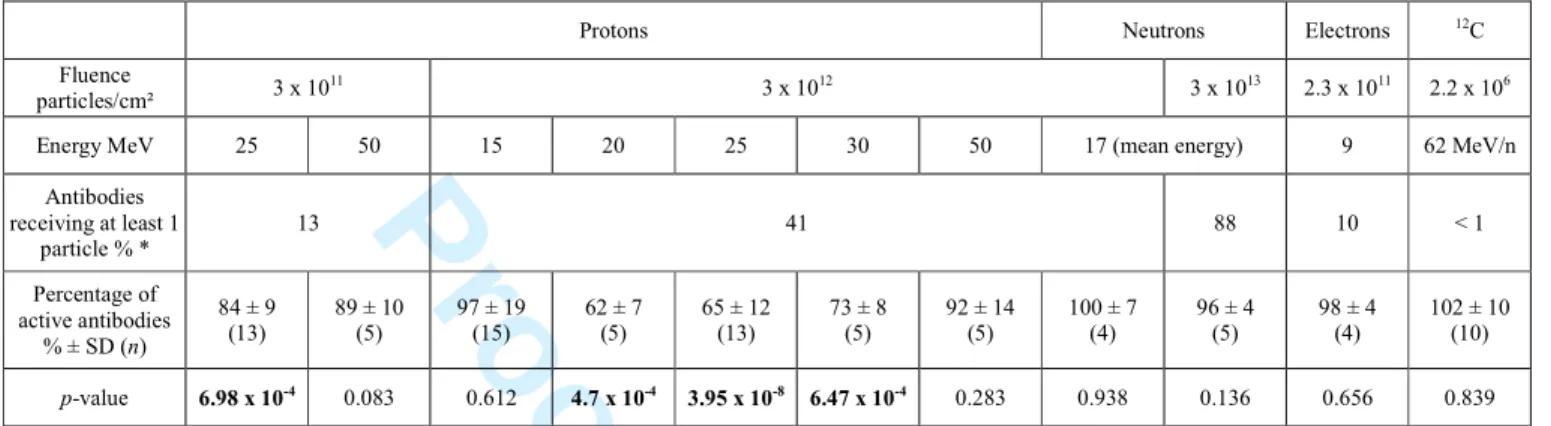
328 4.1. Grafted antibodies 329All the experiments performed on grafted antibodies are summarized in Table 1. This 330
table presents the type, energy and fluence of particles tested. It also specifies the 331
antibody grafting surface density allowing an assessment of the percentage of antibodies 332
receiving at least one particle for each tested fluence. The percentage of antibodies still 333
active after irradiation, calculated against non-irradiated controls (NIC), reveals possible 334
degradation induced only by radiation exposure. Indeed, the effects of transport 335
conditions are evaluated by confronting NIC with reference samples stored in the 336
laboratory (R4°C), as described in paragraph 3.2.2. However, as for all the tested 337
conditions NIC proved to be significantly equal to R4°C (not shown), only irradiation 338
effects are presented here. 339
Irradiation on the other hand had different effects on the tested antibodies. Indeed, 340
although no effect was detected with neutrons, electrons and 12C, significant effects 341
were observed with protons. Surprisingly, for high fluences, protons between 20 and 30 342
MeV significantly altered the antibody recognition performances, with losses around 343
30-35% and p-values between 10-4 and 10-8, but not at lower and higher energies. 344
Similarly, even at a lower fluence 25 MeV protons produced a significant recognition 345
Proof For Review
loss, though limited to only 10-20%, whereas at 50 MeV no significant recognition loss 346
was recorded. In our model the antibody surface density was maintained identical for 347
the different exposure experiments therefore only 13% of antibodies should have 348
received at least one particle at the lowest proton fluence against 42% at the highest. 349
The protons’ energy appears thus as a more damaging factor than the fluence, as only a 350
certain energy range (20-30 MeV) produced significant damage to antibodies regardless 351
of the fluence applied. However, by diminishing the ratio between the antibody surface 352
density and the particles’ fluence by a factor 3 (42% against 13% of antibodies 353
receiving at least one particle between high and low fluences respectively) the effect of 354
irradiation was greatly attenuated for 25 MeV protons (65% against 84% of active 355
antibodies respectively). 356
The other tested particles did not induce significant changes in antibody recognition 357
capabilities even at very high neutron fluence (3 x 10¹³ particles/cm²) or with heavy 358
carbon ions at high energy (62MeV/n). 359
360
4.2. Free antibodies
361
Free antibodies were irradiated by 25 MeV and 50 MeV protons and 17 MeV neutrons 362
at different fluences (3 x 10¹¹ and 3 x 10¹² particles/cm² for protons and 3 x 10¹² and 3 x 363
1013 particles/cm² for neutrons). Results are summarized in Table 2. The irradiation 364
effect was estimated following the methodology described in Baqué et al. (2011a). 365
Briefly, when the half maximal inhibitory concentration (IC50) significantly increases, 366
it indicates that in average, the antibodies have lost recognition capabilities since HRP 367
has only one epitope to which the antibody binds (Moreau et al. 2011). 368
Proof For Review
No modification of free antibody recognition capabilities under proton irradiations at 50 370
MeV was observed. However, at 25 MeV, we highlight here a significant recognition 371
capability loss for free antibodies, leading to a significant increase in IC50 compared to 372
the NIC. The increase in IC50 value indicates that, in average, the antibody activity has 373
been deteriorated by 25 MeV protons irradiation leading to partial or complete antigen 374
recognition site degradation. Based on a simplistic model, which considers that IC50 375
changes are only linked to a total loss of recognition capability, we can however 376
estimate the percentage of active antibodies compared to non-irradiated controls as 377
reported in Table 2. A maximum of 50% of antibodies appear to have lost their 378
recognition capability when irradiated with a high fluence of 25 MeV protons. Although 379
the other recorded changes in IC50 values after proton or neutron irradiation appear also 380
quite high, with 20 to 30 % damaged antibodies (most notably after a high neutron 381
flux), they were not significantly different from the controls. These results however 382
point out a high variability in the samples, which can be problematic for repeatability 383
measurements of future space instruments. 384
385
5. Discussion/Conclusion
386 387
Based on Monte Carlo simulations of the radiation environment faced by a biochip 388
dedicated to explore Mars’ surface (Le Postollec et al. 2009a), our team performed 389
several ground-based irradiation experiments on biochip recognition molecules. Even 390
though protons and neutrons clearly dominate the radiation spectrum during the Earth-391
Mars transit and on the Martian surface, other particles might be equally deleterious to 392
biological molecules such as the antibodies used in biochips. Furthermore, a wide range 393
of particle energy and fluence can be considered according to the envisaged mission to 394
Proof For Review
Mars but also to other planetary bodies of interest in the Solar System. In the present 395
study, the irradiation effects of protons, neutrons, electrons and carbon ions on the 396
recognition capabilities of antibodies were therefore investigated at different energies 397
and fluences. Two antibodies formulations were submitted to irradiation in order to 398
broadly represent any future biochip-based space instruments as both grafted and free 399
antibodies are considered. Our experimental approach consisted of using particle 400
fluences in the same order of magnitude as grafted antibodies surface density in order to 401
measure any damaging effect occurring when a particle interacts with an antibody. 402
Among the tested particles, only protons significantly altered the antibodies recognition 403
capabilities. These damaging effects were however recorded only for a certain energy 404
range between 20 and 30 MeV at both high and low fluences but confirmed for both 405
formulations (free and grafted antibodies). Indeed, at higher and lower protons energies 406
the antibodies recognition capabilities were not significantly altered. Irradiations of free 407
antibodies lead moreover to a high variability in the estimated recognition capabilities 408
of our antibodies samples. 409
Therefore, although the energy range of deleterious particles appears quite limited, a 410
biochip instrument performance would not be affected for a typical mission to Mars, as 411
the fluences of particles in this energy range will be significantly lower than the 412
antibody surface density. However, this result underlines that attention must be paid to 413
the ratio between antibody surface density and particles fluences expected for a given 414
mission. The biochip instrument must be designed so that antibody surface density is 415
much greater than incident protons fluence. Instrument shielding and/or antibodies 416
grafting density should be consequently adapted. 417
Proof For Review
In a similar ground-based study performed on five antibodies freeze-dried in different 419
protective molecular matrices, Derveni et al. (2012) pointed out the more damaging role 420
of processing and packaging than irradiation. Using doses of protons and neutrons at 421
high energies (50 and 47 MeV respectively), comparable to the ones used in the present 422
work, they did not detect any evidence of activity loss due to irradiation for a typical 423
mission dose (1011 to 1012 protons/cm2 and 107 to 108 neutrons/cm²). However, using 424
1013 protons/cm2, most of the antibodies lost their activity. Thanks to these results they 425
suggested that further shielding or alternative radiation protection approaches would 426
need to be considered for long duration missions to other astrobiological targets. Our 427
present work confirms this suggestion. We propose that the ratio between the fluence of 428
protons and the surface density of antibodies has to be much lower than unity to prevent 429
important loss of activity. 430
431
The main limitation of ground-based studies is that each constraint is generally studied 432
individually and for a limited period of time that is not representative of a real space 433
mission. In particular, the effect of cosmic rays is generally studied at a given energy (or 434
a limited range of energies) and for one type of particle in a single experiment. 435
Moreover, additional constraints and hazards are expected for a space instrument. Long 436
term storage, temperature variations, contamination risks, launch, landing and 437
transportations vibrations and shocks should all be taken into account in the design and 438
testing of a space dedicated instrument. For these reasons, a real space exposure of 439
biochip prototypes has been attempted in the past by the LMC team for a short-term 440
mission aboard the BIOPAN platform on a Russian Foton spacecraft (Derveni et al. 441
2013) and ground-based and field studies have been performed for the SOLID prototype 442
(Parro et al. 2008; Sobrado et al. 2014). Furthermore, in the frame of the BiOMAS 443
Proof For Review
project, biochip samples are currently exposed to real space conditions inside the 444
EXPOSE-R2 platform of ESA, part of the Photochemistry on the Space Station (PSS) 445
project, which was installed on the outside of the Zvezda module of the International 446
Space Station (ISS) in August 2014 (Vigier et al. 2013). 447
The long-duration exposure of the EXPOSE missions (Rabbow et al. 2009, 2012, 2015) 448
range from 12 to 18 months in the LEO environment of the ISS. The radiation 449
environment at this altitude, although not equivalent to interplanetary space or the 450
Martian surface, will allow anyway for a much better estimate of the long-term 451
resistance of immunoassays instruments for space applications. 452
Nevertheless, due to the high number of potentially hazardous factors encountered 453
during a space mission, ground-based studies are essential to isolate the most damaging 454
ones and thus propose adequate shielding or handling procedures. 455
456
Thus, our results from ground-based irradiation campaigns globally indicate that cosmic 457
rays might not alter the final performance of a biochip-based instrument in a typical 458
Martian mission, when antibodies are used as binders to detect the presence or the 459
absence of a target compound. The damaging effects of 20-30 MeV protons recorded in 460
the present study should not however be overlooked and further testing on-ground will 461
be necessary to support and interpret data from real space exposure missions. 462
463 464
Acknowledgments
465
The research leading to these results has received funding from the European Union 466
Seventh Framework Programme FP7/2007-2013 under Grant Agreement no 262010 – 467
ENSAR. 468
Proof For Review
We would like to thank the French National Space Agency (CNES) (Convention 469
number DCT/SI/IM/2009-17733) and the Interdisciplinary CNRS program 470
“Environnements Planétaires et Origines de la Vie” for financial support. 471
We also thank the Louvain-la-Neuve cyclotron facility staff, the Institut Bergonié staff 472
and the staff from the Laboratori Nazionali del Sud of the Instituto Nazionale di Fisica 473
Nucleare for their help during irradiation experiments. 474
Proof For Review
References
476 477
Agostinelli, S. et al. (2003) Geant4—a simulation toolkit. Nucl. Instrum. Methods 478
Phys. Res. Sect. Accel. Spectrometers Detect. Assoc. Equip.. 506(3), 250–303.
479
Allison, J. et al. (2006) Geant4 developments and applications. IEEE Trans. Nucl. Sci.. 480
53(1), 270–278.
481
Baqué, M. et al. (2011a) Investigation of Low-Energy Proton Effects on Aptamer 482
Performance for Astrobiological Applications. Astrobiology. 11(3), 207–211. 483
Baqué, M., Le Postollec, A., Coussot, G., Moreau, T., Desvignes, I., Incerti, S., 484
Moretto, P., Dobrijevic, M., & Vandenabeele-Trambouze, O. (2011b) Biochip 485
for astrobiological applications: Investigation of low energy protons effects on 486
antibody performances. Planet. Space Sci.. 59(13), 1490 – 1497. 487
Berger, G., Ryckewaert, G., Harboe-Sorensen, R., & Adams, L. (1997) Cyclone–A 488
multipurpose heavy ion, proton and neutron see test site. In 1997 RADECS 489
Conference Data Workshop. p. 51.
490
Coussot, G., Faye, C., Ibrahim, A., Ramonda, M., Dobrijevic, M., Postollec, A., 491
Granier, F., & Vandenabeele-Trambouze, O. (2011) Aminated dendritic 492
surfaces characterization: a rapid and versatile colorimetric assay for estimating 493
the amine density and coating stability. Anal. Bioanal. Chem.. 399(6), 2295– 494
2302. 495
Coussot, G., Perrin, C., Moreau, T., Dobrijevic, M., Postollec, A.L., & Vandenabeele-496
Trambouze, O. (2011) A rapid and reversible colorimetric assay for the 497
characterization of aminated solid surfaces. Anal. Bioanal. Chem.. 399(3), 1061– 498
1069. 499
Derveni, M., Allen, M., Sawakuchi, G.O., Yukihara, E.G., Richter, L., Sims, M.R., & 500
Cullen, D.C. (2013) Survivability of Immunoassay Reagents Exposed to the 501
Space Radiation Environment on board the ESA BIOPAN-6 Platform as a 502
Prelude to Performing Immunoassays on Mars. Astrobiology, 503
130103060450009. 504
Derveni, M., Hands, A., Allen, M., Sims, M.R., & Cullen, D.C. (2012) Effects of 505
Simulated Space Radiation on Immunoassay Components for Life-Detection 506
Experiments in Planetary Exploration Missions. Astrobiology. 12(8), 718–729. 507
de Diego-Castilla, G., Cruz-Gil, P., Mateo-Martí, E., Fernández-Calvo, P., Rivas, L.A., 508
& Parro, V. (2011) Assessing Antibody Microarrays for Space Missions: Effect 509
of Long-Term Storage, Gamma Radiation, and Temperature Shifts on Printed 510
and Fluorescently Labeled Antibodies. Astrobiology. 11(8), 759–773. 511
Gobet, F. et al. (Submitted) Experimental and Monte Carlo absolute characterization of 512
a medical electron beam. Radiat. Meas. 513
Proof For Review
Hassler, D.M. et al. (2013) Mars’ Surface Radiation Environment Measured with the 514
Mars Science Laboratory’s Curiosity Rover. Science, 1244797. 515
Kim, M.-H.Y. et al. (2014) Comparison of Martian Surface Radiation Predictions to 516
the Measurements of Mars Science Laboratory Radiation Assessment Detector 517
(MSL/RAD). In American Geophysical Union Fall 2014 Meeting. San 518
Francisco, CA, United States. 519
Manchado, J.M., Sebastián, E., Romeral, J., Sobrado-Vallecillo, J., Herrero, P.L., 520
Compostizo, C., Gómez-Elvira, J., & Parro, V. (2015) SOLID SPU: A TRL 5-6 521
Sample Preparation Instrument for Wet Chemistry Analysis on Mars. In Lunar 522
and Planetary Science Conference. p. 1222. 523
Martins, Z. (2011) In situ biomarkers and the Life Marker Chip. Astron. Geophys.. 524
52(1), 1.34–1.35.
525
McKay, C.P. et al. (2013) The Icebreaker Life Mission to Mars: A Search for 526
Biomolecular Evidence for Life. Astrobiology. 13(4), 334–353. 527
McKay, D.S., Steele, A., Allen, C., Thomas-Keptra, K., Schweitzer, M.H., Priscu, J., 528
Sears, J., Avci, R., & Firman, K. (2000) Mars immunoassay life detection 529
instrument (MILDI). Lunar Planet Inst Contrib. (1062 Part 2), 219–220. 530
McKenna-Lawlor, S., Gonçalves, P., Keating, A., Reitz, G., & Matthiä, D. (2012) 531
Overview of energetic particle hazards during prospective manned missions to 532
Mars. Planet. Space Sci.. 63–64(0), 123–132. 533
Moreau, T., Faye, C., Baqué, M., Desvignes, I., Coussot, G., Pascal, R., & 534
Vandenabeele-Trambouze, O. (2011) Antibody-based surfaces: Rapid 535
characterization using two complementary colorimetric assays. Anal. Chim. 536
Acta. 706(2), 354–360.
537
Parro, V. et al. (2005) Instrument development to search for biomarkers on mars: 538
Terrestrial acidophile, iron-powered chemolithoautotrophic communities as 539
model systems. Planet. Space Sci.. 53(7), 729–737. 540
Parro, V. et al. (2008) SOLID2: an antibody array-based Life-detector instrument in a 541
Mars drilling simulation experiment (MARTE). Astrobiology. 8(5), 987–999. 542
Parro, V. et al. (2011a) Classification of Modern and Old Rio Tinto Sedimentary 543
Deposits Through the Biomolecular Record Using a Life Marker Biochip: 544
Implications for Detecting Life on Mars. Astrobiology. 11(1), 29–44. 545
Parro, V. et al. (2011b) SOLID3: a multiplex antibody microarray-based optical sensor 546
instrument for in situ Life detection in planetary exploration. Astrobiology. 547
11(1), 15–28.
548
Le Postollec, A. et al. (2007) Development of a Biochip dedicated to planetary 549
exploration. First step: resistance studies to space conditions. In Journées SF2A 550
2007 Semaine de l’Astrophysique Française 2007.
Proof For Review
Le Postollec, A. et al. (2009a) Monte Carlo Simulation of the Radiation Environment 552
Encountered by a Biochip During a Space Mission to Mars. Astrobiology. 9(3), 553
311–323. 554
Le Postollec, A., Coussot, G., Baqué, M., Incerti, S., Desvignes, I., Moretto, P., 555
Dobrijevic, M., & Vandenabeele-Trambouze, O. (2009b) Investigation of 556
Neutron Radiation Effects on Polyclonal Antibodies (IgG) and Fluorescein Dye 557
for Astrobiological Applications. Astrobiology. 9(7), 637–645. 558
Rabbow, E. et al. (2009) EXPOSE, an astrobiological exposure facility on the 559
International Space Station - from proposal to flight. Orig. Life Evol. Biospheres. 560
39(6), 581–598.
561
Rabbow, E. et al. (2012) EXPOSE-E: an ESA astrobiology mission 1.5 years in space. 562
Astrobiology. 12(5), 374–386.
563
Rabbow, E. et al. (2015) The astrobiological mission EXPOSE-R on board of the 564
International Space Station. Int. J. Astrobiol..14(1), 3–16. 565
Sephton, M.A., Sims, M.R., Court, R.W., Luong, D., & Cullen, D.C. (2013) Searching 566
for biomolecules on Mars: Considerations for operation of a life marker chip 567
instrument. Planet. Space Sci.. 86, 66–74. 568
Sims, M., Cullen, D., Bannister, N., Grant, W., Henry, O., Jones, R., McKnight, D., 569
Thompson, D.P., & Wilson, P. (2005) The specific molecular identification of 570
life experiment (SMILE). Planet. Space Sci.. 53(8), 781–791. 571
Sims, M.R. et al. (2012) Development status of the life marker chip instrument for 572
ExoMars. Planet. Space Sci.. 72(1), 129–137. 573
Smith, H. & Parro, V. (2014) Planetary Protection Plan for an Antibody based 574
instrument proposed for Mars2020. In 40th COSPAR Scientific Assembly. p. 575
3140. 576
Sobrado, J.M., Martín-Soler, J., & Martín-Gago, J.A. (2014) Mimicking Mars: A 577
vacuum simulation chamber for testing environmental instrumentation for Mars 578
exploration. Rev. Sci. Instrum.. 85(3), 035111. 579
Vigier, F. et al. (2013) Preparation of the Biochip experiment on the EXPOSE-R2 580
mission outside the International Space Station. Adv. Space Res.. 52(12), 2168– 581
2179. 582
Wang, J. (2006) Electrochemical biosensors: Towards point-of-care cancer diagnostics. 583
Biosens. Bioelectron.. 21(10), 1887–1892.
584
Zeitlin, C. et al. (2013) Measurements of Energetic Particle Radiation in Transit to 585
Mars on the Mars Science Laboratory. Science. 340(6136), 1080–1084. 586
587 588
Proof For Review
Figure legends
589
Fig. 1: Simulated spectra of particle fluxes, as a function of energy, during the
Earth-590
Mars transit (left) and at Mars’ surface (right) with the energy range of particles 591
investigated in this study (red zone). This figure is an adaptation of Fig. 8 in Le 592
Postollec et al. (2009a). 593
Fig. 2: Top: Proton irradiation using the Light Ion Facility (LIF) at the cyclotron of
594
Louvain-la-Neuve. The source is located on the left in this picture and the samples are 595
placed on the right behind a metal slide with a 10 cm diameter hole. Several removable 596
disks are placed between the source and the samples to allow the modulation of protons 597
energy. Bottom Left: Picture of the facility at the Institut Bergonié where samples were 598
irradiated with 9 MeV electrons. Bottom Right: Mobile device developed to ensure the 599
ELISA plate motion during carbon ions irradiation at LNS (Catania). 600
Proof For Review
Proof For Review
Proof For Review
Table 1. Influence of neutron, proton, electron and carbon radiation effects on grafted
antibodies recognition capability at different fluences. The percentages of active antibodies were normalized using the NIC that were thus fixed at 100%. The percentage of antibodies receiving at least one particle was calculated according to the antibody surface density, the tested fluence and the sample geometry. SD, standard deviation; n is the number of measurements. p-value < 0.05 (in bold) indicate samples that are different to NIC at 95 % of confidence.
Protons Neutrons Electrons 12
C Fluence particles/cm² 3 x 10 11 3 x 10¹² 3 x 10¹³ 2.3 x 10¹¹ 2.2 x 10⁶
Energy MeV 25 50 15 20 25 30 50 17 (mean energy) 9 62 MeV/n
Antibodies receiving at least 1 particle % * 13 41 88 10 < 1 Percentage of active antibodies % ± SD (n) 84 ± 9 (13) 89 ± 10 (5) 97 ± 19 (15) 62 ± 7 (5) 65 ± 12 (13) 73 ± 8 (5) 92 ± 14 (5) 100 ± 7 (4) 96 ± 4 (5) 98 ± 4 (4) 102 ± 10 (10) p-value 6.98 x 10⁻⁴ 0.083 0.612 4.7 x 10⁻⁴ 3.95 x 10⁻⁸ 6.47 x 10⁻⁴ 0.283 0.938 0.136 0.656 0.839 *Antibody surface density is equal to 8.8 x 10¹¹ Ab/cm² for all experiments.
Proof For Review
Table 2. Influence of neutron and proton irradiation on free-antibody recognition capability at
different fluences. IC50 (µg/mL), half maximal inhibitory concentration; SD, standard deviation; n is the number of measurements. The percentages of active antibodies were estimated in comparison with NIC. p-values < 0.05 (in bold) indicate samples that are different to non-irradiated controls at 95 % of confidence.
Protons Neutrons Non-irradiated controls (NIC) Fluence particles/cm² 3 x 10¹¹ 3 x 10¹² 3 x 10¹³
Energy MeV 25 50 25 50 17 (mean energy)
IC 50 (µg/mL) ± SD (n) 3.1 ± 0.2 (4) 4.1 ± 1.0 (7) 4.7 ± 1.0 (7) 3.8 ± 0.8 (8) 3.2 ± 0.2 (4) 4.2 ±1.4 (8) 3.2 ± 0.6 (18) Percentage of active antibodies % 100 71 50 79 105 73 100 p-value 0.946 0.059 0.004 0.085 0.666 0.163