Identifying breast cancer patients at risk for Central
Nervous System (CNS) metastases in trials of the
International Breast Cancer Study Group (IBCSG)
B. C. Pestalozzi
1, D. Zahrieh
2, K. N. Price
3, S. B. Holmberg
4, J. Lindtner
5, J. Collins
6,
D. Crivellari
7, M. F. Fey
8, E. Murray
9, O. Pagani
10, E. Simoncini
11, M. Castiglione-Gertsch
12,
R. D. Gelber
2,3, A. S. Coates
13& A. Goldhirsch
10,14For the International Breast Cancer Study Group
1Department of Oncology, University Hospital, Zu¨rich, Switzerland and the Swiss Group for Clinical Cancer Research (SAKK);2
IBCSG Statistical Center, Dana-Farber Cancer Institute, Boston, MA, USA;3
Frontier Science and Technology Research Foundation, Boston, MA, USA;4
Department of Surgery, SU/Moelndal’s Hospital, Moelndal, Sweden;5
Department of Surgery, Institute of Oncology, Ljubljana, Slovenia;6
Department of Surgery, Royal Melbourne Hospital, Australian New Zealand Breast Cancer Trials Group, Melbourne, Australia;7
Centro di Riferimento Oncologico, Aviano, Italy;8
Department of Medical Oncology, Inselspital, Bern, Switzerland and the Swiss Group for Clinical Cancer Research (SAKK);9Groote Schuur Hospital and University of Cape Town, South Africa;10Oncology Institute of Southern Switzerland, Lugano, Switzerland and the Swiss Group for Clinical Cancer Research (SAKK);11Oncologia Medica-Spedali Civili, Brescia, Italy;12IBCSG Coordinating Center, and Institute of Medical Oncology, Inselspital, Bern, Switzerland;13
The Cancer Council Australia, Australian New Zealand Breast Cancer Trials Group and School of Public Health, University of Sydney, Australia;14
European Institute of Oncology, Milan, Italy
Received 1 December 2005; revised 10 February 2006; accepted 28 February 2006
Background:
We sought to determine whether a high-risk group could be defined among patients with operable breast cancer in whom a search of occult central nervous system (CNS) metastases was justified.Patients and methods:
We evaluated data from 9524 women with early breast cancer (42% node-negative) who were randomized in International Breast Cancer Study Group clinical trials between 1978 and 1999, and treated without anthracyclines, taxanes, or trastuzumab. We identified patients whose site of first event was CNS and those who had a CNS event at any time.Results:
Median follow-up was 13 years. The 10-year incidence (10-yr) of CNS relapse was 5.2% (1.3% as first recurrence). Factors predictive of CNS as first recurrence included: node-positive disease (10-yr = 2.2% for > 3 N+), estrogen receptor-negative (2.3%), tumor size > 2 cm (1.7%), tumor grade 3 (2.0%), < 35 years old (2.2%), HER2-positive (2.7%), and estrogen receptor-negative and node-positive (2.6%). The risk of subsequent CNS recurrence was elevated in patients experiencing lung metastases (10-yr = 16.4%).Conclusion:
Based on this large cohort we were able to define risk factors for CNS metastases, but could not define a group at sufficient risk to justify routine screening for occult CNS metastases.Key words:
breast cancer, central nervous system, adjuvant chemotherapy, competing risks, CMF, metastasesintroduction
In women with breast cancer, a population-based estimate of
the incidence proportion of brain metastases is 5.1% [1].
Based on case series, the incidence of clinically evident central
nervous system (CNS) metastases among women with
metastatic breast cancer is estimated to be 10% to 16% [2, 3].
In autopsy series, brain metastases are found in 20% to 30%
of patients. To our knowledge, the present report is the first
series on a large cohort of patients with long-term follow-up
(median 13 years) from the diagnosis of early-stage breast
cancer which analyzes factors associated with CNS recurrence.
The purpose of this report is to determine whether there is
a patient population with a risk of CNS metastases high
enough to justify routine screening for CNS recurrence.
patients and methods
We analyzed data from 9524 patients with early breast cancer who were entered onto International Breast Cancer Study Group (IBCSG; formerly the Ludwig Breast Cancer Study Group) trials I through IX [4–10] between 1978 and 1999 (Table 1). Clinical, hematological and biochemical assessments of each patient were required every three months for two years, every six months until the end of the fifth year and yearly thereafter until death. All sites of disease relapse, including the first and subsequent events until death, were recorded in the study databases. In trials I-V, chest X-rays and bone scans were required every six months for two years and once yearly up to five years. These tests were recommended beyond the fifth year only if clinically indicated. In trials VI, VII and VIII, chest x-rays and bone scans were required but further tests were performed only
original
article
Correspondence to: Dr B. C. Pestalozzi, Department of Oncology, University Hospital, Ra¨mistrasse 100, 8091 Zu¨rich, Switzerland. Tel:+41 44 255 22 14; Fax:+41 44 255 45 48; E-mail: bernhard.pestalozzi@usz.ch
following clinical indications. In trial IX further tests were recommended only if clinically indicated.
For all trials, ER concentrations of greater than or equal to 10 fmol/mg of cytosol protein by ligand-binding assay were considered positive; lower values were considered negative. Steroid hormone receptor determination by immunohistochemistry was allowed later in trials VI through IX. The cohort of patients with node-positive, ER-negative disease was prospectively defined as high risk population of interest.
Centrally assessed HER2 was available for a subgroup of 3871 patients from trials V, VIII, and IX, and was evaluated using
immunohistochemistry. Central immunohistochemical analysis for patients in trial V (n = 1506) was performed in 1991 using the monoclonal antibody ICR12, which recognizes the external domain of the c-erbB-2 protein. The antibody was titrated to only stain tumors that showed amplification [11] and would thus be equivalent to 3+ using current criteria. For trials VIII–IX (n = 2365) HER2 was evaluated in 2002 according to the 4 tiers (from 0 to 3+) FDA-approved scoring system, taking into account the percentage of immunoreactive cells in the invasive component of the tumors, and the intensity and completeness of membrane staining [12]. For trials VIII-IX 3+ is considered HER2-positive. The subset of patients that have HER2 status available is largely node-negative (81%).
All patient data, including data regarding disease- and survival-related events, were reviewed and classified by the medical study coordinators (A.G. and M.C.-G.).
statistical methods
Cumulative incidence functions were used to estimate the percentage of patients who experienced the various competing events within the study cohorts rather than the overestimated percentages obtained with the Kaplan-Meier method based on the cause-specific hazards [13–15]. Differences between the cumulative incidence functions according to patient subgroups were tested for statistical significance using the procedure of Gray [16]. Analyses were conducted to determine whether the risk of recurrence in CNS increased according to baseline characteristics or after a first recurrence elsewhere. A cumulative incidence function regression model of Fine and Gray [17] was used for multiple regression analyses. Covariates included in the model were nodal status, pathologic tumor size, tumor grade, estrogen receptor status, menopausal status, and age. Overall survival from the time of CNS recurrence was estimated using the method of Kaplan and Meier [18]. P values are two-sided.
categories of sites of recurrence
First breast cancer recurrence events were classified according to their sites. Local recurrences were confined to the ipsilateral conserved breast or ipsilateral chest wall (with or without mastectomy scar involvement). Regional recurrences involved ipsilateral axillary, supraclavicular, and/or internal mammary lymph node metastases. Distant recurrences involved soft tissue, bone, and visceral metastases, including CNS and all other organ involvement and diffuse intra-abdominal metastases. Other first Table 1. Characteristics of IBCSG Trials I through IX
Trial Population Years of accrual No. of eligible patients Treatment groups Median follow-up (years)
I Premenopausal women with 1–3 Pos nodes 1978–1981 491 CMF·12 23 CMFp·12
II Premenopausal women with ‡ 4 Pos nodes 1978–1981 327 CMFp·12 22 Ox + CMFp·12
III Postmenopausal women < 65 years old 1978–1981 463 Observation 23 p + T·12
CMFp + T·12
IV Postmenopausal women 66–80 years old 1978–1981 320 Observation 22 p + T·12
V Pre- or postmenopausal women with Neg nodes 1981–1985 1275 Observation 19 PeCMF
V Pre- or postmenopausal women with Pos nodes 1981–1985 1229 PeCMF 18 CMFpT·6
PeCMF + CMFpT·6
VI Premenopausal women with Pos nodes 1986–1993 1475 CMF·6 13 CMF·6 + reint
CMF·3 CMF·3 + reint
VII Postmenopausal women with Pos nodes 1986–1993 1212 T 13 T + delayed CMF
T + CMF·3
T + CMF·3 delayed CMF VIII Premenopausal women with Neg nodes 1990–1999 1063 Goserelin·24 mos. 8
CMF·6
CMF·6 / Gos·18 mos. IX Postmenopausal women with Neg nodes 1988–1999 1669 T·5 years 9
CMF·3 / T·57 mos.
Abbreviations: Pos, positive; Neg, negative; C, cyclophosphamide 100 mg/m2orally (PO) days 1–14 of each cycle; M, methotrexate 40 mg/m2intravenously (IV) days 1 and 8 of each cycle; F, fluorouracil 600 mg/m2IV days 1 and 8 of each cycle; p, prednisone 7.5 mg/d PO; Ox, oophorectomy; T, tamoxifen 20 mg
PO once daily; PeCMF, perioperative CMF; reint, reintroduction of 3 cycles of CMF; delayed CMF, 3 cycles of CMF 9, 12, and 15 months after randomization; goserelin, monthly subcutaneous implants at 3.6 mg.
events, including contralateral breast cancer, non-breast cancer second malignancies, and death without malignancy, were also recorded. Time to first event was defined as time from randomization to the occurrence of a first event of any type. An event was considered to be a component of a first event if diagnosed within a 2-month time frame.
Occurrence of CNS metastases as the first site of recurrence with or without recurrence at any other site was the event of interest. Diagnosis of CNS recurrence was based on clinical assessment. At the time of relapse CNS imaging was not performed unless there was a clinical suspicion of CNS recurrence. All other sites of first recurrence (not CNS) and any other event, such as, contralateral breast cancer, non-breast cancer second primary tumors, and death without recurrence, were considered competing events. The sum of the cumulative incidence of CNS metastases plus the cumulative incidence of the other competing events equals the cumulative incidence of a first event of any type. In this retrospective analysis it was not possible to distinguish between parenchymal brain metastases and leptomeningeal disease. Since leptomeningeal disease is reportedly five times less frequent than parenchymal brain metastases [2], this study essentially discusses the latter.
In addition to site of first recurrence, we calculated cumulative incidence of CNS events at any time (either as first event or as subsequent recurrences). Time to CNS recurrence at any time was defined as the time from randomization to the time of CNS recurrence whether it was a first or subsequent event. Death before CNS recurrence was considered the only competing event in this analysis. Patients experiencing neither CNS recurrence nor death were censored at the time last known to be alive without CNS recurrence.
We analyzed the cumulative incidence of subsequent CNS metastases according to sites of first recurrence. The time to subsequent recurrence was measured from the time of the first recurrence; death without subsequent CNS recurrence was the only competing event. CNS recurrence was observed at autopsy for 28 patients (0.3%). For this report, these 28 cases were included in the analysis as competing risks for estimating the incidence of CNS recurrence.
results
Included in the analysis were 9524 eligible patients from
IBCSG trials I through IX (Table 1), and patient characteristics
are shown in Table 2. The sites of first event are shown in
Table 3. At a median follow-up of 13 years, 46.2% of all patients
were alive without recurrence; 53.8% (5122 of 9524)
experienced a first event, namely disease recurrence at known
sites (n = 3937), contralateral breast cancer (n = 384), failure
at an unknown site (n = 36), a non-breast cancer second
primary (n = 389), or death without recurrence (n = 376).
Overall, CNS was a component of first recurrence in 1.3%
of patients (126 of 9524). Fifty-five patients (0.6%) experienced
a CNS recurrence concurrently (within 2 months) with other
sites as a first event.
The site-specific cumulative incidences of CNS metastases
and other competing events as first recurrence are shown in
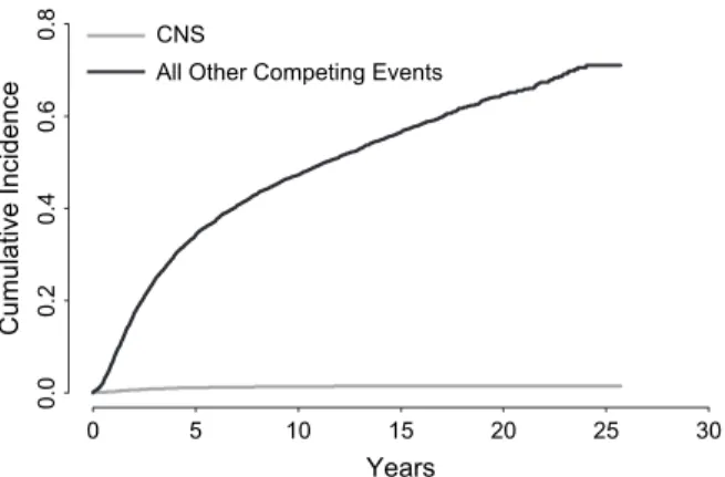
Figure 1. At 10 years from study entry, the cumulative incidence
of CNS metastases as components of first recurrences was 1.3%,
and the cumulative incidence of competing first events was
47.2%. At 10 years, the total cumulative incidence of recurrence
due to any cause as a first event was 48.5% (1.3% + 47.2%).
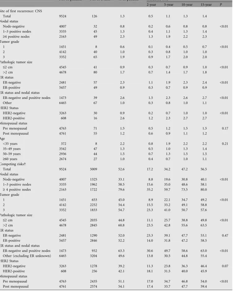
Table 4 lists the cumulative incidences of CNS metastases
as site of first recurrence and competing events at 2, 5, 10 and
15 years from randomization, for the overall population and
for subpopulations defined according to lymph node
involvement, tumor grade, pathologic tumor size, ER status,
the combination of ER and lymph node status (ER-negative
with positive nodes versus other), HER2 status, menopausal
Table 2. Patient and tumor characteristics
No. of patients
Percentage
Total cases 9524 100.0
Mastectomy 7371 77.4
Breast-conserving surgery with radiotherapy 2153 22.6 0 positive nodes 4007 42.1 1–3 positive nodes 3354 35.2 4 or more positive nodes 2163 22.7
Tumor grade 1 1451 15.2
Tumor grade 2 4142 43.5
Tumor grade 3 3352 35.2
Tumor grade unknown 579 6.1
Tumor size £2cm 4545 47.7
Tumor size >2cm 4678 49.1 Tumor size unknown 301 3.2
ER-positive 5457 57.3
ER-negative 2481 26.0
ER-unknown 1586 16.7
ER-negative and positive nodes 1473 15.5
Other* 6465 67.9 ER-unknown 1586 16.6 HER2-negative 3263 34.3 HER2-positive 608 6.4 HER2 unknown 5653 59.4 Premenopausal 4763 50.0 Postmenopausal 4761 50.0 Age < 35 years 372 3.9 Age 35–49 years 3542 37.2 Age 50–59 years 2936 30.8 Age ‡ 60 years 2674 28.1
*Subgroup defined as not node-positive with ER-negative primary tumors.
Table 3. Sites of first event
No. of patients Percentage
Total patients 9524 100.0 Events 5122 53.8 Deaths 4043 42.5 Sites Local 796 8.4 Contralateral breast 384 4.0 Regional 549 5.8
Distant: soft tissue, nodes 158 1.7
Distant: bone 1052 11.0
Distant: viscera* 1382 14.5 Second (non-breast) primary 389 4.1 Death w/o recurrence 376 3.9
Unknown 36 0.4
status, and age. Significant differences across categories of
lymph node status, tumor grade, pathologic tumor size, ER
status, HER2 status, and the combination of ER and lymph
node status were observed in univariate analyses among
patients with recurrence in CNS. Patients with node-positive
disease and ER-negative primary tumors at the time of diagnosis
were prospectively defined as a group of interest expected to
show a higher risk of CNS recurrence. This subgroup had
2- and 10-year cumulative incidence of CNS as first event of
1.5% and 2.6%, respectively (P < 0.01). Among patients
presenting with four or more involved nodes, the cumulative
incidence of CNS metastases as first event was 1.3% at 2 years
and 2.2% at 10 years (P < 0.01). The incidence of CNS disease
was significantly higher among patients with ER-negative
primary tumors, with a 10-year cumulative incidence of 2.3%
noted, compared with an incidence of 0.9% among patients
with ER-positive primary tumors (P < 0.01), and patients
with HER2-positive tumors with a 10-year cumulative incidence
of 2.7% compared with 1.0 for those with HER2-negative
tumors (P < 0.01). Larger tumor size was predictive of
a significantly higher incidence of CNS involvement at 10 years
(1.7% versus 0.9%; P < 0.01). Also, the incidence of CNS disease
was significantly higher among patients with grade 3 tumors,
with a 10-year cumulative incidence of 2.0%. All significant
factors retained statistical significance in the multiple
regression analyses (results not shown).
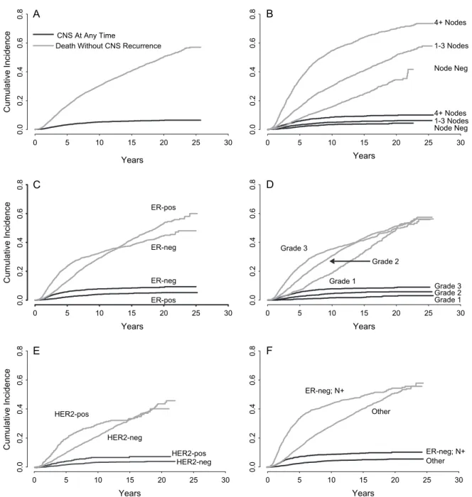
The cumulative incidences of recurrence in CNS at any
time (whether first or subsequent recurrence) are listed in
Table 5. CNS was a site of recurrence at any time in 5.4% of
patients. Fifty-seven percent of patients were still alive without
CNS metastases, and 37.1% had died without known CNS
metastases. The 10-year cumulative incidence of CNS
recurrence at any time was 5.2% (Figure 2A). Among patients
presenting with four or more involved nodes, the cumulative
incidence of CNS metastases at any time was 2.3% at 2 years
from randomization and 8.5% at 10 years (Figure 2B). The
cumulative incidence of CNS metastases at any time was
significantly higher among patients with ER-negative tumors
than among patients with ER-positive tumors (7.8% versus
4.0% at 10 years; P < 0.01) We note with interest the different
patterns of the risk of death across time for cohorts defined
by ER status. (Figure 2C). Among patients with grade 3 tumors,
the cumulative incidence of CNS metastases at any time was
1.9% at 2 years from randomization and 7.8% at 10 years
(Figure 2D). In patients with HER2-positive disease, the 10-year
cumulative incidence of CNS disease was higher than among
patients with HER2-negative disease (6.8% versus 3.5% at
10 years; P < 0.01; Figure 2E). In the high risk cohort of patients
with node-positive disease and ER-negative primary tumors,
the cumulative incidence of CNS metastases at any time was
significantly higher than among patients in the cohort
without node-positive disease and ER-negative primary tumors
(8.7% versus 4.4% at 10 years; P < 0.01; Figure 2F). Younger
patients (< 35 years), those with larger tumors, and
premenopausal patients also had significantly higher incidences
of metastases in CNS (all P < 0.01). Again, these factors
remained statistically significant in the multiple regression
analyses (results not shown).
A total of 391 patients experienced a subsequent CNS
recurrence after a first recurrence elsewhere (Table 6). Site of
first recurrence was recorded as viscera for 135 (34.5%) and
bone for 85 (21.7%) of these 391 patients. When viscera was
the first site of recurrence (n = 1256; excluding the 126 CNS
events classified as viscera as site of first recurrence),
a subsequent CNS recurrence occurred in 10.7% (135 of 1256)
of cases. The cumulative incidence of a subsequent recurrence
in CNS among the 1256 patients whose first recurrence was
viscera was 7.6% at 2 years and 11.4% at 10 years after the first
recurrence. When bone was the first site of recurrence,
a subsequent CNS recurrence occurred in 8.1% (85 of 1052)
of cases. The 2- and 10-year cumulative incidence of
a subsequent recurrence in CNS among the 1052 patients
whose first recurrence was bone was 4.6% and 8.6%,
respectively.
Within the cohort of 577 patients who experienced lung
metastasis as site of first recurrence (excluding 26 patients
who had concurrent sites in the CNS and lung), 90 (15.6%)
patients experienced a subsequent recurrence in CNS. The
cumulative incidence of a subsequent recurrence in CNS
among the 577 patients whose first recurrence was lung was
10.6% at 2 years and 16.4% at 10 years after the first
recurrence (Table 6).
We also examined the relationship between the number
of documented metastatic sites and CNS recurrence. The
proportion of CNS recurrences increased as the number
of metastatic sites increased (data not shown).
Five hundred and seventeen patients experienced a CNS
recurrence at any time (whether first or subsequent
recurrences). The median survival after CNS recurrence was
4 months, and only 20% of these patients survived beyond
1 year (Figure 3). In view of the dismal overall prognosis, it
is surprising that four patients have survived 4.5 years beyond
CNS recurrence. Therapeutic information was available for
three of these four cases. In all three cases, CNS recurrence
was treated with surgical excision, while radiotherapy was
added for two of the three cases.
discussion
Despite treatment, the prognosis after CNS recurrence in
breast cancer is dismal [2, 3], with a survival rate of only 20%
0 5 10 15 20 25 30 0.0 0.2 0.4 0.6 0.8 Cumulative Incidence CNS
All Other Competing Events
Years
Figure 1. Cumulative incidence of CNS metastases and other competing events as first recurrences among 9524 patients. Time was measured from the date of randomization.
Table 4. Site-specific cumulative incidence: site of first recurrence (measured from date of randomization)
No. of patients No. of events % of patients Incidence (%)
2-year 5-year 10-year 15-year P Site of first recurrence: CNS
Total 9524 126 1.3 0.5 1.1 1.3 1.4 Nodal status Node-negative 4007 32 0.8 0.2 0.6 0.8 0.8 <0.01 1–3 positive nodes 3335 45 1.3 0.4 1.1 1.3 1.4 ‡4 positive nodes 2163 49 2.3 1.3 1.9 2.2 2.3 Tumor grade 1 1451 8 0.6 0.1 0.4 0.5 0.7 <0.01 2 4142 40 1.0 0.3 0.8 1.0 1.0 3 3352 65 1.9 0.9 1.7 2.0 2.0
Pathologic tumor size
£2 cm 4545 41 0.9 0.3 0.7 0.9 1.0 <0.01
>2 cm 4678 80 1.7 0.7 1.4 1.7 1.8
ER status
ER-negative 2481 57 2.3 1.1 1.9 2.3 2.4 <0.01
ER-positive 5457 49 0.9 0.3 0.7 0.9 0.9
ER status and nodal status
ER-negative and positive nodes 1473 39 2.6 1.5 2.3 2.6 2.7 <0.01
Other 6465 67 1.0 0.3 0.8 1.0 1.1 HER2 Status HER2-negative 3263 30 0.9 0.2 0.7 1.0 1.0 <0.01 HER2-positive 608 16 2.6 1.2 2.3 2.7 2.7 Menopausal status Pre menopausal 4763 71 1.5 0.5 1.2 1.5 1.5 0.17 Post menopausal 4761 55 1.2 0.6 0.9 1.1 1.2 Age <35 years 372 8 2.2 0.8 1.9 2.2 2.2 0.21 35–49 years 3542 47 1.3 0.5 1.0 1.3 1.4 50–59 years 2936 44 1.5 0.7 1.3 1.5 1.5 ‡60 years 2674 27 1.0 0.4 0.7 1.0 1.1 Competing risks Total 9524 5009 52.6 17.2 34.2 47.2 56.5 Nodal status Node-negative 4007 1325 33.1 8.8 19.6 30.8 40.1 <0.01 1–3 positive nodes 3335 1962 58.5 15.6 35.0 48.6 58.1 ‡ 4 positive nodes 2163 1722 79.6 35.2 59.7 73.5 80.0 Tumor grade 1 1451 653 45.0 8.9 22.1 34.7 49.2 <0.01 2 4142 2252 54.4 15.5 33.2 49.1 58.8 3 3352 1833 54.7 23.3 41.0 50.7 57.6
Pathologic tumor size
£2 cm 4545 2035 44.8 11.1 25.7 38.8 49.8 <0.01
>2 cm 4678 2845 60.8 23.5 42.8 55.6 63.5
ER status
ER-negative 2481 1290 52.0 23.3 39.1 47.7 53.1 0.47
ER-positive 5457 2846 52.2 14.0 31.8 47.2 58.5
ER status and nodal status
ER-negative and positive nodes 1473 932 63.3 30.6 49.7 58.6 63.0 <0.01 Other (excluding ER unknown) 6465 3204 49.6 13.8 30.5 44.8 55.4
HER2 Status HER2-negative 3263 1278 39.2 11.3 23.8 36.3 46.4 0.07 HER2-positive 608 256 42.1 18.1 31.5 40.0 45.9 Menopausal status Pre menopausal 4763 2435 51.1 17.0 34.7 46.8 54.0 <0.01 Post menopausal 4761 2574 54.1 17.4 33.7 47.7 59.4
at 1 year in our series, although a few patients survive for
many years after surgical resection (with or without
radiotherapy), in conformity with a randomized study
indicating that solitary and hence completely resectable brain
metastases should be surgically treated [19]. However, few
patients are eligible for surgical resection. Whole brain
radiotherapy with local boost have been shown to improve
survival compared with whole brain radiotherapy alone for
single metastases and to improve local brain control rate for
up to 3 or 4 metastases [20]. Our finding of an increased
incidence of CNS recurrence among patients with
node-positive disease, negative estrogen-receptor status,
high-tumor grade and younger age is similar to previous
reports [2]. These same factors are well-established prognostic
factors for any type of breast cancer recurrence, and not
specific to CNS recurrence.
The overall 10-year incidence of CNS recurrence (5.2%)
in the present series may seem rather low. However,
similar rates (5.1%) have been found in population-based
studies [1]. Based on case series, the incidence of clinically
evident CNS metastases among women with metastatic breast
cancer is estimated to be 10% to 16% [2, 3]. In advanced
breast cancer, a high incidence of CNS metastases was found
in women treated with taxanes, 14% and 30%, respectively
[21, 22]. Similarly, a high incidence (34%) of CNS
metastases was found among patients treated with
trastuzumab for stage IV breast cancer [23, 24]. Because
trastuzumab does not cross the blood-brain barrier [25], this
higher incidence may reflect prolongation of disease control
in the systemic compartment but not in the CNS. In
addition, HER2-positive tumors are known to behave more
aggressively, and it may be their biological behavior that
results in an increased rate of CNS metastasis. In a nude
mouse model meastasis formation by human breast cancer
cells was dependent on metastasis-specific genes [26].
Alternatively, it is possible that taxanes and/or trastuzumab
could somehow alter the blood brain barrier thereby leading
to an increased rate of CNS metastases. Here we report a large
series of 9524 patients entered onto adjuvant trials at a time
when neither taxanes nor trastuzumab were available for
adjuvant treatment. Our finding of a higher incidence of CNS
recurrence in the HER2-positive cohort among those with
available HER2 status (3871 patients) supports an inherent
increased likelihood of CNS metastasis independent of taxane
or trastuzumab therapy.
Some authors have expressed the hope that it may become
possible to define populations with a risk of CNS recurrence
sufficient to justify brain imaging in asymptomatic patients
[2, 23]. Although such justification involves subjective
judgment, we were unable to define a high-risk group for
which the search for occult CNS metastases seemed to us to
be justifiable. Even in patients experiencing lung recurrence,
the rate of subsequent CNS recurrence was only 10.6% at
2 years and 16.4% at 10 years after the first recurrence.
In our series, HER2 status was available in only 41% of
the patients and gene expression profiles were not available
at all. It is possible that these factors might help to define
groups at high risk for CNS recurrence. The present study is
inherently limited by its retrospective nature. Its strengths
include the large number of patients treated in prospective
protocols which should reduce selection bias thus preventing
falsely elevated incidence rates.
Even if we would be able to define a high risk population
suitable for screening for occult brain metastases at a reasonable
cost, there is at present no evidence that early detection and
treatment of asymptomatic CNS relapse would be of clinical
benefit. A recent series of 155 patients with metastatic disease
screened for occult CNS involvement found 23 (14.8%) patients
with occult CNS disease [27]. Although the prognosis of these
patients was reduced compared to those without occult
CNS disease, there was no evidence that earlier treatment of
CNS disease was beneficial. Nevertheless, patients presenting
with CNS metastases (and their relatives) frequently express
frustration that the metastases were not detected earlier. If
screening for occult brain metastases were easy and cost-free,
we would probably agree to do it even for such a tiny
prospective gain. Therefore, a cost–benefit analysis should be
undertaken. In such an analysis, costs would of course include
not only dollars but the patient burden of undergoing
investigations (including invasive tests in cases which turn out
to be false positives) and the burden of earlier knowledge of
a dire prognosis. Limited-stage small cell lung cancer in
complete remission remains the only disease where
prophylactic cranial irradiation is currently justified [28].
Prophylactic cranial irradiation is not justified for any cohort
of breast cancer patients at this time.
Table 4. (Continued)
No. of patients No. of events % of patients Incidence (%)
2-year 5-year 10-year 15-year P Age
<35 years 372 228 61.3 26.3 49.5 57.7 60.8 <0.01
35–49 years 3542 1762 49.7 16.3 33.7 46.0 53.1
50–59 years 2936 1479 50.4 17.1 33.2 45.3 54.7
‡60 years 2674 1540 57.6 17.2 33.8 49.5 62.8
Abbreviation: ER, estrogen receptor.
The numbers of patients in the subpopulations not shown were as follows: 579 with tumor grade unknown; 301 with pathologic tumors of unknown size; 1586 ER status unknown; 5653 HER2 unknown.
Table 5. Cumulative incidence of recurrence in CNS at any time (measured from date of randomization)
No. of patients No. of events % of patients Incidence (%)
2-year 5-year 10-year 15-year P Recurrence in CNS at any time
Total 9524 517 5.4 1.0 3.3 5.2 5.8 Nodal status Node-negative 4007 130 3.2 0.5 1.9 3.4 3.7 <0.01 1–3 positive nodes 3335 186 5.5 0.8 3.2 5.0 5.7 ‡4 positive nodes 2163 201 9.3 2.3 5.8 8.5 9.3 Tumor grade 1 1451 28 2.0 0.1 0.7 1.5 1.9 <0.01 2 4142 200 4.8 0.6 2.5 4.5 5.3 3 3352 261 7.8 1.9 5.4 7.8 8.2
Pathologic tumor size
£2 cm 4545 179 3.9 0.4 2.1 3.6 4.4 <0.01
>2 cm 4678 324 6.9 1.5 4.4 6.8 7.2
ER status
ER-negative 2481 200 8.1 2.3 5.7 7.8 8.4 <0.01
ER-positive 5457 229 4.2 0.5 2.2 4.0 4.6
ER status and nodal status
ER-negative and positive nodes 1473 138 9.4 2.8 6.8 8.7 9.6 <0.01
Other 6465 291 4.5 0.7 2.5 4.4 5.9 HER2 status HER2-negative 3263 107 3.3 0.5 1.9 3.5 3.8 <0.01 HER2-positive 608 39 6.4 1.3 4.2 6.8 6.8 Menopausal status Pre menopausal 4763 317 6.7 1.0 3.8 6.2 7.1 <0.01 Post menopausal 4761 200 4.2 1.0 2.8 4.2 4.6 Age <35 years 372 40 10.8 1.9 7.3 10.5 10.9 <0.01 35–49 years 3542 225 6.4 0.9 3.5 5.9 6.8 50–59 years 2936 164 5.6 1.3 3.4 5.4 6.0 ‡60 years 2674 88 3.3 0.8 2.3 3.3 3.6
Competing risk: death before recurrence in CNS
Total 9524 3536 37.1 4.9 16.6 30.3 40.7 Nodal status Node-negative 4007 754 18.8 1.8 7.5 15.8 24.6 <0.01 1–3 positive nodes 3335 1405 41.9 4.4 15.5 30.1 40.9 ‡4 positive nodes 2163 1377 63.7 11.5 34.9 54.6 63.8 Tumor grade 1 1451 461 31.8 2.4 6.8 18.8 33.1 <0.01 2 4142 1585 38.3 3.6 14.8 30.7 43.0 3 3352 1310 39.1 7.8 23.3 35.3 41.5
Pathologic tumor size
£2 cm 4545 1356 29.8 2.8 11.0 23.1 34.4 <0.01
>2 cm 4678 2110 45.1 7.0 22.3 37.9 47.3
ER status
ER-negative 2481 913 36.8 8.6 23.4 32.7 37.9 <0.01
ER-positive 5457 1952 35.8 2.9 13.1 29.2 41.8
ER status and nodal status
ER-negative and positive nodes 1473 706 47.9 12.5 32.1 42.7 47.8 <0.01
Other 6465 2159 33.4 3.9 12.8 27.5 38.9 HER2 status HER2-negative 3263 816 25.0 2.9 9.8 21.1 31.8 <0.01 HER2-positive 608 185 30.4 5.8 19.7 29.5 32.3 Menopausal status Pre menopausal 4763 1587 33.3 4.5 15.9 27.9 35.7 <0.01 Post menopausal 4761 1949 40.9 5.3 17.3 32.8 45.9
Table 5. (Continued)
No. of patients No. of events % of patients Incidence (%)
2-year 5-year 10-year 15-year P Age
<35 years 372 159 42.7 7.8 24.0 36.5 42.8 <0.01
35–49 years 3542 1119 31.6 4.2 15.3 26.9 34.3
50–59 years 2936 1023 34.8 5.0 16.2 29.0 38.7
‡60 years 2674 1235 46.2 5.3 17.6 35.4 51.1
See the corresponding footnote in Table 4.
4+ Nodes Node Neg 1-3 Nodes 4+ Nodes 1-3 Nodes Node Neg Grade 3 Grade 2 Grade 1 Grade 3 Grade 2 Grade 1 0 5 10 15 20 25 30 0.0 0.2 0.4 0.6 0.8 0.0 0.2 0.4 0.6 0.8 Years 0 5 10 15 20 25 30 Years 0 5 10 15 20 25 30 Years 0 5 10 15 20 25 30 Years 0 5 10 15 20 25 30 Years 0 5 10 15 20 25 30 Years Cumulative Incidence 0.0 0.2 0.4 0.6 0.8 0.0 0.2 0.4 0.6 0.8 Cumulative Incidence 0.0 0.2 0.4 0.6 0.8 0.0 0.2 0.4 0.6 0.8 Cumulative Incidence CNS At Any Time
Death Without CNS Recurrence
A
ER-neg ER-pos ER-neg ER-posC
D
B
E
ER-neg; N+ ER-neg; N+ Other Other HER2-pos HER2-pos HER2-neg HER2-negF
Figure 2. Cumulative incidence of CNS recurrence at any time and death without CNS recurrence, from the time of randomization, among 9524 patients. (A) Overall results; (B) results according to nodal status (node-negative, n = 4,007; one to three positive nodes, n = 3354; ‡ 4 positive nodes, n = 2,163); (C) results according to ER status (ER-negative, n = 2481; ER-positive, n = 5,457); (D) results according to tumor grade (tumor grade 1, n = 1,451; tumor grade 2, n = 4,142; tumor grade 3, n = 3352); (E) results according to HER2 status (HER2-negative, n = 3,263; HER2-positive, n = 608); and (F) results according to the high risk cohort of node-positive patients with ER-negative primary tumors (n = 1473) versus other (i.e. not node-positive with ER-negative primary tumors) (n = 6465).
acknowledgements
We thank the patients, physicians, nurses and data managers
who participate in the International Breast Cancer Study
Group trials. We thank Rita Hinkle for central data
management. We gratefully acknowledge the initial support
provided by the Ludwig Institute for Cancer Research and
the Cancer League of Ticino and the continuing support for
central coordination, data management and statistics provided
by the Swiss Group for Clinical Cancer Research (SAKK), the
Frontier Science and Technology Research Foundation, The
Cancer Council Australia, Australian New Zealand Breast
Cancer Trials Group (National Health Medical Research
Council), the United States National Cancer Institute
(CA-75362), the Swedish Cancer Society, and the Swiss Cancer
League. We also acknowledge support for the Cape Town
participants from the Cancer Association of South Africa
and for the St Gallen participants from the Foundation for
Clinical Research of Eastern Switzerland.
references
1. Barnholtz-Sloan JS, Sloan AE, Davis FG et al. Incidence proportions of brain metastases in patients diagnosed (1973 to 2001) in the metropolitan Detroit cancer surveillance system. J Clin Oncol 2004; 22: 2865–2872.
2. Lin NU, Bellon JR, Winer EP. CNS metastases in breast cancer. J Clin Oncol 2004; 22: 3608–3617.
3. Boogerd W. Central nervous system metastasis in breast cancer. Radioth Oncol 1996; 40: 5–22.
4. Castiglione-Gertsch M, Johnsen C, Goldhirsch A et al. The International (Ludwig) Breast Cancer Study Group trials I-IV: 15 years follow-up. Ann Oncol 1994; 5: 717–724.
5. Ludwig Breast Cancer Study Group: Combination adjuvant chemotherapy for node-positive breast cancer: Inadequacy of a single perioperative cycle. N Engl J Med 1988; 319: 677–683.
6. Ludwig Breast Cancer Study Group: Prolonged disease-free survival after one course of perioperative adjuvant chemotherapy for node-negative breast cancer. N Engl J Med 1989; 320: 491–496.
7. International Breast Cancer Study Group: Duration and reintroduction of adjuvant chemotherapy for node-positive premenopausal breast cancer patients. J Clin Oncol 1996; 14: 1885–1894.
8. International Breast Cancer Study Group: Effectiveness of adjuvant chemotherapy in combination with tamoxifen for node-positive postmenopausal breast cancer patients. J Clin Oncol 1997; 15: 1385–1394.
9. International Breast Cancer Study Group. Adjuvant chemotherapy followed by goserelin versus either modality alone for premenopausal lymph node-negative breast cancer: A randomized trial. J Natl Cancer Inst 2003; 95: 1833–46.
10. International Breast Cancer Study Group. Endocrine responsiveness and tailoring adjuvant therapy for postmenopausal lymph node-negative breast cancer: A randomized trial. J Natl Cancer Inst 2002; 94: 1054–1065.
11. Birner P, Oberhuber G, Stani J et al. Evaluation of the United States Food and Drug Administration-approved scoring and test system of HER-2 protein expression in breast cancer. Clin Cancer Res 2001; 7: 1669–75.
12. Gusterson BA, Gelber RD, Goldhirsch A. et al. Prognostic importance of c-erbB-2 expression in breast cancer. J Clin Oncol 1992; 10: 1049–1056.
13. Kalbfleisch JD, Prentice RL. The Statistical Analysis of Failure Time Data. New York, NY: Wiley 1980.
Table 6. 391 patients who experienced a subsequent CNS metastasis following a first recurrence
Site of first recurrence No. with site as first recurrence*
No. of CNS mets
% of patients
Cumulative incidence of CNS recurrence after this site (%)
2-year 5-year 10-year
Local 796 58 7.3 2.6 6.6 8.2
Contralateral breast 384 11 2.9 0.5 1.6 3.5
Regional 549 74 13.5 6.9 12.7 13.6
Distant: soft tissue, nodes 158 24 15.2 9.0 15.6 15.6
Distant: bone 1,052 85 8.1 4.6 8.0 8.6
Distant: viscera** 1,256 135 10.7 7.6 10.1 11.4
Second (non-breast) primary 389 4 1.0 0.8 1.2 1.2
Total 4,584 391 8.5 4.9 8.2 9.2
*Unknown sites at first recurrence were excluded (n = 36).
**Excluding the 126 CNS events classified as viscera as site of first recurrence.
Ninety patients (15.6%) experienced a subsequent CNS metastasis following the presence of lung (n = 577) metastasis as site of first recurrence. Two-year, 5-year, and 10-year cumulative incidence of CNS recurrence after a lung metastasis was 10.6%, 14.1%, and 16.4%, respectively.
Years Probability 1.0 0.9 0.8 0.7 0.6 0.5 0.4 0.3 0.2 0.1 0.0 0 1 2 3 4 5 6 7 8 9 10
Figure 3. Time from CNS recurrence to death for the 517 patients with CNS recurrence (whether first or subsequent recurrences). One-year survival following CNS recurrence was 20%. Four patients survived more than 4.5 years after CNS recurrence.
14. Gaynor JJ, Feuer EJ, Tan CC et al. On the use of cause-specific failure and conditional failure probabilities: Examples from clinical oncology data. J Am Stat Assoc 1993; 88: 400–409.
15. Gooley TA, Leisenring W, Crowley J et al. Estimation of failure probabilities in the presence of competing risks: New representations of old estimators. Stat Med 1999; 18: 695–706.
16. Gray RJ. A class of K-sample tests for comparing the cumulative incidence of a competing risk. Ann Stat 1988; 16: 1141–1154.
17. Fine JP, Gray RJ. A proportional hazards model for the subdistribution of a competing risk. J AM Stat Assoc 1999; 94: 496–509.
18. Kaplan EL, Meier P. Nonparametric estimation from incomplete observations. J Am Stat Assoc 1958; 53: 457–81.
19. Patchell RA, Tibbs PA, Walsh JW et al. A randomized trial of surgery in the treatment of single metastases to the brain. N Engl J Med 1990; 322: 494–500. 20. Tsao MN, Mehta MP, Whelan TJ et al. The American Society for Therapeutic
Radiology and Oncology (ASTRO) evidence-based review of the role of radiosurgery for malignant glioma. Int J Radiat Oncol Biol Phys. 2005 Sep 1; 63(1): 47–55. Review.
21. Freilich RJ, Seidman AD, DeAngelis LM. Central nervous system progression of metastatic breast cancer in patients treated with paclitaxel. Cancer 1995; 76: 232–236.
22. Crivellari D, Pagani O, Veronesi A et al. High incidence of central nervous system involvment in patients with metastatic or locally advanced breast cancer treated with epirubicin and docetaxel. Ann Oncol 2001; 12: 353–356.
23. Bendell JC, Domchek S, Burstein HJ et al. Central nervous system metastases in women who receive trastuzumab-based therapy for metastatic breast carcinoma. Cancer 2003; 97: 2972–2977.
24. Burstein HJ, Lieberman G, Slamon DJ et al. Isolated central nervous system metastases in patients with HER2-overexpressing advanced breast cancer treated with first-line trastuzumab-based therapy. Ann Oncol 2005; 16(11): 1772–1777.
25. Pestalozzi BC, Brignoli S. Trastuzumab in CSF. J Clin Oncol 2000; 18: 2350–2351.
26. Minn AJ, Kang Y, Serganova I et al. Distinct organ-specific metastatic potential of individual breast cancer cells and primary tumors. J Clin Invest 2005; 115: 44–55.
27. Miller KD, Weathers T, Haney LG et al. Occult central nervous system involvement in patients with metastatic breast cancer: prevalence, predictive factors and impact on overall survival. Ann Oncol 2003; 14: 1072–1077. 28. Auperin A, Arriagada R, Pignon J-P et al. Prophylactic cranial irradiation for
patients with small-cell lung cancer in complete remission. N Engl J Med 1999; 341(7): 476–484.