HAL Id: hal-02413179
https://hal.archives-ouvertes.fr/hal-02413179
Submitted on 21 Sep 2020
HAL is a multi-disciplinary open access
archive for the deposit and dissemination of
sci-entific research documents, whether they are
pub-lished or not. The documents may come from
teaching and research institutions in France or
abroad, or from public or private research centers.
L’archive ouverte pluridisciplinaire HAL, est
destinée au dépôt et à la diffusion de documents
scientifiques de niveau recherche, publiés ou non,
émanant des établissements d’enseignement et de
recherche français ou étrangers, des laboratoires
publics ou privés.
Developmental Plasticity and Cellular Reprogramming
in Caenorhabditis elegans
Joel Rothman, Sophie Jarriault
To cite this version:
Joel Rothman, Sophie Jarriault. Developmental Plasticity and Cellular Reprogramming in
Caenorhab-ditis elegans.
Genetics, Genetics Society of America, 2019, 213, pp.723 - 757.
�10.1534/genet-ics.119.302333�. �hal-02413179�
| WORMBOOK
CELL FATE, SIGNALING, AND DEVELOPMENT
Developmental Plasticity and Cellular Reprogramming
in Caenorhabditis elegans
Joel Rothman* and Sophie Jarriault†,1
*Department of MCD Biology and Neuroscience Research Institute, University of California, Santa Barbara, California 93111, and
†IGBMC (Institut de Génétique et de Biologie Moléculaire et Cellulaire), Department of Development and Stem Cells, CNRS UMR7104, Inserm U1258, Université de Strasbourg, 67404 Illkirch CU Strasbourg, France ORCID IDs: 0000-0002-6844-1377 (J.R.); 0000-0003-2847-1675 (S.J.)
ABSTRACT While Caenorhabditis elegans was originally regarded as a model for investigating determinate developmental programs, landmark studies have subsequently shown that the largely invariant pattern of development in the animal does not reflect irrevers-ibility in rigidlyfixed cell fates. Rather, cells at all stages of development, in both the soma and germline, have been shown to be capable of changing their fates through mutation or forced expression of fate-determining factors, as well as during the normal course of development. In this chapter, we review the basis for natural and induced cellular plasticity in C. elegans. We describe the events that progressively restrict cellular differentiation during embryogenesis, starting with the multipotency-to-commitment transition (MCT) and subsequently through postembryonic development of the animal, and consider the range of molecular processes, including transcriptional and translational control systems, that contribute to cellular plasticity. These findings in the worm are discussed in the context of both classical and recent studies of cellular plasticity in vertebrate systems.
KEYWORDS reprogramming; transdifferentiation; transdetermination; cell type conversion; stem cells; WormBook
TABLE OF CONTENTS
Abstract 723
Cellular Plasticity 724
Definitions 724
Origin of the concept, relationship with cellular potential 725 Classical examples of cellular plasticity: toti-, pluri-, multi- or uni-potent cells and their demonstration 725 Developmental Programming: Regulative vs. Mosaic Models 726 Programming in the worm: classical view andfixed lineage 726 Early specification suggests plasticity is lost early 726 Founder cells are largely specified by apparent cell-intrinsic mechanisms at the time of their birth 726 Cleavage-arrest experiments and exclusivity of cell fate 727 Importance of DNA replication in specification 727 The C. elegans embryo: regulative development and early cellular plasticity clues 728 Nonlinear segregation of developmental potential 728
Continued
Copyright © 2019 by the Genetics Society of America doi:https://doi.org/10.1534/genetics.119.302333
Manuscript received May 14, 2019; accepted for publication July 25, 2019 Available freely online through the author-supported open access option.
1Corresponding author: IGBMC (Institut de Génétique et de Biologie Moléculaire et Cellulaire), Department of Development and Stem Cells, CNRS UMR7104, Inserm U1258,
CONTENTS, continued
Wide developmental capacity of early blastomere nuclei 728 Blastomere rearrangements and latent developmental potential 728 Early ablations also revealed instances of regulative development 729 Lineage defects in mutants provide strong evidence in favor of regulative interactions 730 Early Embryonic Cellular Plasticity is Restricted to a Defined Time Window: the Multipotency-to-Commitment
Transition 730
Early embryonic founder cells are specified at the time of birth, but remain developmentally plastic
through several divisions 730
Plasticity is retained in early blastomeres during a defined developmental window 731 Molecular events controlling plasticity of early blastomeres and timing of the MCT 731 Cellular Plasticity of Specialized Cells: Reprogramming and Transdifferentiation—an Overview 732 Current Questions in the Field that can be Addressed with Studies of Plasticity in C. elegans 733 What are the cellular and molecular mechanisms underlying natural and induced direct reprogramming? 733 Do the mechanisms of reprogramming differ between different cell types? 733 Does reprogramming occur through gradual, continuous process or discrete steps? 734 What is the range of cell types that can be reprogrammed? 734 To what extent does functional or lineal relatedness predispose cells to reprogramming into a
particular cell type? 734
Current Road Blocks, Conceptual Relevance, and Experimental Contribution of C. elegans 734 C. elegans-specific features provide unique assets to study Td 735 Overall relevance and how C. elegans contributes new concepts and paradigms 735
Natural Reprogramming in the Worm 736
A rectal-to-neuronal cell identity conversion 736
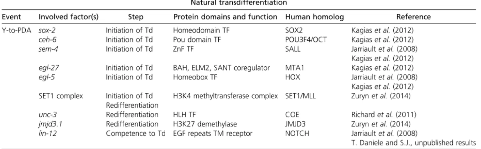
Y-to-PDA Td: cellular steps, molecular processes: 736
Transition occurs through discrete cellular steps: 736
Initiation is controlled by conserved nuclear factors: 737
Invariance of natural Td is ensured by successive layers of histone modifying activities: 737
MCM neurons arise from the amphid socket cells in males 738
Other putative plasticity events 738
Induced Reprogramming in the Worm 739
Germline-to-soma 739
Cytoplasmic control of cell fate in the germline 739
Nuclear control of pluripotency in the germline and germline-to-soma reprogramming: 741
Soma-to-germline reprogramming 743
Soma-to-soma: in vivo induced reprogramming of fully differentiated postmitotic somatic cells 746
Barriers to Reprogramming 747
General Principles Underlying Reprogramming 748
Summary and Future Perspectives 749
Cellular Plasticity Definitions
A
defining property of our tissues and organs is that they remain functional over time. This implies that their main constituents, cells, persistently maintain their specialized identity. This specialized cellular identity is characterized by the combination of the cell’s morphology and function—properties that are underlined by a specific transcriptional program. A major change in the way we view the mainte-nance of the specialized cellular identity has occurred re-cently: while the identity of differentiated cells was originally thought to be foreverfixed after being acquired— a view that was largely held until 2006—it has become clear that it can be altered or entirely changed, even after terminal differentiation. This ability of a cell to give rise to cell(s) with
a different identity is called cellular plasticity. Cellular plas-ticity can occur naturally in an organism, or can be triggered exogenously, either experimentally or by the environment. Although the term has initially been used to describe a cellu-lar path proceeding from a less differentiated to a more dif-ferentiated identity, cellular plasticity entails a variety of processes including retrodifferentiation (the reversal to a lin-eally related progenitor or stem cell identity, and even rever-sal to a pluripotent state), transdetermination (the swap in differentiation commitment), and transdifferentiation (Td, the stable switch from one differentiated identity to another). The concept of cellular plasticity does not imply any direc-tionality, e.g., toward a more or less differentiated state, or what thefinal identity will be (see Box 1 Definitions). Origin of the concept, relationship with cellular potential Cellular plasticity has classically been used as a defining property of stem cells. Stem cells self-renew and can give rise to descendants that have adopted a more differentiated iden-tity. The number of possible alternative identities they can engender represents the cellular potential of the initial stem cell, and is often used to classify stem cells. While concepts and definitions have been largely defined and tested in vertebrate animals (see below), they are used to describe developmental events throughout the animal kingdom (see Box 1 Defini-tions). Thus, cells can be totipotent (i.e., can give rise to all the embryonic and extraembryonic tissues), pluripotent (i.e., can give rise to cells belonging to all three embryonic germ layers—endoderm, mesoderm, ectoderm), multipotent (i.e., can give rise to several types of cells constituting one or more tissues), or unipotent (i.e., can give rise to a specific lineage and differentiate into one cell type only) (see Box 1 Definitions).
By contrast, a differentiated cell is a specialized cell asso-ciated with a particular function in an organ or tissue (see Box 1 Definitions). In most tissues, differentiated cells ultimately become postmitotic, and the differentiated state is classically seen as an end point, a view influenced by Waddington’s epigenetic landscape, in which cells during development are represented as balls rolling down from their highest cel-lular potential, represented as the top of a mountain, to their final differentiated state where they rest, represented as the bottom of a valley (Waddington 1957).
Classical examples of cellular plasticity: toti-, pluri-, multi-or uni-potent cells and their demonstration
When and where are cells with broad cellular potential found in living animals? A large body of work has focused on the vertebrate embryo, in which the early embryonic cells have been shown to be totipotent, after which their cellular poten-tial becomes more restricted as development proceeds.
Such cellular potential has been demonstrated through a variety of approaches. For example, mouse embryonic cells up to the eight-cell embryonic stage are believed to be totipotent, as they retain the ability to contribute to both embryonic and extraembryonic tissues in blastomere aggregation
experi-ments and yield viable pups (Boroviak and Nichols 2014). One natural demonstration of totipotency at the two-cell-stage is the production of identical twins following separation of the two blastomeres in mammals.
A large proportion of embryonic cells is thought to retain the ability to form an implanting embryo until the 16-cell stage, at which stage the cells of the Inner Cell Mass (ICM) start specializing as embryonic epiblast cells or primitive endoderm cells (Morgani and Brickman 2014). Cell lines, called Embryonic Stem (ES) cells, have been derived from these ICM cells of preimplantation embryos. These ES cells express markers of the ICM cells and self-renew over a long time, in contrast with the transient ICM cells from which they are derived, and can be maintained indefinitely in culture (Boroviak and Nichols 2014). Both ICM and ES cells are thought to be pluripotent. Pluripotency is classically function-ally demonstrated by testing the potential of single cells to contribute to normal development. This can be achieved by injecting them into a developing embryo, or through blasto-mere aggregation experiments or tetraploid complementa-tion assays, and by testing their ability to contribute to all lineages. ES cells meet this functional definition. In addition, their differentiation potential can be tested by experimentally inducing differentiation along different lineages in vitro. While ES cells are engineered through isolation of ICM cells in very defined culture conditions and may progress to a specific pluripotent stage during derivation, it has been hy-pothesized that ES cells capture a naïve pluripotency state naturally found in ICM cells (Boroviak and Nichols 2014).
Early embryonic pluripotency is rapidly lost and cells later in development are thought to be multipotent, oligopotent, or unipotent. To characterize these properties, in vitro clono-genic assays, in vitro and/or in vivo phenotyping, and in vivo transplantation assays are classically used (Blanpain and Simons 2013). For instance, in vitro differentiation ap-proaches have been performed using a variety of primary cells—or cell lines—and culture conditions, from embryoid bodies or neurospheres to single cells such as intestinal stem cells, and, more recently, through the use of 3D matrices. In addition, transplantation assays and label-retaining approaches are performed in vivo to identify and follow stem cells and their descendants in their physiological environment.
Stem cells have also been described in adults, where they are thought to contribute to homeostasis, repair, and regen-eration of adult tissues. Classical examples include unipotent satellite cells, which are muscle stem cells (Sambasivan and Tajbakhsh 2015), and the multipotent intestinal crypt stem cells (van der Flier and Clevers 2009).
As highlighted for ICM cells, it should be noted that the cells exhibiting cellular plasticity during development, some with broad cellular potential, exist only transiently, by contrast with the classical definition of stem cells involving long-term self-renewal. These cells are therefore classically called blas-tomeres or progenitors. Thus, the notion of stem cells is, in large part, built on the ability to culture in vitro pluripotent
cell lines (like ES cells), and on the description in several adult tissues of long-term resident stem cells, many of which are unipotent.
Together, this body of work has led to a hierarchical view of development in which early blastomeres in vertebrate em-bryos transition from totipotency to pluripotency and then continue to restrict their cellular potential as they progress along their specialization path, ultimately adopting theirfinal differentiated and fixed identity. This notion of gradual morphing is implicitly suggested by Waddington’s epigenetic landscape graphical representation, although whether differ-entiation paths actually follow an incremental hierarchical process or, on the contrary, proceed through a succession of sharp transitions, remains to be determined.
Developmental Programming: Regulative vs. Mosaic Models
Are the properties of cellular potential, pluripotency, and multipotency, universal and do they apply to the worm? There have been a number of studies addressing the question of cellular potential in Caenorhabditis elegans. This section and the next will focus on those studies examining cellular plas-ticity in the embryo.
Programming in the worm: classical view andfixed lineage C. elegans development has been described at the single cell level in landmark studies [Sulston and Horvitz 1977; Deppe et al. 1978; Krieg et al. 1978; Sulston et al. 1983; see Giurumescu and Chisholm (2011) for more recent auto-mated and semi-autoauto-mated lineage data]. Knowledge of its cellular lineage has revealed that development proceeds in a highly stereotyped way: a generally fixed relationship be-tween cell ancestry and cell fate, as well as largely invariant cell positioning, has been observed throughout embryonic development.
Does the ancestry of the cells constrain their fate and, if so, how? The striking invariance of the cell lineage raised the question of whether differentiation paths are intrinsically (cell-autonomously) determined, a process called mosaic de-velopment, or whether cells in the C. elegans embryo could adapt to changes and interference, indicating that cell fates are dependent on cell–cell interactions, a process called reg-ulative development (Conklin 1905).
As presented in the next section, C. elegans development had initially been seen as mosaic. This view resonated with the apparent mosaic development and segregation of cy-toplasmic determinants described in many invertebrates (Conklin 1905). Initial studies in C. elegans focused on the fate and determination of the early embryonic blasto-meres. The fertilized egg, named P0, divides unequally into a larger anterior daughter, called AB, and a smaller poste-rior one called P1(Sulston et al. 1983), which continues to
divide along different division patterns and contribute to different tissues. Experiments in which the size of AB was reduced through removal of cytoplasm showed that the
differential size of these two blastomeres does not sub-stantially alter their division behavior or fate, suggesting that qualitative cytoplasmic differences dictate their fate (Schierenberg 1984, 1986; Schierenberg and Wood 1985). The AB blastomere will divide into what will become the anterior, ABa, and posterior, ABp, daughters. ABa lineage follows complicated asymmetrical patterns while ABp di-vides mostly through symmetrical patterns, both producing similar and unique cell types. Their descendants will be the major contributors to the cells of the hatching larva, and will generate cells of the nervous system, the hypoder-mis, and the pharynx. P1continues to divide unequally to
give rise to the EMS and P2blastomeres. P2will give rise to
the C and P3blastomeres, and P3to the D and P4
blasto-meres. EMS gives rise to MS, which produces primarily me-sodermal cells and E, which generates only intestine. C generates muscles and hypodermis, and D only muscles. P4is the precursor of the germline (Figure 1). These
blas-tomeres are called founder cells, as they solely or mostly give rise to one tissue.
Early specification suggests plasticity is lost early
A range of evidence from early embryological studies suggested that much of C. elegans embryogenesis is char-acterized by mosaic development, as reviewed in this section.
Founder cells are largely specified by apparent cell-intrinsic mechanisms at the time of their birth
To address the extent to which autonomous specification mechanisms act during embryogenesis, a number of ap-proaches have been undertaken. One of these consisted of isolating specific blastomeres, culturing them in isolation, and assessing which fate they or their descendants adopted. In isolation, the AB blastomere divides and produces recogniz-able hypodermal and neuronal cells, similar to many of the cell types it produces in wild type embryos (Priess and Thomson 1987; Gendreau et al. 1994; Moskowitz et al. 1994). The P1
blastomere obtained after gently bursting the eggshell and eliminating the AB blastomere, is able to divide in culture and generates four cells, one of which shows characteristics of the endodermal precursor cell E, specifically the gut-specific rha-bitin granules (Laufer et al. 1980). When P1 blastomeres
obtained similarly are treated with a cleavage inhibitor, they also give rise to cells with gut granules (Figure 1) (Laufer et al. 1980). These experiments suggested that the ability of P1 to generate gut fate is intrinsic to the P1 cell at the
two-cell-stage. This work also suggested that cell division is not a requirement for expression of at least certain differen-tiation characteristics. Furthermore, if isolated P1
blasto-meres are allowed to divide further, they yield a partial embryo containing a few hundred cells that twitches and that contains muscle cells (Figure 1) (Laufer et al. 1980; Gossett et al. 1982). Again, this suggested that the potential to pro-duce muscle is intrinsically present in the P1blastomere and
as strong evidence that early embryonic specification is largely carried out autonomously and already established in the early blastomeres, consistent with a mosaic pattern of development.
One intrinsic mechanism underlying mosaic development is cell fate determination through the segregation of cytoplas-mic determinants. Early observations supported such a me-chanism at both the organelle and molecular levels. The germline-specific P granules are transmitted specifically to germline precursors during the early asymmetric cell divisions that establish the germline progenitor, P4(Strome and Wood
1983). Similarly, proteins such asPIE-1andMEX-5/6(Mello et al. 1996; Schubert et al. 2000) are distributed unequally in the zygote and subsequent blastomeres prior to cell division (Cowan and McIntosh 1985). The levels of theSKN-1 pro-tein, required in P1descendants to produce pharyngeal cells,
are also higher in the P1blastomere compared to its AB sister
(Bowerman et al. 1993). Caveats to these experiments exist: for instance, a role for cell–cell interactions in specification, preceding cell separation manipulations for example, cannot be definitely excluded; in addition, the impact of cell–cell signaling in rapidly dividing blastomeres may only be seen in one-to-a-few cell generations later. However, collectively, thesefindings made a strong case that cell fate potential is driven largely by cell-intrinsic mechanisms in early C. elegans embryos.
Cleavage-arrest experiments and exclusivity of cell fate The law of exclusivity of differentiation postulated by Weiss (1939) posits that a single cell chooses only one path
of differentiation at a time. This principle was supported in C. elegans through cleavage-arrest experiments on early embryos, which were used both to assess when the potential to differentiate particular cell typesfirst appears, and to ex-amine whether the potential to produce several differenti-ated cell types can coexist in the same blastomere (Cowan and McIntosh 1985). Cowan and McIntosh observed cleavage-arrested isolated P1blastomeres and asked if gut, muscles, and
hypodermis markers, all fates found in the P1progeny, can be
coexpressed in such setting. While they observed that all three markers were found in similar proportion in cleavage-arrested P1 blastomeres, they never found them to be coexpressed
(Cowan and McIntosh 1985). Thesefindings supported the view of exclusivity of differentiation pathways (Weiss 1939) and were interpreted as yet further evidence for mosaic devel-opment in the worm.
Importance of DNA replication in specification
An early model, called the quantal cell cycle model (Holtzer et al. 1975, 1983), posited that progression through the cell cycle, and DNA replication in particular, is a necessary re-quirement for cells to alter their expression profiles and change their commitment or differentiation state. The model postulates that the DNA:cytoplasm ratio controls transcrip-tion through its impact on cell cycle. In the context of embryo-genesis, it implied that even when embryos are competent for transcription, they would start transcribing only once a G1 or G2 phase is added to the cell cycle.
However, cell fusion experiments to produce nondividing heterokaryons, carried out around the same time, have shown
Figure 1 Early embryonic lineage, evidence for cell-intrinsic determination and nonlinear segregation of developmental potential. The fertilized egg, named P0, divides unequally into the AB and P1blastomeres. AB further divides into ABa and ABp, which will follow different division patterns. The
tissue contribution of their descendants is indicated in gray. P1divides unequally to give rise to the EMS and P2blastomeres. P2will give rise to the C and
P3blastomeres, and EMS gives rise to MS and E. The tissue contributions of the descendants of the C, P3, E, and MS blastomeres is indicated on the
right. The cellular phenotype observed (in black) or not (in red) after culture in isolation, with or without cell arrest, of each blastomere up to the four-cell stage is also indicated.
that cells in culture can alter portions of their transcriptional programs in the absence of DNA replication (Blau et al. 1983, 1985; Chiu and Blau 1984), suggesting that this model may not entirely apply, at least in in vitro settings. In an effort to decipher the relationship between DNA replication and line-age specification in an in vivo physiological setting, Edgar and McGhee (1988) tested this hypothesis in live C. elegans em-bryos using pulses of the drug aphidicolin, which instanta-neously blocks DNA replication. Focusing on the E lineage and by comparison it to the hypodermal, and body wall mus-cle lineages, their experiments addressed the requirement for rounds of DNA replication in the expression of intestinal, lineage markers (Edgar and McGhee 1988).
Edgar and McGhee (1988) found that neither the number of replication rounds per se, nor the DNA:cytoplasm ratio or elongation of the cell cycle prevented expression of differen-tiation markers or their timing, suggesting that the quantal cell cycle model did not apply, at least for the lineages exam-ined, in C. elegans embryo. However, their results suggest that thefirst round of DNA synthesis after the sublineage progen-itor has been established (for instance, the E founder cell) is key. This was interpreted by the authors as a critical period during which gut genes are“activated” or licensed for later expression (Edgar and McGhee 1988). This could be achieved via the replication-dependent elimination of nucle-osomes and other histone modifications, thus giving access to key determinants to target regions on the chromosomes, or it could represent a critical period when cytoplasmic gut deter-minants are translocated to the nucleus and thus bind to specific loci on the chromosomes. One implication of these studies is that early blastomeres, rather than being generally open to adopting different differentiation fates, must be made competent to do so in vivo through a precisely timed DNA replication period. However, such an explanation must take into account how markers of several different fates, in-cluding gut, can be observed in cleavage-arrested P1
blasto-meres (which continue to undergo DNA replication and mitosis) (Laufer et al. 1980), as presented in the Cleavage-arrest experiments and exclusivity of cell fate section.
The C. elegans embryo: regulative development and early cellular plasticity clues
While much evidence points to a mosaic mode of development in C. elegans, it is also clear that regulative development also functions during early embryogenesis, and that early blasto-meres likely retain wider developmental potential than they express during wild-type development.
Nonlinear segregation of developmental potential As described above, cleavage-arrested P1blastomeres appear
to differentiate gut, based on the presence of gut granules (Laufer et al. 1980; Cowan and McIntosh 1985). However, cleavage-arrested P0blastomeres never produce gut granules
(Figure 1). This suggests that gut fate determinants are not present, or are not active, in P0, but arise in the P1cell,
per-haps as a result of its interaction with the AB blastomere or of
P0division. Similar observations have been made when
seg-regation of muscle differentiation potential is examined: it is not seen in P0, while it is present in P1and P2blastomeres
(Figure 1). In addition, muscle differentiation potential is not present in cleavage-arrested EMS, while it is present in the P1
mother, and in its MS daughter (Figure 1). These data sug-gest that different developmental potentials are not simply segregated in a linear fashion but can reappear throughout the lineage in daughters of cells that do not exhibit this po-tential (Cowan and McIntosh 1985).
Similarly, the lineal pattern of the worm’s tissues argues against a purely mosaic pattern of development: indeed, with the exception of the intestine and the germline, whose origin can be traced to one early ancestor, C. elegans tissues are polyclonal in origin (Sulston et al. 1983), excluding simple linear segregation of fates along lineages.
Wide developmental capacity of early blastomere nuclei Studies have suggested that there is greater plasticity in the early blastomeres than is evident from the lineage. When the anterior cytoplasm together with the zygote nucleus are extruded from the anterior pole of wild-type 1-cell embryos, it produces an AB-like cell that can divide. If one of the nuclei obtained after two divisions is slipped back into the posterior enucleated part that had remained, the newly recreated cell then divides like a P1blastomere that is able to produce P
derivatives like muscles (Figure 2) (Schierenberg and Wood 1985; Schierenberg 1986). These experiments suggested that an AB-like nucleus still contains P1developmental
po-tential, even after two cell divisions, and thus could be regarded as totipotent. However, another interpretation could be that the posterior enucleated part behaves like a P1
envi-ronment that can reprogram the AB-like nucleus, in analogy to what John Gurdon observed in his landmark nuclear transfer experiments in frogs (Gurdon 1962).
Blastomere rearrangements and latent developmental potential
In another key set of experiments, displacement of meres within the embryo revealed that early sister blasto-meres that normally invariably adopt different fates are actually initially equipotent. For example, the AB daughters generate common but also unique cell types: only ABa gen-erates pharyngeal muscles and inner labial neurons, while only ABp generates the GABA-containing neurons. To exam-ine if ABp retains ABa developmental potential and vice versa, Priess and Thomson swapped their positions by micromanip-ulation before their separation is complete in otherwise nor-mal embryos, and examined the identity of their descendants in embryos, larvae, and adults. All 11 such embryos devel-oped into seemingly normal and viable larvae and adults, and the expected ABa and ABp derivatives were all found at their normal location, though likely generated by the other, in-terchanged, AB daughter (Figure 2) (Priess and Thomson 1987). These striking results strongly suggested that, although the AB daughters stereotypically follow different
developmental paths, they in fact have a broader develop-mental potential than the one they normally express during development. Selective expression of a subset of this de-velopmental potential thus relies on the environment and differential cell–cell interactions that each AB daughter experiences—a conclusion reinforced by the concomitant finding that AB blastomeres cultured in isolation do not gen-erate all expected cell types (Priess and Thomson 1987). Together with the Schierenberg experiments described above, these studies argue that the AB blastomere at least is inherently more potent—and maybe more totipotent— than is apparent during normal development.
Early ablations also revealed instances of regulative development
Laser-mediated ablation of individual blastomeres in embryos comprising 28 cells or more revealed a restricted number of cases of regulative development, that occurred mostly during late embryogenesis (Sulston et al. 1983). These experiments initially reinforced the idea that C. elegans development and early embryogenesis in particular was largely mosaic.
However, further experiments revealed numerous cell–cell interactions that are required for the correct specification of early blastomeres as well as the complete repertoire of all
cells in the C. elegans embryo. Experiments using tempera-ture-sensitive mutants that alter the cell-cycle length of par-ticular blastomeres, in which case the resulting embryo developed abnormally, or of all founder cells, in which case normal development was observed, suggested a requirement for timely cell–cell interactions (Schierenberg et al. 1980). Priess and Thomson (1987) further examined when descen-dants of the AB and P1blastomeres arefirst determined using
ablation experiments and monoclonal antibodies that specif-ically label pharyngeal or body wall muscles (contributed to by both the AB and P1blastomeres) in late embryos. They
showed that the blastomeres that generate these cells appear to be determined by the 28-cell stage. The muscles descen-dants of the P1blastomere did not require AB-derived cells, as
ablation of the AB blastomere at the two-cell stage did not alter the ability of the P1descendants to produce pharyngeal
and body wall muscles (Priess and Thomson 1987). These results are consistent with the observed segregation of mus-cle fate in P1(Laufer et al. 1980). By contrast, removing the
P1blastomere did not allow generation of the expected
pha-ryngeal or body wall muscles in AB descendants development (Figure 2). It was further shown that removal of EMS, a P1
daughter, led to the absence of pharyngeal muscles, while removal of P2still allowed the generation of both body wall
Figure 2 Evidence for the early developmental plasticity window and the control of its timing. Early blastomeres up to the 8E-cell stage can exhibit high plasticity, as revealed by blastomeres swap (x), blastomeres ablations or culture in isolation (*), nuclear transfer (&) or analysis of lineage mutants (£). In
addition, overexpression (OE) of defined TFs during this time-window transforms most, if not all, blastomeres into cells bearing characteristics of the indicated tissues. Transition from the 8E to the 16E-cell stage marks the multipotency-to-commitment transition (MCT). The window can be extended by removing the polycomb PRC2 complex (mes-2,mes-3mutants) or early Notch activity (glp-1mutant).
and pharyngeal muscles (Figure 2) (Priess and Thomson 1987). This strongly suggested that an interaction between P1or its daughter EMS and the AB lineage is necessary for the
formation of muscles in AB descendants.
Along the same lines, the role of inductive interactions during gut development has been examined more closely. The entire gut derives from a single blastomere, the founder E cell in C. elegans. Based on blastomere isolation experiments, it was initially suggested that EMS, the mother of the founder E cell, and even the P1blastomere, have the cell-intrinsic ability
to produce intestinal fate (Laufer et al. 1980). However, sub-sequent studies revealed that isolation of the EMS blastomere at the early four-cell stage blocked its ability to generate gut descendants. It was found that EMS requires a signal from P2
at the four-cell stage to generate gut descendants (Figure 2) (Schierenberg 1987; Goldstein 1992, 1993). During later embryogenesis, several additional cell–cell inductions were also revealed by ablation experiments, such as the regulative interaction between ABprapaapa (aka W) and ABplapaapa (aka G2) or the regulative interactions between ABplpaaaapa (aka excretory duct) and ABprpaaaapa (aka G1) (Sulston and Horvitz 1977). Likewise, several regulative interactions have also been described during larval development, including, but not restricted to, the specification of the anchor (AC) and vulval uterine (VU) cells or of the Vulval Precursor Cells (VPCs) (Greenwald 2012).
Lineage defects in mutants provide strong evidence in favor of regulative interactions
Analysis of mutations affecting embryogenesis further revealed numerous cell–cell interactions that are required for the cor-rect specification of early blastomeres and the complete rep-ertoire of all cells in the C. elegans embryo. We will here focus on three such examples in early embryogenesis. In particular, lineage founders were shown to be differentially specified at the four-cell stage through intercellular directional signals that depend on the geometrical arrangement of the blastomeres. This specification involves a P2 signal to ABp through the
Notch extracellular receptor GLP-1 (Figure 2); both AB de-scendants express the maternally encoded Notch receptor
GLP-1, but only the posterior one contacts P2, which expresses
the Notch ligandAPX-1, and, therefore, only it is induced to acquire an ABp fate (seehttp://www.wormbook.org/chapters/ www_notchembryo/notchembryo.htmland references therein). Further analysis of theglp-1mutants also revealed a signal from the MS blastomere to the two (of the four) ABa de-scendants that it contacts, and which expressglp-1, occur-ring in the 12-cell stage embryo (Figure 2) (Priess and Thomson 1987; Shelton and Bowerman 1996). This inter-action is necessary to induce the ABa descendants to pro-duce pharyngeal cells, and to break the left-right symmetry of the AB lineage (Priess and Thomson 1987; Gendreau et al. 1994; Hutter and Schnabel 1994; Mango et al. 1994; Moskowitz et al. 1994).
The blastomere isolation experiments also revealed a P2to
EMS signal that is necessary for subsequent production of
endoderm (Figure 2) (Goldstein 1992, 1993). Genetic screens and mutant analyses have shown that this signal is mediated by redundant Wnt, src, and MAPK pathways, resulting in the polarization of the EMS cell, and asymmetric concentration of the TCF/POP-1TF in the nucleus of the MS daughter vs. the E daughter (Lin et al. 1995; Thorpe et al. 1997; Bei et al. 2002).
Together, these studies showed that, even in an animal with a stereotyped cell lineage, development is not strictly driven by intrinsic factors, but also relies on timely cell–cell inductive interactions and the embryo’s cellular geometry. They also emphasize that the cellular and developmental potential of single cells should be assessed out of their normal contexts as it can be greater than their fate could predict.
Early Embryonic Cellular Plasticity is Restricted to a Defined Time Window: the Multipotency-to-Commitment Transition
Early embryonic founder cells are specified at the time of birth, but remain developmentally plastic through several divisions
The cell-intrinsic ability of founder cells to remain develop-mentally plastic through several subsequent rounds of cell division was further demonstrated by the ability of them and their descendants to change fate and even lineage commit-ment in response to ectopically driven expression of cell fate-determining transcription factors (TFs). For example, it was found that all somatic lineages could be converted to endo-derm in response to ubiquitous expression of single GATA-type TFs that function in endoderm specification and differentia-tion (Figure 2;END-1,END-3,ELT-2, andELT-7) (Fukushige et al. 1998; Zhu et al. 1998; Maduro et al. 2005; Sommermann et al. 2010), resulting in embryos containing essentially only differentiated gut cells. Similarly, transcription factors for other differentiated cell types are capable of converting most or all cells of embryos into pharynx (driven byPHA-4/FoxA; Horner et al. 1998), muscle (driven by the bHLH factorHLH-1; Fukushige and Krause 2005), or epidermis (driven by the
ELT-1 GATA or LIN-26 ZnF factor; Gilleard and McGhee 2001; Quintin et al. 2001) (Figure 2).
This conversion process, or transdetermination, reflects bonafide redirection of most or all somatic progenitors into the pathway for endoderm (or pharynx, muscle, or epider-mis) differentiation, as other cell-type specific differentiation, characteristic of the other two germ layers, is prevented in the cells that inappropriately express markers of the differ-entiated gut.
Thus, several divisions after founder cell lineages are established, many or all somatic lineages of the early embryo, though normally specified to particular lineages, appear to remain largely pluripotent (capable of contributing to any of the three germ layers). Thesefindings underscore the exclu-sivity of cell fates that had been originally inferred from early experiments on cleavage-arrested embryos (Cowan and
McIntosh 1985): genuine acquisition of one differentiated fate by transdetermination or reprogramming precludes ex-pression of genes characteristic of other fates.
Plasticity is retained in early blastomeres during a defined developmental window
The demonstrated developmental plasticity of early embry-onic cells has provided a useful test bed for exploring in vivo mechanisms that control pluripotency and subsequent devel-opmental commitment. Although early descendants in each founder cell lineage retain the ability to be transdetermined into any of three germ layer types, this multipotency is lost by midembryogenesis, after which embryos undergo essentially normal development and differentiation when challenged with widespread expression of cell fate-determining TFs (Zuryn et al. 2012; Spickard et al. 2018). Thus, embryos transition from a multipotent state into one in which cells resist reprogramming into other cell types. This multipotency-to-commitment transition (MCT), occurs within a few hours of fertilization, at around the 200–300 cell stage, or after the E lineage has divided to produce eight descendants (8E stage, Figure 2; the simple E lineage provides a convenient landmark for scoring various stages in embryonic development). Most, if not all, animal embryos undergo a similar transition from a plastic to a committed state of differentiation (Boroviak and Nichols 2014; Bernardo et al. 2018); hence, the MCT generally marks a major event that occurs during embryogenesis across metazoan phylogeny.
Molecular events controlling plasticity of early blastomeres and timing of the MCT
The molecular processes underlying the MCT have been in-vestigated by analyzing genes whose loss delays its onset, resulting in extension of the period during which cells can be redirected into other developmental pathways (Spickard et al. 2018). Concomitant with this transition into commitment, nuclei undergo a change in structure that was proposed to reflect a global increase in the condensation of chromatin, suggesting conversion to heterochromatin (Yuzyuk et al. 2009). Consistent with this notion, components of the PRC2 chromatin remodeling complex, including MES-2, which encodes the H3K27 methyltransferase catalytic sub-unit, andMES-3, were found to be required for the normal onset of the MCT (Yuzyuk et al. 2009). Embryos deficient for this complex are delayed in commitment to normal pathways of differentiation, as evidenced by the ability to redirect nor-mally nonmuscle cells into muscle when forced to express the muscle-determining factor HLH-1(Figure 2) (Yuzyuk et al. 2009). The delayed transition from a multipotential to a com-mitted state inmes-2mutants is paralleled by a widespread change in gene expression: genes that are normally expressed specifically during early embryogenesis (including the muscle-specific unc-120gene, the endoderm-determin-ing end-1 gene, and the ABa lineage-expressed tbx-37 and -38genes) and whose expression is attenuated after the 4E stage, were found to be overexpressed and maintained
expression after this stage, perhaps reflecting perdurance of an early embryo-like (uncommitted) state (Yuzyuk et al. 2009). The ability to probe developmental plasticity by challeng-ing embryos with forced expression of TFs provided an entry point into identifying genes that, when debilitated, result in a failure of cells to commit at the normal developmental stage. A screen for such genes revealed that Notch signaling contrib-utes to establishing the MCT in the AB lineage. In embryos lacking the Notch receptorGLP-1, the MCT is delayed to well after the 8E stage, allowing END-3to activate widespread, ectopic gut differentiation much later than inglp-1(+) em-bryos (Figure 2) (Djabrayan et al. 2012).
Maternally encodedGLP-1is expressed specifically in AB
and its descendants and Notch signaling is known to specify many cell types during thefirst several divisions of the AB lineage in response to signals from descendants of the P1
blastomere (Mello et al. 1994; Moskowitz et al. 1994; Hutter and Schnabel 1995; Moskowitz and Rothman 1996). Thus, specification of AB descendants per se might result in commitment of these cells, consistent with temper-ature-shift experiments that revealed that the stage at which
GLP-1functions in several early cell fate decisions also cor-related with its temporal requirement in establishing the MCT (Djabrayan et al. 2012). However, some AB-derived cells apparently never receive cell-type-specifying Notch sig-nals during embryogenesis and yet, as with Notch-signaled cells, appear to resist reprogramming into endoderm by
END-3 at the appropriate time. In fact, it was found that the requirement for GLP-1 in determining developmental plasticity is, apparently, not dependent on the AB-extrinsic specification signals from P1descendants, but is autonomous
to the AB lineage: isolated AB-derived partial embryos, which cannot receive signals from P1descendants, continue to show
GLP-1-dependent activation of the MCT at the normal time (Djabrayan et al. 2012). The role ofGLP-1in establishing the MCT in the AB lineage independently of the known signaling ligands led to thefinding that two noncanonical, apparently secreted DSL-like ligands,DSL-1and-3, show a similar re-quirement for establishment of the MCT (Djabrayan et al. 2012). Thus, these putative ligands may act onGLP-1 specif-ically in the AB lineage to direct commitment to nonendo-derm fate without specifying cell identities. As elimination of either PRC2 or Notch function (in the AB lineage) results in superficially similar effects, i.e., delay of the MCT and persis-tence of developmental plasticity, it is conceivable that Notch acts through the PRC2 system to alter chromatin accessibility to reprogramming. Of note, a similar relationship between
GLP-1/Notch and PRC2 has been unraveled in germline-to-soma conversion, as described below (Seelk et al. 2016).
It is believed that deployment of the gene regulatory networks that direct embryonic development progressively restricts the developmental fates of cells through the action of TFs and cis-regulatory sequences. Such networks establish autoregulatory “lockdown” circuits that underlie commit-ment to specific pathways of differentiation (Davidson and Levine 2008; Davidson 2010). Thefindings described above
suggest that additional mechanisms operate independently of these transcriptional networks, and function to provoke developmentally plastic cells to commit to particular differ-entiated states. Given thatmes-2(2) embryos are viable, as
aredsl-1(2) and -3(2) mutants, timely activation of the MCT
does not appear to be critical for successful differentiation and development, at least in conditions in which cell fate acquisition is not challenged. Thus, systems that commit ini-tially developmentally plastic cells to differentiation may function to ensure developmental fidelity per se. As such, these systems may function independently of pathways that control the differentiated fate of cells.
Cellular Plasticity of Specialized Cells: Reprogramming and Transdifferentiation—an Overview
Is cellular plasticity a property restricted to stem cells or to progenitors found in developing embryos? Classical develop-mental experiments highlighted very early on that differen-tiated cells have an inherent cellular plasticity that is not normally expressed but that can be experimentally revealed. For instance, late 19th century studies showed that newts can regenerate their lens after complete removal, and that the likely source of the regenerated lens is pigmented epithelial cells of the dorsal iris (Collucci 1891; Wolff 1895; Kodama and Eguchi 1995). Cellular plasticity was also demonstrated for cells that are not yet fully specialized: grafting the dorsal lip of the blastopore of a frog or newt embryo onto the ventral side of a receiving embryo leads to the induction of a second body axis: the fate of the ventral cells of the receiving embryo is changed by the graft (Mangold and Spemann 1927). Later, John Gurdon found that the transplantation of a differenti-ated somatic nucleus into an enucledifferenti-ated frog egg led to the reprogramming of the differentiated nucleus, allowing the development of fertile frogs (Gurdon 1962; Gurdon and Uehlinger 1966). Thus, very early on, it appeared that, at least under specific conditions, specialized cells could exhibit cellular plasticity. This and subsequent sections will focus on the cellular plasticity of specialized cells, e.g., differentiated tissue cells and germ cells.
In fact, the ability of cells that have acquired specialized identities in multicellular organisms to undergo conversion into other cell types has been observed throughout both the plant and animal kingdoms, including members of the ecdy-sozoan and lophotrochozoan branches of protostomes and the deuterostomes. Cell fate conversion has been described in both natural and experimental settings outside of develop-ment. Examples include regeneration following injury, in which cells of a variety of types, and entire structures, can be provoked to develop from either differentiated cells or multipotential progenitors present in the tissue, as occurs, for example, in regenerating amphibian lens (Henry and Tsonis 2010). In the context of disease, cell type conversions and metaplasias might be at the basis of a variety of cancers (Syder et al. 2004; Means et al. 2005). While resistance to the notion that specialized cells could change their identity
was initially strong, a new way to induce cellular plasticity experimentally has been described more recently in land-mark experiments. Indeed, pluripotent stem cells have been engineered in vitro starting from differentiated cells (Takahashi and Yamanaka 2006) that exhibit similar proper-ties to ES cells, and can also be grown indefinitely in culture. Such feats, called pluripotent reprogramming, necessitate the forced expression of a cocktail of transcription factors endogenously expressed in ES cells and important to main-taining their pluripotency. Although variations in the cocktail composition exist, and small molecules replacements for some of these factors have been attempted, four factors are commonly used: Oct4, Sox2, Klf4, and cMyc (Takahashi and Yamanaka 2006; Takahashi et al. 2007). It should be noted that, while all differentiated cell types used to date have been reprogrammed, the programming success rate is very low. These landmark experiments have an important conceptual implication, as they imply that pluripotency in not an inher-ent property of a stem cell inher-entity, but a state that can be acquired. In addition, these studies, together with the early descriptions of unexpected cellular plasticity events, have de-cisively shown that the differentiated cellular identity is not fixed, and opened the door to the possibility that cellular plasticity may not be a property uniquely found in stem cells or blastomeres.
Various terms can be found in the literature to describe similar or distinct cellular plasticity events (see Box 1 Defini-tions). Cellular reprogramming, transdetermination, and Td all share the common characteristic that the normal fate of a cell is altered to that of another cell type, and generally involve remodeling of the transcriptional state of a nucleus, a process that translates into morphological and functional changes. Though “cellular reprogramming” can also be found in the literature with a very narrow application, that is the reprog-ramming of differentiated cells into an embryonic-like stem cell called induced pluripotent stem (iPS) cell, cellular reprogramming is a more generic term that does not imply the degree of specialization of the initial or final identities. We have thus used “pluripotent reprogramming” to desig-nate reversion from a differentiated identity to an ES-like state. By contrast, transdetermination involves a switch in the fate of progenitor or stem cells from one lineage type into another, and Td applies to events in which cells that have adopted a differentiated fate are converted to another differ-entiated cell type [for the first use of Td in English, see Selman and Kafatos (1974); for definition and criteria, (Kodama and Eguchi 1995)]. It is important to note that the definition of these processes is based on the initial and final identities and their switch after a single inducing event, but not on the cellular or molecular mechanisms underlying the conversion itself, which are in many cases unknown and may vary in different contexts. In addition, “direct reprog-ramming” or “direct cell type conversion” are also found in the literature instead of Td, often to describe experimentally induced processes in cell culture assays. For clarity, we pro-pose to use them indistinctly, but to specify, by using“natural”
or“induced,” when these processes are observed in nature or have been triggered experimentally respectively (see Box 1 Definitions). While applications taking advantage of cellular plasticity have been pushed forward more recently, these concepts have deep roots in developmental biology.
Transdetermination wasfirst used to describe the conver-sion of imaginal disk identity in Drosophila larvae, from leg to wing, for example (Garcia-Bellido 1966; Maves and Schubiger 1999; Worley et al. 2012) as a result of environ-mental changes or injury. This redirection of progenitor cells that have not yet become specialized can be contrasted with bonafide Td events. This latter process, again first described in insects (Selman and Kafatos 1974) has been observed both in dividing and nondividing cells.
The distinction between transdetermination and transdif-ferentation of fully differentiated cells cannot always be pre-cisely defined, the limitation being the precision with which it is possible to define the extent of differentiation of the initial cell prior to its reprogramming. However, alteration of a lineage trajectory before cells have adopted a specific func-tion—for example, when the fates of blastomeres in early C. elegans embryos are redirected as a result of loss of gene function or forced expression of TFs, or as occurs with home-otic mutations and when an eye is induced at the end of a leg by forced expression of eyeless in Drosophila—is definitively transdetermination. When the cell, prior to its conversion, has already performed a differentiated function—as with the rectal epithelial cell in C. elegans, which converts to a specialized neuron (Jarriault et al. 2008), or withfibroblasts that can be converted to muscle cells as a result of forced MyoD expression (Davis et al. 1987; Tapscott et al. 1988)— the event can be definitively considered Td.
Germ cell reprogramming is a special case, complicating the distinction between transdetermination and Td. Develop-ing germ cells are a distinct population of cells, which one may view as very specialized, that are undergoing the process of later transforming into a totipotent embryo. As such, conver-sion of germ cells into differentiated cells may be the result of derepression of somatic differentiation that is poised to occur following fertilization. This might be exemplified by the acquisition of differentiated somatic features of several line-ages by germ cells mutant for translational regulator-coding genes (Ciosk et al. 2006). Though the distinction is partly semantic, and similar molecular mechanisms might be at play, the latter process could be considered premature acti-vation of the normal—and poised—embryonic program, rather than a de novo switch from one transcriptional pro-gram to another, as would be expected for transdetermina-tion or Td.
The goals of studying mechanisms of developmental reprogramming in model organisms such as C. elegans are both fundamental and practical. The intensive genetic ap-proaches available with the system can be highly informative as to the stepwise mechanisms and key molecular regulatory processes underlying conversion of one cell type to another within an in vivo context. In addition, one key aspect and
nagging technical limitation in multicellular organisms is the ability to follow and unambiguously assess the cell type conversion at the single cell level. With its transparency and fully described cellular lineage (Sulston and Horvitz 1977; Sulston et al. 1983), the worm offers a unique model in this respect. Although cell identity can be changed either natu-rally or experimentally, it is of importance to understand at the fundamental level at what stage in the differentiation process there is a restricted or unique pathway that must be followed to acquire that identity. Moreover, it is critical to learn the extent to which the state of the nucleus is identical in two cells that ultimately adopt the same differentiated fate, either during normal development from a multipotent progenitor, or as a result of Td. This information can also guide the adaptation of reprogramming approaches in regen-erative medicine. To safely replace damaged or lost cells resulting from injury or disease with genetically identical cells from a patient that have been created by inducing dif-ferentiation of stem cells or Td of a different cell type requires a complete understanding of the similarities between the natural and induced cells at the level of both gene expression and epigenetic states, and the stability of the induced state once transplanted. Such studies might also advance insights into which source of cells, whether directly from patient-har-vested tissues, or from those cultured for long periods, affects the efficiency and longevity of the transdifferentiated re-placement tissues.
Current Questions in the Field that can be Addressed with Studies of Plasticity in C. elegans
The ability to experimentally induce cellular reprogramming, either to another differentiated identity, or to a pluripotent state, as well as the existence of natural reprogramming events, have triggered a booming newfield of investigations. Here, we summarize the main questions that have emerged, and are or can be pursued using C. elegans as a model system, with a focus on induced and natural direct reprogramming (aka Td).
What are the cellular and molecular mechanisms underlying natural and induced direct reprogramming?
The worm allows for systematic genetic analyses, a powerful tool, which, combined with candidate approaches and cellular analyses, has and further will allow the steps and the require-ments to change the initial identity involved to be deciphered, including barriers and drivers, the importance of cell division, any potential requirement for transient reversal to a lineage-related progenitor/stem cell-like state, and the redifferentia-tion process.
Do the mechanisms of reprogramming differ between different cell types?
Thefield still awaits a systematic side-by-side comparison of different Td events in a given species. Several cell type-specific characteristics could impact the process. For instance,
overexpression of a given TF does not lead to reprogramming of all cell types (Davis et al. 1987), possibly pointing to dif-ferent mechanistic paths. In addition, some natural Td events appear to involve cellular division, while others do not. The actual requirement and contribution of cell division, or of DNA synthesis, to reprogramming and whether it reveals mechanistic differences remains to be determined. Of note, studies on the microevolution of the vulva in nematodes (Srinivasan et al. 2001; Félix 2005) have shown that different mechanistic strategies can lead to the same end point. A re-lated question is to examine to what extent are redirection of a cell’s commitment (i.e., transdetermination) and Td mech-anistically related. In addition, it may be informative to com-pare germ and somatic cells. Indeed, how is germline multipotency regulated and how are germ cells protected from reprogramming? Are these mechanisms similar to how the differentiated state of somatic cells is maintained?
Are the steps, molecular and cellular processes in natural vs. induced reprogramming identical, overlapping, or entirely dis-tinct? Since the end point between different reprogramming events varies (from diverse differentiated identities to a plurip-otent one), it is likely that aspects such as those involved in redifferentiation will differ between reprogramming events. However, it is conceivable that all reprogramming events, nat-ural and induced, require the proper erasure of the initial iden-tity and that the networks allowing this erasure share common features. In fact, thefinding that homologous factors are in-volved in both erasure of the initial identity during natural Td in worms and in the induction of pluripotent reprogramming in mammals argues for the possibility that general principles un-derlying the cellular plasticity of the differentiated identity may be found (Kagias et al. 2012; Becker and Jarriault 2016). Further studies should shed light on whether shared mechanisms will be found and whether induced reprogram-ming events borrow from mechanisms that naturally exist. Does reprogramming occur through gradual, continuous process or discrete steps?
We tend to view differentiation as an incremental process represented by a continuum of very close states, both at the cellular and molecular levels. This view has been very much influenced by the classical representation of differentiation along, for example, the hematopoietic lineage in vertebrates. Does reprogramming, and direct reprogramming in particular, proceed similarly? Related, is a pluri/multipotent intermedi-ate necessary? While some studies of cell populations have suggested a gradual morphing from one identity into another, with some mixed intermediate features after direct reprog-ramming is triggered (Zhou et al. 2008), studies at the single-cell level in the worm suggest a process that involves clear ruptures from one state to the other (Richard et al. 2011). Subsequent transcriptomic analysis on mammalian cell pop-ulations induced to directly reprogram have given support to this hypothesis (e.g., Di Tullio et al. 2011), and single cells transcriptomic data are expected to bring more answers to this debate. Determining the cellular and molecular states
and the nature of their transitions involved during direct reprogramming, may have a broader conceptual impact as it may change as well how we conceptually view the natural differentiation process.
What is the range of cell types that can be reprogrammed? All cell types that have been used for pluripotent reprogram-ming have yielded the production of iPS, albeit with a very low efficiency. However, specific inducing cues used for direct reprogramming have sometimes been successful at reprog-ramming of given cell types but not others, suggesting that a given reprograming cue does not have the same efficiency in different cell types (Davis et al. 1987; Zhou et al. 2008; Riddle et al. 2016). This may be due to the intrinsic cellular context, the micro- and macro-environment, or a combination of both. In addition, the induced reprogramming efficiency is very low, with only some cells in the population seemingly more amenable to changing their identity while most cells do not. It is thus important to address what makes a cell amenable to reprogramming, whether some cells are more prone to it, and why, as this will determine if there are limits to the cellular repertoire that can be targeted for reprogramming, or if any cell can be converted into any other cell type.
To what extent does functional or lineal relatedness predispose cells to reprogramming into a particular cell type?
In other terms, does the initial identity of a cell influence what itsfinal identity can be? For instance, identity swaps between different cell types within the same germ layer, or between cells that perform similar functions, such as epithelia, could be more readily implemented. Many of the described reprogram-ming events that occur naturally during development involve a switch of identities within one germ layer, suggesting that such events might be more easily evolvable (Selman and Kafatos 1974; Monier et al. 2005; Jarriault et al. 2008; Sprecher and Desplan 2008; Gettings et al. 2010). However, examples of trans-germ layer reprogramming have been de-scribed, albeit with low efficiency, when experimentally in-duced (Vierbuchen and Wernig 2011), suggesting that germ-layers boundaries can be crossed, at least experimentally. But if there are barriers to reprogramming, does the strength of these barriers increase with the degree of unrelatedness be-tween thefinal identity and the initial one? Alternatively, it could be that related cell types share factors and properties that maximize the action of a given inducing cue when ex-perimentally forcing reprogramming of one into the other. Such investigations will determine whether there are limits to the repertoire of final reprogrammed identities for each starting cell type.
Current Road Blocks, Conceptual Relevance, and Experimental Contribution of C. elegans
A number of technical road blocks exist that have slowed down obtaining answers to the questions listed in the previous
section. A major limitation at the base of many controversies in the direct reprogrammingfield relates to the ability (or lack thereof) to trace reprogramming events at the single cell level, and unambiguously assess the lineal relationship between the initial and the final identity. Indeed, such an unambiguous relationship is required to exclude that the initial cell pop-ulation was not homogenous and contained undetected pro-genitors of the final identity. Accurate cell-tracing is still a challenge in many models, and, when such tools are available, relies on the reliability of these tools for following precise cells over time. For instance, when based on the use of a promoter that labels a cell population, detailed knowledge is required regarding the range of cell identities, especially within a lineage, that this promoter labels. Another limitation is that in vivo Cre-Lox label-retaining approaches can be subjected to variations (Comai et al. 2014). In addition, the accessibility and transparency of the region observed to tracing and im-aging of transdifferentiating cells over time are other impor-tant factors that arise in in vivo analyses. Furthermore, the efficiency of reprogramming influences the ability to predict which cells will be reprogrammed. Indeed, apart from a few instances that stand out (Xie et al. 2004; Di Tullio et al. 2011), induced direct reprogramming efficiency is usually low, and synchronization of the process can be an issue. This low effi-ciency precludes easily following and imaging the process at the single-cell level. Thus, while population studies can be performed, events or molecules that are unique to the few reprogramming cells in this population may be masked. It is expected that the availability of sensitive approaches that can be performed at the single cell level on multiple cells at once, such as Next Generation Sequencing, will greatly improve the ability to study direct reprogramming dynamically. These challenges have slowed down the ability to study the process, the early steps in particular, and to elucidate the molecular and cellular mechanisms underlying it. Another crucial as-pect is the ability to obtain, and be able to assess the com-pleteness of the cell type conversion, both at the genetic and epigenetic levels. This may affect aspects of the functionality of the cell made, as well as its stability over time. Finally, this stability of thefinal cell, in particular when in a physiological, multi-signal environment, and long after any inducing cue has been removed, is a crucial aspect to evaluate, especially when translational applications are envisioned.
C. elegans-specific features provide unique assets to study Td
Above all, the knowledge of the detailed and stereotyped somatic lineage (Sulston and Horvitz 1977; Sulston et al. 1983; White et al. 1986) of the worm, and its associated cel-lular anatomy, provides unique and unmatched advantages: whether natural or induced direct reprogramming is studied, the cells that have changed their identity can be unambigu-ously identified, as well as the variability of the process in terms of the number of these cells or their identity, from one experiment to another. The early description of the lineage subsequently made it possible to identify many cell- and
tissue-specific markers, further increasing the investigator’s ability to study each identity’s characteristics. In addition, owing to the complete transparency of the worm, the process can be followed in live animals, over time, in given cells. Important questions such as the function of the naturally transdifferentiated cells and their physiological integration, or the ageing properties of the experimentally or naturally converted cell, can thus be directly examined in vivo.
One of the key advantages of the worm, besides knowledge of its cellular lineage, is its amenability to genetic studies. This enables not only the testing of candidates using existing mutants, newly engineered ones via the CRISPR-Cas9 tech-nology, or using reverse genetic approaches including RNAi, but, importantly, it enables the identification of genes involved in the process without preconceived ideas, by conducting large-scale forward genetic screens. Such systematic, inten-sive screens have proven time and again their power to unravel novel concepts and molecular players (Jorgensen and Mango 2002; Pasquinelli and Ruvkun 2002; Greenwald 2012). In addition, owing to the availability of live fluorescent re-porters, these screens can be designed to target one or a few specific cells, as well as to target specific steps or time points in a process. This brings an unparalleled level of pre-cision and allows an effective mutant analysis process as rel-evant primary mutations directly affecting the process are more likely to be isolated.
We should bear in mind, however, that there are a number of limitations associated with the model: while C. elegans is very amenable to in vivo studies, the lack of a long-term cell culture system has limited the ability to study Td in vitro. In addition, owing to the cuticle of the worm acting as an exoskeleton, as well as to the small size of cells and nuclei, cell or nucleus transplantations are not currently possible. This limitation pre-cludes a number of useful tests on cells’ reprogramming abil-ities and functionality. Nuclear transfers would be very useful to assess for instance the reprogramming capacity of a natu-rally transdifferentiating cell’s content. Moreover, cellular transplantation of an experimentally induced reprogrammed cell into the tissue corresponding to itsfinal identity would be very powerful to test whether it can function as such. And transplantation of a naturally transdifferentiating cell at dif-ferent locations, or at the same location but at difdif-ferent de-velopmental stages would make it possible to test the impact of the micro-environment. It is hoped that the technology for such micro-transplantations will become available in the fu-ture to expand the available tools. Of note, blastomere iso-lation and recombination have been used (Goldstein 1992), which, combined with our increasing abilities to purify single cells from whole worms using a combination of fluorescent markers, may allow some of these tests to be performed. Overall relevance and how C. elegans contributes new concepts and paradigms
One important question is whether the study of particular reprogramming events in the worm (or in any given organism) will yield general insights into cellular plasticity that will apply