HAL Id: tel-00833274
https://tel.archives-ouvertes.fr/tel-00833274
Submitted on 12 Jun 2013
HAL is a multi-disciplinary open access
archive for the deposit and dissemination of sci-entific research documents, whether they are pub-lished or not. The documents may come from teaching and research institutions in France or abroad, or from public or private research centers.
L’archive ouverte pluridisciplinaire HAL, est destinée au dépôt et à la diffusion de documents scientifiques de niveau recherche, publiés ou non, émanant des établissements d’enseignement et de recherche français ou étrangers, des laboratoires publics ou privés.
Inheritance and evolution of epigenetic reprogramming
in Mammalian germ cells
Antoine Molaro
To cite this version:
Antoine Molaro. Inheritance and evolution of epigenetic reprogramming in Mammalian germ cells. Populations and Evolution [q-bio.PE]. Université Pierre et Marie Curie - Paris VI, 2012. English. �NNT : 2012PA066109�. �tel-00833274�
Ecole doctorale Complexité du Vivant - ED515
Thèse de Doctorat de l’Université Pierre et Marie Curie, Paris VI
Présentée par
Antoine Molaro
Pour obtenir le grade de DOCTEUR de l’université Paris VI
Intitulé :
Inheritance and Evolution of Epigenetic
Reprogramming in Mammalian Germ Cells
Soutenue le 9 MAI 2012 devant le jury composé de :
Dr. Frédéric Devaux
Président
Dr. Vincent Colot
Examinateur
Dr. Jonathan Weitzman
Rapporteur
Dr. Hervé Seitz
Rapporteur
Dr. Philip Avner
Examinateur
Dr. Gregory Hannon
Directeur
UPMC - Ecole Doctorale Complexité du Vivant, ED 515 7 Quai Saint-Bernard, case 32, 75252 Paris cedex 05, France
Howard Hughes Medical Institute Cold Spring Harbor Laboratory One Bungtown Road Cold Spring Harbor, NY, USA 11724
Résumé:
Héritabilité et Evolution de la Reprogrammation Epigénétique des Cellules Germinales chez les Mammifères
Chez les mammifères, les cellules germinales sont induites à partir des tissus somatiques de l’embryon post-implantatoire. Les cellules germinales primordiales nouvellement induites voient l’ensemble de leurs marques de méthylation de l’ADN intégralement effacées puis rétablies de novo. Cette reprogrammation épigénétique rétablit leur pluripotence et leur permet d’acquérir les marques d’empreintes parentales. Chez les mâles, la méthylation de novo nécessite une voie d’ARN interférence impliquant les protéines PIWI et leurs petits ARNs associés (piRNAs). Les souris mutantes pour les protéines PIWIs sont stériles et présentent une méthylation incomplète des transposons.
Nous avons généré des souris transgéniques permettant d’étudier les signaux nécessaires à la production des piRNAs. Nous montrons que des loci reprogrammés sont capable de produire des piRNAs exogènes. Nous avons ensuite étudié l’impact de la perte des piRNAs sur les profils de méthylation des spermatocytes : alors que la majorité du génome reste correctement méthylé, seul un nombre réduit de transposons, transitoirement réactivés dans les cellules germinales primordiales, semble être affecté. Troisièmement, nous avons identifié chez l’Homme des différences structurelles entre les profils de méthylation de novo des cellules ES et du sperme. Enfin, la comparaison des profils de méthylation du sperme d’Homme et de Chimpanzé a révélé que le génome et l’épigénome évoluent de manière distincte ou concertée selon les régions. Dans leur ensemble, nos résultats illustrent l’étonnante plasticité des interactions existantes entre le génome et l’épigenome au cours du développement et de l’évolution.
Mots-clefs : épigénétique – piRNA - cellule germinale - méthylation – transposon - évolution
Summary:
Inheritance and Evolution of Epigenetic Reprogramming in Mammalian Germ Cells
During mammalian post-implantation development, germ cells are induced from the somatic tissues of the embryo. Following their induction, primordial germ cells undergo a genome-wide erasure and de novo re-establishment of DNA methylation marks. This epigenetic reprogramming re-instates pluripotency and allows parental imprints to be deposited. In the male germ line, a unique RNAi pathway involving PIWI proteins and their associated small RNAs (piRNAs) is
necessary for proper de novo methylation. PIWI mutant mice are infertile and display methylation defects over transposon sequences.
Using a transgenic approach, we investigated the signals necessary for piRNA production. We show that artificial piRNAs can be produced from reprogrammed loci outside of their native context. We then studied the genome-wide impact of piRNA loss on germ cell methylation. Whereas most of the genome is properly methylated, only a small group of transposons transiently reactivated in primordial germ cells is affected. Also we identified important structural differences in de novo methylation profiles between human sperm and ES cells. Finally, we compared sperm methylation profiles between human and chimpanzee and showed that the genome and the epigenome can evolve independently. Taken together, our results highlight the surprising plasticity of genome and epigenome interactions during development and evolution.
Keywords: epigenetic – piRNA – germ cell – methylation – transposon – evolution
Table of Contents
List of figures ... 6
List of Abbreviations ... 6
Preface/Acknowledgments ... 8
Chapter 1: Introduction ... 9
1.1: Induction and Development of Male Germ Cells in Mammals ... 11
1.1.1 Key Aspects of Early Embryonic Development ... 11
1.1.2 Induction of Primordial Germ Cells from Somatic Tissues at E6.5 ... 13
1.1.3 Colonization of Embryonic Gonads by Mitotic and Post Mitotic PGCs between E11.5 and P2 ... 16
1.1.4 From Spermatogonial Stem Cells to Mature Sperm ... 17
1.2: DNA Methylation Dynamics During Mammalian Development ... 20
1.2.1 Key Aspects of DNA methylation ... 21
1.2.2 DNA Methylation: Functional Relevance and Evolutionary Consequences .... 28
1.2.3 Germ Cells and ES Cells: Outcome of Epigenetic Reprogramming ... 31
1.3: Germ Cell Associated Small RNA Pathways ... 39
1.3.1 Overview of RNAi ... 40
1.3.2 PIWI Proteins and Germ Cell Specific RNAi ... 42
1.3.3 PiRNA Biogenesis during Male Germ Cell Development ... 44
1.3.4 Link between piRNAs and De Novo Methylation in Male PGCs ... 49
1.4 Epigenetic Inheritance and Evolution some Open Questions. ... 50
Chapter 2: Results ... 51
2.1 Study of a Transgenic piRNA Cluster in Meiotic Mouse Germ Cells. ... 51
2.1.1 Résumé en Français. ... 51
2.1.2 Specific contribution to the publication ... 52
2.1.3 Publication reference ... 53
2.2 Establishment of De novo Methylation Profiles in Mouse PGCs: interplay between transcription, small RNAs and De novo Methylation. ... 72
2.2.1 Résumé en Français. ... 72
2.2.2 Specific contribution to the work ... 73
2.2.3 Manuscript and figures ... 73
2.3 DNA Methylation Profiles of Chimp and Human Sperm: A Look into Epigenetic Evolution ... 92
2.3.1 Résumé en Français ... 92
2.3.2 Specific contribution to the publication ... 93
2.3.3 Publication reference ... 93
Chapter 3: Discussion and Perspectives. ... 115
3.1 Towards an understanding of piRNA cluster biology ... 116
3.2 Transposon de novo methylation in the male germ line: insight into the ecology of our genomes ... 118
Literature cited ... 124 Appendix ... 140
List of figures
Figure 1.1: Mouse pre-implantation development ... 12Figure 1.2: Primordial germ cell induction in the mouse embryo. ... 14
Figure 1.3: Structure of the mouse seminiferous tubule and mouse meiosis ... 19
Figure 1.4: Establishment of DNA methylation ... 23
Figure 1.5: Reprogramming during mammalian development. ... 32
Figure 1.6: Timing of de novo methylation during male germ cell development. ... 37
Figure 1.7: Mouse piRNA biogenesis and silencing function. ... 48
Figure 2.1: Retro-transposon reactivation upon epigenetic reprogramming. ... 76
Figure 2.2: piRNA-mediated de novo methylation is restricted to distinct transposon copies. ... 81
Figure2.3: Features and biogenesis of piRNAs mediating transposon de novo methylation ... 85 SupFig2.1: ... 87 SupFig2.2: ... 88 SupFig2.3: ... 89
List of Abbreviations
CpG: Cytosine-phophate-Guanine DNA: Deoxyribonucleic AcidDMR: Differentially Methylated Region
(X)dpc/E: (X) Days Post Coitum, can be replaced by E(X)
(X)dpp/P: (X) Days Post Partum, can be replaced by P(X)
ESC: Embryonic Stem Cells ExE: Extraembryonic Ectoderm HMR: Hypomethylated Region ICM: Inner Cell Mass
IP: Immunoprecipitation
LINE: Long Nuclear Interspersed Elements LTR: Long Terminal Repeats
MASC: Multipotent Adult Stem Cells mRNA: Messenger RNA
PGC: Primordial Germ Cells RNA: Ribonucleic Acid RNAi: RNA-intereference RNAse: RNA nuclease
SINE: Short Interspersed Nuclear Element SSC: Spermatogonial Stem Cells
TE: Trophoectoderm VE: Visceral Endoderm WT: Wild Type
Chapter 1: Introduction
In sexually reproducing organisms, germ cells embody the concept of heredity. Upon fertilization, male and female germ cells (or gametes) have the ability to re-create a fully developed and fertile offspring. On the one hand, germ cells constitute the “continuous” link between generations, ensuring the transmission of all the traits accumulated over a species’ evolutionary history. On the other, they re-ignite embryogenesis at each life cycle, allowing the production of a unique individual, different from its parent but still somewhat similar. Germ cells are directed toward the “future”- similar to a new throw of dice in the never-ending game of evolution - however, because they constitute one of the most potent state of a cell, they rewind development and differentiation. Consequently, studying the biology of germ cells implies taking a deep dive into the biology and regulation of genomes as well as into some of the most fundamental aspects of developmental biology.
In 1889, August Weismann was one of the first biologists to popularize the idea that the physical separation between the immortal “germen” (what we would now call germ cells) and the perishable “soma” (constituting the rest of the body) distinguished ontogeny from phylogeny. According to his germ plasm theory, the inheritance of new traits occured exclusively via alterations of the germen, excluding any somatic “soft inheritance”. In light of contemporary molecular, cellular and developmental biology, this vision has become substantially more
refined. Development and evolution cannot be considered as two independent phenomena and our current knowledge on the formation of germ cells points towards a more plastic concept of heredity. However, his initial observation retains something to reflect upon as a key question remains: what makes germ cells so different from somatic cells with their relationship to heredity?
In mammals germ cells undergo an atypical mode of development during which a pool of cells primed toward a somatic fate is induced to become germ cell precursors. These cells are somehow “de-differentiating” from their previous fate or, in other words, “re-acquiring” a totipotent state. This phenomenon of cell fate reprogramming is by essence an epigenetic process and requires the activation of characteristic pathways involving chromatin remodeling and RNA interference machineries. Embedded within this phenomenon lies the core of this Thesis work, where I studied the epigenetic pathways necessary for the formation of a viable male gamete and compared the outcome of these pathways with other cell types as well as between closely related organisms. In the following introductory points I will outline the specification and development of germ cells, followed by an overview the role of DNA methylation during this process, and finish by introducing a small RNA pathway linked with the establishment of methylation marks in male germ cells.
1.1: Induction and Development of Male Germ Cells in Mammals
1.1.1 Key Aspects of Early Embryonic Development
Mammalian embryonic development begins with the fertilization of a fully-grown oocyte by a mature sperm cell. During this event, the transcriptionally silent male and female pronuclei fuse to form a diploid 1-cell zygote. In mouse, this zygote will remain transcriptionally silent for approximately 20 hours and rely on maternally deposited mRNAs for protein synthesis (Figure 1.1, Evsikov et al., 2004; Wang et al., 2004). The maternal-zygotic transition occurs at the mid-2-cell stage. Maternal mRNAs are degraded and zygotic transcription begins (Flach et al., 1982; Latham et al., 1991; Bouniol et al., 1995). The zygote follows a series of rapid cell divisions most of which are asynchronous past the 8-cell stage. As cells divide, they become specified into 2 major lineages: the trophoblast, contributing to extra-embryonic tissues, and the inner cell mass (ICM), contributing to all 3 future embryonic germ layers (endoderm, ectoderm and mesoderm). By the blastocyst stage (around 4.5 dpc in mouse and day 6-7 in human), the trophoblast lineage surrounds the ICM and its adjacent cavity – the blastocoel. By the time of implantation, the blastocyst is composed of 3 cell lineages: the trophectoderm, derived form the trophoblast, the epiblast and the primitive endoderm, derived from the ICM (Figure 1.2).
Figure 1.1: Mouse pre-implantation development
This image depicts a cross section of the female adult gonads and uterine horn. It shows the migration and early steps of pre-implantation development of the mouse zygote. Post fertilization, the zygote undergoes cleavage and compaction at E3.0. Blastocyst specification occurs between E4.0 and E4.5; the ICM is shown in blue and the trophoblast in red. Implantation occurs past E4.5. Image credit: stemcells.nih.gov.
During these key steps of pre- and post-implantation development, signaling pathways and transcriptional programs act coordinately to maintain the developmental potency of the embryo, as exemplified by the well-characterized stem/pluripotent-cell transcriptional network involving Oct4, Nanog and Sox2 (reviewed by Chambers and Smith, 2004; Surani et al., 2007; Silva and Smith, 2008). This network, in addition to key epigenetic modifiers, such as the Ezh2 complex (mammalian polycomb like complex), have been shown to be essential for proper ICM development in mouse embryos as well as for maintaining the
stemness of ICM derived embryonic stem cells (ESCs) ex-vivo (Chambers et al., 2003 and 2007; Mitsui et al., 2003; Nichols et al., 1998; Masui et al., 2007). In the epiblast and in cultured ES cells, Oct4, Nanog and Sox2 dominate the transcriptional network, directing their target genes to continuously repress lineage specification and maintain self-renewal. From the zygote to the establishment of the epiblast, developmental potency decreases (from totipotent to pluripotent), as epiblast cells cannot give rise to trophoblastic tissues following implantation.
1.1.2 Induction of Primordial Germ Cells from Somatic Tissues at E6.5
The induction of primordial germ cells occurs soon after blastocyst implantation. PGCs arise from a competent region located at the junction of the extraembryonic ectoderm (ExE), visceral endoderm (VE) and the adjacent anterior-proximal epiblast (Figure 1.2, Ginsburg et al., 1990; Lawson and Hage, 1994). At ~E6.0 the most proximal epiblast is still pluripotent but is being restricted towards a somatic fate (Saitou et al., 2002; Yabuta et al., 2006; Kurimoto et al., 2008). Indeed, single cell expression profiles revealed that this tissue expresses the key genes priming the development of the embryonic and extra-embryonic mesoderm (T-brachyury, Hoxa1, Hoxb1) and that early PGC precursors initially display these expression features. Interestingly, via heterotopic and orthotopic transplant experiments in mouse, it was shown that
autonomous fashion, and that PGCs can arise from distal epiblast cells transplanted in the proximal region.
Figure 1.2: Primordial germ cell induction in the mouse embryo.
At E4.5 the blastocyst is composed of three cell types, trophoectoderm (TE, purple), primitive endoderm (PE, yellow), and epiblast (Epi, blue). The TE cells in direct contact with the epiblast proliferate and from the extraembryonic ectoderm (ExE) at E5.5. The initial embryonic patterning including anterior–posterior polarity formation, gastrulation, and germ cell specification is mediated by signalings from the ExE- and PE-derived visceral endoderm (VE) that cover the epiblast. Primordial germ cell (PGCs, green) induction occurs between E6.75 and E7.5.
DVE, distal visceral endoderm; AVE, anterior visceral endoderm; ExM, extraembryonic mesoderm; EM, embryonic mesoderm; DE, definitive endoderm. Image adapted form Mitinori Saitou and Masashi Yamaji, 2010.
It is believed that antagonistic signals originating from the proximal VE and BMP4/8 signaling from the ExE restrict a pool of ~40 cells to become PGCs (Lawson et al., 1999; Chang et al., 2001; Tremblay et al., 2001; Ohinata et al., 2009). Between 6.5-7.5 dpc, these PGCs acquire a unique fate characterized by the sequential expression of Blimp1 (B-Lymphocyte induced maturation protein
1, also known as Prmd1), Prmd14 (PR domain containing transcription factor 14), and Stella/Dppa3 (Developmental pluripotency-associated gene 3) (Ohinata et al., 2005; Vincent et al., 2005; Yamaji et al., 2008; Payer et al., 2003). They also stain positively for alkaline phosphatase (Tam et al., 1996). Whereas Blimp1 has been shown to be associated with the suppression of the prior mesodermal-somatic fate (for example down regulating Hoxa1-2-3 and Tbx6), Prmd14 is necessary for the re-acquisition of pluripotency, in particular via the up-regulation of Sox2 (Yamaji et al., 2008).
Following a brief period of proliferation and concomitant with gastrulation, newly induced PGCs migrate along the dorsal mesentrery and colonize the future gonads in the dorsal part of the emryo by ~E10-11.5. In the current model for PGC migration, homotypic cell adhesion and heterotypic repulsion serves to direct cellular migration and directionality (Tanaka et al., 2005). An interferon-inducible transmembrane protein (Ifitm3, also known as Fragilis) is expressed after induction by BMP signaling during PGC specification (Saitou et al., 2002). A similar protein, Ifitm1/Fragilis2, is later expressed in the developing PGC and the nascent mesodermal cells (Tanaka et al. 2001; Lange et al. 2003). A subsequent down-regulation of Ifitm1, but the persistence of Ifitm3 on the cell surface, results in a heterotypic repulsion between the PGC and its surrounding mesoderm. This repulsion restricts the PGC to the endoderm, thus facilitating their passive migration along the hindgut as it elongates (Lawson and Hage, 1994). The subsequent exit of the PGC from the endoderm (towards the developing embryonic gonads) might also be mediated by the activation of Ifitm1 or
deactivation of Ifitm3, eliminating the heterotypic repulsion (Tanaka et al., 2005). However, the model remains disputed, as the knockout of Ifitm3 or the Ifitm cluster does not appear to affect PGC development (Lange et al., 2008).
1.1.3 Colonization of Embryonic Gonads by Mitotic and Post Mitotic PGCs between E11.5 and P2
Between 11.5 and 13.5dpc PGCs proliferate, undergo a first wave of major epigenetic changes (discussed in detail in section 1.2) and acquire their sex-specific fates. By 13.5dpc they are referred to as gonocytes and their total number doesn’t exceed ~10000 cells per embryo or ~5000 cells per gonads. Hereafter, PGCs undergo sex-specific developmental paths, which will lead to the formation of highly dimorphic post-meiotic male and female gametes later in the life of the animal (spermatozoa and oocytes respectively). Briefly, whereas male PGCs enter a long G1 phase at 13.5dpc from which they exit only after birth (~2dpp or P2), female PGCs begin meiosis during embryonic development and arrest at the end of prophase 1 until post-pubertal development.
As PGCs move from their site of induction to colonize the gonad gene expression is extremely dynamic. In addition to the genes essential for lineage restriction and fate specification, once in the gonads, PGC activate the expression of Gcna-1 (germ cell nuclear antigen 1, Enders et al., 1994) and the widely conserved RNA-helicase VASA (Mvh, Mouse vasa homolog, Toyooka et al., 2000) both of which are exclusively found in this lineage. Another important marker of post migratory male PGCs is the sustained expression of Oct4, which
in conjunction with Nanog, has been shown to be essential for PGC survival past 11.5dpc (Chambers et al., 2007; Kehler et al., 2004). These genes and others constitute useful markers for cell staining and/or isolation of PGCs from dissected embryos.
1.1.4 From Spermatogonial Stem Cells to Mature Sperm
Male germ cell development is ultimately achieved soon after birth when PGCs occupy their final niche at the basement membrane of seminiferous tubules and become a dedicated stem-cell population termed spermatogonial stem cells (SSCs, Figure 1.3). SSCs isolated and cultured from mouse or human adult testes have been shown to differentiate into various cell types in-vitro and maintain their ability to form teratomas in immune-compromised mouse models even after multiple passages (Seandel et al., 2007, Guan et al., 2006). More importantly, adult SSCs and their cultured derivative, known as multipotent adult stem cells (MASCs), display an ESC like potential as they can contribute to all 3 germ-layers (endoderm, mesoderm and ectoderm) when injected into early blastocysts (Guan et al., 2006).
Mouse and human adult testis share a common structural organization. Seminiferous tubules are packaged into each gonad, and spermatogenesis occurs in a staged fashion from the basal side to the luminal side of each tubule. The SSCs and their daughters (type A and B spermatogonia) lie basally, they divide throughout the life of the individual. Moving towards the lumen, cells
of mature spermatozoa. Pre-puberty, the first wave of meiosis is believed to occur in a somewhat synchronous fashion (Bellve et al., 1977, see Evaluation of the Testis, Cache River Press 1990) albeit with some discrepancies between tubules. By taking advantage of this synchronized first wave of meiosis, staged isolation of meiotic cells can be achieved and their proprieties characterized. As SSCs divide, their daughters - type-A spermatogonia - move away from the basal membrane, enlarge in size and become type-B spermatogonia. Type-B spermatogonia maintain their mitotic potential and increase in number prior to their entry into meiosis as primary spermatocytes - characterized by fully replicated and condensed chromatin (with a chromosome count of 2n4C, Figure 1.3). During the first meiotic division, homologous chromosomes pair, crossing-overs occur and each homolog moves into distinct secondary spermatocytes (chromosome count of 1n2C). These cells enter the second division of meiosis, where sister chromatid segregate into distinct daughter cells to produce haploid spermatids (chromosome count of 1n1C). During meiosis, germ cell developmental potential decreases and Oct4 expression is progressively lost (Pesce 1998). In the most final stage of sperm differentiation (spermiogenesis), canonical nucleosomes are replaced by protamines, compacting the genome into a highly dense, transcriptionally inactive, structure (Coffigny et al. 1999; Cho et al. 2001).
Figure 1.3: Structure of the mouse seminiferous tubule and mouse meiosis
Schematic representation of a seminiferous tubule cross-section. A schematic view of meiosis is shown on the right together with the corresponding DNA contents. Meiosis occurs in a polarized fashion from the basal membrane to the lumen of the tubule. Spermatogonial stem cell derived Type-A and -B spermatogonia are found at the most basal side of the tubule. Their DNA content is 2n2C - 2 copies of the genome, diploid, with a total of 2 chromatids, one per homolog. Fully replicated spermatogonia enter Meiosis I as primary spermatocytes and exit as secondary spermatocytes – DNA content going from 2n4C (replicated diploid genome) to 1n2C (replicated haploid genome). Secondary spermatocytes undergo meiosis II and exit as round spermatids with DNA content of 1n1C. Adapted form de Rooij, 2003.
Meiosis 2n2C 2n4C 1n2C 1n1C 1n1C
}
1n1C}
Meiosis I Meiosis II1.2: DNA Methylation Dynamics During Mammalian Development
Organogenesis and cell-fate specification is a complex process whereby a pool of cells with equivalent potential progressively acquires a dedicated function in the organism – a process known as differentiation. At the molecular level, differentiation is characterized by cell-type specific transcript expression (of both coding and non-coding RNAs), reflecting the progressive restriction of the genome to a specific state. Chromatin based epigenetic regulation drives the establishment and maintenance of these states through mitosis and in some cases meiosis. Epigenetic modifications rely on direct chemical modification of the DNA molecule (e.g. DNA methylation) as well as post-translational modification of the N-terminal tail of histones, which compact the genome into the chromatin fiber. Histone modifications and DNA methylation unequally mark the genome in a time and context dependent fashion (e.g. at promoters, enhancer, repeats…) and contribute to the regulation of its transcriptional activity.
The previous section overviewed the key developmental steps leading to the formation of mature male germ cells. Prior to their induction, germ cells belong to a population of cells primed to differentiate into somatic tissues. The specification of the germ cell lineage is associated with the re-acquisition of pluripotency, a feature previously seen in the ICM of growing blastocysts. During these reprogramming events the epigenome is particularly dynamic, with genome-wide erasure and re-establishment of histone and DNA methylation marks. Focusing on DNA methylation, the following section will review the key
aspects of the deposition/removal of methylation marks and finish by a detailed description of DNA methylation re-programming in developing PGCs.
1.2.1 Key Aspects of DNA methylation
In mammals, DNA methylation is the deposition of a methyl (-CH3) group on the fifth carbon of cytosines. Methyl-Cs are extremely prevalent across the genome, with 60-70% of all cytosine being methylated in somatic tissues (Bird et al., 1985; Ehrlich et al., 1982). In most of the human and mouse cell types investigated thus far, DNA methylation is primarily detected in the context of a CpG di-nucleotide. In this context, methylation is predominantly found to be symmetrical on both strands, allowing the maintenance of methylation patterns during replication (Holliday et al., 1975; Riggs AD 1975; Wigler et al., 1981a/b). In 1975, Holliday and Pugh postulated that chemical modifications of the DNA molecule at/or around regulatory elements would accompany the modulation of gene transcription involved in controlling cell proliferation and differentiation during development. In addition, they proposed a model explaining how these marks would be deposited and maintained during cell division in a semi-conservative fashion. Their predictions turned out to be valid, with the discovery of maintenance and de novo methyl-transferase enzymes that catalyse the active deposition of methyl marks throughout the genome. Four genes with putative DNA methyl-transferase activity can be found in most mammalian genomes (dnmt1, 2, 3a and 3b). Whereas DNMT1, 3a and 3b have been shown to be
1.2.1a Establishment of DNA methylation in Mammals DNMT1 was the first isolated DNA methyl-transferase in mammals (Bestor et al., 1988). Several lines of evidence suggest that DNMT1 is preferentially involved in the maintenance of methylation marks. During S-phase, DNMT1 is targeted to sites of DNA replication via several N-terminal domains interacting with component of the replication fork such as PCNA and UHRF1 (Leonhardt et al., 1992; Liu et al., 1998; Sharif et al., 2007; Arita et al., 2008). In addition, inhibition of DNMT1, using chemical inhibitors or antisense blocking oligonucleotides, affects the formation of replication forks (Knox et al., 2000). Finally, in vitro experiments on purified DNMT1 have shown that DNMT1 preferentially catalyzes the addition of a methyl group in the context of hemimethylated dsDNA (Zucker et al., 1985; Flynn et al., 1996; Pradhan et al., 1999). Thus the recruitment of DNMT1 during DNA replication is likely responsible for the re-establishment of symmetrical methylation at hemimethylated sites generated upon daughter strand synthesis (Figure 1.4). The recent solving of the crystal structures of DNMT1:DNA complexes provided some evidence for DNMT1 strong preference for CpG sites and showed that, when not engaged in catalysis, DNMT1 folding inhibits its interaction with fully unmethylated sites (Song et al., 2011).
Figure 1.4:
Establishment of DNA methylation
In the context of CpG dinucleotides,
methyl-groups (red) are
symmetrically deposited on both strands of the DNA sequence by de novo methyl-transferases
(top). Upon DNA
replication, the newly synthesized strands are hypomethylated, creating
asymmetric sites
(middle). The recruitment of DNMT1 at replication forks re-establishes symmetrical methylation (bottom).
Because of the inherent dynamic nature of the genome during development and differentiation, methylation marks are often deposited de novo, outside of a maintenance context. Whereas DNMT1 has been shown to have de novo methyl-transferase activity, albeit at low frequency, DNMT3a and b enzymes primarily achieve this function. The de novo methyl-transferase activity of these two enzymes on unmethylated DNA has been shown both in-vivo and in-vitro (Okano et al., 1998 and 1999; Gowher et al., 2001; Hsieh CL, 1999).
CpG CpG CpG CpG CpG CpG CpG me me me me me me me me me me me me CpG CpG CpG CpG CpG CpG CpG CpG CpG CpG CpG CpG CpG CpG DNMT3a/b/l me me me me me DNA replication CpG CpG CpG CpG CpG CpG CpG me me me me me me me
Symmetric DNA methylation
Asymmetric DNA methylation
DNMT1 CpG CpG CpG CpG CpG CpG CpG me me me me me me me CpG CpG CpG CpG CpG CpG CpG me me me me me me me
Symmetric DNA methylation De Novo methylation
Despite their close sequence similarity, DNMT3a and b act on different genomic targets and catalyze DNA methylation in slightly different ways. DNMT3b has been shown to be essential for de novo methylation of peri-centromeric regions and act as a processive enzyme (Okano et al., 1999; Gowher et al., 2002). In contrast, DNMT3a was proposed to act preferentially on intergenic and genic single copy loci and requires de novo targeting following each methylation event (Lin et al., 2002; Hata et al., 2002).
Interestingly, de novo methyltransferases require the presence of a non-catalytic co-factor named DNMT3L to be functional in-vivo. DNMT3L is strongly up-regulated in zygotes and developing gonads and its presence in DNMT3 complexes enhances the catalytic activity of the enzymes (Hata et al., 2002; Chedin et al., 2002; Suetake et al., 2004). The structural analysis of DNMT3a:DNMT3L complexes revealed that through its C-terminal interaction with the catalytic site of DNMT3a, DNMT3L promotes the formation of a tetramer including 2 copies of the DNMT3a:3L complex and enhances targeting to the chromatin fiber (Jia et al., 2007; Jurkowska et al., 2008).
1.2.1b Erasure of DNA Methylation Marks: Current Models
DNA methylation is a reversible modification. Two modes of demethylation have been proposed in the literature: active and passive. Passive demethylation occurs when methyl marks are not re-established during cell division and get diluted out after several rounds of division. Recently, a flurry of studies suggested
that, similar to plants, hydroxylation of methyl-cytosines and base excision repair mechanisms could be involved in active DNA demethylation in mammals (reviewed by Bhutani et al., 2011). In this model, hydroxylation of methylcytosines by TET proteins (ten-eleven translocation) is followed by an AID/APOBEC mediated deamination into hydroxymethyluridine. This base is then recognized by the base pair excision pathway, which mediates its replacement with unmethylated cytosines. For example, these studies showed that knocking-down TET or AID (Activation Induced Deaminase) proteins impaired ES cell differentiation and induced pluripotent stem cell reprogramming, demonstrating that removal of DNA methylation memory was an essential step in the transition between distinct cellular states (Ficz et al., 2011; Bhutani et al., 2010). Moreover, animal mutants for TDG (a glycosyase involved in the base excision repair pathway) display demethylation defects during early embryogenesis (Cortellino et al., 2011).
There is still much debate as to which mode of demethylation is more prevalent during development. Because of its unspecific nature, passive demethylation seems like an attractive model to explain a fast and global erasure of methylation profiles. Active demethylation, on the other hand, seems more adapted to target a small subset of regulatory regions that fluctuate in methylation state during stem cell maintenance and differentiation. The reality might be more complex as both processes could co-occur in the same cellular context (see for example Rougier et al., 1998; Mayer et al., 2000; Inoue et al., 2011).
1.2.1c Interaction between DNA Methylation and other Chromatin Modifications
DNA methylation is functionally linked to the modification of histone tails, which can be methylated, acetylated, ubiquitinated or phosphorylated (reviewed by Cedar and Bergman, 2009). Briefly, whereas di- and tri-methylation of histone H3 lysine 9 (H3K9me) is associated with constitutive heterochromatin and transcriptional silencing, H3K4 and/or H3K36 methylation and H3K9 acetlyation mark actively transcribed regions in euchromatic domains. Other marks, such as H3K27 methylation, are found across regions displaying a more plastic pattern of expression named “bivalent domains” – switching rapidly from permissive to repressive (Bernstein et al., 2006). DNMTs are found in complex with histone modifiers such as de-acetylases (HDACs, Fuks et al., 2001; Rountree et al., 2000; Jones et al., 1998; Nan et al., 1998), methyltransferases (e.g. SUV39, G9a) and the heterochromatin-associated protein HP1 (Tachibana et al., 2002; Fuks et al., 2003; Esteve et al., 2006; Smallwood et al., 2007), showing how the interplay between higher order chromatin and DNA methylation dynamically structure the genome.
Several studies have now shown that un-methylated DNA is generally associated with H3K9acetylated/H3K4methylated enriched domains (see for example Edwards et al., 2010; Hashimoto et al., 2010). On the other hand, methylated regions are often found overlapping HP1, H3K9me2 and H3K9me3 heterochromatin. It is still unclear which chromatin signal (DNA methylation or
histone modifications) triggers the establishment of the other. Both epigenetic signals could act in a self-enforcing loop as seen in plants and fungi (Tariq et al., 2003; Tamaru et al., 2001; Jackson et al., 2002; Henckel et al., 2009). For example, inhibition of de novo methylation or the ectopic introduction of hypomethylated DNA reduces H3K9me deposition, enhances histone de-acetylation and recruits H3K4 methyl-transferases (Hashimshony et al., 2003; Nguyen et al., 2002; Thomson et al., 2010). In turn H3K4 methylation inhibits DNMT3L recruitment at promoters and prevents de novo methylation (Ooi et al., 2007).
1.2.2 DNA Methylation: Functional Relevance and Evolutionary Consequences
1.2.2a Function of DNA Methylation during Mammalian Development
For the past 30 years, DNA methylation has been studied in a wide variety of developmental contexts, including human and mouse differentiated cells, stem cells and whole tissues. These studies were focused on establishing the profiles, and studying the regulatory effects, of DNA methylation over a limited set of genomic loci or over ectopically introduced sequences – such as integrated viruses and transfected plasmids. These studies revealed that the maintenance of gene expression profiles during cell fate restriction and differentiation largely relies on methylation gain and losses at promoters or other regulatory elements (see Bird A, 2002; Goll and Bestor, 2005). In addition, mono-allelic and imprinted gene expression are mediated by the methylation of control regions on the silenced allele during embryonic and germ cell development (Li et al. 1993; reviewed in Surani MA, 1998); the most extreme example of this is X-chromosome inactivation, where, in females, an entire X-chromosome is mono-allelically expressed. The establishment and maintenance of X-chromosome inactivation depends on epigenetic modifications including a chromosome wide enrichment for methyl-Cs (for example see Kaslow et al., 1987; Csankovszki et al., 2001; Panning et al., 1996). Finally, and probably most importantly, the silencing of genomic repeats rely almost exclusively on DNA methylation. Preventing the establishment of DNA methylation over transposons leads to their
transcriptional up-regulation and induces genomic damage by non-homologous recombination (Okano et al., 1999; Walsh et al., 1998; Bourc’his and Bestor, 2004; Yoder and Bestor, 1997).
The essential role of DNA methylation is further highlighted by the deleterious effect of dnmt knockouts. Targeted deletion of DNMT1, 3a and 3b all lead to embryonic lethality (Li et al., 1992; Okano et al., 1999), albeit with different phenotypes. Interestingly, while none of these enzymes have been shown to be required for the maintenance of ES cell self-renewal, but instead affect differentiation in culture. As expected, dnmt1 targeting leads to a 3-5 fold genome-wide reduction of cytosine methylation. More importantly, homozygous mouse embryos arrest their development between 9dpc and 11dpc and die in-utero. Similarly, dnmt3a/b targeted deletions lead to severe embryonic phenotypes. Interestingly, whereas dnmt3b null embryos can’t survive past 9.5dpc, dnmt3a nulls develop to term but die soon after birth at around day 4. This strongly supports the notion that these two enzymes have essential and non-redundant roles. A deeper look at methylation defects harbored in these mutants revealed that peri-centromeric and transposon methylation is more affected in dnmt3b and double nulls than in dnmt3a mutants alone. The methylation of imprinted loci in whole-embryos is not affected in these mutants; however, proper imprinting fails to be established during ES cell differentiation in culture.
Because of the early embryonic lethal phenotypes displayed by these mutants, it has been hard to study their direct effect on germ cell methylation,
which occurs past 13.5dpc in mouse. The answer came from the study of dnmt3l null animals and revealed that this cofactor was essential for male germ line development and for the establishment of maternal imprints in developing oocytes (Bourc’his et al., 2001 and 2004). Dnmt3l nulls fail to establish de novo methylation during PGC development and display a strong up-regulation of retro-transposon transcripts. These germ cells enter meiosis but undergo apoptosis around the pachytene stage of meiosis 1. On the other hand, heterozygous embryos born from homozygous females die a mid-gestation due to biallelic expression of imprinted loci.
1.2.2b Evolutionary Impact of CpG Methylation
One interesting long-term effect of DNA methylation is its genome-wide impact on C to T transitions over evolutionary time scales (Duncan and Miller, 1980; Bird, A 1980; Cooper et al., 1989; Ehrlich et al., 1990; Schorderet et al., 1992). Methylated Cs can undergo spontaneous deamination into uracil and are subsequently replaced by thymines. Consequently, CpGs are strikingly under represented in mammalian genomes (Human and Mouse genome sequencing consortium). However, mammalian genomes retain areas of relatively high CpG density, called “CpG islands” (CGIs) (Gardiner-Garden and Frommer, 1987). These regions are conserved across vertebrate genomes and have somehow avoided CpG depletion over evolutionary time. In mouse and humans, CGIs typically overlap other important genomic elements such as transcriptional start sites (TSS) (Takai and Jones 2002, Gardiner-Garden and Frommer, 1987).
There is still much mystery as to what evolutionary force shapes CGIs over short and long evolutionary periods. One likely explanation is that hypomethylated regions display such enrichment over time because of lower rates of deamination, especially if hypomethylated in germ cells (Bird A, 1985). Alternatively, higher CpG densities could regulate transcription factor binding affinity or be retained due to selective pressure in the course of speciation, independently of methylation. Recently, Cohen, Kenigsberg and Tanay (Cohen et al., 2011) explored all possible scenarios, and suggested an interesting model in which the combination of CGI genomic context (promoters, intergenic…), methylation status (in germ cells or somatic tissues) and positive selection at individual CpG sites over regulatory or coding regions could explain the current CGI content of primate genomes. Of course only a detailed profiling of methylation across species and tissues would put these models to the test.
1.2.3 Germ Cells and ES Cells: Outcome of Epigenetic Reprogramming
As highlighted in section 1.1, early PGCs and the ICM of pre-implantation embryos share some interesting molecular and developmental features - the most striking of them being the shared expression of the stem-pluripotent transcriptional network mediated by Oct4, Nanog and Sox2. In addition to these overlapping transcriptional programs, both lineages undergo a wave of
genome-specification (Monk et al., 1987; reviewed by Reik et al., 2001; Surani et al., 2007). The function of this epigenetic reprogramming still remains elusive, but it is associated with the establishment and maintenance of pluripotency and is needed for the acquisition of the unique chromatin signature of the ICM and the establishment sex specific epigenetic states in PGCs (Figure 1.5).
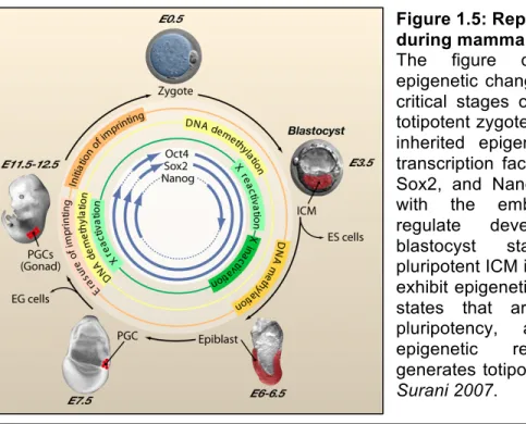
Figure 1.5: Reprogramming during mammalian development.
The figure depicts the main epigenetic changes occurring during critical stages of development. The totipotent zygote contains maternally inherited epigenetic modifiers and transcription factors, including Oct4, Sox2, and Nanog. These, together with the embryonic transcripts, regulate development to the blastocyst stage, where the pluripotent ICM is established. PGCs exhibit epigenetic and transcriptional states that are associated with pluripotency, and the ensuing epigenetic reprogramming re-generates totipotency. Adapted from Surani 2007.
1.2.3a DNA Methylation Reprogramming in Pre-implantation Embryos
In the mouse 1-cell zygote, several reports have shown that the paternal genome undergoes a rapid wave of DNA demethylation prior to the onset of the first cleavage (Mayer et al., 2000; Oswald et al., 2000; Santos et al., 2002; Wossidlo et al., 2010). Using 5mC antibodies as well as targeted bisulfite sequencing, these studies have shown that, as protamine-to-histone exchange
occurs in the paternal pronucleus, overall levels of DNA methylation decrease at most genic and intergenic sequences with the exception of imprinted loci. In contrast, the maternal genome gradually loses methylation as cleavage occurs in what looks like a replication dependent process (Howlett et al., 1991; Kafri et al., 1992; Rougier et al., 1998; Inoue et al., 2011). By the 8-cell stage most zygotic methylation marks have been erased, and only imprinted methylation can be detected.
Re-acquisition of DNA methylation is observed in the developing ICM; however, the trophoblast lineage seems to be re-methylated to a lesser extent (Monk M, 1990; Santos et al., 2002). DNMT expression is very dynamic during pre and post-implantation development. DNMT3b is the first de novo methyl-transferase to be expressed in the ICM between 4.5 and 7.5dpc (Watanabe et al., 2002; Hirasawa et al., 2008). Past 9.5dpc DNMT3b is replaced by DNMT3a, which is then detected throughout the embryo during the rest of development. There is still much to be learned about the precise methylation profiles the ICM harbors after de novo methylation. An attempt to answer this question come from the study of methylation patterns in cultured ES cells, which are derived from the ICM and are thought to preserve its developmental potency. ES cells express all 3 DNMTs and display dynamic change in methylation, resembling those seen during organogenesis, upon in-vitro differentiation. The recent study of human H1 and H9 ES cell methylomes revealed that ES cells have methylation levels approaching, but not reaching, those seen in somatic tissues, indicating a global de novo methylation (Lister et al., 2009, Laurent et al., 2010). They also
confirmed earlier observations suggesting that, in ES cells, promoters are generally hypomethylated at developmentally regulated genes (e.g. HOX clusters). By comparing ES cells to various differentiated tissues, these studies showed that reduced methylation at promoters and high methylation in gene bodies was observed at transcriptionally active genes. Interestingly, they also found regions displaying non-CpG methylation, a feature thought to be restricted to plant and fungi. Although still under investigation, this form of cytosine methylation could affect about 20% of cytosines found outside a CpG context in undifferentiated ES cells.
1.2.3b Reprogramming in Germ Cells
During PGC induction and development, an important remodeling of the chromatin fiber precedes the erasure of DNA methylation genome-wide, which is completed by 13.5dpc in mouse (Hajkova et al., 2008; Seki et al., 2005 and 2007; Popp et al., 2010). Following their induction and during G2 arrest (between ~7.5 and ~9.5dpc), the nuclei of PGCs progressively enlarge and immunofluorescence stainings for H3K9me2/3, H3K9ac and H3K27me3, are greatly reduced. By 10.5dpc, PGC chromatin is believed to be extremely loose but pervasive transcription is maintained at a low level via RNA polymerase II Serine 5 and Serine 2 dephosphorylation (Seki et al., 2007). Histone variants are also transiently incorporated (e.g. H3.3 and H2A.Z), and the canonical histone linker H1 is lost, indicating that deeper changes in nuclear chromatin might also prevent aberrant gene expression (Hajkova et al., 2008 and 2010). Again using
immunofluorescence, it was shown that by 12.5dpc the genome wide distribution of repressive histone modifications (e.g. H3K27me3 and H3K9me2/3 but not H3K9ac), histone variants, linker H1 and active RNA polII, revert to the levels seen in surrounding somatic tissues. A detailed picture of the distribution of these chromatin changes is still lacking due to the low abundance of these cells and the lack of culture models recapitulating these events.
In 7.5dpc PGCs, focused studies revealed that imprinted loci, transposons and single copy genes still bear substantial methylation indicative of their somatic origin – one X chromosome is inactivated in females (Sugimoto et al., 2007; Tam et al., 1994) and at least some copies of LINE-1s and IAP retro-transposons retain over 70% of methylated Cs at their regulatory sequences (Hajkova et al., 2002).
As PGCs enter G2 arrest, DNMT3a and b are transcriptionally down regulated. In contrast, Dnmt1 is still present albeit at low levels (Seki et al., 2005; Kurimoto et al., 2008). During this time frame, the global levels of 5mC staining begin to reduce (Seki et al., 2005) and reach their lowest when PGC resume proliferation past ~9.5dpc suggesting that a portion of the genome is loosing methylation. Detailed analysis of the methylation status of PGCs between 10.5 and 13.5dpc using methylation sensitive restriction assays and focused bisulfite sequencing showed that the dynamics of DNA demethylation of imprinted loci, single copy genes and transposons is more heterogeneous than previously thought (Walsh et al., 1998; Lees-murdock et al., 2003; Lane et al., 2003; Hajkova et al., 2002; Lee et al., 2002). Some copies of LINE-1 and IAP
retrotransposons retain substantial methylation until 11.5, and loose most of this signal between 12.5 and 13.5dpc. Notably, more copies of IAP seem to retain methylation even after 13.5dpc and demethylation is less prominent in female PGCs (Lees-murdock et al., 2003; Lane et al., 2003). In addition, whereas some single copy genes and imprinted loci (maternally imprinted Nnat, and paternally imprinted Peg3/5 and H19) show partial demethylation as early as 10.5dpc, most achieve full demethylation rapidly between 12.5 and 13.5dpc (Xist promoter, Peg10…, Lee et al., 2002; Hajkova et al., 2002). These data suggest that both passive and active demethylation could be involved in the drastic erasure of methylation reaching its lowest at 13.5dpc (Figure 1.6). To gain further insight into PGCs methylation reprogramming, Popp and colleagues recently reported a low coverage survey methylation of PGCs at E13.5 using bisulfite sequencing, and compared them to sperm and ES cells (Popp et al., 2010). Despite the low coverage of this study, they were able to show that by 13.5dpc overall methylation levels are below 10% in PGCs compared to over 70% in all other tissues analyzed. They also provide evidence for the involvement of AID in active demethylation of early PGCs as AID deficient mice show a small but significant increase in methylation at 13.5pdc compared to WT.
Figure 1.6: Timing of de novo methylation during male germ cell development.
During PGC migration to the gonads, DNA methylation levels (red) are erased in both male and female PGCs (black box). During male gametogenesis, de novo methylation occurs between 13.5dpc and SSC establishment post-birth. Methylation levels remain high throughout meiosis. The relative timing of PIWI protein expression and piRNA abundance is also depicted (see section 1.3).
Male PGCs start de novo methylation soon after their entry into embryonic gonads. In contrast, the genome of female PGCs remains hypomethylated until arrested oocytes resume growth and meiosis after birth. In male PGCs, DNMT3L and DNMT3a start to accumulate at ~13.5, peak by ~15.5 and revert to their
CpG-me
7.5dpc 13.5dpc 16.5dpc 14dpp
Migration De-novo Methylation Birth, SSCs Meiosis
Paternal Imprints Repeat silencing
Female and Males Males
High
Low
MILI
MIWI2
MIWI
DNMT3B and DNMT1 are found at much lower levels throughout PGC development (La Salle et al., 2004). Consequently, transposon sequences and imprinted loci initiate a rapid wave of de novo methylation between 14.5 and 18.5dpc. However, de novo methyl-marks are continually deposited until day 2 post-birth when PGCs colonize their niche as SSCs (Walsh et al., 1998; Ueda et al., 2000; Kato et al., 2007; Lees-Murdock et al., 2003; Kuramochi-Miyagawa et al., 2008). Methylation profiles are believed to undergo little if any changes during male meiosis. DNMT3B and DNMT1 levels are elevated in SSCs and reduce both during meiosis and spermiogenesis as sperm nuclei become transcriptionally silent. Nucleosomes a exchanged for protamines late in spermiogenesis. Hammoud and colleagues recently showed in human sperm that about 4% of the genome in still packaged in nucleosome retaining domains (Hammoud et al., 2009). These nucleosomes consist of either canonical or histone variants, such as the testes-specific histone H2B (TH2B). Interestingly, regions protected from protamine-exchange were also shown to be under methylated, further connecting the methylation status of the genome to higher order chromatin structures.
1.3: Germ Cell Associated Small RNA Pathways
In addition to protein coding messenger RNAs, eukaryotic cells express a wide range of non-coding transcripts ranging from 19-30bp, for small regulatory RNAs, to several hundred base pairs (e.g. long intergenic non-coding RNAs, lincRNAs). They often engage in transcriptional and post-transcriptional gene regulation, relying on RNA:DNA or RNA:RNA interactions (reviewed by Mercer et al., 2010 and Ghildiyal et al., 2010). During metazoan development, germ cells are characterized by the expression of a distinct RNAi pathway involving PIWI proteins and their 24-30bp associated piRNAs (PIWI-interacting RNAs). Male mice deficient for piRNAs are infertile, as germ cells fail to undergo productive meiosis (Deng et al., 2002; Kuramochi-Miyagawa et al., 2001 and 2004; Carmel et al., 2007). It is believed that the interplay between piRNAs and de novo methylation drives the re-methylation of retro-transposons during PGC development (Aravin and Bourc’his 2008; Aravin and Hannon 2008). Considering the extreme sequence diversity of piRNAs, and the high abundance of retro-elements in mammalian genomes, this model provides an interesting framework to study the connection between transposon dynamics and germ cell development.
The previous section highlighted the dynamic character of DNA methylation patterns during germ cell development at both single copy loci and repeated sequences. However, beyond 12.5dpc, PGCs are transcriptionally
piRNAs and other coding and non-coding transcripts are prior to de novo methylation. The following points cover the known biogenesis pathways leading to the production of small interfering RNAs in various cellular contexts, and focus on the newly characterized piRNA pathway in germ cells, mode of action of which still remains uncharacterized.
1.3.1 Overview of RNAi
From the characterization of silencing phenotypes induced by the introduction of multi-copy transgenes in plants, or the cloning of the first micro-RNA (mimicro-RNA) in C.elegans (lin-4, Lee et al., 1993), micro-RNA interference has been shown to be a highly conserved biological pathway used to dampen gene expression via the targeting of cellular mRNA and, sometimes, via transcriptional inhibition. At the core of this pathway lies the interaction between a small RNA and an Argonaute protein. Whereas small RNAs guide this RNA Induced Silencing Complex (RISC) to the cognate targets using base pairing, Argonaute proteins mediate silencing via mRNA cleavage, translational inhibition or chromatin remodeling (on the latter see Volpe and Martienssen 2011). Different classes of small RNA have now been characterized in unicellular and multicellular eukaryotes as well as in prokaryotes and archea (Hannon 2002; Marraffini and Sontheimer 2010). Over the past 20 years, small RNAs have been shown to regulate development, cell cycle, epigenetic inheritance and hundred small RNAs have been show to be mis-expressed in various diseases including
cancer and are currently being used for diagnosis and therapy (Silva et al., 2004).
In metazoans, small interfering RNAs (siRNA) and miRNAs are typically 19 to 23nt in size and constitute the most ubiquitously expressed classes of small RNAs. A third class, termed piRNAs (for PIWI interacting RNAs, also known as repeat-associated-small-interfering RNA, Aravin et al., 2003), is known to be exclusively expressed in germ cells and range form 24 to 31nt. miRNAs and siRNA share common steps in their biogenesis as both of them require the endonuclease III activity of Dicer to produce fully functional single stranded small RNAs from double stranded (ds) RNA structures (Bernstein et al., 2001; reviewed in Carmell and Hannon, 2004). They differ mostly in the nature of their precursors: siRNA are produced from long ds-RNA whereas miRNA are processed from a single stranded primary transcript that folds into a stem loop structure cleaved by the nuclear RNAse III enzyme Drosha (Lee et al., 2004). In contrast, piRNAs are thought to be initially processed from long primary single stranded transcripts by an unknown, Dicer independent, mechanism (Vagin et al., 2006; Houwing et al., 2007).
Following these processing steps, single stranded small RNAs are loaded into an Argonaute protein forming a functional RISC. Argonautes constitute a large conserved RNA binding protein family. They possess two related domains: the PAZ domain interacting with the 3' end of a small RNA and the PIWI domain, interacting with the 5'end, which forms the RNAseH catalytic domain (Song et al., 2004, Liu et al., 2004). Based on sequence analysis, Argonaute proteins can be
separated into two closely related clades: the Ago and the Piwi subfamily (Carmell et al., 2002). Argonautes in the Ago clade are most similar to ARGONAUTE1 found in Arabidopsis thaliana, and primarily interact with miRNAs and siRNA. The Piwi clade however, shares most similarities with the drosophila PIWI protein.
In mammals, the association of Argonaute proteins with a small RNA is primarily occurring in the cytoplasm where targeting is also thought to happen. RISC can mediate target cleavage 10nt away from the 5' end of the small RNA when perfectly matched. However, in most cases, mismatched interactions leads to translational inhibition (Olsen and Ambros, 1999). When engaged in silencing, RISC is found in processing and stress bodies throughout the cytoplasm (Liu et al., 2005a/b, reviewed by Leung and Sharp 2006). These bodies contain numerous structural and catalytic proteins promoting the silencing function of RISCs associated with miRNAs, siRNAs and even piRNAs (on the latter see Siomi and Aravin 2011).
1.3.2 PIWI Proteins and Germ Cell Specific RNAi
In contrast to the ubiquitously expressed Agos, the Piwi clade is restricted to the germ line. Preliminary studies in drosophila revealed that the PIWI proteins, piwi and aubergine, are essential for gametogenesis and germline stem cell renewal (Cox et al., 1998 and 2000; Schmidt et al., 1999). Disruption of the piwi gene causes improper stem cell divisons in the male and female germline,
and disrupts gametogenesis progression preventing cyst formation and meiosis. The mouse and human genomes encode three conserved PIWI proteins: MIWI/HIWI (PIWIL1), MILI/HILI (PIWIL2) and MIWI2/HIWI2 (PIWIL4). During mouse gametogenesis PIWI proteins are detected in PGCs as early as 12.5-13.5 dpc. In the male germ-line, MILI is expressed throughout spermatogenesis until the round spermatid stage following meiosis, first occurring around 20dpp (Figure 1.6). Similarly, MILI is detected in the female germ-line soon after PGC migration and is continuously expressed in both arrested and growing oocytes (Aravin et al., 2008). MIWI2 expression starts at ~15.5dpc and stops prior to SSC establishment after birth. Finally, MIWI expression starts at the pachytene stage of meiosis, at around 14 dpp, and stops at the round spermatid stage. Cellular localization of these proteins addressed by immuno-fluorescence or using GFP-fused transgenic animals showed that while MILI and MIWI are found exclusively in the cytoplasm, MIWI2 can shuttle to the nucleus when loaded with piRNAs (Aravin et al., 2008).
Disrupting either mili or miwi genes both causes male sterility. However, while mili-KO leads to in meiotic arrest at the pachytene stage, miwi-KO animals undergo meiosis properly, but germ cells fail to develop beyond the haploid round spermatid stage (Deng et al., 2002; Kuramochi-Miyagawa et al., 2001 and 2004). These data suggest that MILI and MIWI have non-redundant functions in spermatogenesis. Recently, two miwi2 deficient mice were independently generated and revealing that MIWI2 is essential for SSC maintenance, with MIWI2 loss resulting in a progressive depletion of undifferentiated germ cells in
adult testes (Carmell et al., 2007, Kuramochi-Miyagawa et al., 2008). miwi2-KO animals also fail to undergo full meiosis and arrest cell division before the pachytene stage. Considering MIWI2 expression during embryogenesis, this effect is certainly due to defects occurring before meiosis.
In addition to germ line defects, it has been shown that Drosphila piwi and aub and mouse miwi2 and mili mutant animals derrepress transposons in the germ line (Sarot et al., 2004; Brennecke et al., 2007; Carmell et al., 2007; Aravin et al., 2007). It was therefore suggested that PIWI proteins might play a critical role in transposon silencing in the germ line, and that defects observed during gametogenesis are mainly caused by the reactivation of transposable elements.
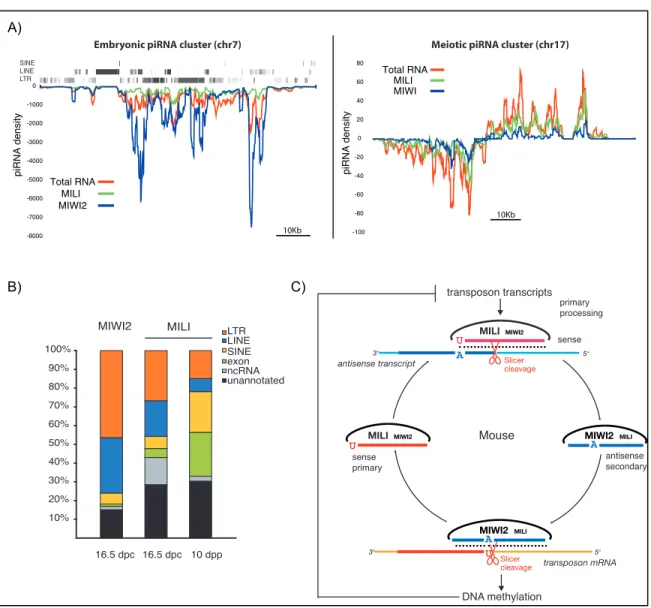
1.3.3 PiRNA Biogenesis during Male Germ Cell Development In mammals, two distinct waves of piRNA expression have been described so far (Girard et al., 2006; Aravin et al., 2007 and 2008). The first wave of piRNA expression peaks at around the time when MIWI2 and MILI are co-expressed in PGCs (between 15.5dpc and birth) and those piRNAs are referred to as embryonic piRNAs. The second wave occurs during MIWI and MILI co-expression when cells enter the first division of meiosis; those are referred to as meiotic piRNAs (Figure 1.6). The purification of PIWI protein complexes and subsequent cloning and sequencing of their associated piRNAs revealed that these two waves differ mostly in the type of transcripts they can target.
Embryonic piRNAs are strongly, but not exclusively, enriched for transposable elements sequences, including all three classes of
retro-transposons (LINEs, SINEs and LTRs, Figure 1.7). Consequently, a large fraction of these piRNAs maps to multiple locations in the genome, offering the potential for robust and redundant silencing of transposons during epigenetic reprogramming of PGCs. Unlike the strong strand preference seen for the different PIWI complexes in drosophila (Brenecke et al., 2007), MILI and MIWI2 only show a slight bias toward sense and anti-sense piRNAs respectively (Aravin et al., 2008). In addition, MILI and MIWI2 have distinct piRNA size preferences: 26-27nt for MILI and 28-29nt for MIWI2. Interestingly, in a MILI mutant background MIWI2 fails to load piRNAs and to localize in the nucleus, suggesting that MILI functions upstream of MIWI2 (Aravin et al., 2008, Kuramochi-Miyagawa et al., 2008). Meiotic piRNAs are enriched for sequences mapping to large un-annotated portions of the genome both in rat, mouse and human (Lau et al., 2006, Girard et al., 2006, Aravin et al., 2006). MILI and MIWI bind virtually the same piRNA sequences though each complex is associated with its characteristic piRNA size - ~26nt for MILI and ~30nt for MIWI. The lack of information about the cellular function of the transcripts regulated by meiotic piRNAs makes it challenging to study their function, especially in an attempt to explain the MIWI sterility phenotype (Deng et al., 2002). Finally, between these two waves, MILI is continuously expressed in late PGCs, SSCs and pre-meiotic spermatogonia. Despite the absence of any partner, MILI is loaded with a “transition” population switching from transposons rich piRNAs to a much more gene enriched population in SSCs and ultimately meiotic piRNAs as cells enter differentiation (Aravin et al., 2007).
There is still much mystery regarding how piRNAs are processed from primary transcripts. Key signatures of piRNA sequences include a 5’Uracil and a 2’-O-methyl group at the 3’ end, both of which have been shown to be important for their functional association with PIWI proteins (Horwich et al., 2007; Saito et al., 2007; Kirino et al., 2007a/b). The analysis of piRNA sequences uniquely mapping to the genome revealed the existence of large piRNA loci, 10kb to a 100kb in size, producing both sense and antisense reads (Brenecke et al., 2007; Girard et al., 2006; Aravin et al., 2006, 2007 and 2008). These large piRNA clusters produce about 10-20% of all embryonic piRNAs (the remaining piRNAs being produced from individual transposons or genes) and more than 90% of meiotic piRNAs (see Figure 1.7A). Whereas embryonic piRNA clusters can generate primary transcripts on both strands, meiotic piRNA clusters are transcribed from one strand but often show a typical bidirectional structure with a central promoter firing in opposite directions. Several lines of evidence from drosophila and mouse suggest that primary piRNA 5’end production requires the activity of a phospholipase D, MITOPLD in mouse and Zucchini in drosophila (Watanabe et al., 2011; Haase et al., 2010). Mutant animals for these proteins accumulate cluster transcripts and display sterility phenotypes. Recently, an in-vitro study using insect cells lysates, provided evidence that 3’ ends of piRNAs are generated by the trimming of long transcript loaded into PIWIs by an unknown 3’ to 5’ exonucelase (Kawaoka et al., 2011). These observations provide an attractive model to explain the size and sequence preference of piRNAs bound to PIWI proteins.