Int. .I. Radiation Oncology Eiol. Phys.. Vol. 38, No. 2, pp. 343-350, 1997 Copyright 0 1997 Elsevier Science Inc. Printed in the USA. All rights reserved
0360.3016/97 $17.00 + .OO ELSEVIER
l
Clinical Investigation
PI1 SO360-3016( 97) 00031-X
SEMINOMA
ARISING
IN CORRECTED
AND UNCORRECTED
INGUINAL
CRYPTORCHIDISM:
TREATMENT
AND PROGNOSIS
IN 66 PATIENTS
YE-XIONG LI,
M.D.,*
PHILIPPEA. COUCKE, M.D.,? Tu-NAN QIAN, M.D.,*
YI-RONG HUANG,M.D.,*
DA-ZHONG
Gu, M.D.,
* RENCOLIVIER MIRIMANOFF,M.D.t
AND ZI-HAOYu, M.D.*
*Department of Radiation Oncology, Cancer Hospital and Institute, Chinese Academy of Medical Sciences, Beijing, P. R. China: and ‘Department of Radiation Oncology, Centre Hospitalier Universitaire Vaudois, Lausanne, Switzerland
Purpose: The purpose of this study was to analyze prognosis and treatment results for seminoma arising in corrected and uncorrected inguinal cryptorchidism (SCIC and SUIC).
Methods and Materials: We reviewed 66 patients with inguinal seminomas between June 1958 and December
1991 at the Cancer Hospital and Institute of Chinese Academy of Medical Sciences. Of these patients, 23 had
prior orchiopexy and 43 presented with an inguinal form of cryptorchidism. At presentation, 17 of 66 (26%)
patients had nodal metastases. This nodal involvement was 30% (7 of 23) for SCIC and 23% (10 of 43) for SUIC,
respectively. These numbers are comparable with those in a series of patients treated for scrotal seminoma at
our institution (26% vs. 20%). However, 3 of 23 (13%) patients who had prior orchiopexy presented with
inguinal nodal metastasis as compared with 0 of 43 patients with SUIC or 4 of 237 patients with scrotal seminoma
(p < .05). There were 49 stage I, 5 stage IIA, 8 stage IIB, 3 stage III, and 1 stage IV patients. All patients underwent radical orchiectomy and received further radiotherapy, chemotherapy, or both. Patients with stage I
and stage II disease were treated primarily with radiotherapy, whereas patients with stage III and IV disease
were treated with chemotherapy.
Results: The overall and disease-free survival at 5 and 10 years was 94% and 92%, 89% and 87%, respectively.
The overall 5- and lo-year survival by stage was 100% and 100% for stage I, and 77% and 68% for stage II, respectively @ < .05). There was no significant difference in survival between SUIC and SCIC (93% vs. 96%
at 5 years). Four patients developed relapse. Two of these four patients experienced relapse at the inguinal area,
due to a marginal miss. Three of four patients with relapse were successfully salvaged, and one died of disease.
Conclusion: Our results indicate that prognosis for inguinal seminoma is excellent and similar to that of scrotal
seminoma. Postorchiectomy radiotherapy can be considered as the standard treatment for stage I and IIA inguinal
seminoma. We recommend routinely including the para-aortic and ipsilateral pelvic nodes. 0 1997 Elsevier
Science Inc.
Seminoma, Cryptorchidism, Radiotherapy.
INTRODUCTION
Testicular tumors account for only 1% of all malignancies, but are among the most common cancers in young men (4, 12). Moreover, it is well known that cryptorchidism is a risk factor in the development of germ-cell tumors (7, 24, 25, 40). Seminomas are more common in unde- scended testis, with more than 80% of testicular tumors being histologically labeled as seminomas in abdominal (pelvic), more than 60% in inguinal, and 50% in normally descended testis ( 1,2, 14). Several studies have suggested that early orchiopexy reduces the risk of seminoma de- velopment (1, 14, 17).
Data in the literature on inguinal seminomas are rare
and the numbers of patients in published series are small. Inguinal seminoma is generally discussed under the topic of germ-cell tumor in cryptorchid testes or in
combination with seminoma in abdominally unde-
scended testis (2, 10, 20, 35). Seminoma arising in an undescended inguinal cryptorchid testicle is extremely rare in Western countries because inguinal cryptorchi- dism is systematically corrected ( 14, 30). In contrast to patients with scrotal seminoma, clinical studies report a high proportion of advanced stage disease in patients with cryptorchid seminoma. The optimal management after orchiectomy for cryptorchid seminoma is less well established and the prognosis is controversial ( 1, 2, 10,
18, 20, 32). It remains unclear whether the poor prog- nosis observed in inguinal seminoma is due to advanced stage or other factors. In this study, we retrospectively reviewed our experience with inguinal seminoma, in an attempt to analyze clinical features, pathway of nodal spread, and prognosis, and also to determine the treat- ment option for this rare entity.
Reprint requests to (present address): Ye-Xiong Li, M.D., De- taire Vaudois, 101 1-Lausanne, Switzerland. partment of Radiation Oncology, Centre Hospitalier Universi- Accepted for publication 5 November 1996.
344 I. J. Radiation Oncology l Biology l Physics Volume 38, Number 2, 1997
METHODS AND MATERIALS
From June 1958 to December 1991, 373 patients with testicular seminomas were treated at the Cancer Hospital and Institute of Chinese Academy of Medical Sciences, Beijing, China. The diagnosis of pure seminoma was con- firmed in all cases by histopathologic review. Of these 373 patients, 136 had a history of cryptorchidism, of whom 25 (23 inguinal and 2 pelvic) had prior orchiopexy. Sixty- eight patients with seminomas arising in pelvic unde- scended cryptorchid testis were reported elsewhere (22). Of the remaining 66 patients, 43 patients had primary tu- mor located distally or proximally in the inguinal canal, and 23 patients who underwent prior orchiopexy for in- guinal cryptorchidism presented with primary tumor in the scrotum. These 66 patients, with seminoma arising in sur- gically corrected (n = 23) and uncorrected (n = 43) in-
guinal cryptorchidism (SUIC and SCIC), form the basis
of this study. The characteristics of these 66 patients were compared with 237 patients with seminoma arising in a normally descended testis. For ease of discussion the latter cohort of patients is labeled “scrotal seminoma.”
Staging
Patients were staged according to Table 1. Clinical stag- ing included a medical history, physical examination, blood chemistry, and chest X-ray. Intravenous pyelogra- phy (IVP) was done in 40 patients. Since 1982, computed tomography scans (CT) and/or ultrasound of abdomen and pelvis were carried out in 34 patients. Tumor markers including a-fetoprotein (crFP) and P-human chorionic go-
nadotrophin (PHCG) have been determined in 35 pa-
tients. Treatment
All but four patients underwent inguinal radical or- chiectomy. These 4 of 23 patients with prior orchiopexy had a scrotal incision. Two patients had inguinal node dissection as well. After orchiectomy, all patients received postoperative radiotherapy, chemotherapy, or both. The treatment options were dependent on the stage of the dis- ease. Patients with stage I and II disease were usually treated with radiotherapy, whereas in patients with stage III and IV chemotherapy was considered the primary treat- ment with or without radiotherapy. As shown in Table 2, of 49 patients with stage I disease, 45 patients were treated with radiotherapy alone and 4 with chemotherapy alone.
Among the 13 stage II patients, the primary treatment was
radiotherapy in 7, and chemotherapy in 6. Three of the latter received chemotherapy only. Three patients with stage III and IV disease received chemotherapy with or without radiotherapy and only one stage III patient re- ceived radiotherapy alone.
Radiotherapy was initially given with a Cobalt-60 unit and, after 1982, with an 8-MV linear accelerator. Daily dose varied from 150 to 200 cGy. Patients with stage I and IIA disease had their para-aortic nodes irradiated with
Table 1. Stage for testicular seminoma Stage I II IIA: IIB: III IV Definitions
Primary tumor confined to the testis or extending to adjacent tissue or organ
Infradiaphragmatic node metastases <5 cm in diameter
~5 cm in diameter
Supradiaphragmatic node metastases Extralymphatic metastases
parallel opposed anterior-posterior (AP-PA) fields. The
ipsilateral pelvic nodes were routinely treated. Before 1987, the treatment for pelvic nodes was delivered through a single anterior field. In most cases, this field included the medial and upper inguinal nodes, as previ- ously described (3 I). After 1987, parallel opposed fields (dog-leg) were used and inguinal nodes were excluded in
the treatment volume. The infradiaphragmatic dose for
stage I and IIA disease ranged from 15 to 40 Gy with a median of 30 Gy. Of the six stage IIB patients receiving radiotherapy, four were treated with a para-aortic and ip- silateral pelvic field similar to stage I patients, but the treatment portals were tailored to cover the bulky abdom- inal disease. Two stage IIB patients were treated with ei-
ther whole abdominal-pelvic irradiation (WAPI) or lo-
cal-field radiotherapy following chemotherapy. In contrast to stage I, higher total doses (median 35 Gy) were applied to stage IIB patients. Of the two stage III patients who received radiotherapy, one was treated with para-aortic, pelvic, and mediastinal irradiation, and one with WAPI following chemotherapy. One stage IV patient was treated with combination chemotherapy followed by whole lung irradiation.
Seven patients received the ipsilateral inguinal irradia-
tion and one patient with bilateral inguinal invasion re-
ceived elective irradiation to the bilateral inguinal and pel- vic nodes. Ipsilateral hemiscrotum was irradiated in only four cases of scrotal incision. Prophylactic mediastinal and left supraclavicular irradiation was given to 2 of 13 pa- tients with stage II disease.
Thirteen patients were treated with chemotherapy (Ta- ble 2). Eight of these patients were treated with chemo- therapy alone and five with a combination of chemother-
apy and radiotherapy. Before 1985, chemotherapy
Table 2. Radiotherapy and chemotherapy according to stage Stage
I IIA IIB III IV Total
RT alone 45 3 4 1 0 53
Chemo alone 4 1 2 I 0 8
Chemo + RT 0 1 2 1 1 5
Treatment for inguinal seminoma 0 Y. X. LI er al. 345
Table 3. Clinical features of 66 patients with inguinal
seminoma
Characteristic Number
Age (years) Median Range
Site of primary tumor Right Left Stage I IIA IIB III IV Presenting symptom Painless mass Painful mass Opposite cryptorchidism Inguinal Pelvic 32 18-58 30 36 49 5 8 3 1 50 16 11 3
consisted of N-formylsarcolycin at 30 mg/day orally for 1-2 months (8 patients). Thereafter, a combination reg-
imen of cisplatin. vinblastine, and bleomycin (PVB ) was
applied to the five patients. Follow-up and statistics
Patients were followed every 3 months for 1 year, and thereafter every 6 months or once yearly. At each follow- up, they were submitted to physical examination, com- plete blood count, serum biochemistry, and chest X-rays.
Abdominal-pelvic CT scan and/or ultrasound were per-
formed since 1982.
The follow-up time ranged from 3 to 34 years with a median of 12 years. The overall survival was calculated from the date of surgery. Disease-free survival was cal- culated from the date of surgery until evidence of recur- rence or progression. Updating of the medical files was done in December 1994 and survival data were calculated according to these. Survival curves were calculated by the Kaplan-Meier product-limit method ( 19) and differences between survival curves were analyzed with the log rank
test (29). Numerical data were compared with Student’s t-test and qualitative data with the Chi-square test or Fisher’s exact test.
RESULTS Patient characteristics
Patient ages varied from 18 to 58 years with a median of 32 years. Most patients (46 of 66, 70%) were between 30 and 50 years of age. There were 30 right and 36 left primary sites. Fourteen of 66 (2 1% ) patients had a history of contralateral cryptorchidism. Correction of an ipsilat- era1 inguinal hernia was performed in seven patients. Ob- vious invasion of the surrounding soft tissue was observed in three cases. The most frequently encountered symptom at presentation was an inguinal or scrotal mass, which was painful in 16 patients. Clinical characteristics are sum- marized in Table 3.
The size of the primary tumor arising in an inguinal testes ranged from 3 to 12 cm, with mean and median sizes of 6.5 cm and 7 cm, respectively. These are similar to data of scrotal seminoma (range 2- 15 cm, median 6 cm).
Seventeen of 66 (26%) patients with inguinal semi-
noma had lymph node metastases. Patients with SCtC had
a 30% (7 of 23) incidence of nodal metastases, not sig- nificantly different from the 23% (10 of 43) incidence encountered in SUIC. In Table 4, the stage distribution of inguinal seminoma has been compared with that of scrotal seminoma. Again, there was no significant difference in nodal involvement between the two groups (20% for scro- tal vs. 26% for inguinal seminoma) .
Fourteen of 17 patients with nodal disease had para- aortic involvement without pelvic or inguinal node me- tastases. However, three patients with SCIC had inguinal
nodal metastases without pelvic or para-aortic node in- volvement. Two of these three patients with inguinal nodal involvement experienced this condition unilaterally, whereas the third presented with bilateral nodal involve- ment. However, none of the patients with SUIC showed an inguinal nodal presentation. The difference in inci- dence of inguinal involvement between SCIC (3 of 7, 43%) and SUIC (0 of 10) is significant, as is the differ- ence between SCIC and scrotal seminoma (4 of 48, 8%).
Table 4. Stage distribution of inguinal seminoma compared with that of scrotal seminoma
Stage
I II 111 IV
No. (%I No. (%) No. (%) No. (%I
SUIC 33 (77) 8 (19) 1 (2) 1 (21
SCIC 16 (70) 5 (22) 2 (9) 0 (0)
Scrotal 189 (80) 29 (12) 5 (2) 14 (6)
SCIC = seminoma in corrected inguinal cryptorchidism; SUIC = seminoma in uncorrected inguinal cryptorchidism.
Total
43 23 237
346 I. J. Radiation Oncology 0 Biology 0 Physics Volume 38, Number 2, 1997
Survival
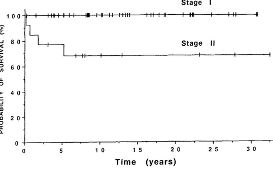
Five patients died of inguinal seminoma, and one of a cerebrovascular accident. The overall 5- and lo-year sur- vival for all 66 patients was 94% and 92%, respectively. The corresponding disease-free survival was 89% and
87% (Fig. 1). The overall 5- and IO-year survival by stage was 100% and 100% for stage I, and 77% and 68% for stage II, respectively (Fig. 2). The difference in survival between stage I and II was statistically significant (p < .05). All five stage IIA patients were alive with no evi- dence of disease, whereas 4 of 8 stage IIB patients died of disease. We treated only four patients with stage III and IV disease and one stage III patient who received radio- therapy alone died of progression. Due to the small num- ber of patients, no actuarial survival was calculated for the latter groups. For 43 patients with SUIC, the overall and disease-free survival at 5 and 10 years was 93% and 90%, and 88% and 85%, respectively. The overall and disease- free survival for 23 patients with SCIC at 5 years was 96% and 91%, respectively. No significant difference in sur- vival was observed between SUIC and SCIC.
Recurrence
Four patients relapsed at 8, 13, 18, and 42 months after initial therapy. One stage I patient who was initially treated with N-formylsarcolycin developed abdominal re- currence. He was salvaged with radiotherapy and is still alive 26 years after salvage treatment. One stage IIB pa-
tient developed liver metastases and died of progressive
disease. Two patients who received infradiaphragmatic ra- diation experienced relapse at the ipsilateral inguinal node. One patient had pelvic relapse as well. One patient presented with stage II SUIC and one with stage 1 SCIC. Both patients were initially treated with a single anterior pelvic field. Therefore, this site of recurrence can be con- sidered as a geographic miss. They were salvaged with radiotherapy. However, one of these patients developed a second relapse in the pelvis. The iatter patient underwent pelvic and inguinal node dissection, and was finally suc- cessfully salvaged with a PVB combination.
Complications
Long-term complications were observed in two pa- tients. There was one case of radiation-related hemor- rhagic cystitis. This patient presented with stage I disease and received 35 Gy at the midline level to the para-aortic and ipsilateral iliac areas. The pelvic area was irradiated by a single anterior field given with a Cobalt-60 unit. The estimated irradiation dose to the bladder was 41-45 Gy. He presented with hemorrhagic cystitis 43 months after irradiation, confirmed by endoscopy. The other patient, retreated for inguinal recurrence by radiotherapy, devel- oped an ipsilateral leg edema and muscular atrophy. The cumulative dose reached 90 Gy to the pelvis and inguinal area. Both patients are still alive without evidence of dis- ease.
Three patients had a second malignant tumor. Two pa- tients had synchronous contralateral scrotal seminoma. One patient with stage I inguinal seminoma developed a contralateral testicular mixed tumor (seminoma plus ter- atoma) 6 years after radiotherapy.
DISCUSSION Presentation
In the current study, we retrospectively reviewed 66 patients with inguinal seminoma. Forty-three of these pa- tients had primary tumor in the inguinal area, and 23 had prior orchiopexy and thus they presented with their pri- mary tumor in the scrotum. Inguinal seminoma is appar- ently more common in China than in Western countries ( 10, 35, 36). The age peak, chief complaint, presenting signs, and symptoms are comparable with seminoma in normally descended testes. In accordance with other series (10, 13, 32), we found 21% bilateral cryptorchidism in patients with inguinal seminoma and 19% in patients with pelvic cryptorchid seminoma.
The size of primary tumor in inguinal seminoma is sim- ilar to that found in scrotal seminoma, but it is much smaller than that seen in pelvic cryptorchid seminoma (22). This can easily be explained by early detection and diagnosis of tumor development in case of inguinal and scrotal seminoma.
Lymphatic metastases
The incidence of lymphatic metastases in the present
series is consistent with that observed by Shi et al. ( 17%) (36). Gauwitz et al. (10) confirms this observation and reports a 33% (3 of 9) incidence in SUIC, not significantly different from the 25% incidence observed in scrotal sem- inoma, but significantly lower than in patients with pelvic cryptorchid seminoma (76%, 13 of 17 ) . These data, how- ever, are in contrast with most other published reports on
nodal metastases in cryptorchid seminoma ( 1, 2, 20, 32).
However, the latter reports contain a smaller number of patients as compared with the former.
As it is the case in scrotal seminoma, the nodal spread in patients with inguinal seminoma is primarily to the para-aortic nodes. However, we more frequently found inguinal nodal involvement in patients with SCIC as com- pared with SUIC or scrotal seminoma. This observation is consistent with the hypothesis that prior orchiopexy or any previous inguinal surgery disrupts the normal lym- phatic drainage (26,40). Therefore, those patients should routinely have the pelvis and inguinal regions examined. Prognosis and treatment
The prognosis of inguinal and pelvic seminoma is not well established and conflicting results have been reported (Table 5 ) . Because of the rarity of this disease, the sur- vival of inguinal seminoma as an entity has not been an- alyzed extensively in the literature. The reported survival rates of patients with seminoma in cryptorchid testis have
Treatment for inguinal seminoma l Y. X. LI et al. 341
Table 5. Summary of the relationship between stage and survival in the literature Stage
Stage 1 Stage II III/IV
Investigators and year Years of Number of
(reference number) treatment Diagnosis patients No. (%) No. (%) No. (%) Five-year survival
Abratt et al. 1992 (1) 1970- 1991 cs 20 11 (55) 4 (20) 5 (25) 76% for all patients; 100% for 13 stage I and IIA Batata ef al. 1980 (2) 1934- 1975 cs 54 33 (61) 14 (26) 7 (13) 78% for all patients;
97% for stage I Gauwitz and Zagars 1992 (10) 1960-1990 PCS 17 4 (24) 11 (65) 2 (12) 92% for all patients
SUIC 9 6 (67) 3 (33) 0 (0)
Sham et al. 1990 (35) 1972-1986 PCSlSUIC 9 6 (67) 2 (22) 1 (11) 6 stage I patients alive at 2 years Kulkami and I&mat 1991 (20) 1980- 1986 PCSWJIC 12 4 (33) 6 (50) 2 (17) 100% for 8 stage I
and nonbulky II Shi et al. 1987 (36) 1960-1980 PCS 39 27 (69) 11 (28) 1 (3) 89% for all patients;
SUIC 64 53 (83) 7 (11) 4 (6) 97% for stage I Li et al. (current series) 1958- 1991 PCS 60 34 (57) 17 (28) 9 (15) 92% for PCS;
100% and 94% for stage I and II, resp.
1958 1991 SUIC 43 33 (77) 8 (19) 2 (4) 93% for SUIC SCIC 23 16 (70) 5 (22) 2 (9) 96% for SCIC;
100% for stage I
CS: cryptorchid seminoma, including all seminomas in corrected and uncorrected pelvic/inguinal testis; PCS: pelvic cryptorchid seminoma, seminoma in uncorrected abdominal/pelvic testis; SUIC: seminoma in uncorrected inguinal cryptochidism; SCIC: seminoma in corrected inguinal cryptorchidism.
been reported to range from 76% to 92% at 5 years ( 1, 2, 10,36). However, more detailed analyses of survival data, including ours, show that early stage disease in cryptor-
chid seminoma carries an excellent prognosis with 97- 100% 5year survival after radiation therapy (1, 2, 20, 36)) which is in accordance with those obtained for scrotal
seminoma(3, 11, 15,37,38,41).Thelowlevelofoverall
survival in most series is mainly explained by the poor results in more advanced-stage disease, due particularly
to the lack of an effective chemotherapy combination in previous years.
For stage I patients primarily treated with radiotherapy the selection of an optimal treatment volume is an impor- tant consideration. Based on our experience, we recom- mend treating the para-aortic and ipsilateral iliac nodes for patients with SUIC or SCIC. Radiation dose should not exceed a total of 25-30 Gy. There is no convincing evi- dence for treating the inguinal lymph nodes and the tumor
Disease-free survival
0 I 1 I I 6 I ! I
0 5 IO 15 20 25 30 35
Time (years)
348 I. J. Radiation Oncology 0 Biology 0 Physics
bed in patients with seminoma in undescended inguinal testis, because the para-aortic lymph nodes remain the first pathway and the rate of inguinal soft tissue invasion is very low. As stated by other investigators, the inguinal lymph nodes should be routinely treated only in those pa- tients who had prior inguinal or scrotal surgery, because of their high risk of nodal involvement (3, 37, 39,41) . In the present series, of 14 stage I patients with SCIC treated with radiotherapy, 9 patients received partial inguinal ir- radiation by a single anterior pelvic field (before 1987) and only one patient received complete inguinal irradia- tion. As a general rule, inguihal and scrotal radiation should be avoided whenever possible. The scattered dose to the remaining testis contributes to a higher risk of in- fertility and perhaps to a second testicular cancer as well. No patient in the present series had scrotal relapse, sug- gesting that the use of scrotal irradiation may not be nec- essary except in instances of overt tumor involvement of scrotal tissues. Regarding the uncertainty of lymph drain- age after corrective surgery in SCIC, adjuvant chemo- therapy may be an effective alternative in these patients (8, 28).
Patients with stage IIA disease should receive irradia- tion to the para-aortic and ipsilateral pelvis. However, if the inguinal nodes or inguinal surrounding soft tissues (tu- mor bed) are involved, then the ipsilateral inguinal nodes and the tumor bed should be treated.
Despite the fact that only two patients received prophy- lactic mediastinal irradiation, no relapse episodes were found in the mediastinal and supraclavicular regions in stage II patients. As is the case with scrotal seminoma (3, 6, 2 1, 34, 39)) prophylactic mediastinal irradiation for in- guinal seminoma has been abandoned.
Combination chemotherapy, especially platinum-based
regimens, which are currently used for bulky stage II scro-
Volume 38, Number 2, 1997
tal seminoma (5,27,41> , are recommended for stage IIB inguinal seminoma.
No definite conclusions can be made concerning stage III and IV inguinal seminoma because of the small number of patients. However, there is no reason to believe that the treatment of choice for stage III and IV inguinal semi-
noma, that is, combined platinum-based chemotherapy,
should be different than in the case of scrotal seminoma at the same stage (8, 9, 23).
Complication
Late complications can be avoided by carefully select- ing adequate techniques. The unique case of hemorrhagic cystitis was observed in a patient with a 35Gy dose ( spec- ified at the midline). This untoward effect on the bladder at this low dose level can be explained by the use of an inappropriate technique in previous years. In this partic- ular patient, the target volume was treated by a single an- terior field given with a Cobalt-60 unit. This implies that there was at least a 20-30% dose heterogeneity in the treated volume, and a large fraction size on the anterior port to the bladder, resulting in the late complication. Proper field arrangement and definition of target volumes are prerequisites for tumor cure, and, in case of relapse after radiotherapy, as was shown in this and many other series, combined chemotherapy should be considered for salvage therapy.
CONCLUSION
Seminoma arising in uncorrected or corrected inguinal cryptorchid testis is an uncommon clinical problem, but a higher incidence of this tumor was observed in China. The frequency of nodal involvement and its spread are similar to that of scrotal seminoma. Inguinal nodes in patients
Stage I
Stage II
0 I I I ' I I I
0 5 10 15 20 25 30
Time (years)
Treatment for inguinal seminoma 0 Y. X. LI et al. 339
with prior orchiopexy (SCIC) are more frequently ob- be included in the treatment ports only when they are in-
served than in patients with noncryptorchid seminoma or volved or if there is obvious involvement of surrounding
seminoma arising in uncorrected inguinal testes. In early- tissues by the primary tumor. In more advanced-stage dis-
stage disease (stage IA and IIA ) , postorchiectomy radio- ease, the treatment of choice remains combination che-
therapy should be performed, and should include para- motherapy. Provided that the treatment is adequate, the
aortic and ipsilateral pelvic nodes. Inguinal nodes should prognosis is excellent and stage-dependent.
REFERENCES
1. Abratt, R. P.; Reddi. V. B.: Sarembock, L. A. Testicular cancer and cryptorchidism. Br. J. Urol. 70:656-659; 1992. 2. Batata, M. A.; Chu, F. C. H.; Hilaris, B. S.; Whitmore,
W. F.; Golbey, R. B. Testicular cancer in cryptorchids. Can- cer 49:1023-1080; 1982.
3. Bayens, Y. C.: Helle. P. A.; Van Putten. W. L. J.; Mali, S. P. M. Orchidectomy followed by radiotherapy in 176 stage I and II testicular seminoma patients: Benefits of a IO-year follow-up study. Radiother. Oncol. 25:97-102;
1992.
4. Boyle, P.; Kaye, B. S.: Robertson. A. G. Changes in testic- ular cancer in Scotland. Eur. J. Cancer Clin. Oncol. 23:827-
830; 1987.
5. Dosmann, M. A.: Zagars. G. K. Postorchiectomy radiother- apy for stages I and II testicular seminoma. Int. J. Radiat. Oncol. Biol. Phys. 26:381-390; 1993.
6. Evensen, J. R.; Fossa. S. D.; Kjellevold, K.; Lien, H. H. Testicular seminoma: Analysis of treatment and failure for stage II disease. Radiother. Oncol. 4:55-61; 1985.
7. Farrer, J. H.; Walker, A. H.; Rajfer, J. Management of the postpubertal cryptorchid testis: A statistical review. J. Urol.
134:1071-1076; 1985.
8. Fossa, S. D.; Droz, J. P.: Stoter, G.; Kaye, S. B.; Vermey- len, K.; Sylvester, R. Cisplatin, vincristine and ifospham- ide combination chemotherapy of metastatic seminoma: Results of EORTC trial 30874. Br. J. Cancer 71:619-624; 1995.
9. Friedman. E. L.: Garnick, M. B.; Stomper, P. C.; Mauch, P. M.; Harington, D. P.; Richie, J. P. Therapeutic guidelines and results in advanced seminoma. J. Clin. Oncol. 3: 1325-
1332; 198.5.
10. Gauwitz, M. D.: Zagars, G. K. Treatment of seminoma aris- ing in cryptorchid testes. Int. J. Radiat. Oncol. Biol. Phys. 24:153-159; 1992.
11. Giacchetti. S.; Raoul, Y.; Wibault, P.: Droz. J. P.; Court, B.; Eschwege, F. Treatment of stage I testis seminoma by radio- therapy: Long-term results-a 30-year experience. Int. J. Ra- diat. Oncol. Biol. Phys. 27:3-9; 1993.
12. Gilliland, F. D.; Key, C. R. Male genital cancers. Cancer 75:295-315; 1995.
13. Gross, R. E.; Jewett, T. C. Surgical experiences from 1222 operations for undescended testes. JAMA 160:634;
1956.
14. Halme. A.; Kellokumpu-Lehtinen, P.: Lehtonen. T.; Teppo, L. Morphology of testicular germ cell tumours in treated and untreated cryptorchidism. Br. J. Urol. 64:78-83; 1989. 15. Hanks. G. E.; Peters, T.; Owen, J. Seminoma of the testis:
Long term beneficial and deleterious results of radiation. Int. J. Radiat. Oncol. Biol. Phys. 24:913-919; 1992.
16. Horwich, A.; Dearnaley, D. P.; Duchesne, G. M.; Williams, M.: Brada, M.; Peckham. M. J. Simple nontoxic treatment
of advanced metastatic seminoma with carboplatin. J. Clin.
Oncol. 7: 1150- 1156: 1989.
17. Jones. B. J.: Thornhill. J. A.; O’Donnell, B.; Kelly, D. G.; Walsh, A.: Fennelly. J. J.; Fitzpatrick, J. M. Influence of
prior orchiopexy on stage and prognosis of testicular cancer.
Eur. Ural. 19:201-203; 1991.
18. Jonsson, K.; Wallace, S.; Jing, B. S.; Boyle, L. E.; John- son, D. E. Lymphangiography in patients with malig- nancy in anon-descended testicle. J. Urol. 119:614-617: 197s.
19. Kaplan, E. L.; Meier, P. Nonparametric estimation from in- complete observations. J. Am. Stat. Assoc. 53:457-481; 1958.
10. Kulkarni. J. N.; Kamat, M. R. Tumors in undescended testis. J. Surg. Oncol. 46:257-260: 1991.
21. Laukkanen, E. L.; Olivotto, I.; Jackson, S. Management of seminoma with bulky abdominal disease. Int. J. Radiat. On- col. Biol. Phys. 14:227-233; 1988.
22. Li, Y. X.; Coucke, P. A.; Qian, T. N.; Huang, Y. R.; Gu, D. Z.; Mirimanoff, R. 0.; Yu, Z. H. Clinical characteristic, prognosis and treatment for pelvic cryptorchid seminoma Int. J. Radiat. Oncol. Biol. Phys. (in press).
23. Loehrer, P. J.; Birch, R.; Williams, S. D.: Greco. F. A.; Ein-
horn, L. H. Chemotherapy of metastatic seminoma: The
Southeastern Cancer Study Group experience. J. Clin. Oncol. 5:1212-1220; 1987.
24. Martin, D. C. Malignancy in the cryptorchid testis. Urol. Clin. North. Am. 9:371-376; 1982.
25. Martin, D. C.; Menck, H. R. The undescended testis: Man- agement after puberty. J. Ural. 114:77-79; 1975.
26. Mason, M. D.; Featherstone. T.; Olliff, J.; Horwich. A. In- guinal and iliac lymph node involvement in germ cell tum- ours of the testis: Implications for radiological investigation and for therapy. Clin. Oncol. 3: 147- 150; 1991.
27. Mason, B. R.: Kearsley. J. H. Radiotherapy for stage II tes- ticular seminoma: The prognostic influence of tumor bulk. J. Clin. Oncol. 6: 1856- 1862; 1988.
28. Oliver, R. T. D.; Edmonds, P. M.; Ong, J. Y. H.; Ostrowski,
M. J.; Jackson, A. W.; Baille-Johnson, H.; Williams. M. V.;
Wiltshire, C. R.; Mott. T.; Pratt, W. R.; Trask, C. W. L.; Hope-Stone, H. F. Pilot studies of 2 and 1 course carboplatin as adjuvant for stage I seminoma: Should it be tested in a randomized trial against radiotherapy? Int. J. Radiat. Oncol. Biol. Phys. 29:3-8; 1994.
29. Peto, R.; Pike, M. C.; Armitage, P.; Breslow. N. E.; Cox.
D. R.; Howard, S. V.; Mantel, N.; McPherson, K.; Peto, J.; Smith, P. G. Design and analysis of randomized clin- ical trials requiring prolonged observation of each patient. Part II. analysis and examples. Br. J. Cancer 35:1-39; 1977.
30. Pike. M. C.; Chilvers, C.; Peckham, M. J. Effect of age at
orchidopexy on risk of testicular cancer. Lancet 1: 1246- 1248; 1986.
31. Qian, T. N.; Hu. Y. H.; Chen, C. X.; Qi. Y. Q.; Gu, D. Z.;
Gu, X. Z. Radiation therapy of seminoma of the testis. Int. J. Radiat. Oncol. Biol. Phys. 7:717-720: 1981,
32. Raina, V.: Shukla. N. K.; Gupta, N. P.; Deo, S.; Rath. G. K. Germ cell tumours in uncorrected cryptorchid testis at Insti- tute Rotary Cancer Hospital. New Delhi. Br. J. Cancer 71:380-382; 1995.
33. Raja, M. A.; Oliber, R. T. D.; Badenoch. 5.: Blandy, J. P.
Orchidopexy and transformation of seminoma to non-semi- noma. Lancet 339:930: 1992.
350 I. J. Radiation Oncology l Biology l Physics Volume 38, Number 2, 1997
34. Sagerman, R. H.; Kotlove, D. J.; Regine, W. F.; Chung, C. T.; King, Cl. A. Stage II seminoma: Results of postor- chiectomy irradiation. Radiol. 172565-568; 1989. 35. Sham, J. S. T.; Choy, D.; Chan, K. W.; Choi, P. H. K. Sem-
inoma of normally-descended and cryptorchid testis. Eur. J. Surg. Oncol. 16:33-36; 1990.
36. Shi, X. H.; Liu, T. F. Radiotherapy for seminoma arising in undescended testis: Analysis of 103 cases. Tumor 7:113-
115; 1987 (in Chinese).
37. Vallis, K. A.; Howard, G. C. W.; Duncan, W.; Combleet, M. A.; Kerr, G. R. Radiotherapy for stages I and II testicular seminoma: Results and morbidity in 238 patients. Br. J. Ra- diol. 68:400-405; 1995.
38. Warde, P.; Gospodarowicz, M. K.; Panzarella, T.; Catton, C. N.; Sturgeon, J. F. G.; Moore, M.; Goodman, P.; Jewett, M. A. S. Stage I testicular seminoma: Results of adjuvant ir- radiation and surveillance. J. Clin. Oncol. 13:2255-2262; 1995. 39. Willan, B. D.; McGowan, D. G. Seminoma of the testis: A 22-year experience with radiation therapy. Int. J. Radiat. On- col. Biol. Phys. 11:1769-1775; 1985.
40. Wobbes, T.; Schraffordt, K. H.; Oldhoff, J. The relation be- tween testicular tumours, undescended testes, and inguinal hernias. J. Surg. Oncol. 14:45-51; 1980.
41. Zagars, G. K.; Babaian, R. J. Stage I testicular seminoma: Rationale for postorchiectomy radiation therapy. Int. J. Ra- diat. Oncol. Biol. Phys. 13:155-162; 1987.