ORIGINAL ARTICLE
Comparative effects of teriparatide and ibandronate on spine
bone mineral density (BMD) and microarchitecture (TBS)
in postmenopausal women with osteoporosis: a 2-year
open-label study
C. Senn&B. Günther&A. W. Popp&R. Perrelet&
D. Hans&K. Lippuner
Received: 22 November 2013 / Accepted: 25 March 2014 / Published online: 24 April 2014 # International Osteoporosis Foundation and National Osteoporosis Foundation 2014
Abstract
Summary Treatment effects over 2 years of teriparatide vs. ibandronate in postmenopausal women with osteoporosis were compared using lumbar spine bone mineral density (BMD) and trabecular bone score (TBS). Teriparatide induced larger increases in BMD and TBS compared to ibandronate, suggesting a more pronounced effect on bone microarchitecture of the bone anabolic drug.
Introduction The trabecular bone score (TBS) is an index of bone microarchitecture, independent of bone mineral density (BMD), calculated from anteroposterior spine dual X-ray absorptiometry (DXA) scans. The potential role of TBS for monitoring treatment response with bone-active substances is not established. The aim of this study was to compare the effects of recombinant human 1–34 parathyroid hormone (teriparatide) and the bisphosphonate ibandronate (IBN), on lumbar spine (LS) BMD and TBS in postmenopausal women with osteoporosis.
Methods Two patient groups with matched age, body mass index (BMI), and baseline LS BMD, treated with either daily subcutaneous teriparatide (N=65) or quarterly intravenous IBN (N=122) during 2 years and with available LS BMD measurements at baseline and 2 years after treatment initiation were compared.
Results Baseline characteristics (overall mean ± SD) were similar between groups in terms of age 67.9±7.4 years, body mass index 23.8±3.8 kg/m2, BMD L1–L4 0.741±0.100 g/ cm2, and TBS 1.208±0.100. Over 24 months, teriparatide induced a significantly larger increase in LS BMD and TBS than IBN (+7.6 %±6.3 vs. +2.9 %±3.3 and +4.3 %±6.6 vs. + 0.3 %±4.1, respectively; P<0.0001 for both). LS BMD and TBS were only weakly correlated at baseline (r2=0.04) with no correlation between the changes in BMD and TBS over 24 months.
Conclusions In postmenopausal women with osteoporosis, a 2-year treatment with teriparatide led to a significantly larger increase in LS BMD and TBS than IBN, suggesting that teriparatide had more pronounced effects on bone microarchitecture than IBN.
Keywords Bone mineral density . Open-label study . Osteoporosis . Parathyroid hormone . Teriparatide . Trabecular bone score . Treatment
Introduction
Osteoporosis and osteoporosis-related fractures represent a worldwide disease burden, especially in North America and Europe [1–5]. With much of this burden stemming from the morbidity and mortality related to the roughly nine million osteoporotic fractures that occur each year [4,5], the primary goal of treatment has long been fracture prevention [6–8].
As recommended by the World Health Organization (WHO) since 1994 [9], bone mineral density (BMD), mea-sured by dual X-ray absorptiometry (DXA) is the current gold standard for diagnosing osteoporosis and monitoring treat-ment, supported by the fact that BMD is a major determinant of bone strength and fracture risk [10]. However, considerable
D. Hans codirected equally the TBS study.
C. Senn
:
B. Günther:
A. W. Popp:
R. Perrelet:
K. Lippuner (*) Department of Osteoporosis, Inselspital, Berne University Hospital and University of Berne, CH-3010 Berne, Switzerlande-mail: [email protected] D. Hans
Center of Bone Disease, Bone and Joint Department, Lausanne University Hospital,
overlap exists between BMD values in individuals who de-velop fractures and those who do not [11], indicating that other factors influence both bone strength and fracture risk. To cite just but a few: macrogeometry of cortical bone, microarchitecture of trabecular bone, as well as bone microdamage, mineralization, and turnover [12–14].
The trabecular bone score (TBS) is derived from a simple anteroposterior LS DXA scan and can be used for the nonin-vasive assessment of intravertebral cancellous bone microarchitecture [15–18]. The TBS was shown to discrimi-nate between patients with incident hip, nonvertebral, or ver-tebral fracture and nonfractured patients with osteoporosis in several prospective and retrospective cohort studies [19–24], with odds ratios ranging between 1.6 and 2.05 with a similar order of magnitude than lumbar spine BMD [19,21,22,24]. In addition, in these studies, the combination of TBS and lumbar spine BMD (LS BMD) was generally superior to either measurement alone with regard to fracture risk predic-tion [19,20,22–24]. Furthermore, the TBS was responsive to treatment with antiresorptive drugs in a large cohort study [25] and in the retrospective analysis of a randomized placebo-controlled trial with the aminobisphosphonate zoledronate [26].
Daily subcutaneous injections of recombinant human 1– 34 N-terminal fragment of parathyroid hormone (teriparatide) were shown to increase BMD and to reduce the risk of new vertebral and nonvertebral, but not hip, fractures in patients with postmenopausal and glucocorticoid-induced osteoporo-sis [27, 28]. The observed increases in LS BMD with teriparatide accounted for approximately 30–41 % of the achieved vertebral fracture risk reduction, suggesting other mechanisms accounting for the remainder [27, 29]. Teriparatide was recently shown to improve trabecular microarchitecture in iliac crest bone biopsies of postmeno-pausal women [29], confirming earlier preclinical data show-ing improved histomorphometric cancellous bone parameters and vertebral bone strength in ovariectomized rats [30] and monkeys [31]. The effects of teriparatide on the TBS are unknown.
Bisphosphonates, such as the aminobisphosphonate ibandronate (IBN), are inhibitors of bone resorption belonging to the mainstay of osteoporosis treatment. Histomorphometric data in postmenopausal women treated during 2 years with intravenous ibandronate showed no increase in trabecular number or volume and no decrease in intertrabecular separa-tion [32]. In line with these findings, an earlier study per-formed with the intravenous aminobisphosphonate zoledronate in postmenopausal women showed an only mod-est increase in TBS consistent with a preservation of vertebral bone microarchitecture [26].
The aims of this study were: (1) to compare the effects of subcutaneous teriparatide and intravenous ibandronate on LS BMD and TBS in postmenopausal women with osteoporosis;
(2) to assess whether the changes in TBS are independent of those of BMD; and (3) to evaluate the changes in TBS in terms of possible clinical relevance at the individual patient level.
Methods
The study was conducted at the Department of Osteoporosis of the University Hospital of Berne, Switzerland as an open-label, retrospective, nonrandomized, treatment-controlled study comparing the effects of an up to 2-year treatment with subcutaneous teriparatide (Forsteo®, Eli Lilly, USA) vs. a 2-year treatment with intravenous IBN (Bonviva®, Roche, Switzerland) on LS BMD and TBS in two groups of post-menopausal women with osteoporosis matched for age, body mass index (BMI), and LS BMD.
Study population and treatment schemes
Postmenopausal women with primary osteoporosis referred for evaluation to the osteoporosis consultation of the Depart-ment of Osteoporosis of University Hospital of Berne, Swit-zerland, between 2007 and 2009, who were subsequently treated with teriparatide 20μg self-injected daily, were eval-uated if they had LS BMD measurements performed by DXA at baseline and after 2 years of therapy (n=70). Women treated with teriparatide usually had experienced a vertebral fragility fracture during a prior therapy with an antiresorptive, inde-pendently of their LS BMD value. The reimbursed duration of treatment with teriparatide was increased from 18 to 24 months during the course of the study. As a consequence, one sixth of the patients treated with teriparatide were treated during 18 months followed by an intravenous infusion of zoledronate 5 mg, the other five sixth were on teriparatide during 24 months. The control group consisted of postmenopausal women with primary osteoporosis in whom a treatment with intravenous ibandronate 3 mg every 3 months was initiated between 2007 and 2009 and monitored during at least two following years at the Department of Osteoporosis of Berne. Women treated with ibandronate usually had a BMD T-score at or below−2.5 or one or more prevalent vertebral fractures. Women on ibandronate were matched for age, BMI, and LS BMD with women in the teriparatide group, following a 2:1 ratio (n=140). All subjects were vitamin D-replete and re-ceived adequate calcium and vitamin D3 supplementation. Women currently on glucocorticosteroids or presenting other secondary forms of osteoporosis were not eligible. Prior ther-apies with bisphosphonates, estrogens, or other bone active substances including vitamin D were allowed. Only women with evaluable DXA scans for both LS BMD and TBS at baseline and after 2 years in the teriparatide and the ibandronate groups, respectively, were included in the analysis.
Measurement of bone mineral density
Bone mineral density (BMD) was assessed by DXA (Hologic QDR 4500A®, Hologic, Bedford, MA, USA) at the single study centre of the Department of Osteoporosis of the Univer-sity Hospital of Berne, Switzerland. All DXA scans were performed in accordance with manufacturer recommendations. Lumbar spine BMD measurements were recorded for L1 through L4 (L1−L4). BMD was expressed as grams per square centimeter of hydroxyapatite and as T-scores (standard devia-tion [SD] from the mean of a healthy young female popula-tion). The manufacturer’s normative database was used as reference for the LS after analysis according to International Society for Clinical Densitometry (ISCD) rules [33]. Individ-ual vertebrae were excluded in case of fractures or degenerative changes, in accordance with ISCD rules for individual verte-brae exclusion (more than 1 standard deviation from immedi-ately adjacent vertebrae). Quality control was performed daily (anthropometric spine phantom supplied by the manufacturer).
Measurement of trabecular bone score
The trabecular bone score (TBS) is a grey-level texture mea-surement that can be applied to DXA images for quantifying local variations in grey level [15–17]. Using experimental variograms of two-dimensional (2D) projection images, TBS can differentiate between three-dimensional (3D) microstruc-tures that exhibit the same bone density, but different trabecular characteristics [15, 18]. The TBS is obtained by direct (re-)analysis of an acquired lumbar spine DXA image, without need for further imaging. All TBS determinations were per-formed in a blinded manner within the Bone Disease Unit at the University Hospital of Lausanne, Lausanne, Switzerland using TBS iNsight® Software version 1.8.2 (Med-Imaps, Bordeaux, France). Lumbar spine TBS (LS TBS) was evaluated in the same vertebrae and regions of measurement as those used for LS BMD, with LS TBS calculated as the mean value of the individual measurements for vertebrae L1−L4. The coefficient of variation for LS BMD measurements at the Department of Osteoporosis of the University Hospital of Berne is 0.90 % when applying with ISCD recommendations (15 outpatients representative of our daily routine with triplicate measurements after repositioning) with a corresponding coefficient of varia-tion of 1.12 % for TBS. Thus, the least significant change (LSC) is 2.49 % for LS BMD and 3.10 % for TBS.
Statistical analysis
Descriptive analysis included means and percentages with standard deviations. The percent changes in BMD and TBS were calculated for each subject as the absolute change from baseline to 2-year follow-up, divided by the baseline value. Bivariate intergroup comparisons were performed between
those treated with IBN vs. teriparatide using Student’s t tests and Pearson χ2 analysis for continuous and noncontinuous variables, respectively. Pearson correlation coefficients were calculated for BMD vs. TBS and for change from baseline in BMD vs. change from baseline in TBS. All inferential tests were two-tailed and P<0.05 was set as the threshold for statistical significance. All statistical analyses were performed using Stata® software (Version 12, StataCorp, Texas, USA).
Results
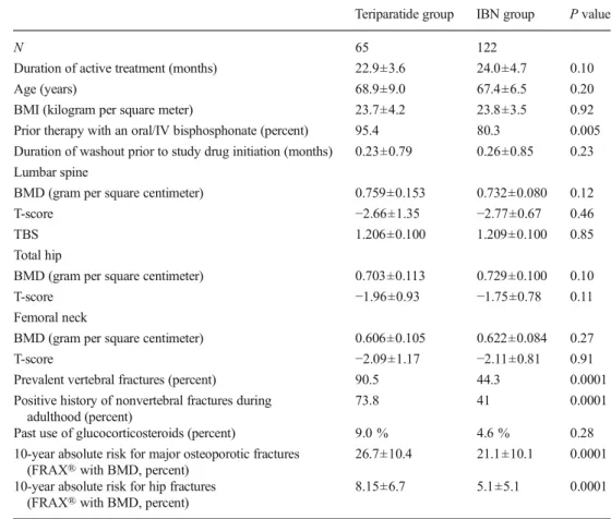
Overall, 65 (93 %) and 122 (87 %) patients had evaluable DXA scans for LS BMD and TBS at baseline and after 2 years in the teriparatide and the ibandronate groups, respectively, and were included in the analysis. In total, 40, 40, and 20 % of the patients had four, three, and two vertebrae evaluated, respec-tively. As a result of matching, baseline characteristics (mean ±SD) were similar between groups in term of age, body mass index, baseline LS BMD T-score and LS TBS (Table 1). Patients in the teriparatide group were more likely having had prior therapy with a bisphosphonate (95.4 vs. 80.3 %, P=0.005), having prevalent vertebral fractures or a positive history of fracture during adulthood (90.5 vs. 44.3, and 73.8 vs. 41.0 %, respectively; P=0.0001 for both), and had a significantly higher clinical fracture risk score for hip and major osteoporotic fractures assessed by FRAX®.
As shown in Fig.1, after 24 months of therapy, LS BMD and TBS increased significantly more with teriparatide com-pared to IBN (+7.6 %±6.3 vs. +2.9 %±3.3 and +4.3 %±6.6 vs. +0.3 %±4.1, respectively; P<0.0001 for both). Compared to baseline, increases in LS BMD were significant in both the teriparatide and IBN group (P<0.0001 for both), while in-creases in LS TBS were significant in the teriparatide group only (P<0.0001).
Baseline spine BMD and TBS were only weakly correlat-ed, (r2=0.04), indicating that only 4 % of the variance in one parameter was explained by the other. There was no correla-tion between the 2-year changes in BMD and TBS from baseline (r2=0.01).
As shown in Table2, LS BMD was more sensitive than LS TBS with regard to the proportion of patients achieving an increase above least significant change (LSC) in both treat-ment groups: 78.5 vs. 61.5 % (McNemar test P<0.01) and 51.6 vs. 26.3 % (P<0.001) with teriparatide and IBN, respec-tively. Interestingly, while only 11.0 % of the patients did not respond in terms of TBS below the LSC with teriparatide, this proportion reached 27.0 % with IBN. Furthermore, in the teriparatide group, 51.0 % of the patients were above the LSC for both LS BMD and TBS vs. only 28.0 % in the IBN group (results not shown in Table 2), suggest-ing a stronger effect on bone microarchitecture with the former.
Discussion
In postmenopausal women with primary osteoporosis, a 2-year treatment with teriparatide increased LS BMD and TBS significantly more and in a significantly greater proportion of patients than a 2-year treatment with intravenous ibandronate.
As the increase in LS TBS was largely independent from the BMD response, these results suggests that LS TBS may contribute to assess the effects of bone anabolic agents on vertebral microarchitecture.
Only few studies have investigated the effect of bone active substances on LS TBS [25, 26]. Taken together with the
Table 1 Baseline Demographics and Clinical Characteristics (mean ± SD)
All values are means ± SD, except indicated otherwise
Teriparatide group IBN group P value
N 65 122
Duration of active treatment (months) 22.9±3.6 24.0±4.7 0.10
Age (years) 68.9±9.0 67.4±6.5 0.20
BMI (kilogram per square meter) 23.7±4.2 23.8±3.5 0.92 Prior therapy with an oral/IV bisphosphonate (percent) 95.4 80.3 0.005 Duration of washout prior to study drug initiation (months) 0.23±0.79 0.26±0.85 0.23 Lumbar spine
BMD (gram per square centimeter) 0.759±0.153 0.732±0.080 0.12
T-score −2.66±1.35 −2.77±0.67 0.46
TBS 1.206±0.100 1.209±0.100 0.85
Total hip
BMD (gram per square centimeter) 0.703±0.113 0.729±0.100 0.10
T-score −1.96±0.93 −1.75±0.78 0.11
Femoral neck
BMD (gram per square centimeter) 0.606±0.105 0.622±0.084 0.27
T-score −2.09±1.17 −2.11±0.81 0.91
Prevalent vertebral fractures (percent) 90.5 44.3 0.0001 Positive history of nonvertebral fractures during
adulthood (percent)
73.8 41 0.0001
Past use of glucocorticosteroids (percent) 9.0 % 4.6 % 0.28 10-year absolute risk for major osteoporotic fractures
(FRAX® with BMD, percent)
26.7±10.4 21.1±10.1 0.0001 10-year absolute risk for hip fractures
(FRAX® with BMD, percent)
8.15±6.7 5.1±5.1 0.0001 0.0% 2.0% 4.0% 6.0% 8.0% 10.0% 12.0% 14.0% Mean per cen t chang e ov er 24 mon ths Spine BMD Spine TBS p< 0.0001 p< 0.0001 p< 0.0001 p< 0.0001 p< 0.0001 Teriparatide Ibandronate p= 0.86 Fig. 1 Percent change in lumbar
spine BMD and TBS at month 24 after treatment with teriparatide (22.9 months) and ibandronate (24 months). Mean values ± standard deviation. P values above the bars refer to significance vs. baseline
present findings, these earlier reports are consistent with the concept that bisphosphonates allow for “positive mainte-nance” of bone microarchitecture rather than a major improve-ment in microarchitecture. For almost two decades, bisphosphonates have been the therapy of choice to treat osteoporosis and to prevent fractures, relying on solid evi-dence with regard to fracture risk reduction [2,34–38]. How-ever, the long-term bone safety of bisphosphonates has been recently questioned [39]. On one hand, bisphosphonates in-crease bone strength by increasing the mineralization of remodelled bone units, reducing cortical porosity and decreas-ing focal stress. On the other hand, they suppress the genera-tion of new bone remodelling units and reduce bone turnover [39]. In the present study, the increase in LS BMD and TBS observed with IBN was of a lower order of magnitude than expected. In earlier studies, LS TBS increased by 0.25 to 0.5 % per year under antiresorptive therapy [25,26], which is clearly more than the 0.3 % over 2 years reported in the present study. One of the possible explanations may be related to the fact that more than 80 % of the “real life” women included had been on bisphosphonate therapy before being switched to intravenous ibandronate after a very short or no washout period. This suggests not unexpectedly that, in pa-tients under prior antiresorptive therapy, bisphosphonates may be more likely to maintain than to restore vertebral microarchitecture.
Teriparatide exerts primarily bone anabolic effects, which include increasing cancellous bone volume and connectivity, increasing cortical bone thickness, and enhancing trabecular morphology [39,40]. Since the inaugural publication by Neer et al. [27] in 2001, several smaller studies have confirmed that teriparatide allows for strong BMD increases and for fracture risk reduction, suggesting that it may become an attractive alternative to bisphosphonates for strengthening and possibly restoring bone microarchitecture [41–44]. In the present study, a 2-year therapy with subcutaneous teriparatide induced a statistically significant increase in LS BMD and TBS of large
magnitude. The latter (+4.3 % in only 2 years) exceeds by far the TBS increases reported with antiresorptive substances to date [25,26]. Furthermore, the ratio of BMD to TBS increase was approximately 2:1 with teriparatide and 9:1 with ibandronate in the present study, compared to 10:1 with bisphosphonates in a retrospective study of 534 postmeno-pausal women treated with antiresorptive therapy in the Ca-nadian province of Manitoba [25], and 4:1 in a 3-year ran-domized, controlled study with yearly intravenous zoledronate [26]. Taken together with the absence of a signif-icant correlation between changes from baseline in BMD and changes from baseline in TBS [25,26], these observations indicate that BMD and TBS measure different characteristics of bone and bone strength.
With regard to individual therapy monitoring, only 12– 35 % of patients on bisphosphonates had an increase in TBS that was exceeding the LSC in earlier studies [25, 26], as compared to 26 % in the present analysis. In contrast, about 62 % of the patients on teriparatide were above LSC, primarily indicating that LS TBS may be more suitable for monitoring the effects of bone anabolic substances than for monitoring the effects of antiresorptives.
To date, only one direct comparison between teriparatide and a bisphosphonate (risedronate) has been published in postmenopausal women with osteoporotic spine compression fractures. In that study, teriparatide yielded a significantly greater increase in BMD from baseline in the lumbar spine and femoral neck and was associated with a lower incidence of vertebral fractures at 18 months (4 vs. 9 %, respectively; P=0.01) and with less severe vertebral fractures (P=0.04) [45]. There have, on the other hand, been several studies assessing the effects of teriparatide in patients in whom bis-phosphonate treatment has failed or otherwise been terminat-ed. In the most recently published study, postmenopausal women with severe osteoporosis who had failed treatment with a bisphosphonate responded well to 18 months of treat-ment with daily parathyroid hormone, with a 37 % reduction in the incidence of fractures in their second year of treatment relative to their first 6 months of therapy, and a 76 % reduction relative to baseline subsequent to this. Patients also reported reduced back pain and improved health-related quality of life while taking teriparatide [46]. These results are consistent with the results of several prior studies demonstrating some benefit of parathyroid hormones in the aftermath of bisphos-phonate therapy [41,44,47] although it is the first time that results are reported with TBS.
The findings of the present study are limited by the retro-spective nature of the analysis. Pretreatment with antiresorptives may have partially blunted some of the expect-ed effects on LS BMD and TBS. In addition, the two groups were not comparable with respect to osteoporosis severity, which may have influenced the results. Keeping these limita-tions in mind, the results show, for the first time, that larger
Table 2 Percentage of patients above, within and below the LSC for both teriparatide and IBN groups and LS BMD and LS TBS
Above LSC (percent) Within LSC (percent) Below LSC (percent) Teriparatide LS BMD 78.5 20.0 1.5 LS TBS 61.5 27.7 10.8 Ibandronate LS BMD 51.6 42.6 5.8 LS TBS 26.3 46.7 27.0
LSC least significant change, LS lumbar spine
Within LSC for LS BMD is between−2.49 and +2.49 % Within LSC for LS TBS is between−3.10 and +3.10 %
effects on trabecular microarchitecture assessed by TBS may be expected when using bone anabolic substances and may open the way for future research in this direction including, but not limited to, the place of TBS alone and/or in combina-tion with BMD and/or clinical risk factors for the choice and monitoring of treatments with bone-active substances and the identification of more individualized treatment schemes for patients with osteoporosis at increased risk of fracture.
Conclusions
In women with postmenopausal osteoporosis, a 2-year treat-ment with teriparatide exerted more beneficial effects on lumbar spine BMD and microarchitecture assessed by TBS than ibandronate. Changes in LS BMD and TBS from base-line were not correlated, confirming that these two parameters measure different responses of bone to therapy. At the indi-vidual patient level, TBS was significantly more sensitive to bone anabolic substances than to antiresorptives, with almost two thirds of the patients on teriparatide showing TBS in-creases above the least significant change.
Acknowledgments We are grateful to Philippe Kress, MD, for reviewing and commenting on our manuscript.
Conflicts of interest Didier Hans is coowner of the TBS patent and has corresponding ownership shares. Christoph Senn, Beatrice Günther, Albrecht W. Popp, Romain Perrelet, and Kurt Lippuner declare that they have no conflict of interest.
References
1. Czerwinski E, Badurski JE, Marcinowska-Suchowierska E, Osieleniec J (2007) Current understanding of osteoporosis according to the position of the World Health Organization (WHO) and International Osteoporosis Foundation. Ortop Traumatol Rehabil 9: 337–356
2. Cooper C, Reginster JY, Cortet B, Diaz-Curiel M, Lorenc RS, Kanis JA, Rizzoli R (2012) Long-term treatment of osteoporosis in post-menopausal women: a review from the European Society for Clinical and Economic Aspects of Osteoporosis and Osteoarthritis (ESCEO) and the International Osteoporosis Foundation (IOF). Curr Med Res Opin 28:475–491
3. Dhanwal DK, Dennison EM, Harvey NC, Cooper C (2011) Epidemiology of hip fracture: worldwide geographic variation. Indian J Orthop 45:15–22
4. Burge R, Dawson-Hughes B, Solomon DH, Wong JB, King A, Tosteson A (2007) Incidence and economic burden of osteoporosis-related fractures in the United States, 2005–2025. J Bone Miner Res 22:465–475
5. Johnell O, Kanis JA (2006) An estimate of the worldwide prevalence and disability associated with osteoporotic fractures. Osteoporos Int 17:1726–1733
6. NIH (2001) Consensus development panel on osteoporosis preven-tion, diagnosis, and therapy, March 7–29, 2000: highlights of the conference. South Med J 94:569–573
7. American College of Rheumatology Ad Hoc Committee on Glucocorticoid-Induced Osteoporosis (2001) Recommendations for the prevention and treatment of glucocorticoid-induced osteoporosis: 2001 update. Arthritis Rheum 44:1496–1503
8. Tucci JR (2006) Importance of early diagnosis and treatment of osteoporosis to prevent fractures. Am J Manag Care 12:S181–S190 9. Report of a WHO Study Group (1994) Assessment of fracture risk
and its application to screening for postmenopausal osteoporosis. World Health Organ Tech Rep Ser 843:1–129
10. Johnell O, Kanis JA, Oden A et al (2005) Predictive value of BMD for hip and other fractures. J Bone Miner Res 20:1185–1194 11. Hordon LD, Raisi M, Aaron JE, Paxton SK, Beneton M, Kanis JA
(2000) Trabecular architecture in women and men of similar bone mass with and without vertebral fracture: I. Two-dimensional histol-ogy. Bone 27:271–276
12. Link TM, Majumdar S (2004) Current diagnostic techniques in the evaluation of bone architecture. Curr Osteoporos Rep 2:47–52 13. Rubin CD (2005) Emerging concepts in osteoporosis and bone
strength. Curr Med Res Opin 21:1049–1056
14. Dalle Carbonare L, Giannini S (2004) Bone microarchitecture as an important determinant of bone strength. J Endocrinol Invest 27:99– 105
15. Hans D, Barthe N, Boutroy S, Pothuaud L, Winzenrieth R, Krieg MA (2011) Correlations between trabecular bone score, measured using anteroposterior dual-energy X-ray absorptiometry acquisition, and 3-dimensional parameters of bone microarchitecture: an experimental study on human cadaver vertebrae. J Clin Densitom 14:302–312 16. Pothuaud L, Barthe N, Krieg MA, Mehsen N, Carceller P, Hans D
(2009) Evaluation of the potential use of trabecular bone score to complement bone mineral density in the diagnosis of osteoporosis: a preliminary spine BMD-matched, case-control study. J Clin Densitom 12:170–176
17. Pothuaud L, Carceller P, Hans D (2008) Correlations between grey-level variations in 2D projection images (TBS) and 3D microarchitecture: applications in the study of human trabecular bone microarchitecture. Bone 42:775–787
18. Silva BC, Boutroy S, Zhang C et al (2013) Trabecular bone score (TBS)—a novel method to evaluate bone microarchitectural texture in patients with primary hyperparathyroidism. J Clin Endocrinol Metab 98:1963–1970
19. Del Rio LM, Winzenrieth R, Cormier C, Di Gregorio S (2013) Is bone microarchitecture status of the lumbar spine assessed by TBS related to femoral neck fracture? A Spanish case–control study. Osteoporos Int 24:991–998
20. Krueger D, Fidler E, Libber J, Aubry-Rozier B, Hans D, Binkley N (2013) Spine trabecular bone score subsequent to bone mineral density improves fracture discrimination in women. J Clin Densitom. doi:10.1016/j.jocd.2013.05.001
21. Boutroy S, Hans D, Sornay-Rendu E, Vilayphiou N, Winzenrieth R, Chapurlat R (2013) Trabecular bone score improves fracture risk prediction in non-osteoporotic women: the OFELY study. Osteoporos Int 24:77–85
22. Iki M, Tamaki J, Kadowaki E, Sato Y, Dongmei N, Winzenrieth R, Kagamimori S, Kagawa Y, Yoneshima H (2013) Trabecular bone score (TBS) predicts vertebral fractures in Japanese women over 10years independently of bone density and prevalent vertebral defor-mity: the Japanese population-based osteoporosis (JPOS) cohort study. J Bone Miner Res
23. Briot K, Paternotte S, Kolta S, Eastell R, Reid DM, Felsenberg D, Gluer CC, Roux C (2013) Added value of trabecular bone score to bone mineral density for prediction of osteoporotic fractures in post-menopausal women: The OPUS study. Bone 57:232–236
24. Hans D, Goertzen AL, Krieg MA, Leslie WD (2011) Bone microarchitecture assessed by TBS predicts osteoporotic fractures independent of bone density: the Manitoba study. J Bone Miner Res 26:2762–2769
25. Krieg MA, Aubry-Rozier B, Hans D, Leslie WD, Manitoba Bone Density P (2013) Effects of anti-resorptive agents on trabecular bone score (TBS) in older women. Osteoporos Int 24:1073–1078 26. Popp AW, Guler S, Lamy O, Senn C, Buffat H, Perrelet R, Hans D,
Lippuner K (2013) Effects of zoledronate versus placebo on spine bone mineral density and microarchitecture assessed by the trabecular bone score in postmenopausal women with osteoporosis: a three-year study. J Bone Miner Res 28:449–454
27. Neer RM, Arnaud CD, Zanchetta JR et al (2001) Effect of parathy-roid hormone (1–34) on fractures and bone mineral density in post-menopausal women with osteoporosis. N Engl J Med 344:1434– 1441
28. Saag KG, Shane E, Boonen S, Marin F, Donley DW, Taylor KA, Dalsky GP, Marcus R (2007) Teriparatide or alendronate in glucocorticoid-induced osteoporosis. N Engl J Med 357:2028–2039 29. Chen P, Miller PD, Recker R, Resch H, Rana A, Pavo I, Sipos AA (2007) Increases in BMD correlate with improvements in bone microarchitecture with teriparatide treatment in postmenopausal women with osteoporosis. J Bone Miner Res 22:1173–1180 30. Arita S, Ikeda S, Sakai A, Okimoto N, Akahoshi S, Nagashima M,
Nishida A, Ito M, Nakamura T (2004) human parathyroid hormone (1–34) increases mass and structure of the cortical shell, with resul-tant increase in lumbar bone strength, in ovariectomized rats. J Bone Miner Metab 22:530–540
31. Chen P, Jerome CP, Burr DB, Turner CH, Ma YL, Rana A, Sato M (2007) Interrelationships between bone microarchitecture and strength in ovariectomized monkeys treated with teriparatide. J Bone Miner Res 22:841–848
32. Recker RR, Ste-Marie LG, Langdahl B, Czerwinski E, Bonvoisin B, Masanauskaite D, Rowell L, Felsenberg D (2010) Effects of inter-mittent intravenous ibandronate injections on bone quality and micro-architecture in women with postmenopausal osteoporosis: the DIVA study. Bone 46:660–665
33. Hans D, Downs RW Jr, Duboeuf F, Greenspan S, Jankowski LG, Kiebzak GM, Petak SM (2006) Skeletal sites for osteoporosis diag-nosis: the 2005 ISCD official positions. J Clin Densitom 9:15–21 34. Black DM, Delmas PD, Eastell R et al (2007) Once-yearly zoledronic
acid for treatment of postmenopausal osteoporosis. N Engl J Med 356:1809–1822
35. Lee YK, Nho JH, Ha YC, Koo KH (2012) Persistence with intrave-nous zoledronate in elderly patients with osteoporosis. Osteoporos Int 23:2329–2333
36. Lindsay R, Cosman F, Lobo RA, Walsh BW, Harris ST, Reagan JE, Liss CL, Melton ME, Byrnes CA (1999) Addition of alendronate to ongoing hormone replacement therapy in the treatment of
osteoporosis: a randomized, controlled clinical trial. J Clin Endocrinol Metab 84:3076–3081
37. Rakel A, Boucher A, Ste-Marie LG (2011) Role of zoledronic acid in the prevention and treatment of osteoporosis. Clin Interv Aging 6:89– 99
38. Reid DM, Hughes RA, Laan RF, Sacco-Gibson NA, Wenderoth DH, Adami S, Eusebio RA, Devogelaer JP (2000) Efficacy and safety of daily risedronate in the treatment of corticosteroid-induced osteopo-rosis in men and women: a randomized trial. European corticosteroid-induced osteoporosis treatment study. J Bone Miner Res 15:1006– 1013
39. Seeman E, Delmas PD (2006) Bone quality—the material and struc-tural basis of bone strength and fragility. N Engl J Med 354:2250– 2261
40. Jiang Y, Zhao JJ, Mitlak BH, Wang O, Genant HK, Eriksen EF (2003) Recombinant human parathyroid hormone (1–34) [teriparatide] improves both cortical and cancellous bone structure. J Bone Miner Res 18:1932–1941
41. Jobke B, Muche B, Burghardt AJ, Semler J, Link TM, Majumdar S (2011) Teriparatide in bisphosphonate-resistant osteoporosis: microarchitectural changes and clinical results after 6 and 18months. Calcif Tissue Int 89:130–139
42. Fahrleitner-Pammer A, Langdahl BL, Marin F et al (2011) Fracture rate and back pain during and after discontinuation of teriparatide: 36-month data from the European Forsteo Observational Study (EFOS). Osteoporos Int 22:2709–2719
43. Finkelstein JS, Wyland JJ, Leder BZ, Burnett-Bowie SM, Lee H, Juppner H, Neer RM (2009) Effects of teriparatide retreatment in osteoporotic men and women. J Clin Endocrinol Metab 94:2495– 2501
44. Trevisani VF, Riera R, Imoto AM, Saconato H, Atallah AN (2008) Teriparatide (recombinant human parathyroid hormone 1–34) in postmenopausal women with osteoporosis: systematic review. Sao Paulo Med J Rev Paul Med 126:279–284
45. Hadji P, Zanchetta JR, Russo L et al (2012) The effect of teriparatide compared with risedronate on reduction of back pain in postmeno-pausal women with osteoporotic vertebral fractures. Osteoporos Int 23:2141–2150
46. Jakob F, Oertel H, Langdahl B et al (2012) Effects of teriparatide in postmenopausal women with osteoporosis pre-treated with bisphosphonates: 36-month results from the European Forsteo Observational Study. Eur J Endocrinol 166:87–97
47. Keel C, Kraenzlin ME, Kraenzlin CA, Muller B, Meier C (2010) Impact of bisphosphonate wash-out prior to teriparatide therapy in clinical practice. J Bone Miner Metab 28:68–76