Int. .I. Radiation Oncology Biol. Phys., Vol. 38. No. 2, pp. 351-357, 1997 Copyright 0 1997 Elsevier Science Inc.
Printed in the USA. All tights reserved 0360.3016/97 $17.00 + .OO ELSEVIER
l
Clinical Investigation
PI1 SO360-3016( 97)00052-7
CLINICAL
CHARACTERISTICS,
PROGNOSIS,
AND TREATMENT
OF PELVIC
CRYPTORCHID
SEMINOMA
YE-XIONG LI, M.D.,*
PHILIPPEA. Coucm, M.D.,? Tu-NAN QIAN, M.D.,* YI-RONG HUANG, M.D.,*
DA-ZHONG Gu, M.D.,* RENGOLIVIER MIRIMANOFF, M.D.+
ANDZI-HAO Yu, M.D.*
*Department of Radiation Oncology, Cancer Hospital and Institute, Chinese Academy of Medical Sciences, Beijing, P.R. China; and
‘Department of Radiation Oncology, Centre Hospitalier Universitaire Vaudois (CHUV), Lausanne, Switzerland
Purpose: To analyze the clinical characteristics, prognosis, and treatment outcome of pelvic cryptorchid seminoma (PCS), and to determine whether whole abdominal-pelvic irradiation for Stage I disease is necessary.
Methods and Materials: From 1958 to 1991,60 patients with PCS were treated at the Cancer Hospital of Chinese Academy of Medical Sciences, Beijing. They presented with a lower abdominal mass and showed a predominance for the right side. A high proportion of patients with PCS [ 26 of 60 (43% )] had metastatic disease, compared to 20% of those with scrotal seminoma, and there was a tendency toward a higher frequency of pelvic nodal me- tastases. There were 34 Stage I, 6 Stage IIA, 11 Stage IIB, 5 Stage III, and 4 Stage IV patients. Of these 60 patients, 56 underwent laparotomy with or without cryptorchiectomy (37 radical orchiectomy, 7 partial orchiec- tomy, and 12 biopsy of the primary or cervical node), and 4 cervical node biopsy only. All patients were further treated with radiotherapy, chemotherapy, or a combination of both. Patients with Stage I and II disease received radiotherapy, whereas patients with Stage III and IV were treated with chemotherapy.
Results: The overall and disease-free survivals at 5 and 10 years were 92% and 87%, and 88% and 84%, respectively. The 5- and lo-year survivals were 100% for Stage I, 94% and 87% for Stage II, and 56% and 42% for Stage III/IV, respectively @ < 0.05). Volume of irradiation, i.e., whole abdominal-pelvic radiotherapy ( 10 patients), versus hockey-stick encompassing paraaortic, ipsilateral iliac nodes and the primary tumor or tumor bed (17) did not influence outcome in Stage I patients. Five patients relapsed within 2-12 years after treatment, and four of these patients were successfully salvaged. Four patients developed a second malignant tumor and died.
Conclusion: Stage I and II PCS can he adequately controlled by radiotherapy regardless of the surgical procedure. Whole abdominal-pelvic irradiation for Stage I and IIA disease is not required, and fields can be limited to the paraaortic, ipsilateral iliac nodes and primary tumor or tumor bed. We recommend platinum-based chemother-
apy for Stage IIB-IV PCS. 0 1997 Elsevier Science Inc.
Seminoma, Cryptorchidism, Radiotherapy.
INTRODUCTION
The incidence of tumors arising in cryptorchid testes is 15 45 times higher than that occurring in a scrotal testis (4, 8, 14, 18, 24, 34). Moreover, in undescended testes located in the abdomen which comprise 15% of cryptorchid testes, the risk of tumor development is even six times higher than for inguinal location (23). Most of those tumors arising in ec-
topic testes are pure seminoma ( 1, 14). However, in com-
parison to scrotal seminoma, they show a tendency to a higher frequency of nodal metastases ( 11, 19).
The prognosis for seminoma arising in cryptorchid testes is still a matter of debate. Early clinical study suggests a worse prognosis compared to scrotal seminoma. However, a number of recent series report an excellent outcome when patients receive appropriate treatment ( 11, 20. 29 ) .
In contrast to postorchiectomy radiation to paraaortic
and ipsilateral pelvic nodes, which is considered the stan-
dard treatment for scrotal pure seminomas (2, 5, 6, 12,
35 ) , there is a controversy concerning the adequate vol-
umes to be treated in the case of Stage I pelvic cryptorchid
seminoma (PCS ) ( 11) . However, owing to the rarity of
the disease and the variety of treatment options, definite
conclusions cannot be drawn ( 11,20,29). The aim of this
retrospective review was to define overall prognosis and
to assess if whole abdominal-pelvic radiotherapy is re-
quired for the treatment of Stage I PCS.
METHODS AND MATERIALS
Between June 1958 and December 199 1, 373 patients
were treated for a testicular seminoma at the Cancer Hos-
Reprint requests to: Ye-Xiong Li, M.D., Department of Ra-
diation Oncology, Centre Hospitalier Universitaire Vaudois (CHUV ), loll-Lausanne, Switzerland. Accepted for publication 29 November 1996.
Volume 38. Number 2. 1997
352 I. J. Radiation Oncology l Biology 0 Physics
Table 1. Stage for testicular seminoma
Stage Definitions I II IIA IIB III IV
Primary tumor confined to the testis or extending to adjacent tissue or organ
Infradiaphragmatic node metastases
<5 cm in diameter 2.5 cm in diameter
Supradiaphragmatic node metastases
Extralymphatic metastases
pital, Chinese Academy of Medical Sciences (CAMS), Beijing, China. The diagnosis of pure seminoma was con- firmed in all cases by histopathologic review. Of these patients, 136 had a history of cryptorchidism, but 25 of them had undergone prior orchiopexy (23 inguinal and 2 pelvic cryptorchidism). From the remaining 111 patients, there were 43 inguinal localizations and 68 presented with a pelvic form of cryptorchidism. Of these 68 patients with PCS, 60 patients were primarily treated at our hospital and 8 patients were referred for recurrence after previous treat- ment elsewhere. The former 60 patients form the basis of this retrospective review. The characteristics and outcome of these 60 patients were compared to those observed in the 237 patients with seminoma arising in normally de- scended testes (scrotal seminoma) .
All patients were staged according to the system de- scribed in Table 1. Patient workup included medical his- tory, physical examination, blood chemistry, and chest X ray. Radiological examinations for infradiaphragmatic disease was done in 45 patients. Intravenous pyelography was performed in 18 patients, bipedal lymphangiography in 3, abdominal-pelvic computed tomography and/or ul- trasound in 28, and renogram in 30. Tumor markers in- cluded alphafetoprotein (AFP) , and P-human chorionic gonadotrophin (PHCG) in 20. Staging laparotomy with careful examination of abdominal and pelvic nodes was done in 25 patients.
Treatment
Surgery. All patients with PCS underwent laparotomy, except for four patients who had cervical node biopsy. The technique and extent of surgery for the primary was de- cided individually according to the tumor extension and presence of adhesions. Of the 56 patients with laparotomy, 37 had complete orchiectomy, 17 had partial orchiectomy or biopsy of the primary tumor, and 2 underwent cervical node biopsy. In Table 2, the extent of the resection, partial or complete, refers to the type of surgery for the primary tumor. The lymph nodes were not resected except for two Stage I patients who had retroperitoneal node dissection. The rate of complete resection of the primary tumor de- creased with increasing stage: 79% for Stage I, 83% for Stage IIA, 36% for Stage IIB, and 11% for Stages III and IV.
Radiotherapy. The treatment option was dependent on the stage, size, and extent of nodal metastases. Patients
Table 2. Surgical approaches for primary tumor
according to stage
Stage
I IIA IIB III IV Total
Laparotomy combined with
Radical cryptorchiectomy 27 5 4 0 1 37
Partial cryptorchiectomy 3 0 2 I I 7
Biopsy of the primary 4 1 4 I 0 10
Cervical node biopsy
only 000 20 2
No laparotomy Cervical node biopsy
only 0 0 I* 1 2 4
* This patient had no histologic confirmation prior to therapy
and developed cervical node metastases during chemotherapy.
with Stage I and II diseases were usually treated with rad-
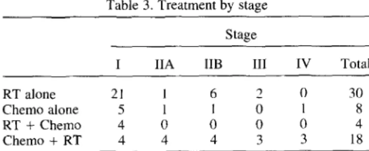
ical radiotherapy with or without consolidation chemo- therapy, whereas in patients with Stage III and IV the treatment choice was chemotherapy with consolidation ra- diotherapy. Fifty-two of 60 patients were treated with ra- diotherapy, and in 30 of them this was the sole treatment after surgery. In 22 patients radiotherapy was combined with chemotherapy. Only eight patients received chemo- therapy alone after the initial diagnosis and/or surgical management (Table 3 ) .
Before 1982, the patients were treated with a cobalt-60 unit, and thereafter, a linear accelerator (8 MV ) was used. The volume was irradiated by parallel opposed anterior- posterior and posterior-anterior fields. The daily dose per fraction varied from 1 to 1.8 Gy. The primary tumor site and areas at risk were approached by either whole abdom-
inal-pelvic irradiation (WAPI) or a hockey-stick tech-
nique encompassing paraaortic, ipsilateral iliac nodes and the primary tumor or tumor bed. In case of WAPI, the whole abdominal-pelvic dose was raised to 15-20 Gy. However, liver and spleen were excluded from the start of WAPI, whereas kidneys received up to 15-20 Gy. At this dose level, a standard hockey stick technique replaced the WAPI. Forty-four of 51 patients with Stages I and II received radiotherapy with or without chemotherapy. Ta- ble 4 summarizes the treatment volumes and specifies the addition or not of chemotherapy to these patients. Of five
Table 3. Treatment by stage Stage
I IIA IIB III IV Total
RT alone 21 I 6 2 0 30
Chemo alone 5 1 I 0 I 8
RT + Chemo 4 0 0 0 0 4
Chemo + RT 4 4 4 3 3 18
Treatment of pelvic cryptorchid seminoma 0 Y. X. LI ef nl. 353
Table 4. Radiation target volume for Stage I and II patients
with or without chemotherapy
Hockey stick WAPI
Local RT
Stage Alone Chemo Alone Chemo plus chemo
I 11 6 10 0 2
IIA I 3 0 0 1
IIB 1 9 5 I I
Total 13 1; 1s 1 4
RT = Radiotherapy; Chemo = chemotherapy; WAPI = whole abdominal-pelvic irradiation.
patients with Stage III, two patients were treated with ra- diotherapy alone and three with combination of chemo- therapy and radiotherapy. Three patients with Stage IV disease received radiotherapy to residual tumor following chemotherapy.
In the early years of this study, prophylactic mediastinal and left supraclavicular irradiation was given to 2 of 34 Stage I and 3 of 17 Stage II patients.
The total dose to the target volume ranged from 20 to 40 Gy (median 30) for Stage I. In Stage II disease, the infradiaphragmatic dose was usually raised to 35 Gy. Af- ter orchiectomy. a dose of 30-40 Gy was applied to the tumor bed. For those patients considered unresectable, the total dose to the primary tumor reached 40-60 Gy.
Chemotherapy. Thirty of 60 patients were treated with chemotherapy. Eight of them received chemotherapy alone and 18 received a combined treatment of chemo- therapy followed by radiotherapy. Only in four patients was chemotherapy given as consolidation treatment after radiotherapy (Table 3 ). During the earlier years of this study, N-formylsarcolycin, a nitrogen mustard analogue, was used alone ( 22 ) or combined with cyclophosphamide or vincristine (5). From 1985 on, more effective chemo- therapy, i.e., platinum-based regimens, were used in three patients.
Follow-up and statistics
Patients were followed up every 3 months for 1 year and thereafter every 6 months or once yearly. At each
follow-up, they underwent a physical examination,
complete blood count, serum biochemistry, and chest X rays. Since 1982, abdominal-pelvic CT scan and/or ul- trasound were added to the routine follow-up proce- dures. Tumor markers. i.e., PHCG and AFP, were not routinely repeated. The median follow-up time was 18 years (range 3 - 36 ) .
The overall survival (OS) was calculated from the date of surgery. Disease-free survival (DFS) was calculated from the date of surgery until evidence of recurrence or progression. Updating of the medical files was done in December 1994, and survival data were calculated ac- cording to these. Survival curves were calculated accord- ing to the Kaplan-Meier method and differences between
survival curves were analyzed with the log rank test. Nu- merical data were compared with Student’s t-test and comparison of table was done by chi-square analysis.
RESULTS
Patient characteristics
The median age of PCS patients was 38 years (range 17 -70 ) , whereas for those with scrotal seminoma (con-
trol group) it was 34 years (range 21-82). The peak
age for PCS was between 30 and 50 years. The most frequently presenting symptoms were low abdominal mass and mass effect. The right to left ratio was 2: 1. Thirteen patients had a contralateral cryptorchidism. One patient had synchronous contralateral scrotal sem- inoma (Table 5 ) .
The size of the primary tumor ranged from 5 to 28 cm, with a median size of 10 cm. There was no correlation between stage and size: The median maximum diameter for Stages I, II, and III was 10 cm, and for Stage IV it was 1 I cm. In contrast, scrotal seminoma had a median di- ameter of 6 cm (range 2- 15), significantly smaller than those in PCS. Eighteen of 60 (30%) patients had invasion of the primary tumor into neighboring structures, espe- cially the bowel ( 11 of 18 ) , the bladder ( 8 of 18 ) , and the iliac vasculature (5 of 18). In 67% ( 12 of 18) of patients who underwent intravenous pyelography, we ob- served an incomplete ( 10) or complete (2) ureteral ob- struction with hydronephrosis. The kidney function re- turned to normal levels after treatment in all patients.
Forty-three percent (26 of 60) of patients with PCS had metastatic disease to the lymph nodes, whereas only 20% (48 of 237) of patients from control group presented with
Table 5. Clinical characteristics of pelvic cryptorchid seminoma Characteristics Location Right Left Stage I IIA IIB III IV Presenting symptom Lower abdominal mass Painful mass Acute pain Other symptom Leg edema Urinary frequency Urodynia Difficult urination Contralateral cryptorchidism Pelvic Inguinal No. of patients 41 19 34 6 11 5 4 49 10 1 5 1 1 1 9 4
354 I. .I. Radiation Oncology 0 Biology l Physics Volume 38, Number 2, 1997
Table 6. Stage distribution in patients with pelvic cryptorchid seminoma compared to that in patients with scrotal seminoma
Stage
I II III IV
n (%I n @) I1 (%) I1 (%I Total
Pelvic 34 (57) 17 08) 5 (8) 4 (7) 60
Scrotal 189 (80) 29 (12) 5 (2) 14 (6) 237
nodal disease (p < 0.0 1) . Table 6 summarizes the differ- ences in stage distribution between PCS and the control group. The anatomical distribution of nodal and/or distant metastases for PCS is shown in Table 7. Pelvic node in- volvement was unilateral except for one patient. Only one patient had pelvic node involvement without paraaortic node involvement. Pelvic nodal invasion in case of nodal metastasis was encountered in 23% (6 of 26) of patients with PCS, whereas only 13% (6/48) of the control group had pelvic nodal involvement. However, this difference was not significant (p = 0.23).
No patient with PCS had inguinal node metastasis, ex- cept for one patient who had pathologically confirmed right-sided inguinal node metastases. The latter patient presented with left-sided PCS and synchronous right- sided scrotal seminoma. He had a bulky primary tumor in the pelvis and had no prior inguinal or scrotal surgery. At staging laparotomy, there were neither paraaortic nor pel- vic nodes metastases.
Survival
The overall and disease-free survival rates at 5 and 10 years were 92% and 87%, and 88% and 84%, respectively. The overall 5-year and IO-year survival rates were 100% and 100% for Stage I, 94% and 87% for Stage II, and 56% and 42% for Stage III/IV, respectively (Fig. 1) . The dif- ferences in survival between Stages I, II, and III/IV were
Table 7. Anatomical distribution of nodal and distant metastases in patients with pelvic cryptorchid seminoma
Sites n Stage IIA Paraaortic only 6 Stage IIB Paraaortic only 8 Pelvic only 1
Paraaortic and pelvic 2
Stage III
Paraaortic and mediastinal 2
Paraaortic, mediastinal, and supraclavicular 2
Paraaortic, pelvic, mediastinal, and supraclavicular 1 Stage IV
Paraaortic, mediastinal, supraclavicular, and liver 1 Paraaortic, mediastinal, liver, kidney, and lung 1
Paraaortic, pelvic, and liver 1
Paraaortic, pelvic, mediastinal, supraclavicular, and lung 1
statistically significant (p < 0.05). For 17 Stage II pa- tients, all 6 Stage IIA patients were alive without evidence of disease, whereas 2 of 11 Stage IIB patients died of disease. The 5year and IO-year survival rates were 100% and 100% for Stage IIA, and 91% and 80% for Stage IIB, respectively. Owing to the small number of patients, there was no significant difference between Stages IIA and IIB.
Eleven patients died of other causes: four of a second malignant tumor, three of hypertension, two of cardiores- piratory disease, one of suicide, and one of unknown causes.
Radiotherapy for Stages I and II
To evaluate the potential impact of treated volume on outcome in Stage I PCS, we compared 17 patients treated prior to 1985 with a hockey-stick technique to 10 patients treated on whole abdominal-pelvic irradiation ( WAPI) . None of these patients with WAPI received chemother- apy, whereas 6 of 17 patients with the hockey-stick tech- nique received chemotherapy (Table 4). The remaining seven Stage I patients were not considered for the com- parison between the hockey-stick technique and WAPI, because five of them received chemotherapy alone and two received local irradiation after chemotherapy. Our intent was basically to compare two modalities of ex- tended field irradiation in Stage I disease. There was no difference between the two groups in locoregional re- currence whether DFS or OS is considered. Only one patient developed distant metastases after hockey-stick radiotherapy.
Fig. 1. Overall survival by stage in PCS: Stage I (n = 34); Stage II (n = 17); and Stage III/IV (n = 9). Log rankp < 0.05.
Treatment of pelvic cryptorchid seminoma 0 Y. X. LI et ul. 355
1n Stage IIA, five of six patients received radiotherapy. Four of them were treated with the standard hockey-stick technique and only one was irradiated locally. One patient received chemotherapy alone. As mentioned in Tables 3 and 4, chemotherapy was given prior to irradiation in all except one patient. All patients were alive and well. Of 11 Stage IIB patients, 6 were treated with WAPI, 3 with the hockey-stick technique, 1 with local irradiation plus chemotherapy, and one with chemotherapy alone. In the
subgroup of patients with Stage IIB who were treated with WAPI, there were no cancer-related events. One patient treated with hockey-stick-field therapy alone died from
abdominal progressive disease. Recurrence
Five of 60 patients (8% ) with PCS developed a relapse. The time to relapse ranged from 2 to 12 years, and in two patients, it occurred beyond 5 years.
Two Stage I patients developed relapse in infradia- phragmatic nodes after N-formylsarcolycin, and one Stage I patient had liver relapse after WAPI. One Stage IIA pa- tient, initially treated with N-formylsarcolycin, experi- enced supradiaphragmatic relapse. One patient with Stage III disease, who received radiotherapy alone, developed relapse in the abdomen. Four patients were salvaged with radiotherapy or chemotherapy, and one died of disease. Complications
One patient treated with orchiectomy, right hemicolec- tomy and radiotherapy reported chronic intermittent di- arrhea, probably due to colectomy-induced malabsorption. Seven patients had bowel obstruction due to abdominal adhesion. Two of them occurred immediately after lapa- rotomy but prior to radiation, and five occurred between 10 months and 21 years after treatment. Surgery was re- quired to correct bowel obstruction in four cases.
Four patients developed a second malignant tumor (SMT) within 4-27 years after treatment of PCS. They died from scrotal seminoma, hepatic carcinoma, squa- mous cell carcinoma of the lung, and rectal adenocarci- noma. Three patients were initially treated with chemo- therapy alone, and only one patient with apical lung cancer was previously treated with infradiaphragmatic radiation.
DISCUSSION
Seminoma arising in cryptorchid testes is rare, account- ing for l-7% of all seminomas (4, 11, 20, 22). PCS is even more exceptional, with only 5% seminomas arising in an abdominally located undescended testis ( 11). How- ever, a higher incidence has been reported in China (29, 30). TO our knowledge, this series represents the largest cohort of PCS ever reported. In our experience, seminoma in cryptorchid testis represents 30% of testicular semi- noma, with 18% PCS and 12% inguinal cryptorchid sem- inoma.
In accordance with Gilliland and Key, there is a trend toward a higher incidence in the third decade ( 13). AS
observed by Willan and McGowan, there is a right pre- dominance which seems also to be the case in scrotal sem- inoma (33). In contrast to scrotal seminoma, PCS tends to present initially with bulky primary disease, often in- vading or compressing surrounding organs. This results in a unique clinical presentation, characterized by leg edema, urinary frequency, and urodynia. Furthermore, PCS, by its location in the pelvis and retroperitoneum and its propen- sity for lymph node metastases, can impair kidney drain- age more frequently than scrotal seminoma.
Pathways of tumor spread
Lymph node metastases are more frequent in PCS than in scrotal seminoma. An incidence of 30-76% has been reported in the literature ( 11, 27, 29, 30). In the present series, the incidence of 43% is significantly higher than the nodal involvement encountered in our control group of scrotal seminoma (20%). This higher incidence of nodal involvement may be explained by a delay in diag- nosis in PCS, compared to scrotal and/or inguinal semi- noma.
The pattern of lymphatic spread is comparable to that of scrotal seminoma, with the major pathway being the paraaortic nodes. Furthermore, our data from PCS reveal that the iliac or paraaortic metastases may occur alone or in combination, regardless of the type of prior surgical intervention. The incidence of pelvic nodes is higher than for scrotal seminoma (23% vs. 13%), and therefore, pa- tients should be routinely investigated at the pelvic level. Inguinal nodal metastases are rare in PCS. Only one pa- tient who presented with synchronous PCS and scrotal seminoma had inguinal nodes. In comparison, 4 of 237 patients with scrotal seminoma had inguinal disease. All five of these patients had bulky abdominal masses without prior inguinal or abdominal surgery. This potentially il- lustrates the disruption of the normal lymphatic pathways resulting in inguinal nodal involvement. Peritoneal metas- tases are rare, and in contrast to Gauwitz and Zagars, who found an 18% (3 of 17) incidence at presentation ( 11)) only one patient in the present series had proven peritoneal implants at autopsy.
Management and prognosis
Surgery alone is not considered to be curative in PCS. The resection rate for primary tumor in the present series was 62% (37 of 60). Frequently resection is made difficult by dense fibrosis, abdominal adhesion, and local inva- siveness of the tumor itself. Nevertheless, the regional control rate was lOO%, owing to the addition of radio- therapy and/or chemotherapy. Primary tumor burden and extent of residual tumors after surgery do not affect out- come; seminoma is highly sensitive to cytoreductive treat- ment. From our data, a complete resection of primary tu- mor is thus not a basic requirement for patients with PCS.
356 I. J. Radiation Oncology 0 Biology 0 Physics
Patients with Stage I PCS have an excellent prognosis after WAPI or hockey-stick radiation. Although nonran- domized, the comparison between patients treated with a hockey-stick technique and those treated with WAPI yields no significant difference in OS and DFS. Therefore, to reduce the potential risk of treatment-related late com- plication ( 16, 32), we recommend a hockey-stick tech- nique encompassing paraaortic, ipsilateral iliac nodes and the primary tumor or tumor bed for Stage I and HA PCS. It must be emphasized that these patients did not in fact receive whole abdominal-pelvic irradiation, since the liver and spleen were shielded from the start of treatment and at 15 -20 Gy, WAPI was replaced by a hockey-stick tech- nique. The differences in treatment volumes merely illus- trate the lack of knowledge in biology and outcome in PCS. Furthermore, in contrast to Gauwitz and Zagars ( 11)) we feel that whole pelvic irradiation is not required unless there is bilateral inguinal, pelvic nodal involve- ment, or obvious peritoneal implants.
For patients with Stage IIB scrotal seminoma, the lit- erature reports excellent survival figures, provided those patients are treated with whole abdominal irradiation (3 1, 33). It is interesting to note that five of the six patients with Stage IIB PCS treated with WAPI alone are alive with no evidence of disease (NED), whereas the single one treated on the paraaortic field only died from disease. However, in bulky seminoma, platinum-based chemo- therapy is currently favored by most authors (6, 25, 26).
Prophylactic mediastinal and/or supraclavicular irra- diation is no longer required in patients with Stage II dis- ease. None of the patients with infradiaphragmatic radio- therapy in this series developed a supradiaphragmatic relapse. This is in accordance with the recent therapeutic trends in bulky Stage II seminoma (7, 21, 25, 28, 33).
Survival for Stage III and IV PCS was poor, merely illustrating the lack of effective chemotherapy in earlier
years. Current standard treatment for advanced stage sem-
inoma includes platinum-based chemotherapy, with response rates reported to be around 85% (9, 10, 17, 22, 26).
The absence of in-field recurrence in Stage I PCS illus- trates the fact that doses in the range of 30-35 Gy are
Volume 38, Number 2. 1997
effective. AS is the case for scrotal seminoma, smaller
doses are probably able to control subclinical disease
( 12), but we do not have data to support this hypothesis. On the other hand, the local control rates obtained with 40-60 Gy on bulky disease or residual primary suggests that 40 Gy is probably enough.
Relapse/complications
Two patients with PCS developed a relapse beyond 5 years, suggesting that late relapse is not a rare event. Therefore, patients with PCS should be followed up on a regular basis even after 5 years (3 ) .
In this series, there were neither abdominal nor pelvic relapses in patients with Stage I and II PCS treated with radiotherapy. However, two of eight patients treated with N-formylsarcolycin alone developed an abdominal or pel- vic recurrence. More effective chemotherapy with plati- num-based regimens might probably have prevented those relapses.
Abdominal complications, especially bowel obstruction are frequently encountered among patients with PCS. This can partly be explained by surgical intervention or loca- tion of the primary, which may result in abdominal ad- hesion.
CONCLUSION
The prognosis in PCS is excellent regardless of the ex- tent of the primary surgery, provided the patients are treated with curative radiotherapy and/or chemotherapy. In case of unresectable primary, a high local control rate can be obtained with radiotherapy alone. For patients with Stage I and IIA disease, the absence of a significant sur- vival benefit for WAPI argues in favor of the limitation of treatment volumes to paraaortic and ipsilateral iliac nodes including the primary tumor or tumor bed. For Stage IIB disease we would favor platinum-based che- motherapy, although in the present retrospective review, WAPI yielded excellent results. As is the case for Stage III and IV scrotal seminoma, platinum-based chemother- apy is the treatment of choice for advanced-stage PCS.
REFERENCES 1. 2. 3. 4. 5.
Abratt, R. P.; Reddi, V. B.; Sarembock, L. A. Testicular
cancer and cryptorchidism. Br. J. Ural. 70:656-659; 1992.
Babaian, R. J.; Zagars, G. K. Testicular seminoma: The MD
Anderson experience. An analysis of pathoiogical and pa- tient characteristics and treatment recommendations. J. Urol.
139:31 l-314: 1988.
Baniel, J.; Foster, R. S.; Gonin, R.; Messemer, J. E.; Dono- hue, J. P.; Einhorn, L. H. Late relapse of testicular cancer. J. Clin. Oncol. 13:1170- 1176; 1995.
Batata, M. A.; Chu, F. C. H.; Hilaris, B. S.; Whitmore, W. F.; Golbey, R. B. Testicular cancer in cryptorchids. Can-
cer 49:1023-1080; 1982.
Bayens, Y. C.: Helle, P. A.; Van Putten, W. L. J.; Mali, S. P. M. Orchidectomy followed by radiotherapy in 176
stage I and II testicular seminoma patients: Benefits of a IO- year follow-up study. Radiother. Oncol. 25:97-102; 1992. Dosmann, M. A.; Zagars, G. K. Postorchiectomy radiother- apy for Stages I and II testicular seminoma. Int. J. Radiat. Oncol. Biol. Phys. 26:381-390; 1993.
Evensen, J. R.; Fossa, S. D.; Kjellevold, K.; Lien, H. H. Testicular seminoma: Analysis of treatment and failure for stage II disease. Radiother. Oncol. 4:55-61; 1985. Farrer, J. H.; Walker, A. H.; Rajfer, J. Management of the postpubertal cryptorchid testis: A statistical review. J. Urol.
134:1071-1076; 1985.
Fossa, S. D.; Droz, J. P.; Stoter, G.; Kaye. S. B.; Ver- meylen, K.; Sylvester, R. Cisplatin, vincristine and ifos- phamide combination chemotherapy of metastatic sem-
Treatment of pelvic cryptorchid seminoma l Y. X. LI et al. 357 10. 11. 12. 13. 14. 15. 16. 17. 18. 19. 20. 21. 22.
inoma: Results of EORTC trial 30874. Br. J. Cancer Southeastern Cancer Study Group experience. J. Clin. Oncol.
7l:619-624; 1995. 5: 1212- 1220; 1987.
Friedman, E. L.; Gamick, M. B.; Stomper, P. C.; Mauch, P. M.; Harington, D. P.; Richie, J. P. Therapeutic guidelines and results in advanced seminoma. J. Clin. Oncol. 3: 1325-
1332; 1985.
Gauwitz, M. D.; Zagars, G. K. Treatment of seminoma aris- ing in cryptorchid testes. Int. J. Radiat. Oncol. Biol. Phys. 24:153-159; 1992.
Giacchetti, S.; Raoul, Y.; Wibault, P.; Droz, J. P.; Court, B.; Eschwege, F. Treatment of Stage I testis seminoma by ra- diotherapy: Long-term results. A 30-year experience. Int. J. Radiat. Oncol. Biol. Phys. 27:3-9; 1993.
Gilliland, F. D.; Key, C. R. Male genital cancers. Cancer 75:295-315; 1995.
Halme, A.; Kellokumpu-Lehtinen, P.; Lehtonen, T.; Teppo, L. Morphology of testicular germ cell tumours in treated and untreated cryptorchidism. Br. J. Urol. 64:78-83; 1989. Hamilton, C.; Horwich, A.; Easton, D.; Peckham, M. J. Ra- diotherapy for stage I seminoma testis: Results of treatment and complications. Radiother. Oncol. 6:115- 120; 1986. Hanks, G. E.; Peters, T.; Owen, J. Seminoma of the testis: Long term beneficial and deleterious results of radiation. Int. J. Radiat. Oncol. Biol. Phys. 24:913-919; 1992.
Horwich, A.; Dearnaley, D. P.; Duchesne, G. M.; Williams, M.; Brada, M.; Peckham, M. J. Simple nontoxic treatment of advanced metastatic seminoma with carboplatin. J. Clin. Oncol. 7:1150-1156; 1989.
Jones, B. J.; Thornhill, J. A.; O’Donnell, B.; Kelly, D. G.; Walsh, A.; Fennelly, J. J.; Fitzpatrick, J. M. Influence of prior orchiopexy on stage and prognosis of testicular cancer. Eur. Ural. 19:201-203; 1991.
Jonsson, K.; Wallace, S.; Jing, B. S.; Boyle, L. E.; Johnson, D. E. Lymphangiography in patients with malignancy in a non-descended testicle. J. Ural. 119:614-617; 1978. Kulkarni, J. N.; Kamat, M. R. Tumors in undescended testis. J. Surg. Oncol. 46:257-260; 1991.
Laukkanen, E. L.; Olivotto, I.; Jackson, S. Management of seminoma with bulky abdominal disease. Int. J. Radiat. On- col. Biol. Phys. 14:227-233; 1988.
Loehrer, P. J.; Birch, R.; Williams, S. D.; Greco, F. A.; Ein- horn, L. H. Chemotherapy of metastatic seminoma: The
23. 24. 25. 26. 27. 28. 29. 30. 31. 32. 33. 34. 35.
Martin, D. C. Malignancy in the cryptorchid testis. Ural. Clin. North Am. 9:371-376; 1982.
Martin, D. C.; Menck, H. R. The undescended testis: Man- agement after puberty. J. Ural. 114:77-79; 1975.
Mason, B. R.; Kearsley, J. H. Radiotherapy for stage 2 tes- ticular seminoma: The prognostic influence of tumor bulk. J. Clin. Oncol. 6: 1856- 1862; 1988.
Mencel, P. J.; Motzer, R. J.; Mazumdar, M.: Vlamis, V.; Bajorin, D. F.; Bosl, G. J. Advanced seminoma: Treatment results, survival, and prognostic factors in 142 patients. J. Clin. Oncol. 12: 120- 126; 1994.
Raina, V.; Shukla, N. K.; Gupta, N. P.; Deo, S.; Rath, G. K. Germ cell tumours in uncorrected cryptorchid testis at Insti- tute Rotary Cancer Hospital, New Delhi. Br. J. Cancer 71:380-382; 1995.
Sager-man, R. H.; Kotlove, D. J.; Regine, W. F.: Chung, C. T.; King, G. A. Stage II seminoma: Results of postor- chiectomy irradiation. Radiology 172:565-568; 1989. Sham, J. S. T.; Choy, D.; Chan, K. W.; Choi. P. H. K. Sem- inoma of normally-descended and cryptorchid testis. Eur. J. Surg. Oncol. 16:33-36; 1990.
Shi, X. H.; Liu, T. F. Radiotherapy for seminoma arising in undescended testis: Analysis of 103 cases. Tumor 7: I 13-
115; 1987 (in Chinese).
Smalley, S. R.; Evans, R. G.; Richardson, R. L.; Farrow, G. M.; Earle, J. M. Radiotherapy as initial treatment for bulky stage II testicular seminomas. J. Clin. Oncol. 3: 1333-
1338; 1985.
Vallis, K. A.; Howard, G. C. W.; Duncan, W.; Combleet, M. A.; Kerr, G. R. Radiotherapy for stages I and II testicular seminoma: Results and morbidity in 238 patients. Br. J. Ra- dial. 68:400-405; 1995.
Willan, B. D.; McGowan, D. G. Seminoma of the testis: A 22-year experience with radiation therapy. Int. J. Radiat. On- col. Biol. Phys. 11: 1769- 1775; 1985.
Wobbes, T.; Schraffordt, K. H.; Oldhoff, J. The relation be- tween testicular tumours, undescended testes, and inguinal hernias. J. Surg. Oncol. 14:45-5 1; 1980.
Zagars, G. K.; Babaian, R. J. Stage I testicular seminoma: Rationale for postorchiectomy radiation therapy. Int. J. Ra- diat. Oncol. Biol. Phys. 13:155-162: 1987.