Lv
I/5’/’/(.
J
Université de Monfréal
Étude de
l’évolution de la structure
des génomes mitochondriawc chez
les
Euglenowa
par
bannie Roy
Département de biochimie
Faculté de médecine
Mémoire présenté à la Faculté des études supérieures
en vue de
l’obtentiondu grade de Maître ès Sciences
en
BiochimieAoût2006 t.
. ri.». •o
(I
‘7-LL)
‘I
Université
de Montréal
Direction des bibliothèques
AVIS
L’auteur a autorisé l’Université de Montréal à reproduire et diffuser, en totalité ou en partie, par quelque moyen que ce soit et sur quelque support que ce soit, et exclusivement à des fins non lucratives d’enseignement et de recherche, des copies de ce mémoire ou de cette thèse.
L’auteur et les coauteurs le cas échéant conservent la propriété du droit d’auteur et des droits moraux qui protègent ce document. Ni la thèse ou le mémoire, ni des extraits substantiels de ce document, ne doivent être imprimés ou autrement reproduits sans l’autorisation de l’auteur.
Afin de se conformer à la Loi canadienne sur la protection des
renseignements personnels, quelques formulaires secondaires, coordonnées ou signatures intégrées au texte ont pu être enlevés de ce document. Bien que cela ait pu affecter la pagination, il n’y a aucun contenu manquant. NOTICE
The author of this thesis or dissertation has granted a nonexclusive license allowing Université de Montréal to reproduce and publish the document, in part or in whole, and in any format, solely for noncommercial educational and research purposes.
The author and co-authors if applicable retain copyright ownership and moral rights in this document. Neither the whole thesis or dissertation, nor substantial extracts from it, may be printed or otherwise reproduced without the author’s permission.
In compliance with the Canadian Privacy Act some supporting forms, contact information or signatures may have been removed from the document. While this may affect the document page count, t does flot represent any loss of content from the document.
Université de Montréal Faculté des études supérieures
Ce mémoire intitulé
Étude de l’évolution de la structure des génomes mitochondriaux chez les Euglenozoa
présenté par: Joannie Roy
a été évalué par un jury composé des personnes suivantes
Normand Brisson président-rapporteur Gertraud Burger directeur de recherche David Morse membre du jury 2
RÉSUMÉ
Un des génomes mitochondriaux les plus atypiques connus est celui des Kinetoplastea (Euglenozoa). Leur génome mitochondrial est composé de deux types de molécules les maxicercles et les minicercles. Chez les trypanosomes (Kinetoplastea), ces molécules sont entrelacées et forment un corps compact, nommée le kinétoplaste. La prévalence de ces caractéristiques chez les autres groupes des Euglenozoa, soit les Diplonemea et les Euglenida, est incoirnue. La première partie du travail, établissant les bases préalables à la suivante, porte sur la description de Rhynchopus euÏeeides n. sp. (Diplonernea). La seconde partie concerne la distribution et la structure de l’ADN mitochondrial chez cette espèce ainsi que chez plusieurs autres membres des Euglenida. Les résultats montrent que la présence de chromosomes multiples est une propriété commune aux génomes mitochondriaux des différents membres des Euglenozoa alors que l’organisation spatiale compacte de l’ADN mitochondrial est restreinte au sous-groupe des Kinetoplastea.
Mots clés
Diplonemea, Euglenida, Rhynchopus, PetaÏomonas, Peranema, Entosiphon, protiste, morphologie, ultrastructure. taxonomie
ABSTRACT
One of the rnost unusual mitochondrial genomes is that of Kinetoplastea (Euglenozoa), which is cornposed of two types of DNA molecules: maxicircles and minicircles. In trypanosomes (Kinetoplastea), mitochondrial DNA circles are interlocked into a single compact network. the kinetoplast. Whether these peculiar features prevail throughout Euglenozoa (Kinetoplastea and its neighbour clade: Euglenida and Diplonernea) is unknown. The work first includes a description of Rhynchopus euleeides n. sp. (Diplonemea). This provided the basis for an investigation on mitochondrial DNA packaging and structure of this species and several members of Euglenida. Our findings indicate that multiple mitochondrial chromosomes are shared by ail Euglenozoa studied, whule a compacted spatial organization of mitochondrial DNA is restricted to Kineropiastea.
Key words:
Diplonernea, Euglenida, Rhynchopus, Fetalomonas, Peranema, Entosiphon, protist, rnorphology, ultrastructure, taxonorny
TABLE DES MATIÈRES
RÉSUMÉ (p. 3) ABSTRACT (p. 4)
TABLE DES MATIÈRES (p. 5) LISTE DES FIGURES (p. 6) LISTE DES TABLEAUX (p. 7) LISTE DES ABRÉVIATIONS p. 2) REMERCIEMENTS (p. 10)
CORPS DE L’OUVRAGE (p. 11) 1. Introduction (p. 11)
1.1. La mitochondrie et ses différentes fonctions (p. 11) 1.2. L’origine de la mitochondrie (p. 12)
1.3. Les différentes lignées eucaryotes (p. 13) 1.4. Le génome mitochondrial (p. 15)
1.4.1. La structure des génomes mitochondriaux (p. 15) 1.4.1.1 .Les animaux, les plantes et les champignons (p. 16) 1.4.1.2.Les protistes (p. 16) 1.4.1.2.1. Les Euglenozoa (p. 16) 1.4.1.2.1.1.Les Kinetoplastea (p. 17) 1.4.1.2.1.2.Les Diplonemea (p. 12) 1.4.1.2.1.3.Les Euglenida (p. 19) 1.5. Hypothèse de travail (p. 19) 2. Résultats (p. 21)
2.1. Article 1 Description of Rhynchopus euleeides n. sp. (Diplonemea), a Free-living Marine Euglenozoan (p. 22)
2.2. Article 2 : Unusual Mitochondrial Genome Structure throughout Euglenozoa (p. 46)
LISTE DES FIGURES
Introduction
Figure 1. L’endosyrnbiose de la mitochondrie. (p. 13)
f igure 2*. Arbre schématique des grandes lignées eucaryotes. (p. 14) Figure 3* Arbre schématique des Euglenozoa. (p. 17)
*Les figures 2 & 3 proviennent d’articles dont Gertraud Burger est co-auteur.
Résultats
Article 1:
Fig. 1--10. Light and scanning electron rnicroscopy of Rhynchopus euleeides, n. sp. Celi morphology and DNA distribution. (p. 42)
Fig. 11--18. Transmission electron microscopy of Rhynchopus euleeides, n. sp. Architecture ofthe flagellar and feeding apparatuses. (p. 43) Fig. 19--29. Transmission electron microscopy of Rhynchopus euleeides. n.
sp. Architecture of feeding apparatus, organelles. and cytoskeleton. (p. 44)
f ig. 30--33. Schematic three-dimensional representation of Rhynchopus euleeides n. sp. 30. Outer view ofa cell in the trophic stage. (p.45)
Article 2
Figure 1. DNA distribution within cells. (p. 6$)
f igure 2. Transmission electron rnicroscopy ofmitochondria and other vesicles. (p. 69)
Figure 3. DNA separation and identification. (p. 70)
Figure 4. DNA spreading observed by election microscopy. (p. 71)
LISTE DES TABLEAUX
Article 1
Table 1. Comparative features ofthe described Diplonernea species. (p. 41)
Article 2
LISTE DES SIGLES ET DES ABRÉVIATIONS
environ Degré
micro A : Adénine
ADN t acide désoxyribonucléique ADNc : ADN complémentaire ADNk : kinétoplaste
ADNmt : ADN mitochondrial ARN : acide ribonucléique
ASCT: acetate :succinate CoA transférase
ASWP : eau de mer artificielle pour protistes (en anglais) ATCC : American Type Culture Collection (en anglais) ATP t adénosine triphosphaté
AUF t Agence universitaire de la francophonie C t celsius
C t cytosine
CCAP t Collection Culture ofAlgae and Protozoa (en anglais) cDNA : ADN complémentaire (en anglais)
CIAR: Institut canadien de recherches avancées (en anglais) CIHR: Instituts de recherche en santé du Canada (en anglais) DAPI t 4’,6’-diamidino-2-phenylindole
EM : microscopie électronique (en anglais)
et al. t locution latine et ahi. En français : et autres. FBS t sérum de foetus bovin (en anglais)
Fig. t figure g: gramme G : guanine
GA t glutaraldéhyde
gRNA: ARN guide (en anglais)
h heure
HS : Sérum de cheval (en anglais) k kilo
kbp kilobase (en anglais) kDNA kinétoplaste (en anglais) kpb kilobase
litre
in milli M molaire
min minute
mtDNA : ADN mitochondrial (en anglais) n : nano
n. sp. s nouvelle espèce
P3 s tampon phosphate (en anglais) PBS : tampon salin phosphate (en anglais) PDH s pyruvate deshydrogénase
PFGE s gel d’électrophorèse en champs pulsées (en anglais) PFO : pyruvate :ferrodoxine oxidoréductase
pH s potentiel hydrogène
PNO s pyruvate :NADP+ oxydoréductase RNA s acide ribonucléique (en anglais) rpm s rotation par minute
T : thymine U s uridine
UGA s Université de l’état de Georgie (en anglais) V s volt
x g s force gravitatioimel
* le genre des noms d’organismes est abrégé à leur première lettre à partir de leur
deuxième apparition dans le texte. Parexemple, Trjpanosoma brucel devient T brucef.
REMERCIEMENTS
Je désire remercier chaleureusement ma directrice de recherche, Gertraud Burger, pour son soutient constant durant ces deux dernières années. Je tiens à la remercier en particulier pour sa rigueur, sa disponibilité, ses encouragements et son encadrement. Elle a grandement participé à la réussite de ce projet.
Ma reconnaissance va également à Franz Lang ainsi quatlx membres des laboratoires de Gertraud et de f ranz pour leur constante disponibilité et pour avoir partagé ces belles aimées. Plus particulièrement, merci à Shona Teijeiro, William Marande et Lise Forget pour leur participation active dans mes projets.
J’exprime ma gratitude à Julius Luke et Drahomira Faktorovâ, ainsi qu’aux autres membres de Féquipe de Julius pour leur collaboration étroite et pour rnavoir accueillie en République Tchèque durant Fété 2005. Mon passage là-bas fut très enrichissant tant au niveau professionnel que personnel.
Je remercie spécialement David Morse pour m’avoir parrainée et pour ses encouragements. Merci également au personnel du département de biochimie, du centre Robert-Cedergren et de la maison internationale.
finalement, la réalisation de ce projet n’aurait pas été possible sans le soutien financier du Ministère de l’éducation pour sa bourse de mobilité, de l’Agence universitaire de la francophonie et des Instituts de recherche en santé du Canada.
1. INTRODUCTION
1.1. La mitochondrie et ses différentes fonctions
La mitochondrie est un organite ubiquitaire chez les cellules eucaryotes aérobies. Engagée dans différentes réactions biochimiques, la mitochondrie est délimitée par une double membrane et possède un bagage génétique indépendant du noyau. Son rôle principal est de supporter le métabolisme énergétique. En effet. le pyruvate. préalablement produit par la glycolyse. y est oxydé grâce à la phosphorylation oxydative et entre ensuite dans le cycle de Krebs. Finalement, les produits de ce cycle permettent la respiration cellulaire, générant en tout jusqu’à 3$ moles d’ATP par glucose métabolisé (Saraste 1999).
Chez les eucaryotes anaérobies et aérobies facultatifs, il existe des variantes métaboliques de la mitochondrie classique ainsi que des organites dérivés nommés hydrogénosomes et mitosomes (Andersson et Kurland 1999; Tovar et al. 1999). Chez ces espèces, le métabolisme du pyruvate, réalisé soit dans
ces organites soit dans le cytoplasme. s’effectue à l’aide de complexes enzymatiques différents dont les produits sont subséquemment convertis grâce â un processus de fermentation (Martin et Millier 199$; Andersson et Kurland 1999; Homeret al. 1999; Martin 2005).
Mis à part ce rôle central dans le métabolisme énergétique, la mitochondrie est aussi le lieu de biogenèse des co-facteurs de fer/soufre. importants dans plusieurs réactions enzymatiques majeures et dans le transfert d’électrons (Liii et Kispal 2000). Elle participe aussi à d’autres fonctions cellulaires comme l’homéostasie des ions et l’apoptose (Logan 2006).
1.2. L’origine de la mitochondrie
Malgré cette apparente diversité de fonctions métaboliques, les mitochondries sont issues d’un évènement ancestral commun. Outre la similarité des réactions biochimiques y siégeant et l’histoire évolutive des enzymes impliquées dans le métabolisme énergétique. l’origine ancestrale commune de ces organites est appuyée par certains indices comme la présence d’une double membrane. Cette caractéristique suggère que la mitochondrie provient de l’endocytose d’une bactérie par l’ancêtre de la cellule eucaryote (f igure 1). En fait, c’est en 1890 (Altmann 1890; Williamson 2002) que l’on a pour la première fois émis cette idée. Ce n’est toutefois qu’à partir des années 1960, avec la découverte d’ADN dans les mitochondries (ADNrnt) de cellules de mammifères (Nass et Nass 1963; Williamson 2002) que Fhypothêse de Fendosymbiose a été confirmée. Depuis ce temps. l’étude des ADNmt dans les différents règnes du vivant ainsi que de leur contenu en gènes ont permis de reconstruire partiellement l’histoire évolutive de cet organite. Ainsi, la nature des partenaires impliqués et les modalités de cette interaction hôte/symbionte sont de mieux en mieux comprises.
Selon les hypothèses les plus couramment acceptées, c’est la modification de l’environnement ambiant qui aurait poussé les deux partenaires à interagir symbiotiquement. Une de ces hypothèses stipule que l’hôte serait devenu strictement dépendant des déchets métaboliques dune bactérie productrice d’hydrogène suite à la diminution de ce gaz provenant des sources géologiques (Martin et Muller 1998). Ceci suppose un évènement unique qui aurait eu lieu dans une population il y a environ un milliard d’années. C’est par la suite que les différentes lignées eucaryotes contemporaines auraient divergé pour posséder maintenant soit une mitochondrie, soit des reliques de celle-ci, comme l’hydrogénosome et le mitosome. C’est aussi pendant cette évolution qu’une partie des gènes de l’ancêtre mitochondrial original a migré vers le noyau. d’une manière plus ou moins importante selon l’espèce.
I
24Ç
3
Ç)
figure 1. L’endosyrnbiose de la mitochondrie.
1.3. Les différentes lignées eucaryotes
La classification des eucaryotes et la terminologie employée pour celle-ci ayant changé à plusieurs reprises au cours des années, les taxonomistes ont récemment recommandé et redéfini certains termes, en tenant compte des connaissances actuelles de l’histoire évolutive du vivant (Adl et al. 2005; Keeling et al. 2005). Les nouveaux schémas reconnaissent cinq à six grands groupes chez les eucaryotes. Chacun de ces groupes est en majorité formé de protistes, desquels ont divergé les lignées d’organismes multicellulaires les plus connues, comme les plantes, les animaux et les champignons. Les protistes, historiquement désignés par les termes protozoaires, protophyta, infusoires, animalcules, protoctista, etc., se définissent communément par exclusion. Selon cette définition, les protistes comprennent les eucaryotes qui ne sont ni des plantes, ni des animaux, ni des champignons.
Selon le schéma (figure 2) de Keeling et al (2005), qui est communément utilisé, les cinq grands groupes eucaryotes qui prévalent aujourd’hui sont les Plantae, les Chromalveolées, les Unikontes, les Rhizaria et les Excavata. Panrii ceux-ci, certains sous-groupes sont mieux connus. Par exemple, les animaux et les champignons font partie du groupe des Unikontes. Les plantes terrestres et les algues vertes et rouges, quant à elles, appartiennent aux Plantae. On retrouve ensuite les Chromalveolées, dont les espèces les plus connues sont sans doute le
parasite causant la malaria, PÏasmodium ainsi que les ciliés. Les Rhizaria, pour leur part, comprennent les radiolaires, protistes bien connus des micropaléontologues à cause de leur capacité à se fossiliser. Finalement, il y a les Excavata, dont font partie le parasite causant la maladie du sommeil, Tiypanosoma brucel, et l’organisme modèle EugÏena gracilis. C’est aussi dans ce groupe qu’on retrouve les eucaryotes ayant les branches phylogénétiques les plus courtes et les plus basales, tels que les jakobides et les retortamonades.
TENDSo
Figure 2. Arbre schématique des grandes lignées eucaryotes (Keeling et al. 2005).
1.4. Le génome mitochondrial
Jusqu’à maintenant, c’est chez un protiste membre des Excavata, le jakobide Reclinomonas americana, que l’on a retrouvé le génome mitochondrial le pius ressemblant à celui d’une bactérie. Cette découverte a permis de mieux caractériser l’ancêtre commun des mitochondries. puisque le génome de R. americana ressemble à celui d’une eubactérie du groupe des alpha protéobactéries, plus précisément. aux Rickettsiae. Le génome mitochondrial de ce protiste est une molécule circulaire de 69 kpb contenant 97 gènes codant pour des ARN fonctionnels et des protéines (Lang et al. 1997).
1.4.1. La structure des génomes mitochondriaux
La similarité du génome mitochondrial à celui d’une eubactérie ne prévaut pas chez tous les organismes. En effet, il existe toute une variété de génomes mitochondriaux. Ceux-ci varient d’une espèce à l’autre selon la taille du génome. la forme et la conformation, le biais de composition en nucléotides. le nombre de chromosomes. le contenu génique ainsi que le code génétique utilisé dans la traduction.
1.4.1 .1 .Les animaux, les plantes et les champignons
Suite à la première découverte, chez les mammifères. d’un génome mitochondrial, les chercheurs ont découvert une grande diversité de génomes chez d’autres organismes pluricellulaires, tant chez les animaux que chez les plantes et les champignons.
À
plusieurs égards, les génomes mitochondriaux des animaux et ceux des plantes terrestres ont évolué dans des directions opposées. D’un côté,monornère circulaire de 14 à 20 kpb qui est principalement composé d’ADN codant.
À
Vautre extrême, les génomes des plantes. quoique énormes. de 180 à 600 kpb. n’ont qu’une capacité codante de 10-20%. Us ont une structure complexe. variable selon l’espèce. où les molécules passent parfois d’un état circulaire à linéaire. (Sederoff 1984; Backert et aï. 1997; Nosek et Tomaska 2003; Lynch et al. 2006). Le génome des champignons est quant à lui qualifié de polydispersé. c’est-à-dire composé principalement de molécules linéaires, qui peuvent être arrangées en tandem, de contenu et de complexité variables (Nosek et aÏ. 1995; Tomaska et aÏ. 2001; Nosek et Tornaska 2003).1 .4.1 .2.Les protistes
C’est chez les génomes mitochondriaux des protistes que les plus grandes variations de structure ont eu lieu. à l’image de la diversité biologique qu’ils représentent eux-mêmes aujourd’hui. Par exemple. le génome de l’apicomplexe P. faïciparum. l’agent causant le paludisme. est le plus petit génome connu présentement. composé d’une molécule linéaire de 6 kpb (Feagin et aï. 1991; f eagin 1992; 2000) et codant pour trois protéines et deux ARN ribosomaux.
À
l’opposé, le génome de Amoebidium parasiticum est d’environ 200 kpb et est composé de quelques centaines de chromosomes linéaires (Lang et aÏ. 2002; Burger et ai. 2003).1.4.1.2.1. Les Euglenozoa
Parmi toute cette variété observée chez les protistes. c’est celle des trypanosomes qui a intrigué le plus profondément les scientifiques. Les trypanosomes font partie des Kinetoplastea. eux-mêmes sous-ensemble des Euglenozoa qui eux, appartiennent aux Excavata. On retrouve trois sous-groupes chez les Euglenozoa: les Kinetoplastea. les Diplonema et les Euglenida (figure 3). Les espèces les plus
étudiées sont, comme ci-haut mentionné, le parasite causant la maladie du sommeil en Afrique, T brucei. celui causant la maladie de Chagas en Arnérique du Sud, T cruzi, et ceux responsables de la leishrnaniose, Leishrnania sp. De plus, parmi les quelques autres membres des Euglenozoa connus, notons le flagellé photosynthétique E. graciÏis,
membre
du sous-groupe des Euglenida, qui sert communément de modèle en biologie.1.4.1.2.1.1 .Les Kinetoplastea
Chez les trypanosomes, le génome mitochondrial est composé d’un grand chromosome circulaire (appelé maxicercle) et de milliers de différents petits chromosomes circulaires (appelés minicercles). Le maxicercle est d’une taille de 20 à 40 kpb alors que la multitude de minicercles mesurent de 0,5 à 10 kpb. Ces
j
0/ Lu —J 0 D uJ frdIbJ __.:::..._.,\‘
\.
tDlu)
çr. f)iptnrma pupithJhrns figure 3. 2005)le kinétoplaste (ADNk). Par ailleurs, les gènes sont encodés sur les maxicercles et leurs ARN messagers respectifs sont massivement édités (RNA editing) afin de rendre la traduction possible. Cette édition, effectuée par l’éditosome, consiste en une insertion et/ou une délétion d’uracile. La matrice d’ARN du complexe de l’éditosome est nommée l’ARN guide et est encodée sur les minicercles (Lukes ct cii. 2002: Sirnpson et aÏ. 2002).
La composition et la structure de l’ADNk varient peu au sein des différentes espèces de trypanosomes. Toutefois, des variantes sont retrouvées chez les membres du taxon voisin, les bodonides, qui forment avec les trypanosomes le groupe des Kinetoplastea. Malgré ces quelques exceptions chez les Kinetoplastea, peu d’autres membres des Euglenozoa sont connus du point de vue de leur génome mitochondrial.
1.4.1.2.1 .2.Les Diplonemea
Récemment, un génome mitochondrial unique a été mis en évidence chez Diionema papiiiaturn, du groupe des Diplonemea (Euglenozoa), des flagellés phagotrophes et osmotrophes. En effet, chez D. papillalum, le génome est composé de plusieurs molécules circulaires de 6 et 7 kpb (Marande et aÏ. 2005). De plus. les gènes encodés sur ces chromosomes sont fragmentés (un fragment par gène) et un mécanisme d’épissage en trans des ARN messagers est nécessaire à leur maturation complète. Toutefois. contrairement au kinétoplaste. ces molécules ne forment pas de corps dense visible au microscope. L’autre genre appartenant aux Diplonemea est Rhynchopus. Il s’agit d’un genre peu étudié dont quelques espèces seulement ont été décrites ultrastructuralernent.
1.4.1.2.1.3.Les Euglenida
Chez les Euglenida, seulement le génome mitochondrial de E. gracilis a été étudié. Malgré de nombreuses études (Manning et aÏ. 1971; Nass et aÏ. 1974; Talen et aï. 1974; Buetow 1989; Hayashi et Ueda 1989; Hayashi-Isimaru et al. 1993; Tessier et aï. 1997; Yasuhira et Simpson 1997: Gray et aÏ. 2004), la structure de ce génome est peu comprise. En effet, selon le groupe de recherche, la taille des molécules observées varie de 1 à 70 kpb. Par contre, toutes les études décrivent les molécules comme étant majoritairement linéaires et complexes. Mis à part cette description partielle du génome de E. gracilis, la seule autre information disponible dans la littérature à propos du génome mitochondrial des Euglenida est l’observation par microscopie électronique, chez deux espèces de Petalomonas, d’une structure dense semblable à celle des kinétoplastes (Leander et aï. 2001).
1.5. Hypothèse de travail
La quantité d’information disponible relative à la mitochondrie et à la diversité de son génome est biaisée d’un groupe phylogénique à l’autre. Ce problème s’observe tant, à grande échelle, entre des grands groupes taxonomiques, qu’à plus petite échelle, à travers certains sous-groupes. Par exemple. les plantes, les animaux et les champignons sont beaucoup plus étudiés que les protistes. Le même problème s’applique également aux trois sous-groupes des Euglenozoa, où les génomes mitochondriaux des Kinetoplastea sont beaucoup mieux caractérisés que ceux des Diplonemea et des Euglenida. L’objectif de ce mémoire est donc d’établir l’étendue de la diversité des génomes mitochondriaux retrouvées à travers les Euglenozoa. Plus précisément, les questions abordées dans ce travail sont les suivantes i- Le type de chromosomes circulaires multiples retrouvés chez D. papillatum est-il unique à cette espèce ou est-il répandu à travers les genres
composant les Diplonemea? ii- L’existence d’un génome mitochondrial sous forme d’un kinétoplaste est-elle restreinte qu’aux Kinetoplastea?
La systématique des Diplonernea est controversée et se fonde principalement sur quelques membres du genre DipÏonema. Ainsi, avant de qualifier la structure du génome mitochondrial retrouvé chez les Diplonernea, il est nécessaire de clarifier les relations regroupant les deux genres qui le composent. soit D1p/ol?emcl et Rhynchopus.
À
ce sujet. certains auteurs ont déjà soulevé la nécessité de recueillir plus «informations morphologiques et moléculaires sur le genre Rhynchopus. Simpson (1997) et Von der Heyden (2004) s’entendent sur la difficulté actuelle d’établir la distinction entre les deux genres. Afin d’éclaircir cette problématique, le premier article présenté dans la section suivante met à jour les connaissances disponibles sur les Diplonemea. plus particulièrement sur la distinction entre les deux genres. en décrivant la nouvelle espèce Rhynchopits euÏeeides n. sp. Cet article permet ainsi d’établir les bases sur lesquels s’appuie l’analyse comparative subséquente des génomes mitochondriaux.Le deuxième article présente et compare des données sur la structure
physique du génome mitochondrial. tant chez R. euÏeeides que chez certaines espèces du groupe des Euglenida (P. cantuscygni, F. trichophorum et E. suÏcatum). Les génomes mitochondriaux de R. euÏeeides et de D. papiÏÏatiirn sont premièrement comparés. Ensuite, l’étude des génomes des Euglenida permet de mettre en perspective ce groupe par rapport aux deux autres taxons voisins, les Diplonemea et les Kinetoplastea.
ACCORD DES COAUTEURS
1. Identification de l’étudiante et du programme bannie Roy, M. SC. Biochimie
2. Description de l’Article
Joannie Roy, Drahomira Faktorovi, Oldfich Benada, Julius Luke et Gertraud Burger. ‘Rhynchopus euleeides n. sp. (Diplonernea), a Free-living, Marine Euglenozoan’, soumis à la revue ‘Journal of Eukaryotic Microbiology’ le 7 avril 2006.
3. Déclaration de tous les coauteurs autres que l’étudiant
À
titre de coauteur de l’article identifié ci-dessus, je suis d’accord pour que Joannie Roy inclue cet article dans son mémoire de maîtrise qui a pour titre Étude de l’évolution de la structure des génomes mitochondriaux chez les Euglenozoa.j / 3 / J I
i ‘t c L tvt
Coauteur Sigfiature Date
6ïkcci urJt
1
f’w
Ô
Coauteur Sigi tuf Date
Jt/t
rL
Coauteur gnatu
(J
DateI Î
ROY ET AL.---RHYNCHOPUS EULEEIDE$ N. S?.
Description of
Rhynchopus eutecides
n. sp. (Diplonemea), a Free living Marine EuglenozoanJOANNIE ROY, DRAHOMiRA FAKTOROV 2,OLD1{ICH BENADAC, JULIUS LUKE and GERTRAUD BURGER
aCentre Robert Cedergren, Bioinforrnatics & Genom les, Département de biochimie, Université de Montréal, Montréal, QC, H3T 1J4, Canada
bBio/o Centre, Institute ofParasitology, C’zech A cademy ofScience, and Faculty of BioÏogy, University of South Bohemia, 37005 eské Budéjovice (Budweis), Czech RepubÏic
clnstilule ofMicrobiology, C’zech Academy of Sciences, 142 20 Frague 4, Czech
Repïtblic
dCanadian Institute Jàr A dvanced Research, Program in EvoÏutionaiy BioÏogy
Corresponding author: G. Burger, Université de Montréal, 2900 Boul. Édouard Montpetit, Montréal QC, H3T 1J4 Canada - Telephone: (514) 343-7936; fAX:
(514) 343-2210; e-mail:
These authors contributed equally to this work. 2
ABSTRACT. We describe Rhynchopus euÏeeides n. sp., using light and electron rnicroscopy. Tue free-living flagellate, which has been isolated earlier from a marine habitat, can be grown axenically in a rich medium. In the trophic stage, ceils display radical changes in form (metaboly); at rest they are lemon-shaped. Gliding is the predominant mairner of locomotion. The two flagella are short stubs of unequal length. and typically concealed in the pocket. Swanrier cells, which form only occasionally. are smaller. disk-shaped. with two flagella of more than two-times the body length. flagella have conventional axonemes. but appear to lack a paraxonemal iod. Cells have a single sub-apical opening that is decorated with a prominent apical papillum. Both the flagellar pocket and the adjacent feeding apparatus merge together into this opening. The most likely single peripherally Iocated mitochondrion is reticulated and contains only a few larnellar cristae. Mitochondrial DNA is abundant and evenly distributed throughout the organelle. Morphological synapornorphies confirrn the affiliation of the species with the genus RÏiynchopus (Diplonernea. Euglenozoa). We discuss the characters which distinguish Rhynchopus from Diplonema. and the validity of the two genera.
Key Words: Diplonemea, Euglenozoa, flagellates, morphology, ultrastructure, taxonorny.
The genus Rhynchopus was first described in 1948, based on the observation ofa single species. Rhynchopus amitus (Skuja 194$; see also A1-Qassab et al. 2002). This free-living flagellate was isolated from pelagic water inthe Erken Lake ofthe east coast of Sweden. Up to now, the only member of this genus that has been characterised ultrastructurally is the parasitic Rhynchopus coscinodiscivorus, which feeds on the cyloplasrn of planktonic diatoms (Schnepf 1994). Otherwise. only sketchy descriptions are available for a few additional isolates. some reported as parasites of clams and lobster larvae. and others free-living (Bodammer and Sawyer 1981; Kent et al. 19$?; Sirnpson 1997; Vickerman 2000). In fact, given the scarce details of these descriptions, it is difficuit to verify whether these specimens are Rhynchopus spp. or rather Dilonema spp.
Initially, Rhvnchopus was considered an unusual euglenid and placed in the Euglenida (Skuja 1948). Since then, classification of this group has undergone numerous modifications (in the following. we wiII use the taxonomical scheme of Adi et al. (2005)). Most importantly, the genus Rhynchopus has been rernoved from the order Euglenida and placed. together with the genus Diplonema. - which
now also comprises species previously named Isonema (Schuster et al. 196$; Patterson and Brugerolle 198$; Triemer and Ott 1990), into the Diplonemea sensu Cavalier-Smith, 1993 (Simpson 1997). Now, Diplonemea and Euglenida, together with Kinetoplastea (commonly named diplonernids, euglenids and kinetoplastids), constitute the phylum Euglenozoa. A higher-level assemblage unites Euglenozoa and Heterolobosea into Discicristata (Patterson 1994; Cavalier-Smith 199$). Furthermore. Euglenozoa, Heterolobosea and jakobids have been merged into Excavata (Simpson and Patterson 2001; Adl et al. 2005). However, solid phylogenetic evidence for the rnonophyly of these higher order groupings rernains to be demonstrated.
Here, we describe in detail the morphology of Rhynchopus euÏeeides n. sp., deterrnined by light and electron microscopy. Based on several features, which set this organism apart from the other members of the genus, we propose the creation ofa new species. With this report. we aim to fil in a critical lack ofmorphological
underlined by others a decade ago (Schnepf 1994; Sirnpson 1997). A detailed morphological description of this group is rnost timely. Due to the recent discovery of a unique mitochondrial gene and genorne structure in Diplonema papilÏatum (Marande et al. 2005), diplonemids are now attracting attention beyond
the protist community.
MATERIALS AND METHODS
Isolation and cultivation. Rhynchopus etiÏeeides n. sp. (previously named ‘Rhynchopus sp.1’ - ATCC 50226) was obtained from the American Type Culture
Collection, where it was deposited by T. Nerad in 1986. Ceils were cultivated axenically in modified artificial sea water consisting of 3.3% sea saits (Instant Ocean). vitamins (0.5 jig/mI biotin. 0.5 j.tg/ml 312. 100 ig/liter thiamine-HCI). and trace metal elements (4.36 jig/liter Na2EDTA. 3.15 jig/liter FeC136 H20. 9.2 tg/1iter CuSO45H2O. 22 jig/liter ZnS047H2O, 10 jig/liter CoC126H2O. 1 8 jig/liter MnC124H2O. 6.3 tg/liter NaMoO42H2O), supplemented with 10% horse (HS) or foetal bovine (FBS) serum. Cultures were grown without shaking at room temperature in adhesion-treated flasks (Corning). A cell scraper (Sarstedt) was used for harvesting the ceils from the flask bottom. A mix of antibiotics (160 jig/ml streptomycin, 160 ig/m1 kanamycin, 280 ig/ml penicillin-G (Wisent)) was added for long-terni maintenance of the strain. but not for cuÏtuies used in experiments. Stocks were conserved in liquid nitrogen in rnodified artificial sea water containing 5% dimethyl sulfoxide. For the test of phagotrophy, the serum was replaced by either 0.1-1% ofcrystallized egg yolk or 10/nil bacteria (a mix
of Enterobacier aerogenes, Silicibacter sp., BacilÏus sp. and Pseudomonas sp.).
The titre was deterrnined by counting the cells in a hernacytorner.
Light and fluorescent microscopy. Live celis were observed with an inverted Nikon Eclipse TE2000-U microscope and pictures were taken with MetaMorph 6.3r3 (Molecular Devices). for DAPI (4’6’-diamidino-2-phenylindole)-staining, cells were flxed for 10 min at room temperature in 4% parafornialdehyde diluted
in 3.3 % artificial sea water. Fixation was stopped by spinning the celis down and
resuspending them in 3.3% artificial sea water. Afier the celis were allowed to adhere onto poly-L-lysine-coated siides for 2 h in a hurnidity chamber. the siides were stained with 1 jig/rnl DAPI in phosphate-buffered saline (PBS) pH 7.2 for 5
min. The stained celis were washed, mounted with the antifade reagent (0.233 g
I ,4-diazabicyclo-(2.2.2)octane; I ml 0.2 M Tris-HC1. pH 8.0; 9 ml glycerol) and examined with a Zeiss Axioplan 100 microscope.
Electron microscopy. for transmission electron rnicroscopy, cells were washed twice in artificial sea water, centrifuged at low-speed (-1200 x g). and fixed following two different protocols: (i) 2% glutaraldehyde (GA) in 0.25 M phosphate buffer (PB) pH 7.2 ovemight at 4 °C: (ii) sarne as (i) but the concentration of PB was 0.1 M. Afier fixation, celis were washed in the
respective buffer supplernented with 4% glucose, pelleted. and embedded in 2% agarose. Post-flxation was done with 2% 0s04 in PB for 2 h at room temperature, followed by a washing step with PB. Afler dehydration in graded series of ethanol, the ceils were embedded in Epon-Araldite. Ultrathin sections were stained with lead citrate and uranyl acetate and examined under a JEOL JEM 1010 microscope. For scaiming electron microscopy observations. cells were grown on coverslips for a few hours. fixed in 5% glutaraldehyde in 0.25M PB pH 7.2 for 5 min at 37
°C in a PELCO 3440 Max laboratory Microwave Oven (Ted Pella; Reddin, CA),
washed in the same buffer, followed by a post-fixation step with 1% osmium tetroxide in 0.IM PBS, pH 7.2 for 5 min and dehydrated in graded series of ethanol. Coverslips were air-dried, rnounted on aluminum stubs with conductive carbon paint (SPI supplies) and observed without any ftirther treatrnent with a JEOL JSM-7400F high resolution Field Emission Scaiming Electron Microscope
RESULTS
Light microscopical observations. The feeding behaviour of Rhynchopus euÏeeides n. sp. depends on available nutrients. In a serum-based medium, ceils feed osmotrophically; at high concentrations of serum (‘-10%), the culture tums blackish due to an unidentified compound secreted by the celis once they reach the stationary phase
(—
i0 ceils/rnl). Altematively. in a medium containing crystallized egg yolk or bacteria. the flagellates will feed by phagotrophy. Generation time of axenic cultures in rnodified artificial sea water supplernented with 10% serum is approxirnately $ hours. Division occurs iongitudinally, from the anterior to the posterior end (Fig. 1).Trophic ceils at rest are typically 10 -- 25 jim long and 4 -- $ jim wide in the
center, of alrnost symmetrical elliptical shape. yet flattened dorso-ventrally (Fig. 7--9). Ceils gilde on the surface. but with increasing celi density. a small percentage of them wiIl bat and cluster together. Locomotion of the trophic stage is accornpanied by peristaltic-like contractions, expansions and contortions of the body, terrned “metaboly’. The anterior portion ofthe celi appears to be responsible for motion and steering (Fig. 2--6). Cysts. which forrn readily in aging cultures. are rounder and smaller than trophic celis. Free-swimming (swarmer’) ceils are rare. and have been observed only at two occasions both in starved cultures. Swarmer ceils are —5 jim long and rather disk-shaped. While the flagella are invisible in trophic and cyst stage, they can be clearly distinguished in the swarmers stage, where they are about 2.5 tirnes longer than the body and used for fast propulsion (Fig. 32).
Fluorescent microscopy of DAPI-stained cells reveais the nucleus, enclosing a single. less fluorescent nucleolus. A large amount of mitochondrial DNA (mtDNA) is seen peripheraÏly, apparentÏy distributed uniformly throughout the reticulated organelle (Fig. 7, 8). Clearly, a kinetoplast-like structure is absent.
Electron microscopical observations. Scanning microscopy shows that the cell surface is smooth. although in some young cells, the posterior end displays a
spiral-like pattem (data flot shown). A single opening is located sub-apically. with its rim folded into a hp and protruding at the dorsal side to form an apical papillum (Fig. 9, 10). In all >100 ceils inspected, only a single opening bas been observed. Flagella remain concealed in glider cells from young and axenic cultures, but a structure resembling a single. short, barely ernerging flagellum bas sometirnes been observed in an otder. non-axenic culture (data flot shown).
Transmission microscopy proved to be difficuit, since the cells ultrastructure collapsed easily during fixation, probably due to their poor resistance to osmotic change. Among the different protocols tested. the fixation with GA diluted in
0.25M PB yielded the best resuits. The obtained images show that celis possess a sub-apical cavity that encloses the feeding apparatus and the flagellar apparatus. Longitudinal sections of the whole cell reveal that both apparatuses occupy up to half of the total body length (Fig. 11, 12. 21, see scale bar), and are generally oriented parallel to one another (Fig. 14). but depending on the cehl shape, they may appear as well in perpendicular orientation (Fig. 15). The two flagella, which are inserted in their pocket in parallel orientation (Fig. 12. 17, 18). consist of conventional 9 (2) + 2 axonemes and lack a paraxonemal rod (Fig. 13--1 7).
Transversal sections ofien show only one flagetium within the pocket, indicating that the two flagella are of unequal length (Fig. 14, 16). The flagehlar root system features a transitional zone bounded by two plates (Fig. 12, 13, 15, 17, 18). The two basal bodies are supported by surrounding microtubules (Fig. 12. 18).
The feeding apparatus, which is positioned adjacent to the flagellar pocket. displays a cytopharyngeal component that is coated on the interior with vanes (ribs) and supporting rods on one side. and with a row of multidirectional microtubules on the opposite side (Fig. 14. 15. 19--22). In a longitudinal view. the cytopharynx appears as a horn-like structure, through which nutrients enter the cell (Fig. 21).
Beneath the cell membrane lies a single layer of tightly packed microtubules oriented longitudinally. This component of the cytoskeleton is thought to be a major player in metaboly and locomotion (Fig. 23, 24). The (most likely single)
shape and encloses sparse and mostly longitudinally oriented lamellar cristae (F ig. 25. 26). A fibrillar array of mtDNA was not detected. corroborating the absence of a kinetoplast-Jike structure. The Golgi apparatus generally assumes a linear forrn. but occurs also in a circularized forrn (Fig. 27, 28). The round nucleus contains a single centrally located electron-dense nucleolus and chromosomes that seem to be permanently condensed (Fig. 29). No structures resembling trichocysts or extrusomes have been observed.
DISCUSSION
Rhvnchopus sp. 1 (ATCC 50226) is a species widely used in molecular phylogenies that aim at the elucidation of the evolutionary history of Euglenozoa (Moreira et al. 2001; Busse and Preisfeld 2002: Simpson et al. 2002; Busse and Preisfeld 2003; Simpson et al. 2004; von der Heyden et al. 2004). In this report. we fui a gap in the knowledge of this key flagellate by providing a detailed morphological description, sumrnarized in Fig. 30--33. The new narne we propose, Rhynchopus euleeides n. sp.. means maggot-like’ and is inspired by the ceil shape. movement and inclination to congregate.
Members of the genus Rhynchopzis bear in their trophic stage two short flagella that are barely ernerging from. or completely buried in, the flagellar pocket. whule in the swarrner stage, flagella are long and fully motile (Vickerman 2000; von der Heyden et al. 2004). The features tliat distinguish individual species within the genus are manifold (see Table 1). First, the habitat varies. Many members of the genus, including R. euÏeeides n. sp., live in marine environments. whereas the type species R. amitus occurs in fresh water. We propose that R. amilus derives from a marine taxon. since the Erken Lake. from which it lias been isolated. was originally part of the Baltic sea and turned into an enclosed basin only relativeiy recently (Weyhenmeyer 199$). Second. the lifestyle of Rhynchopus species is quite diverse. Some members are free-living (Rhynchopus sp. 2 (ATCC5O23O), R. amitus, R. euleeides n. sp.), R. coscinodiscivorus is an intracellular parasite of
diatoms, and several poorly described species were reported to be ectoparasites or ectocommensals (Bodammer and Sawyer 1981; Kent et al. 1987; Sirnpson 1997; Vickerman 2000). Yet. it is possible that these latter taxa are 011131 opportunistic parasites rather than obligate ones. Third. the flagella are quite diverse across the genus. In the trophic stage. they are of about equal length in R. coscinodiscivorus.
whule in R. euÏeeides n. sp., one flagetium appears to be shorter than the other.
Notably. the axonerne is arranged irregularly in R. coscinodiscivorus (Schnepf 1994). whereas in R. euÏeeides n. sp.. it displays a canonical organization. finally. what sets R. euÏeeides n. sp. conspicuously apart from the well-described R. amitiés and R. coscinodiscivorzts is a regular lernon-like shape of its resting celis, contrasting with the pear-shaped celis of the latter two species. Taken together, the particular combination of ecological. morphological and ultrastructural characteristics observed in R. euÏeeides n. sp. qualifies this taxon cÏearly as a new species.
Diplonernea. comprising by the genera Rhynchopiis and DipÏonema, share several features. Both display pronounced metaboly. feature an oval to sack-like ceil shape at rest. possess two short flagella, and contain an exceptionally large arnount of mt DNA ((Maslov et al. 1999: Marande et al. 2005): this work). In addition. the single reticuÏated mitochondrion of diplonernids encloses only a few larnellar, parallel-arranged cristae (with the exception of DipÏonema ambuÏator, which was reported to have nurnerous cristae (Triemer and Ott 1990)). Moreover, the flagellar and feeding apparatuses are very similar in Rhynchopus and DtÏonema (Montegut-Felkner and Triemer 1994; Schnepf 1994; Montegut fe1kier and Trierner 1996). Members of both genera have their feeding apparatus longitudinally arranged and connected with the flagellar apparatus (Trierner and Ott 1990). This complex structure has been first classified as a type II feeding apparatus known from euglenids (Triemer and Farmer I 991a; Linton and Trierner 2001), but a detailed three-dirnensional ultrastructural analysis did not support this notion (Montegut-Felkner and Triemer 1996).
Morphological distinctions are usuaÏÏy based on the relative iength of their two flagella, which are unequal in iength in rnost Rhynchopus species, but equal in all Diplonema species described so far. Sorne authors consider the occurrence of a swarmer stage with fully motile flagella as an additioirnal distinctive trait of the genus Rhynchopus (von der Heyden et al. 2004), but this character is of limited practical use for taxonomic identification, as swaniers foini rarely, at least in R. euÏeeides n. sp. As surnmarized in Table 1. the available morphologicai information on Diplonema and Rhynchopus species is too limited to rigorously distinguish the two genera. However. our resuits indicate that the presence of a single sub-apical opening could be a feature characteristic for Rhynchopus, since ail Diplonerna species exarnined by scairning electron rnicroscopy were reported to have two separate openings (Porter 1973; Trierner and Fariner 1991b). To confirm this hypothesis, a comprehensive scanning electron rnicroscopy study of Rhynchopus and Diplonema species will be necessary.
Rhynchopus and Dilonenia were initially placed in the Euglenida (Griessrnann 1914: Skuja 1948). Indeed. there are several features common to euglenids and diplonemids, yet. most are also shared with kinetop]astids. For example. the feeding and flagellar apparatuses are organized in a similar manner, as are the flagella. Moreover, ail three groups possess a microtubule-reinforced feeding apparatus, a flagellar foot system consisting of two basal bodies and a microtubular root system (Simpson 1997). Similarly, the giant reticulated mitochondrion inciuding long and parallel larneÏÏar cristae appears to be wide spread in Euglenozoa (Simpson 1997; Simpson et ai. 2002). aithough earlier reports describe mitochondria of kinetopiastids and euglenids as being of moderate size and containing densely packed and rather short discoidal cristae (Brugerolle et ai. 1979; Pellegrini 1980; Farmer and Triemer 1994).
The phylogenetic relationships within the Euglenozoa remain controversiai (Moreira et aI. 2001; Busse and Preisfeld 2002; Simpson et al. 2002; Busse and Preisfeld 2003; Moreira et al. 2004; Sirnpson and Roger 2004; von der Heyden et aÏ. 2004; Marande et al. 2005). Recent single-gene phylogenies provide reasonable support for the sister reiationship of Diplonernea and Kinetoplastea, with
Euglenida branching prior to the divergence of the two former clades (Simpson and Roger 2004). Yet, Rhynchopus and Diplonerna are flot aiways recovered as sister groups, with Rhynchopus species sometimes ernerging from within the genus Diplonema (von der Heyden et al. 2004). This means that at present, neither morphological nor molecular phylogeny data support a clear-cut distinction between these genera. raising the question whether DipÏonenîa and Rhvnchopus should be merged. Resolving this issue will require comparative ultrastructural studies of previously characterized taxa. as suggested above. as wefl as phylogenetic analyses based on multiple protein-coding genes.
The relationships within Diplonernea might also be revealed by comparative mitochondrial genomics. As we have shown recently, the mitochondrial genome of D. papilÏatum has a unique multi-partite structure with gene fragments encoded on distinct circular chromosomes (Marande et al. 2005). Interestingly, a sirnilar genome structure is found in R. etiÏeeides n. sp. (W. Marande. J. Roy and G. Burger, unpubl. resuits). Work is in progress to determine how broadly this unusual kind of genome and gene structure is distributed across Diplonemea and related flagellates.
Taxonomie summary
Phylum Euglenozoa Cavalier-Srnith, 1981 Class Diplonemea Cavalier-Smith, 1993 Genus Rhynchopus Skuja. 1948
Rhynchopus eti/eeides. n. sp. Roy. Faktorovâ. Luke et Burger 2006
Diagnosis. We describe Rhynchoptis sp.1’ (ATCC 50226) and rename it Rhynchopus euÏeeides n. sp. The description is based on morphological and physiological data.
Description. In the trophic stage, celis are slightly elongated and 10 -- 25 tm in
length by 4 -- 8 tm in width. In resting ceils, the anterior and posterior ends are
swimming. Flagellar and feeding apparatuses are arranged parallel to the longitudinal axis of the celi and merge together into a single sub-apical opening. The mitochondrion, lining the celi periphery, bas few and long longitudinally oriented cristae of lamellar shape. In serum-supplemented medium, a black pigment is secreted as the culture ages.
In vitro cultïvation. Axenic cultures can be obtained in modified artfficial sea water medium, supplemented with mineral trace elements, vitamins and serum. Cells should be grown in adhesion-treated culture flask in horizontal position, with passage to new medium twice a week. Alternatively, serum can be replaced with bacteria or crystallised egg yolk, resulting in siower grow rate.
Type Iocality. Isolated from sea water, New Bedford, Maine, USA.
Etymology. Rhynchopus euÏeeides n. sp. has been named for its similarity to maggots in shape, movement and gregarious behaviour.
Type material. The type culture bas been deposited by T.A. Nerad in 1986 at the American Type Culture Collection. under the accession number ATCC 50226. The hapantotype bas been deposited at the International Protozoan Type Slide Collection under the accession number USNM 1091283.
ACKNOWLEDGMENTS
This research was supported by a grant (MOP-79309) from the Canadian Institutes of Health Research (CIHR) Genetics Program to G.B., by fellowsbips of the Agence universitaire de la francophonie (AUF), the Ministry of Education of Québec and CIHR to J.R., by grants 5022302 and Z60220518 from the Grant Agency of the Czech Academy of Sciences and grant 6007665801 from the Ministry of Education ofthe Czech Republic to J.L., and by Institutional Research Concept Z50200510 to 0.3. We gratefully acknowledge the Canadian Institute for Advanced Research (CIAR) Program for Evolutionary Biology, for salary and interaction support to G.3. We also thank Shona Teijeiro (Université de Montréal) for the art work, and the electronic microscopy unit of Antonio Nanci (Médecine
dentaire, Université de Montréal) for usage of their facility. J.R. thanks Hugo S. Doyer (Université de Montréal) and Jean-Michel Lavoie (Université Lavai) for etymologicai advice.
LITERATURE CITED
Adi, S. M., Simpson, A. G.. Farmer, M. A., Andersen, R. A., Anderson, O. R.. Barta, J. R.. Bowser, S. S., Brugerolle, G., Fensome, R. A., F redericq, S., James, T. Y., Karpov, S., Kugrens, P., Krug, J., Lane, C. E., Lewis, L. A., Lodge, J., Lynn, D. H., Marin, D. G., McCourt, R. M.. Mendoza, L., Moestrup, O., Mozley Standridge, S. E.. Nerad, T. A., Shearer, C. A., Smirnov, A. V., Spiegel, F. W. & Tayior. M. F. 2005. The new higher level classification of eukaryotes with ernphasis on the taxonorny of protists. I Etikaryot. MicrobioL, 52:399-451.
Ai-Qassab. S., Lee , W. J.. Murray. S.. Simpson, A. G. 3. & Patterson, D. J. 2002.
Flagellates from Stromatolites and Sunounding Sediments in Shark Bay, Western Australia. Acta ProtozooÏ., 41:91-144.
Bodammer, J. E. & Sawyer, T. K. 1981. Aufwuchs protozoa and bacteria on the guis of the rock crab, Cancer irroratus Say: a survey by light and electron rnicroscopy. J ProtozooÏ., 28:35-46.
Brugerolle, G., Lom. J., NohQnkovâ. E. & Joyon, L. 1979. Comparaison et évolution des structures cellulaires chez plusieurs espèces de bodonidés et cryptobiidés appartenant aux genres Bodo. Crjptobia et TiypanopÏasma (Kinetoplastida. Mastigophora). Frotistologica, 15:197-221.
Busse, I. & Preisfeid, A. 2002. Phylogenetic position of Rhynchopus sp. and Diplonema arnbuÏator as indicated by analyses of eulenozoan srnall subunit ribosomal DNA. Gene, 284:83-91.
Busse, I. & Preisfeid, A. 2003. Systematics of prirnary osmotrophic euglenids: a molecular approach to the phyÏogeny of Distigma and Astasia (Euglenozoa). hit.
Cavalier-Smith, T. 1992. A revised six-kingdom system of life. Biol. Rev. Camb. Philos. Soc., 73:203-66.
Farrner, M. A. & Triemer, R. E. 1994. An ultrastructural study of Lentomonas appÏanaïum (Preisig) n.g. (Euglenida).I Eukaiyoi. Microbiol., 41:112-119.
Griessmann, K. 1914. Ueber marine Flagellaten. Arch. Protistenkd, 32:1-72. Kent, M. L., Elston, R. A.. Nerad, T. A. & Sawer, T. K. 1927. An Isonema-like flagellate (Protozoa: Mastigophora) infection in larval Geoduck clams, Panope abrupta. I lm’ertebr. PathoÏ.. 50:221-229.
Linton. E. W. & Trierner. R. E. 2001. Reconstruction of the flagellar apparatus in PÏoeoiia coslala (Euglenozoa) and its relationship to other euglenoid flagellar apparatuses. I Eukaiyot. Microbiol., 48:88-94.
Marande, W., Luke, J. & Burger, G. 2005. Unique mitochondrial genome structure in diplonemids. the sister group of kinetoplastids. Eukaiyot. (‘eh, 4:1137-46.
Masïov. D. A., Yasuhira, S. & Simpson, L. 1999. Phylogenetic affinities of DipÏonema within the Euglenozoa as inferred from the SSU rRNA gene and partial COI protein sequences. Protist, 150:33-42.
Montegut-felkner. A. & Trierner. R. 1994. Phylogeny ofDplonenia ambulator
(Larsen and Patterson) 1. Homologies of the flagellar apparatus. Europ. J Protistol., 30:227-23 7.
Montegut-Felkner, A. & Triemer, R. 1996. Phylogeny of Dzilonema ambulator (Larsen and Patterson) 2. Homologies of the feeding apparatus. Europ. J Protistol.. 32:64-76.
Moreira. D., Lopez-Garcia. P. & Rodriguez-Valera. F. 2001. New insights into the phylogenetic position of diplonemids: G+C content bias, differences of evolutionary rate and a new environmental sequence.
mi.
I Syst. Evol. Microbiol.,51:2211-9.
Moreira, D.. Lopez-Garcia. P. & Vickerman. K. 2004. An updated view of kinetoplastid phylogeny using environmental sequences and a doser outgroup: proposal for a new classification of the class Kinetoplastea. Int. I $yst. Evol. Microbiol., 54:1861-75.
Patterson, D. J. 1994. Protozoa: Evolution and Systematics. In: Hausrnann, K. & Hiilsmann, N. (eUs.), Progress in Protozoology. Gustav Fischer Verlag, Stuttgart
-Jena- New York. pp. 1-14.
Patterson, D. J. & Brugerolle, G. 1988. The ultrastructural identity of Stephanopogon cipogon and the relatedness of this genus to the other kinds of protists. Europ. I ProtistoÏ., 23:279-290.
Pellegrini, M. 1980. Three-dirnensional reconstruction of organelles in EugÏena graciÏis Z. II. Qualitative and quantitative changes of chloroplasts and mitochondrial reticulum in synchronous cultures during bleaching. I CeÏÏ. Sel.. 46:313-40.
Porter, D. 1973. Isonema papillatum sp. n., a new colorless marine flagellate: A
light- and electron microscopic study. I Protozool., 20:35 1-6.
Sclmepf. E. 1994. Light and electron microscopical observations in Rhynchopus coscinodiscivorus spec. nov.. a colorless. phagotrophic euglenozoon with concealed flagella. Arch. Protistenkd. 144:63-74.
Schuster, F., Goldstein, S. & Hershenov, B. 196$. Ultrastructure of a flagellate, Isonerna nigricans nov. gen. nov. sp., from a polluted marine habitat.
Protistologica, 4:141-154.
Sirnpson. A. G. B. 1997. The identity and composition of the Euglenozoa. Arch. ProtistenkcL, 148:318-28.
Simpson, A. G. B., Gui, E. E., Callahan, H. A., Litaker, R. W. & Roger, A. J. 2004. Early evolution within kinetoplastids (Euglenozoa), and the Jate ernergence oftrypanosomatids. Protist. 155:407-22.
Simpson. A. G. B.. Luke. J. & Roger. A. J. 2002. The evolutionary history of kinetoplastids and their kinetoplasts. Mol. BioÏ. Evol., 19:2071-83.
Simpson, A. G. B. & Patterson, D. J. 2001. On core jakobids and excavate taxa: the ultrastructure ofJakoba incarcerata. I Eukaiyot. MicrobioÏ., 48:480-92. Simpson, A. G. B. & Roger. A. J. 2004. Protein phylogenies robustly resolve the deep-level relationships within Euglenozoa. Mol. PhyÏogenet. Evol., 30:201-12. Skuja, H. 194$. Taxonomie des Phytoplanktons einiger Seen in Uppland,
Triemer, R. & Farmer, M. A. 1991 a. The ultrastructural organization of the heterotrophic euglenids and its evolutionary implications. In: Patterson, D. & Larsen, J. (eds.), The biology of free-living heterotrophic flagellates. Clarendon Press, Oxford, U.K. pp. 185-205.
Triemer, R. E. & Farmer, M. A. 1991b. An ultrastructural comparison of the mitotic apparatus. feeding apparatus. flagellar apparatus and cytoskeleton in euglenoids and kinetoplastids. ProtopÏasina. 164:91-104.
Triemer, R. E. & Ott. D. W. 1990. Ultrastructure of Diplonema ainbuiÏator Larsen & Patterson (Euglenozoa) and its relationship to Isonema. Europ. J Profisiol., 25:316-320.
Vickerman, K. 2000. Diplonernids. In. Lee, J. J., Leedale. G. F. & Bradbury, P. (eds.), An illustrated guide to the protozoa. Allen Press, Lawrence, Kansas. pp.
1157-1159.
von der Heyden. S.. Chao. E. E., Vickerman. K. & Cavalier-Smith. T. 2004. Ribosomal RNA phylogeny of bodonid and diplonemid flagetiates and the evolution of Euglenozoa. J Eukari’ot. MicrobioÏ.. 51:402-16.
Weyhenrneyer. G. A. 199$. Resuspension in Jakes and its ecological impact - a
review. Arch. Hydrobiol., 51:1$5-200.
FIGURE LEGENDS
Fig. 1--10. Light and scanning electron rnicroscopy of Rhynchopus euleeides, n. sp. Ceil morphology and DNA distribution. 1. Final stage ofcell division. Anterior end is indicated by an asterisk. 2--6. Movement of a single celi (direction of the movement is shown by arrows), tirne interval 7 s. Note large refractile vacuoles. 7. DAPI-stained cell revealing nuclear (N) and mitochondrial (arrowheads) DNA. 8. Same cell as in Fig. 7 in Nornarski interference contrast. 9. Whole cdl displaying a smooth surface. 10. Enlarged anterior part of the cdl showing the sub-apical pocket (O) and the apical papillum (P). Bar = 10 tm (Fig. 1-8) and 2 jim (Fig
9-10).
Fig. 11--18. Transmission electron rnicroscopy of Rhynchopus euÏeeides, n. sp. Architecture of the flagellar and feeding apparatuses. 11. Longitudinal section through the anterior part of the cell showing the feeding apparatus (FA) that is supported by multiple fibres (a;iows) and the apical papillum (P) surrounding the sub-apical opening (0). 12. Longitudinal section through the anterior part of the cell showing the flagellar pocket (F1) re-inforced apically by fibers (arrows), the apical papillum (P), the sub-apical pocket (O), the transitional plate (arrowheads), the basal body (B) and the flagellar root (R). 13. Detail of Fig. 12 showing the transitional zone of the flagellum (arrowheads). 14. Transverse section of the flagellar pocket (F1) and the feeding apparatus (FA), both structures being supported by fibres (arrows). 15. Section through the flagellar and feeding apparatuses. Transitional plates (anowheads) and supporting fibres (anows) are visible. 16. Transverse section of a 9 (2) + 2 ordered axoneme. Only a single flagellum was seen in this section. 17. Transverse section of the flagellar pocket containing two flagella. The lefi flagellum shows the transitional zone, characterized by a 9 (2) + O tubular arrangement. 18. Section through the flagellar
(arrowheads). Bar = 500 nm; the fixation protocol (j) was used (see Materials and Methods).
Fig. 19--29. Transmission electron microscopy of Rhynchopus euÏeeides, n. sp. Architecture of feeding apparatus, organelles. and cytoskeleton. 19. The feeding apparatus (FA) at the very apical end of the celi. Note the prorninent fibres surrounding the cytostome (arrow) and the structured apical papillum (P). 20. Oblique view of the feeding apparatus (FA) showing its horn shape. Note the apical papillum (P) 21. Longitudinal view ofthe apical papillum (P) rnerged with the feeding apparatus that forms a J-shaped tube lined by two-directional fibres (arrows). 22. Transverse section of the feeding apparatus (FA) containing a number of microtubules and plicate vanes (arrows) surrounding the central cytostome. 23. Tranverse section of the cytoskeletal corset (C) of the celi. 24. Longitudinal section of the cytoskeletal corset (C). 25. Longitudinally sectioned peripherally-located mitochondrion (M) with prominent cristae (arrow). 26. Cross sectioned peripherally-located mitochondrion (M) with scarse cristae (arrows). 27. Linear forrn of the Golgi apparatus (G). 28. Multirnembraneous vesicles, interpreted as circularized Golgi apparatus (G). 29. Transverse section of the cell showing a large nucleus (N) with condensed chromosomes and a prorninent nucleolus (nu). Bar = 500 nni; the fixation protocols (i) (Fig. 19--24, 27-29) and (ii) (Fig. 25, 26) were used (see Materials and Methods).
Fig. 30--33. Schematic three-dimensional representation of Rhynchopus euÏeeides n. sp. 30. Outer view of a cell in the trophic stage. Cell shape suggests snapshot in movement. Note the single sub-apical opening. 31. Top view ofthe anterior end of the cell represented in Fig. 30 illustrating its dorso-ventral flatness. 32. Swarmer stage. Note its small size and long flagella. 33. Inner view of the cell represented in Fig. 30 and 31. Note that the flagellar and feeding apparatuses merge together into a single microtubule-reinforced area. Bar 2 im.
3
D
1. Comparative features ofthe described Diplonemea species. Emerging Pigment Equal length of Ordered Paraxo-Tricho Swarmer CetI dimensions Apical Open Description Habitat Parasi-tism3 produc-length of stage (jim) papillum ing flagella flagella axoneme nemal rod cyst tion (tim) O(T); yes (T) no (T) no this report marine no yes yes 10-25 x 4-8 yes I no (T) 12 (S) fresh Skuja 194&’ water, nob ? yes 2025bx 79 yes ? ? O (T); 7 7 —5-1 1 (S) marine Schnepf l994’ marine yes 7 ? 20-25 x 10-12 yes ? yes (T) ? no (T) no (T) no see Simpson marine ? ? yes ? ? ? 7 ? yes (S) yes (S) 1997 Griessmann marine no 7 no 2$-35 x 8-10 yes ? yes —8-10 ? ? ? 1913 Schuster marine no yes no 40-50 x 3-7 no ? yes 3 yes no yes et al. 196$ Porter 1973 marine no no no 10-24 x 4-8 yes 2 yes 5.5-7 yes no no Larsen & fresh Panerson water, yes yes no 17-24 x 6-10 yes 2 yes 2-3 yes no no l99O marine Larsen & Patterson marine ? ? no 30-48 x? no ? ? —10 ? ? 1990 no information available; T, trophic stage; S, swarrner stage. are lacking to distinguish between opportunistic and obligate parasitism; b see also Al-Qassab et al. 2002; C see also Triemer & Ott, 1990i•
E-r P q :t, ;;. s’ e -q 2
j
J-1 T’ b. r’ j;,. 21 *28 — 29 1C
)
//
z
+420 395310388
l6.SRP.2006 9:21 +420 385310388 PPRRZ.IJBTRV CBU
C. 690 P.1/1
ACCORD DES COAUTEURS
L Identification de l’étudiante et du programme
Joannie Roy, M. $C. Biochimie
2. Description de l’Article
Joannie Roy, DrahomfraFaktorovi, Julius Luke et Gertraud BurgeT. ‘Unusual
Mitochondria Genome Structure throughout Euglenozoa’, en préparation pour
ta soumission à larevue ‘Protist’.
3. Déclaration de tous les coauteurs autres que l’étudiant
À
titre de coauteur de l’article identifié ci-dessus, je suis d’accord pour queJoannie Roy inclue cet article dans son mémoire de mattrise qui a pour titre
Étude de l’évolution dc la structure des génomes mitochondriaux chez les
Euglenozoa.
/It7M/k,9-
7Z7,’)
//71/,I%
Coauteur Siiature Date
-
//
Coauteur ‘ Date
tQ
Unusual Mitochondrial Genome Structure throughout Euglenozoa Running titie: Euglenozoan Mitochondrial Genomes
Joannie Roya, Drahomira Faktorov, Julius Luke, and Gertraud Burger”1
aCentre Robert Cedergren, Bioinformatics & Genomics, Département de biochimie, Université de Montréal, Montréal. QC, H3T 1J4, Canada
bBjology Centre, Institute of Parasitology, Czech Academy of Sciences and Faculty of Biology, University of South Bohemia. 370 05 eské Budéjovice (Budweis), Czech Republic
Corresponding author: fax 1 (514) 343-2210 e-mail
One of the most unusual mitochondrial genomes lias been found in Kïnetoplastea. Whule typical mîtochondrial DNA is made up of a single chromosome, that of Kinetoplastea is composed of multiple chromosomes: The maxicircle that bears ‘regular’ genes, and numerous minicircles that specify guide RNAs involved in RNA editing. In trypanosomatids (Kinetoplastea), UNA circles are compacted into a single dense body, the kinetoplast. In this report, we address the question whether mitochondrial genomes with multiple chromosomes and aggregated spatial organization are restricted to Kinetoplastea or prevail throughout the whole Euglenozoa
-Kinetoplastea, Euglenida and Diplonemea. Until now, we have examined the Diplonemea - Rhynchopus euleeides and the Euglenida - Peta!onwnas
cantuscygili, Peranema tricliopitorum and Entosiphon stdcattun, using light and electron microscopy and molecular techniques. Our findings indicate that compact mtDNA packaging is conflned to trypanosomatids, while multi chromosome mitochondrial DNA exists in ail euglenozoan species studied 50 far.
Introduction
Given a common aipha-proteobacterial ancestor of ail contemporary mitochondria, a wide variety of evolutionary pressures must have triggered the tremendous diversity observed across eukaryotic lineages. In the rnost extensively studied taxa, genome structure and gene content vary from monomeric circular molecules of 14-20 kbp coding for two dozen ofgenes in Metazoa (Wolstenholme 1992). to multirneric linear, circular-mapping rnoÏecules of enormous size (Nosek and Tornaska 2003), but containing only up to twice as rnany genes in plants (Backert et al. 1997). More recent extension ofthese studies to protists not only provided striking support for the endosymbiont theory of the origin of mitochondria by the discovery of the most bacteria-like mitochondrial genome in Reclinomonas ainericana. but also revealed an astounding diversity of mitochondrial genome structures (Lang et al. 1999; Burger et al. 2003: Gray et al. 2004). Regarding shape. size, conformation or ploidy of protist mitochondrial genome. anything goes.
Mitochondrial genomes of the protist group Kinetoplastea have intrigued the scientific community rnost profoundly. Their mitochondriat DNA (rntDNA) referred to as the kinetopiast DNA (kDNA) consists of a maxicircle, which generally ranges from 20 to 40 kbp in size depending on the species. and encodes typical mitochondrial genes that are encrypted. Kinetoplasts also contain numerous minicircles of a size varying. across kinetoplastids. between 0.5 to 10 kbp and encode guide RNAs. the decryption’s template used in RNA editing (Lukes et al. 2002).
The physical architecture of kDNA (the kinetoplast), observable by transmission electron microscopy, is manifold across kinetoplastids (Table 1). The first and best described structure is the eu-kDNA of Tiypanosoma and Crithidia, a single dense body consisting of a highly organized catenated network of maxi and minicircles. In Bodo saïtans, a pro-kDNA has been described, whose single dense body is composed of tightly packed, but un-catenated circles. Moreover, in Bodo
caudatus and C’iyptobia helicis. a pan-kDNA is present. characterized by loose and large conglomerates of monomeric circles. fihling rnost of the mitochondrial lumen. A fourth structure. poly-kJNA. which consists of monomeric circles packed in multiple bundies that are scattered throughout the mitochondrial lumen, is found in DimastigeÏÏa mimosa, D. tiypaniformis and Cruzella marina. Finally, an unusual form of kDNA is the mega-kDNA. present in Tlypal7opÏasma and Jarellia, where the DNA is distributed evenly in the lumen but where the minicircles are tandernly link together. Apart from their structure, an interesting characteristic ofthe kinetoplast is its physical connection to the basal bodies ofthe flagella and its segregation during celi growth (Ogbadoyi et al. 2003).
Recently, two important findings brought to light the possibility that the muti-chromosome organization observed in kinetoplastids might be prevalent in ah three euglenozoan clade. i.e.. also in diplonemids and euglenids. Indeed. Leander et al. (2001) observed by electron rnicroscopy an inclusion body in mitochondria of Petalomonas canliiscygni and P. mediocaneÏlata. reminiscent of a kinetoplast. In addition, we recently described mitochondrial chromosomes of 6 and 7 kbp in Diplonema papilÏalum (Marande et al. 2005). In this case, no kinetoplast-like structure was observed but instead, we found that the mitochondrial chromosomes are monorneri e circles evenly distributed throughout the mitochondrial lumen.
To investigate whether multi-particle mitochondrial chromosomes are indeed prevalent across ail Euglenozoa. we chose to survey the poorly studied euglenids and diplonemids. Within the euglenids, we examined: Petalomonas cantuscygni, Peranema trichophorum and Entosihon sulcatum, three heterothrophic members of the most basal clade, the Heteronematina (Adi et al. 2005). Within diplonernids. we examined Rhynchopus euÏeeides which was recently described using light, transmission and scanning electron rnicroscopy (Roy et al. submitted). It was formaliy named Rhynchoptts sp 1 ATIC 50226 and will be referred in this article to Rhynchopus euÏeeides. Together with the previously characterized D. papiÏÏalum. both diplonemid genera will then be
Here we report the structure of mitochondria and the physical and molecular organization of mtDNA in these Euglenozoa species using fluorescent microscopy, electron microscopy and molecular biology techniques.
Results
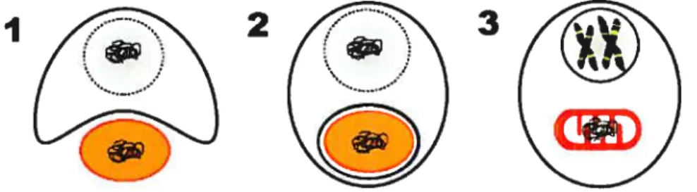
mtDNA Distribution within the Celis
for detection and quantification of rntDNA of Euglenozoa, we used DAPI which binds to A+T-rich DNA preferentially by minor-groove interaction (Manzini et al.
1983; Trotta et al. 2003).
Rhynchopus celis stained with DAPI (fig. 1, lA-C) show abundant rntDNA in a thin (250-500 mn wide) reticulated pattem. Inspection of different focal planes indicates that the network lines the periphery of the cytoplasm, which is confirmed by electron microscopy (see below).
In Fetalornonas (fig. 1, 2A-C), we observed a distribution of DNA similar to Rhynchopus. Again, large arnounts of mtDNA forrn a dense, but much thicker (0.5-1.5 .tm wide) network meandering through the ceil. Surprisingly, the fluorescence of mtDNA is much stronger than that of the nucleus, regardless of the focal plane. The area in the cell that systernatically lacks fluorescence corresponds to the feeding and flagellar apparatuses.
Compared to the two above species, the extra-nuclear DNA of Entosihon (Fig. 1, 3A-C) and Feranema (fig. 1, 4A-C) appears much less abundant. Enlosiphon displays small diffusely fluorescent spheres of 0.2 trn diameter in certain areas of the celi, whereas in others, the fluorescence pattem is volurninous and cloudy. Peranerna ceils reveal fluorescent bodies of 1 .0 jtm length, with distinct oval to rod-like shapes ofien arranged in rosettes. Electron microscopy confirms that these bodies are indeed mitochondria (see Fig 2D).