Université de Montréal
Neural correlates of affordance competition
in dorsal premotor cortex
par
Alexandre Pastor-Bernier
Département de Physiologie Faculté de Médecine
Mémoire présentée à la Faculté des études supérieures en vue de l’obtention du grade de doctoral (Ph. D)
en Sciences Neurologiques
Août, 2012
Université de Montréal
Faculté des études supérieures et postdoctorales
Ce mémoire intitulé:
Neural correlates of affordance competition in dorsal premotor cortex
présenté par: Alexandre Pastor-Bernier
a été évaluée par un jury composé des personnes suivantes:
Trevor Drew président-rapporteur Paul Cisek directeur de recherche Numa Dancause membre du jury Steven Wise examinateur externe Daniel Lévesque
To the brave that dare adventure in science,
“… Men Wanted: For hazardous journey. Small wages, bitter cold, long months of complete darkness, constant danger, safe return doubtful. Honour and recognition in
case of success …”
— Ernest Shackleton (1874—1922)
Newspaper announcement before his Endurance Expedition
“… No one really starts anything new. Everyone builds on other men’s failures. What each man contributes to the sum of knowledge is what counts…”
Daniel Keyes (1927) Excerpt from Flowers for Algernon
RÉSUMÉ
Le travail présenté dans cette thèse porte sur le rôle du cortex prémoteur dorsal (PMd) au sujet de la prise de décision (sélection d’une action parmis nombreux choix) et l'orientation visuelle des mouvements du bras. L’ouvrage décrit des expériences électrophysiologiques chez le singe éveillé (Macaca mulatta) permettant d’adresser une fraction importante des prédictions proposées par l'hypothèse des affordances
concurrentes (Cisek, 2006; Cisek, 2007a). Cette hypothèse suggère que le choix de toute
action est l’issue d'une concurrence entre les représentations internes des exigences et des atouts de chacune des options présentées (affordances; Gibson, 1979).
Un intérêt particulier est donné au traitement de l'information spatiale et la valeur des options (expected value, EV) dans la prise de décisions. La première étude (article 1) explore la façon dont PMd reflète ces deux paramètres dans la période délai ainsi que de leur intéraction. La deuxième étude (article 2) explore le mécanisme de décision de façon plus détaillée et étend les résultats au cortex prémoteur ventral (PMv). Cette étude porte également sur la représentation spatiale et l’EV dans une perspective d'apprentissage. Dans un environnement nouveau les paramètres spatiaux des actions semblent être présents en tout temps dans PMd, malgré que la représentation de l’EV apparaît uniquement lorsque les animaux commencent à prendre des décisions éclairées au sujet de la valeur des options disponibles. La troisième étude (article 3) explore la façon dont PMd est impliqué aux “changements d'esprit“ dans un procès de décision. Cette étude décrit comment la sélection d’une action est mise à jour à la suite d'une instruction de mouvement (GO signal).
Les résultats principaux des études sont reproduits par un modèle computationnel (Cisek, 2006) suggérant que la prise de décision entre plusieurs actions alternatives peux se faire par voie d’un mécanisme de concurrence (biased competition) qui aurait lieu dans la même région qui spécifie les actions.
Mots-clés: décisions, biais, concurrence, affordances, sélection d'action, spécification des actions, cortex prémoteur, PMd, PMv, EV, valeur relative, valeur absolue, distance, paramètres spatiaux, apprentissage, électrophysiologie, singe.
ABSTRACT
This thesis examines the role of the dorsal premotor cortex (PMd) in the process of decision making (action selection) and visual guidance of arm movements. The work describes electrophysiological experiments conducted in awake monkeys (Macaca
mulatta) and tests a number of important predictions suggested by the affordance
competition hypothesis (Cisek, 2006; Cisek, 2007a). This hypothesis suggests that
decisions can be viewed as the result of a competition between internal representations of conflicting demands and opportunities for actions or affordances (Gibson, 1979).
Specific interest is given to the interaction between spatial information and expected value (EV) in a proposed affordance competition mechanism for action selection. The first study presented (article 1) explores how EV is represented during the delay period in PMd. This study also describes how this area reflects the spatial metrics of the options and examines the interaction between value and spatial information. The second study (article 2) explores the mechanism of action selection in more detail and extends the results to ventral premotor cortex (PMv). This study also addresses the nature of value and spatial representations from a learning perspective. In a novel environment the spatial metrics of the actions seem to be invariably present in PMd, meanwhile EV representations appear only once the animals make behaviorally informed decisions about the value of the available options. The third study (article 3) explores how PMd is involved in “changes of mind” in which action selection is updated following a movement instruction (GO signal).
The major findings in all these studies are reproduced by a computational model (Cisek, 2006) suggesting that decisions between actions can be made through a biased competition process that takes place in the same region that specifies the actions.
Keywords: decisions, bias, competition, affordances, selection, action-specification, premotor cortex, PMd, PMv, EV, relative value, absolute value, distance, spatial parameters, learning, electrophysiology, monkey.
TABLE OF CONTENTS RÉSUMÉ...I ABSTRACT...III LIST OF FIGURES... IX LIST OF TABLES ... XI LIST OF ABBREVIATIONS...XII ACKNOWLEDGEMENTS... XIV I. PREAMBLE ... 1 II. INTRODUCTION... 3
1. General philosophical introduction ... 3
2. The motor role of the central nervous system, a brief historical perspective... 12
3. The anatomical organization of the premotor cortex ... 14
4. The role of PMd in visual guidance of movements: visual processing pathways... 24
5. The role of PMd in visual guidance of movements: the parieto−frontal network. ... 29
6. Parietal structures implicated in visuo-motor transformations and guidance of movements... 34
7. Parietal structures implicated in action-selection and guidance of movements ... 37
8. The basal ganglia and premotor cortex... 38
9. PMd role in visual guidance of movements: learning studies... 42
10. Role of dorsal premotor cortex in guidance of arm reaching movements: a traditional cognitive view ... 45
10.1. Decisions among actions in PMd ... 46
10.2. Action planning in PMd... 48
10.3. Movement preparation in PMd... 51
10.4. Movement execution in PMd ... 52
11. Inconsistencies in the traditional cognitive view: perception, cognition and action... 54
12. Alternatives to the cognitive view ... 57
13. Frontoparietal specification of potential actions and the foundations of the affordance competition hypothesis... 59
14. Sources of biasing signals for action selection ... 61
14.1. The dopamine system... 61
14.2. The basal ganglia ... 63
14.3. Prefrontal cortex ... 67
14.4. Ventral stream structures... 70
15. The affordance competition hypothesis ... 72
16. A computational model for reaching decisions: achievements and predictions... 79
III. OBJECTIVE, HYPOTHESIS AND PREDICTIONS OF THE THESIS...84
IV. ARTICLE 1 ...88
NEURAL CORRELATES OF BIASED COMPETITION IN PREMOTOR CORTEX...88
ABSTRACT ... 89
INTRODUCTION... 90
MATERIALS AND METHODS ... 91
RESULTS ... 93
Behavior... 93
Neural activity in PMd... 94
Population analyses ... 95
Gain effect of distance over relative value... 97
A biased competition model reproduces the results... 97
DISCUSSION ... 98
REFERENCES... 101
SUPPLEMENTAL MATERIALS... 109
Task apparatus and recording sites ... 110
Calculation of directional tuning ... 111
Additional repetitions in single-cell recordings... 112
Statistics for the assessment of value and distance effects ... 112
Determination of a unique value for latency of effects ... 114
V. ARTICLE 2... 120
SPACE MATTERS: TRADING ACTION METRIC AND VALUE REPRESENTATIONS IN PREMOTOR CORTEX ... 120
ABSTRACT ... 121
INTRODUCTION... 122
MATERIALS AND METHODS ... 127
Task apparatus and recording sites ... 129
RESULTS... 132
Behavioral results... 132
Neural results... 133
PMd activity predicts free choices ... 134
PMd activity reflects the relative value of potential actions ... 134
PMd activity reflects the angular distance between potencial actions... 136
Neural activity across premotor cortex follows similar trends... 138
Relative value modulation for novel instructional cues can be acquired through learning... 138
DISCUSSION ... 141
Theoretical background... 141
Premotor activity is modulated by relative value with full normalisation ... 144
Evidence that decisions between actions are made within sensorimotor circuits... 145
Convergence of specification and selection systems in premotor cortecs... 148
Is modulation of neural activity simply a result of motivational changes? ... 151
Concluding remarks ... 152
REFERENCES ... 154
SUPPLEMENTAL MATERIALS ... 183
VI. ARTICLE 3... 197
DORSAL PREMOTOR CORTEX IS INVOLVED IN SWITCHING MOTOR PLANS ... 197
ABSTRACT... 198
INTRODUCTION ... 199
MATERIALS AND METHODS... 203
Instrumentation and technical procedures... 203
Behavioral task ... 204
Kinematic analysis ... 205
Cell tuning and relative value discrimination ... 206
Plan-switch analysis ... 207
Computational modeling ... 210
BEHAVIORAL RESULTS... 211
NEURAL RESULTS ... 213
PMd activity predicts switching of motor plans ahead of movement onset... 213
PMd contribution to kinematics prior to movement onset (initial direction) is observed in situations where there is no relative value bias... 215
A biased competition model can reproduce the dynamics of the plan-switch... 216
DISCUSSION ... 217
REFERENCES ... 226
VII. GENERAL DISCUSSION... 242
1. Recapitulation of results ... 242
3. Decisions among actions in basal ganglia... 247
4. Representations of value or probability of choice: LIP vs PMd ... 249
5. Absolute and relative value representations: LIP vs PMd... 251
6. Absolute and relative value representations: FEF vs PMd ... 255
7. Distribution of effects in PMd and comparison with human tasks... 256
8. Competition among action representations takes place in the same areas involved in guidance of movements ... 258
9. Simultaneous action and specification extends beyond the time a movement is instructed ... 259
10. Decisions among non-spatially guided actions (NOT ADDED) VIII. FUTURE PERSPECTIVES ... 261
1. Action cost during action selection: interaction between context and value ... 261
2. Neural encoding of action costs ... 263
2.1. Basal ganglia ... 263
2.2. Anterior cingulate cortex... 264
2.3. Premotor cortex ... 266
3. The cognitive debate in neuroeconomics... 267
4. Recent models for decision making ... 273
IX. CONCLUSIONS... 277
LIST OF FIGURES
Figure 1. Schematic decision-making scenarios during natural behavior………10
Figure 2. Architectonic areas of relevance to the present study in the human brain and in the macaque brain………...15
Figure 3. Frameworks of visuospatial processing……….27
Figure 4. The parietofrontal network. A summary of connectivity from posterior parietal cortex primarily to PMd and M1………...31
Figure 5. Summary of the major corticocortical visual pathways to premotor cortex.…32 Figure 6. Basic circuitry of the basal ganglia………...39
Figure 7. Sketch of the proposed neural substrates of the affordance competition hypothesis, in the context of visually guided movement. ……… 74
Figure 8. Computational model..………..80
ARTICLE I. NEURAL CORRELATES OF BIASED COMPETITION IN PREMOTOR CORTEX Figure 1...105 Figure 2...106 Figure 3...107 Figure 4...108 Supplemental Figure 1………...………..116 Supplemental Figure 2……….117 Supplemental Figure 3...118 Supplemental Figure 4...119
ARTICLE II. SPACE MATTERS: TRADING ACTION METRIC AND VALUE REPRESENTATIONS IN PREMOTOR CORTEX Figure 1...160
Figure 3...163 Figure 4...165 Figure 5...167 Figure 6...169 Figure 7...171 Figure 8...173 Figure 9...174 Figure 10...176 Supplemental Figure 1...184 Supplemental Figure 2...185 Supplemental Figure 3. ...186 Supplemental Figure 4...187 Supplemental Figure 5...189 Supplemental Figure 6...191 Supplemental Figure 7...193 Supplemental Figure 8...195
ARTICLE III. DORSAL PREMOTOR CORTEX IS INVOLVED IN SWITCHING MOTOR PLANS Figure 1...232 Figure 2...233 Figure 3...235 Figure 4...237 Figure 5...238 Figure 6...239
LIST OF TABLES
ARTICLE I. NEURAL CORRELATES OF BIASED COMPETITION IN PREMOTOR CORTEX
Supplemental Table 1...115 ARTICLE II. SPACE MATTERS: TRADING ACTION METRIC AND VALUE REPRESENTATIONS IN PREMOTOR CORTEX
Table 1. Classification of recorded neurons in dorso-lateral PMd……….178 Table 2. Classification of recorded neurons in PMv………..178 Table 3. Classification of cell activity in PMd according to observed effects………...179 Table 4. Classification of cell activity in PMd ………..180 Table 5. Classification of PMd neurons collected in the learning condition…………..181 Table 6. Predicted firing rates for relative value effects according to
a completely divisive normalization model………182
ARTICLE III. DORSAL PREMOTOR CORTEX IS INVOLVED IN SWITCHING MOTOR PLANS
Table 1. Classification of cells………240 Table 2. Population latencies obtained with sliding ANOVA………....241
LIST OF ABBREVIATIONS
ACC: anterior cingulate cortex AG: angular gyrus
AI: inferior arcuate sulcus AIP: anterior intraparietal area AS: superior arcuate sulcus BG: basal ganglia
C: central sulcus Ca: calcarine fissure CHT: center hold time
CVML: conditional visuomotor learning Cx: cerebral cortex
DA: dopamine
DLPFC: dorsolateral prefrontal cortex DMPFC: dorsomedial prefrontal cortex EV: expected value
FEF: frontal eye field
GABA: gamma aminobutyric acid GPe: external segment of the globus pallidus
GPi: internal segment of the globus pallidus
IDV: initial direction vector IF: inferior frontal sulcus IFc: inferior precentral sulcus IO: inferior occipital sulcus IP: intraparietal sulcus IPL: inferior parietal lobe IPS: intraparietal sulcus L: lateral fissure
LIP: lateral intraparietal cortex Lu: lunate sulcus
M1: primary motor cortex MIP: medial intraparietal area MST: mediosuperiotemporal cortex MT: mediotemporal cortex
MTL: medial temporal lobe OC: striate cortex
OFC: orbitofrontal cortex OT: occipitotemporal sulcus P: principal sulcus
PCC: posterior cingulate cortex PCS: precentral sulcus
PFC: prefrontal cortex PMd: dorsal premotor cortex PMv: ventral premotor cortex PO: parieto-occipital visual area Pos: parieto-occipital sulcus PPC: posterior parietal cortex PT: preferred target
pre-SMA: pre-supplementary motor area
PRR: parietal reach region RF: receptive field
RSC: retrosplenial cortex RT: reaction time
RV: relative value
S: spur of the arcuate sulcus S1: primary somatosensory cortex
S2: secondary somatosensory cortex SC: superior colliculus SEF: supplemental eye field. SF: superior frontal sulcus SFy: sylvian fissure
SMA: supplementary motor area. SMG: supramarginal gyrus SNc/SNpc: substantia nigra pars compacta
SNr: substantia nigra pars reticulata
SNdl: dorsolateral substantia nigra pars reticulata
SPc: superior precentral sulcus SPD: superior precentral dimple
SPL: superior parietal lobule SSRT: stop signal reaction time ST: superior temporal sulcus STN: subthalamic nucleus Str: striatum
STS: superior temporal sulcus TE: rostral inferior temporal cortex TEO: posterior inferior temporal cortex
Th: thalamus
V1: primary visual cortex VIP: ventral intraparietal cortex VLPFC: ventrolateral prefrontal cortex
ACKNOWLEDGEMENTS
This work was carried out at the department of Physiology in the Groupe de Recherche du Système Nerveux Central (GRSNC), Faculty of Medecine, University of Montreal, under the supervision of Dr. Paul Cisek. I am very grateful to my supervisor for his precious advice, patience and engagement in research. Without his help this work would have never been possible. I would also like to thank my lab colleagues for their friendly and positive attitude during my stay as well to our lab technician, Marie-Claude Labonté for her sincere friendship and continuous support. Finally, I would like to dedicate this work to my beloved wife, Elena Samarova for her unconditional love and constant encouragements as well as to our little Trevor for his joyful attitude and precocious interest in science.
I. PREAMBLE
The organization of this thesis is as follows. A general philosophical introduction lays out the main concepts that will be treated in further detail through the entire work. The introduction is followed by an historical perspective section on the anatomy of the main structure studied here, the dorsal premotor cortex (PMd). Subsequently, a
physiology review section recapitulates the anatomy and discusses in detail the fundamental concepts presented in the general introduction. This section is split in two parts. The first part introduces the function of PMd according to a “cognitive view” and describes its advantages and drawbacks. The second part is more general and describes alternatives to the cognitive view. This part introduces the foundations of the affordance
competition hypothesis.
The affordance competition hypothesis is central in this dissertation and its main concepts are treated in full detail (i.e. a description of fronto-parietal visual processing pathways, bias competition concepts, source of bias information and supporting data in computational modeling). The general hypothesis makes specific predictions that are described after the physiology review section and are tested in three included articles.
The discussion section consists of several parts. The first part consists in a global recapitulation of the predictions and results. The second part addresses the competition hypothesis in PMd and basal ganglia (BG). The third part compares relative value encoding in PMd with other cortical areas such as the lateral intraparietal cortex (LIP) and the frontal eye fields (FEF). A fourth part extends these observations to human
studies. The last part revisits two aspects of the competition process: the location and timing of the process in basis of observations conducted by different research groups.
A future perspectives section follows the discussion. This section introduces the notion of effort and action cost in the context of potential upcoming studies within the frame of the affordance competition hypothesis. A recent debate about the emerging field of neuroeconomics is briefly inspected. A conclusion section finalizes the present work.
II. INTRODUCTION
1. General philosophical introduction
The work presented in this thesis addresses several important aspects of the neurophysiologic process known as decision making. Decision making is a deliberative process that results in the commitment to a categorical proposition (Gold and Shadlen, 2007). It can be seen as motivated procrastination (Cisek et al., 2007) or a situation in which one succumbs to the preponderance of one set of influences over another (Bierce, 1911). However, to favour one option over another or to engage in one action or another might be completely different things. We term decisions in a similar fashion whether we deal with an abstract process, such as choice of university or career, or whether we ponder among two concrete actions, such as braking or accelerating upon the sight of an amber light in our drive to work.
The classical view of decision making (Fodor, 1983; Pylyshyn, 1984) has this process belonging to “cognition”, a separate process from sensory motor control. For instance, few processes can be as removed from sensory-motor control as playing strategy games such as chess. For a chess player, decisions are portrayed best by the time commitment of one pondered strategy over another with no obvious link between a reported movement and the internal sequences of action-outcomes that the player has in mind (Newell and Simon, 1972). The mental process has to be covert if the player wants to stand a chance since guessing is a main feature in the game. The view of decision
making as entirely abstract (“cognitive process”) is deeply rooted in history, starting from classic Greek scholarship (Hicks, 1907).
Aristotle for instance, argued that the mechanism responsible for purposive human behavior was conveyed exclusively by a “nonmaterial substrate” like the human soul. This influential idea was not challenged by any alternative view until Descartes (Descartes, 1649, 1664) proposed a dual deterministic and cognitive explanation of human behavior. In contrast with Aristotle, fully deterministic decisions included very elegant and stereotyped sensory-motor reflexes that had a very tangible substrate in the human body. Although cognitive behavior was still relegated to a “nonmaterial substrate”, the very foundations for empirical research, binding the neural mechanisms linking sensation and action, were laid down at this time. Notably, the notion of information being transferred from sensory nerves to muscles through basic reflexes flourished with 20th century physiologists such as Sherrington (Sherrington, 1906).
The characterization of the primary sensory and motor systems along with recent physiological data provided the foundations for novel paradigms in psychology like
behaviorism (Skinner, 1974). Behaviorists attempted to explain even the more abstract
process on the basis of essential sensory-motor behaviors and argued that all processes (covert or non-covert such as thinking or walking respectively) should have similar observational correlates. Supporters of behaviorism proposed to treat physical and psychological disorders with qualified experimental methods such as operant-conditioning (Skinner, 1974).
In response to the behaviorist view, Lachman and Butterfield proposed a “cognitive” alternative to explain processes such as decision making (Lachman et al.,
1979). The cognitive view relegates decision making, including decisions among actions, to an exclusive stage of abstract processing that takes place in a serial information processing architecture. This view further suggests a spatial and temporal decoupling of the decision process from perception and action. Although the disagreement between behaviorism and cognitivism went beyond the scope of the decision making it is important to emphasize that this point was particularly crucial.
The dispute between these two psychology schools continued until the second half of the 20th century when it was found that both views could complement each other for the treatment of psychological pathologies such as phobias, posttraumatic stress disorders and addictions (Hofmann and Smits, 2008; Rachman, 1997; Santrock, 2008). Moreover, the interaction of the cognitive and behaviorist points of view led to important advances in developmental child psychology (Piaget, 1954).
However, a certain ambiguity still remains today attending the characterization of decision making. Although the cognitive view might still be popular, from an ecological perspective decision making cannot be seen as an entirely abstract process, since the majority of living creatures are faced with a world of actions that need both spatial and temporal overt behavior. The point of view that a decision can be made between overt actions is an outstanding feature that has evolutionary implications as well. All animals display a capacity to decide among actions, whether the decisions are very simple, as it is the case in quasi-reflexive local feeding responses observed in primitive organisms like flatworms (i.e. Planocera gilchristi, Gruber and Ewer, 1962) or whether the decisions involve complex foraging behaviour as it is the case in primates and humans (Bautista et al., 2001; Janson, 1998; Kacelnik, 1997; Noser and Byrne, 2007; Tinbergen, 1951;
Stevens et al., 2005). Although decisions among actions can vary greatly in complexity it is reasonable to believe that they are governed by similar principles that allow accumulation of evidence, comparison of the potential options and commitment as more abstracts form of decisions (Glimcher, 2003; Gold and Shadlen, 2007). For instance, early oculomotor studies of saccade selection have shown that even simple kinds of decisions might confer insights into higher cognitive functions (Schall, 2004a). Consequently, a main and very general question regards how decisions among actions
are currently characterized within the existent framework of theoretical, anatomical and physiological knowledge.
Several attempts to address this question have been proposed and lead to the current debate in neuroeconomics. Neuroeconomics is an emerging discipline that bridges neuroscience and economics and suggest that humans make decisions between different options by integrating all relevant factors such as expected gains, potential risks and action costs, into a single variable reflecting the subjective value of each offer (Friedman, 1953; Simon, 1947, 1983). However, some neuroeconomists go a step further in this assumption and postulate that all decisions are made according to classic
economic-utility postulates for economic value (Padoa-Schioppa, 2011; Von Neumann and Morgenstern, 1944). Among other assumptions, economic-utility postulates require decisions to be independent of any contextual parameter such as the metrics and cost of the actions.
The economic-utility conjecture appears to be grounded on particular neurophysiology studies suggesting the orbitofrontal cortex (OFC) as the neural correlate for economic value and economic choice (Padoa-Schioppa and Assad, 2006, 2008).
According to this point of view, a decision is made exclusively in abstract terms and is a serial process dictated by the OFC, wherefrom the outcome of the decision radiates to all other structures. This view is substantiated in the goods-based model (Padoa-Schioppa, 2011).
This model has several important caveats as it proposes that decisions are made before action costs can be taken into account for instance, and cannot explain human non-transitive behavior (Güth et al., 1982; Kahneman and Tversky, 1982). Alternatives to this view have been proposed (Cisek, 2012).
In Cisek’s view, decisions among actions can be seen as the consequence of a dynamic interaction between perception and action rather than sequestering the decision process to an abstract stage like “cognition”. This view proposes that decisions can be
made through a distributed consensus in which action costs and spatial parameters of the
action can be taken into account when decisions are made among actions. The
framework in which the decision process is made is the affordance competition
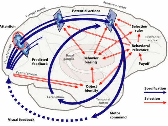
hypothesis. This hypothesis suggests that decisions can be viewed as the result of a competition between internal representations of conflicting demands and opportunities for action, also called affordances (Gibson, 1979). The competition between potential actions plays out within reciprocally interconnected areas of the parietofrontal system (Matelli and Luppino, 2000) that can represent different aspects of movement (Jones et al., 1978; Marconi et al., 2001; Pandya and Kuypers, 1969).
Sensorimotor areas such as dorsal premotor cortex (PMd) are sensible candidates for being causally involved in action selection. For instance, Song et al. (2011) showed that reversible inactivation of the superior colliculus (SC), a sensorimotor structure just
two synapses away from the motor neurons that move the eye (Basso and Wurtz, 1998), could affect saccade target selection in non-human primates. The animals performed in a 4-target visual search task and after SC inactivation, made fewer saccades to the targets in the affected zone. These deficits were not simply motor as they were mostly absent when only a single target was presented and inactivation was conducted. In this case the animals were still able to saccade to the single target.
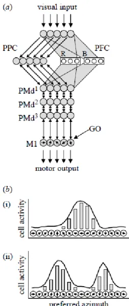
The affordance competition hypothesis also suggest that multiple potential actions can be represented simultaneously within a given cortical area as has been indirectly shown for the arm system (reach and grasp) and oculomotor system (Baumann et al., 2009; Cisek and Kalaska, 2005; Glimcher, 2003; McPeek and Keller, 2002; Scherberger and Andersen, 2007). For instance, cell population data in PMd suggests that two mutually-exclusive potential reaching actions can be simultaneously represented until a choice can be made, at which time the activity corresponding to the non-chosen option becomes suppressed. This simultaneous specification of multiple potential actions is also supported by several behavioral studies of reaching movements made in presence of distractors (Song and Nakayama, 2006, 2008; Song et al., 2008; Tipper et al., 1992) and lays out the framework for a competition based mechanism for action selection.
The affordance competition hypothesis further suggests that the competition process between the options takes place within the same regions that specify the actions (Cisek, 2006, 2007a). This suggestion is in agreement with proposed mechanisms of
selective attention, in which the selection process also takes place through competition in
the very sensory areas that process the percepts (Boynton, 2005; Desimone and Duncan, 1995; Treue, 2001). In both selective attention and affordance competition mechanisms,
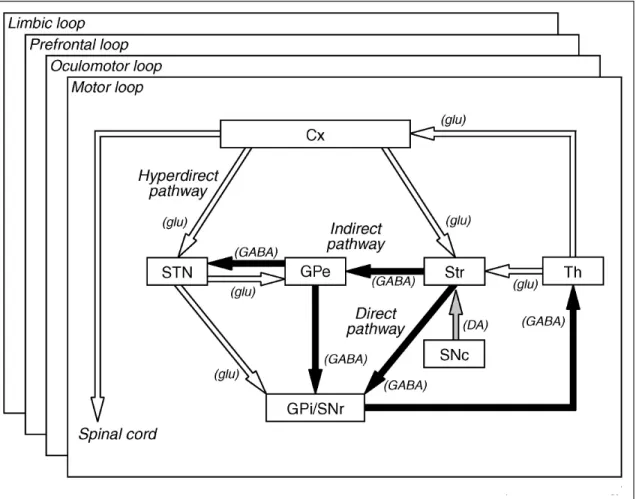
cells with different percept preferences (stimuli orientation or movement preference, respectively) mutually inhibit each other, creating a competition between potential actions or percepts. In the affordance competition hypothesis, the competition process is gradually resolved throughout the parieto-frontal system as new information is incorporated into the process: the bias. The notion of bias arises from the different excitatory inputs that the fronto-parietal system receives from diverse cortical and subcortical structures that project to it (e.g. prefrontal cortex, PFC and BG) conveying particular sets of information (e.g. appetitive, hedonic value) about the options for action. These biases become integrated with sensory or motor variables. For instance, LIP integrates expected utility, local income, hazard rate and relative subjective desirability along with specific spatial parameters of the actions such as saccade probability (Dorris and Glimcher, 2004; Janssen and Shadlen, 2005; Platt and Glimcher, 1999; Sugrue et al., 2004). Expected utility is an economic concept that represents the betting preferences of an individual as a function of the payout, probability of outcome, risk and subjective value of the options (i.e. utility of the options). The hazard rate represents the probability of an event to happen in time and local income the reward history (actual vs past rewards).
The mechanism proposed in the affordance competition hypothesis is not surprising from the point of view of interactive behavior where decisions among actions must take account of the “dynamic” aspects of the environment in which the actions are played out. Considering, for instance, the situation in Figure 1A, a predator may be initially faced with two potentially useful/attractive pursuit actions, but as soon as the chase begins the metrics of the actions and the estimates of their relative value may change, as it is the case if one of the targets (zebras) splits in two groups. Furthermore,
decisions between actions are strongly dependent upon their geometrical relationship. For example, if an animal seeking escape (zebra in Figure 1B) is faced with two opposite routes (large obstacle ahead), the choice has to be all-or-none to a given direction. If the escape routes are similar (small obstacle ahead) then the best strategy may consist in mixing both options and delay making the choice until the last moment. This implies that when choosing to reach between two nearby targets the nervous system can mix their neural representation and start moving between them. The observation that decisions among actions can be affected by the metrics of the actions is both consistent with human psychometric data (Chapman et al., 2010; Favilla, 1997; Ghez et al., 1997) and with a number of oculomotor neurophysiology studies (Louie et al., 2011; Schall, 2004a), although this particular issue remains a central topic in the present work and will therefore be examined in detail.
Figure 1. Schematic decision making scenarios during natural behavior.
A. The environment around the lion provides information on both the spatial metrics and relative values of potential pursuit actions (arrows, with value indicated by width). During ongoing activity, this information is constantly changing and what was once a single action may sometimes split into two (bottom). B. When faced with two opposite escape routes (top), the zebra must make an all-or-none decision, but when the escape routes are similar (bottom), it may mix them initially and veer toward one or the other in-flight (adapted from Cisek, 2012).
2. The motor role of the central nervous system, a brief historical perspective
From the earliest western medical writings, it was thought that the movement of the body was controlled by the brain. In the Edwin Smith Surgical Papyrus originating in the Pyramid age (3000 BC) there are a number of descriptions of motor dysfunctions after head injury (Breasted, 1930). A literal citation of one of the cases reads as follows “the subject walks shuffling with the sole on the side of him having that injury which is to the skull”. This contralateral symptom was interpreted as the result of a blow to one side of the head causing the brain to impact on the inside of the contralateral skull (contrecoups syndrome). Later on, Hippocratic doctors (500 BC) would write more extensive treaties on head wounds showing good awareness that head injuries could produce contralateral symptoms. However, the primary interest was diagnosis and not the study of the underlying anatomy or physiology (Courville, 1946). Aretaeus, a Greek physician who practiced in Rome and Alexandria (200 BC), went a step further and distinguished paralysis due to head injury from paralysis due to spinal injuries, an observation that led him to postulate that some kind of crossing must take place above the craniovertebral junction. However, where exactly the crossing occurred remained a mystery for centuries(Louis, 1994) only to be revealed much later by the Pisan scholar Domenico Mistichelli (1675-1715) in his “trattato dell’apoplessia” and by the French military surgeon Dr François Pourfour du Petit (1664-1741) with complementary field work (Thomas, 1910).
One of the most important events in the history of the study of the motor functions of the cortex was the discovery by Fritsch and Hitzig in 1870 that electric
stimulation of the cerebral cortex could produce discrete movements. These results were in agreement with the ideas of John Hughlings Jackson (1815-1911), often considered the “father of English clinical neurology”. Hughlings Jackson reasoned that the cortex had basic sensory-motor functions and also gathered clinical evidence substantiating this view (Young, 1970). In studying epileptic seizures, he noticed a systematic spread of convulsions from one body part to its immediate next. These observations led him to suggest that different areas of the cortex could be involved in the control of particular muscle groups and that these areas would be arranged in a way that mimic the organization of the body (Hughlings Jackson, 1873; Temkin, 1971; Young, 1970). Hughlings Jackson’s work was notably expanded by Ferrier (1874a, b) who explored the effects of electrical stimulation in extensive areas of the cerebral cortex, well beyond what is considered today’s motor cortex (M1) and including prefrontal areas such as FEF. Ferrier observed that electrical stimulation could induce not only seizures but also discrete movements. The later observation was particularly interesting because these discrete movements seemed to cluster on particular areas. Ferrier reported that electrical stimulation on these areas evoked movements of eyelids, face, mouth, tongue, ear, neck, hand, foot and tail. The early work by Ferrier was conducted in a wide range of animals such as dogs, jackals, rabbits and cats, substantiating the generality of Jackson’s assumptions. A growing interest in the primate brain would then lead Ferrier to extend research to non-human primates, delineating not less than 19 centers related to different movements including walking, arm retraction, extension and flexion of the wrist, mouth opening and protrusion of the tongue, sneering expression of the face and eye movements (Ferrier, 1874-1875). Ferrier (1875) also conducted lesion studies of the motor centers
reporting a correlation between the size and location of a lesion with the type and severity of the resulting paralysis. As techniques for electrical stimulation improved, Ferrier’s results were confirmed in humans. The Canadian Dr. Penfield and colleagues applied electrical stimulation along the precentral cortex in patients during surgery for the removal of tumors and epileptic foci (Penfield and Boldrey, 1937; Penfield and Rasmussen, 1950). Their results revealed a disproportionate somatotopic map of the body that is commonly depicted as Penfield’s motor homunculus with very similar results in non-human primates (Woolsey’s semiusculus; Woolsey et al., 1952). These pioneering works are the foundation of the modern functional and anatomical definition of the motor and premotor cortices.
3. The anatomical organization of the premotor cortex
Early human anatomical work by Brodmann (1909) revealed that a significant fraction of the frontal lobe lacks a clearly defined internal granular layer (IV) and is commonly referred as agranular cortex. This area can be further parceled into a well defined field with large pyramidal cells including exclusively Betz cells (Betz, 1874), lying in the anterior bank of the precentral sulcus (area 4), and a wide region spanning the precentral gyrus and the posterior portion of the superior frontal gyrus on both the lateral and medial surfaces in the primate brain (area 6) (Figure 2A-B).
Pre-SMA SMA M1 PMd PMv SEF ACC OFC HUMAN MACAQUE
C.
PPC PMd yFigure 2. Architectonic areas in the human (A) and macaque brain (B) along with expanded mesial and lateral views of the macaque brain (C). The numbers on the left-side picture correspond to Brodmann and Walker's classification, meanwhile the right-side picture uses the classification of Barbas and Pandya (1987). Notice the consistent mapping of area 6 (blue shades) and area 4 in both humans and macaques despite the different representation of gyrus sulci in both species, in particular for macaque principal sulcus (P) that becomes the superior frontal sulcus (SF) and the inferior frontal sulcus (IF) (orange ellipse in the right-side pictures). The figures on the right show the location of the dorsal premotor cortex (PMd) and its homologous subdivisions in humans and macaques. PMd can be subdivided into rostral PMd (pre-PMd in humans and F7 in the macaque, pink ellipses) and caudal (pre-PMd ((pre-PMd proper in humans and F2 in the macaque, blue ellipse). On the top left figure: PCS, precentral sulcus; CS, central sulcus; SMG, supramarginal gyrus; IPS, intraparietal gyrus; STS, superior temporal gyrus; SFy, Sylvian fissure; AS, arcuate sulcus; PS, principal sulcus. On the top right figures: dlPFC, dorsolateral prefrontal cortex; SPc, superior precentral sulcus; C, central sulcus; PPC, posterior parietal cortex (green ellipses); IPc, inferior precentral sulcus; AS, superior arcuate sulcus, AI, inferior arcuate sulcus; S, spur of the arcuate sulcus; C, central sulcus; SPD, superior precentral dimple; C, central sulcus; IP, intraparietal sulcus (adapted from Aboitiz and Garcia, 1997; Abe and Hanakawa, 2009). The green rectangle depicted in C shows the parcellation of the motor cortex, posterior parietal, and cingulate cortices. The motor and premotor areas are defined according to Matelli et al. (1985, 1991). IP, intraparietal sulcus, AG, annectant gyrus; C, central sulcus; Ca, calcarine fissure; Cg, cingulate sulcus; IO, inferior occipital sulcus; L, lateral fissure; Lu, lunate sulcus; P, principal sulcus; POs, parieto-occipital sulcus; ST, superior temporal sulcus; FEF, frontal eye field; SEF, supplemental eye field; PMd, dorsal premotor cortex; PMv, ventral premotor cortex, M1; motor cortex; pre-SMA, pre-supplementary motor area; SMA, supplementary motor area. AI, inferior arcuate sulcus; AS, superior arcuate sulcus; C, central sulcus; Cg, cingulate sulcus; ACC, anterior cingulate cortex; OFC, orbitofrontal cortex; DLPFd, dorsolateral prefrontal cortex, dorsal; DLPFv, dorsolateral prefrontal cortex, ventral; MIP, medial intraparietal cortex; LIP, lateral intraparietal cortex; VIP, ventral intraparietal area; Anterior intraparietal area, AIP; PE, PEc, PEIp, PF,
PFG and PG are arbitrary names given to cytoarchitectonically distinct areas defined by von Bonin and Bailey (1947). Note that area 6 corresponds roughly to PMd, PMv (green and red shaded areas), SMA and pre-SMA (blue and orange shaded areas). Area 4 corresponds to M1 (beige shaded area F1) (adapted from Rizzolatti and Luppino, 2001).
Evidence from clinical observations and cortical ablation experiments conducted by Fulton (1935) led to the original view of a primary motor cortex corresponding to
area 4, whereas Woolsey et al. (1952) along with Penfield and Welch (1951) further
established the concept of a non-primary motor cortex revealing a physiologically distinct region, the supplementary motor cortex.
The supplementary motor cortex is located in the medial aspect of area 6 and represents today’s supplementary motor area (SMA) and pre-supplementary motor area (pre-SMA). Hines (1929) defined the remaining of area 6 as “premotor cortex”, although research in this region would linger for nearly half a century mainly because of experimental shortcomings. For instance, Woolsey et al. (1952) and Travis (1955) concluded that the premotor cortex was not a part of the motor system because cortical stimulation of the area in deeply anesthetized monkeys didn’t evoke movements. Although it was known that the level of anesthesia is a critical variable for cortical excitability (Bucy and Fulton, 1933), knowledge of the connectivity pattern of the premotor region and spinal cord were at that time very limited and so were the chances to obtain an ideal combination of stimulation parameters and probing locations.
It is known today that low intensity electrical stimulation (≥40µA) of regions of the premotor cortex that have corticospinal projections (F2, PMd, Figure 2C) (Dum and Strick, 1991; He et al., 1993) can evoke everything from finger twitches to complex
arm-to-mouth movements (Godschalk et al., 1995; Graziano et al., 2002; Raos et al., 2003). Extensive stimulation of the premotor cortex has also helped characterizing areas involved in oculomotor control such as the supplemental eye field (SEF; Tehovnik and Lee, 1993) and FEF (Bruce et al., 1985; Robinson and Fuchs, 1969; Schlag and Schlag-Rey, 1987). We can parcel the premotor cortex of non-human primates in at least seven non-primary motor areas involved in controlling arm movements such as reaching and grasping (Barbas and Pandya, 1987; Geyer et al., 2000; Matelli et al., 1985, 1991; Vogt, 1919; Von Bonin and Bailey, 1947) and two further areas involved in oculomotor control (Barany et al., 1923; Ferrier, 1875) (Figure 2C).
The first two regions are located in Brodmann’s area 6 and are namely the dorsal and ventral premotor cortices (PMd and PMv) which are caudal to the arcuate sulcus and rostral to M1. The SMA and pre-SMA are located in the superior frontal gyrus and loosely correspond to Woolsey’s supplementary motor cortex (Woolsey et al., 1952). Another three non-primary motor areas can be further identified in the banks of the cingulate cortex, namely the rostral, dorsal and ventral cingulate motor areas (CMAr, CMAd and CMAv, respectively)(Dum and Strick, 1991; He and Strick, 1995; Picard and Strick, 1996). The two oculomotor areas are FEF and SEF. The first region, FEF (also called area 8 of Brodmann) is located within the anterior wall of the arcuate sulcus and encompasses a triangular area next to the spur junction of both branches of the arcuate sulcus (Barany et al., 1923; Ferrier, 1875). The second area, SEF is a post-arcuate region located in the rostral bank of the arcuate sulcus close medially to pre-SMA (Schlag and Schlag-Rey, 1987; Woolsey et al., 1952). Although these two areas are classified traditionally as belonging to the prefrontal cortex, the FEF in particular has functional
features that are similar to PMd (Thura et al., 2011). The FEF is involved in action specification and competition-based mechanisms of goal selection in the oculomotor system while the PMd may perform similar functions in the arm system (McPeek et al., 2006; Cisek and Kalaska, 2005).
The modern organization of the premotor cortex is full of nuances and a number of alternative ways of subdividing area 6 have been proposed (Barbas and Pandya, 1987; Matelli et al., 1991; Muakkassa and Strick, 1979). These alternative divisions are worth mentioning in order to address the functional differences observed within the region. For instance, Barbas and Pandya (1987) delineated the premotor cortex (area 6) mainly on the basis of cytoarchitectonic and myeloarchitectonic features and subdivided it into a dorsal sector (PMd) and a ventral sector (PMv) at the spur of the arcuate sulcus.
These authors reported hints of an “emergent” layer IV in PMv and higher myelin content in PMd, although the most striking results were observed studying the connectivity patterns within these two sectors using anterograde and retrograde tracers. A
rostral portion of PMd had specific frontal connections restricted to the neighboring dorsal frontal regions, whereas a caudal portion of PMd sent projections to the motor cortex. These results would be later complemented by tracing studies conducted by Strick and colleagues detailing inputs from the dorsolateral prefrontal cortex (DLPFC) to rostral PMd (Lu et al., 1994) and projections from the caudal sector of PMd to not only M1 but also directly to the spinal cord (Dum and Strick, 1991; He et al., 1993).
In contrast, the PMv was found to be connected extensively both with PFC and M1. For instance, Carmichael and Price (1995a, b) and Strick and colleagues (Lu et al., 1994) showed that both OFC, DLPFC and the dorsomedial prefrontal cortex (DMPFC)
share reciprocal projections with PMv. The DLPFC projections to PMv are extensive in contrast to the projections of DLPFC to PMd that are limited to only a portion of the arm area. Matsumura and Kubota (1979) and Muakkassa and Strick (1979) also showed projections of PMv to M1.
The idea that anatomical differences could be observed not only in PMd and PMv but also within each of these cortical areas separately motivated yet another parcelation of area 6, incorporating a growing bulk of information from neuroanatomical and physiological studies (Matelli et al., 1985, 1991). According to Matelli et al. (1985, 1991), the dorsal part of the agranular frontal cortex could be subdivided into three areas: area F1, corresponding to the primary motor cortex (M1), and areas F2 and F7 which together correspond to PMd (Figure 2). Area F2 occupies the caudal two-thirds of superior area 6 and is bordered caudally by area F1 and extends rostrally up to the border with area F7. Area F7 is located about 3 mm in front of the genu of the arcuate sulcus and extends medially to the superior limb of the arcuate sulcus until area F6 (pre-SMA). Area F7 corresponds roughly with the rostral division of PMd of Barbas and Pandya (1987) and incorporates a distinct eye movement representation in its rostro-medial aspect (SEF) and a motor representation of a forelimb field with a minority of other body parts representations embedded in it (Tachibana et al., 2004).
These results are congruent with physiological studies (Boussaoud et al., 1998; Fujii et al., 2000; Mitz and Godschalk, 1989) suggesting that this area is involved in visual guidance of arm reaches. Area F2 is medially delimited by area F3 (SMA) and laterally by the spur of the arcuate sulcus, which separates it from areas F4 and F5, that together correspond to Barbas and Pandya’s PMv. F2 corresponds roughly to the region
referred to as the caudal part of PMd in physiological studies (Wise et al., 1997). The topography of the corticospinal projections (Dum and Strick 1991; He et al. 1993), the data from single-neuron recording studies (Kurata, 1989) and intracortical microstimulation studies (Godschalk et al., 1995; Raos et al., 2003) suggest that area F2 has a gross somatotopic arrangement, with a hindlimb field located medially to the superior precentral dimple and a forelimb field located laterally to it (SPP, Figure 2B). Area F2 contains a significant proportion of cells (16%) with visual responses and seems implicated in visual guidance of arm reaches. The visual responses in area F2 are not just perceptual but appear to be driven by the instructional significance of the stimulus for motor behaviour (Wise et al., 1996a). Most visually driven neurons are concentrated within the rostrolateral sector of the forelimb representation of area F2 (which is the region of F2 spanning from the precentral dimple to the spur (Fogassi et al., 1999).
Tanji’s group (Fujii et al., 2000) reported similar visual responses and evoked saccades in the PMd region corresponding roughly to the entire area F7 plus a rostral portion of F2. It has been shown that gaze modulation effects observed in PMd for arm-reaching movements are modest when the animals are instructed to perform in a free gaze condition, in comparison to when the animals have to move the eyes after an oculomotor instruction (Boussaoud et al., 1998; Cisek and Kalaska, 2002). These results are in agreement with the work from Tanji’s group (Fujii et al., 2000). Their study reported that saccadic responses elicited by either visual or electrical stimulation in PMd were functionally different from responses observed in SEF and FEF, further indicating the role of PMd in coordination of eye and arm movements in a context that requires cognitive behavioral control.
The border between PMd and its neighboring areas has also been an issue of debate mainly concerning PMv. A precentral polysensory zone caudally located from the spur separating area F2 and F4 (between caudal PMd and PMv) has been recently proposed by Graziano and Gandhi (2000). Graziano suggests a role in the guidance of movements in basis of tactile, visual and auditory information for this particular region.
According to Matelli, PMv can also be subdivided in anatomical and physiologically distinct regions, namely areas F4 and F5. Area F4 constitutes the caudal part of PMv (Matelli et al., 1985). It is connected with posterior parietal areas such as the ventral intraparietal area (VIP), the intraparietal sector of area PE, and the secondary somatosensory cortex (S2) (Rizzolatti and Lupino, 2001, for a review). F4 neurons discharge according to specific body part movements and electrical stimulation of this area evokes neck, arm, and face movements with often a combination of two or three body parts (Fogassi et al., 1996; Gentilucci et al., 1988). Most F4 neurons are activated by somatosensory, visual or auditory stimuli (Fogassi et al., 1996; Gentilucci et al., 1988; Graziano et al., 1999). This area presents bimodal, somatosensory and visual neurons that have RF within a reaching distance (peripersonal space) and code actions in extrinsic (spatial) coordinates, rather than in intrinsic limb coordinates (Kakei et al., 2001). It has therefore been suggested that area F4 transforms specific positions in peripersonal space into arm, neck, and face/mouth movements and is also involved in space perception (Fogassi et al., 1996; Rizzolatti et al., 1996).
Area F5 occupies the most rostral part of PMv in the macaque monkey, and contains a motor representation of distal hand movements (Hepp-Reymond et al., 1994; Kurata and Tanji, 1986; Rizzolatti et al., 1988). The neurons of this area discharge during
specific goal-directed hand movements such as grasping, holding and tearing. This area is also directly connected with M1 and receives rich inputs from S2, parietal area PF (7b), and from a parietal area located inside the intraparietal sulcus, the anterior intraparietal area (AIP) (Godschalk et al., 1984; Luppino et al., 1999; Matelli et al., 1986; Matsumura and Kubota, 1979; Muakkassa and Strick, 1979) which is traditionally associated with grasping (Baumann et al., 2009). There are two classes of visuomotor neurons in monkey area F5: canonical neurons, which respond to the presentation of an object, and “mirror” neurons, which respond when the monkey sees object-directed action (Rizzolatti and Luppino, 2001). “Mirror” neurons require a meaningful interaction between an effector (e.g. hand or mouth) and an object (e.g. edible fruit) in order to be active (Gentilucci et al., 1988).
Early studies characterize these cells according to the naturalistic motor acts that they prefer and classify them into proximal and distal classes involving diverse effectors such as arm, hand and mouth. For instance, Gentilucci et al., (1988) reports “mirror” neurons in the distal classes as "Grasping-with-the-hand-and-the-mouth neurons", "Grasping-with-the-hand neurons", "Holding neurons" and "Tearing neurons". The proximal classes are: "Reaching neurons" and "Bringing-to-the-mouth-or-to-the-body neurons". More recently it has been demonstrated that area F5 also harbors “mirror” neurons that discharge during the execution and observation of mouth actions.
Most of mouth “mirror” neurons become active during the execution and observation of mouth ingestive actions such as grasping, sucking or breaking food. Some of them respond during the execution and observation of oral communicative actions such as lip smacking (Ferrari et al., 2003). In general, “mirror” neurons have gathered
considerable attention in the scientific community as they might be involved in diverse processes such as action understanding, language, communication and learning by imitation (Rizzolatti and Luppino, 2001). Despite the appeal of this hypothesis “mirror” neurons cannot be regarded as the only type of cells in which the observation of external events can generate motor representations of the actions associated with those events. Unlike “mirror” neurons, PMd cells do not respond to direct observation of naturalistic behaviors but are implicated in the prediction of impending actions or events based on arbitrary cue-response associations. However, Cisek and Kalaska (2004) found PMd cells that are active during action observation and seemed involved in mental rehearsal of action plans (Cisek and Kalaska, 2004). This property might contribute to abstract functions underlying the assessment and understanding of observed events and suggest a relation with PMv “mirror” cells: although PMd cells and PMv “mirror” cells may have very different properties, it is plausible that both groups are required in processes such as action learning and understanding.
4. The role of PMd in visual guidance of movements: visual processing pathways
Early studies indicated that the occipital cortex lacks direct access to the primate frontal lobe (Jones and Powell, 1970; Pandya and Kuypers, 1969), although it has been known for a long time that striate and extrastriate visual areas can relay visual information to premotor areas via the parietal cortex (Critchley, 1953a; Milner and Goodale, 1993). The notion of a parietal relay for visual information has its foundations in the classic “dorsal and ventral processing streams” hypothesis proposed by
Ungerlieder and Mishkin (1982). However, the idea that the visual system is divided into two main streams of information may not be entirely novel and can be tracked back to the work of Max Schultze (1866) and to the Duplicity Theory proposed by von Kries (1895).
According to Ungerlieder and Mishkin’s proposal, visual processing can be segregated into two anatomically and functionally distinct pathways originating in the striate cortex, namely an occipito-parietal dorsal stream specifying spatial location and an occipito-temporal ventral stream specifying object identity (Figure 3A).
The dorsal stream travels through the occipitoparietal cortex (V1-V4) to the caudal part of the posterior parietal cortex (PPC), the inferior parietal lobule (IPL), and extends further to DLPFC. The ventral stream travels through the occipitotemporal cortex to the inferior temporal cortex (TE) and extends further to the ventrolateral prefrontal cortex (VLPFC, Figure 3A).
Lesions of the ventral and dorsal streams in monkeys produced selective deficits in object vision and spatial vision, respectively, leading to their characterization as ‘What’ and ‘Where’ pathways (Macko et al., 1982; Mishkin et al., 1983). Human patients with PPC lesions are able to recognize objects, but not their spatial relationship (Andersen, 1987; Critchley, 1953a, b). For instance, optic ataxia is characterized by a specific deficit in localizing visual targets with respect to the body and results from lesions centered around the intraparietal sulcus (IPS) and the superior parietal lobule (SPL) (Rondot et al., 1977). Consequently, patients suffering from optic ataxia are able to identify objects properly, although they cannot accurately perform a goal directed action. However, it is important to mention that the functional distinction between ventral and dorsal stream is not entirely clear-cut and a number of additional clinical studies in
humans have revealed nuances for grasping actions (James et al., 2003; Milner et al., 1991; Read et al., 2010). For instance, a patient with agnosia (patient D.F.) who had a large bilateral lesion of the occipitotemporal cortex and a small left-sided lesion of the occipitoparietal cortex (James et al., 2003; Milner et al., 1991) presented impaired perception of objects but intact ability to grasp them (Figure 3B).
In fact, patients like D.F. can pre-shape the hand to reflect size, shape and orientation of objects and are able to both orient and transport the hand to an intended reach location in space. However they cannot indicate the orientation of their own hand in space (and by extension pantomime an action) despite being aware of spatial depth information (Read et al., 2010). These findings, combined with the connectivity patterns between the posterior parietal and frontal premotor areas (Wise et al., 1997 for a review), have led to the proposal of a ‘How’ instead of a “Where” function for the dorsal stream (Goodale and Milner, 1992).
Recent studies account better for these nuances by segregating the dorsal
parieto-frontal stream into three functionally and anatomically distinct, major pathways: a parieto–prefrontal pathway, a parieto–premotor pathway and a parieto–medial temporal pathway (Figure 3C). The parieto–prefrontal pathway has its strongest sources in the LIP, VIP, the mediotemporal (MT) and mediosuperiotemporal (MST) regions, and links the occipito–parietal circuit with two areas, namely a pre-arcuate region (i.e. FEF) and the caudal portions of the banks of the principal sulcus in PFC (i.e. DLPFC) (Cavada and Goldman-Rakic, 1989; Schall et al., 1995). This pathway is basically involved in control of eye movements, spatial working memory and highly cognitive processing. The
V1-V4
IPL
DLPFC
Figure 3 Frameworks of visuospatial processing. A. The original formulation of the dorsal and ventral streams in the macaque monkey. The ventral stream is a multisynaptic pathway projecting from the striate cortex (area OC) to area TE in the inferior temporal cortex, with a further projection from area TE to VLPFC (i.e. FDv from Bonin and Bailey, 1947). The dorsal stream is a multisynaptic pathway projecting from striate cortex to area PG in the inferior parietal lobule, with a further projection from area PG to dorsal DLPFC (i.e. FDD from Bonin and Bailey, 1947). On the basis of the effects of lesions in monkeys, the ventral stream was termed a ‘What’ pathway supporting object vision, whereas the dorsal stream was labeled a ‘Where’ pathway supporting spatial vision. B. The top panel depicts the location of the lesions in patient D.F. (shown in blue and indicated by white arrows) that led to impairment in object perception but not in the accuracy of orienting her hand when reaching to the same objects. This pattern of results led to the proposal depicted in the bottom panel, that the dorsal stream is more accurately characterized as a ‘How’ pathway supporting visually guided action than as a perceptual ‘Where’ pathway. C. The new neural framework for dorsal stream function that is proposed by Kravitz et al. (2011). At least three distinct pathways emanate from the posterior parietal cortex. One pathway targets the PFC (shown by a dashed green arrow) and supports spatial working memory (the parieto–prefrontal pathway); a second pathway targets the premotor cortex (shown by a dashed red arrow) and supports visually-guided actions (the parieto– premotor pathway); and the third targets the medial temporal lobe, both directly and through the posterior cingulate and retrosplenial areas (shown by a dashed blue arrow), and supports navigation (the parieto–medial temporal pathway). PCC, posterior cingulate cortex; RSC, retrosplenial cortex; TE, rostral inferior temporal cortex; TEO, posterior inferior temporal cortex; V1, visual area 1. (A and B are adapted from Kravitz et al.,
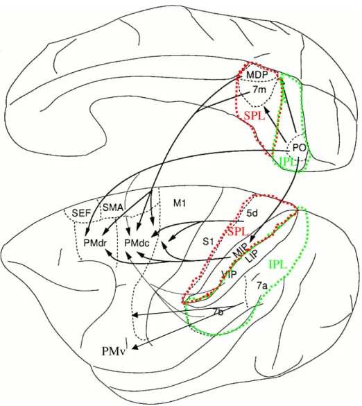
One projection has its major source in SPL areas V6A and the medial intraparietal region, MIP. This projection targets PMd areas F2 and F7 (Gamberini et al., 2009; Matelli et al., 1998). The second projection arises primarily from area VIP and projects to PMv areas F4 and F5 (Rozzi et al., 2006). This pathway mediates eye movements (Nachev et al., 2008), as well as numerous forms of visually guided actions such as reach and grasp (Colby and Duhamel, 1991; Duhamel et al., 1998; Fattori et al., 2001, 2009, 2010; Galletti et al., 1991, 1995, 1997, 2001). The parieto–medial temporal pathway links the IPL with the medial temporal lobe (MTL) including the hippocampus. This pathway is involved in processing of navigationally relevant information, distant-space perception, route learning and spatial long-term memory (Kravitz et al., 2011). The two former pathways traditionally constitute the parieto-frontal network which is highly relevant for both visual guidance of movements and decision making processes.
5. The role of PMd in visual guidance of movements: the parieto−frontal network.
The anatomical and functional organization of the parieto-frontal network underlying arm reaching is well documented (Caminiti et al., 1996; Johnson et al., 1993, 1996) and particular emphasis will be given to the role of the main structures in PPC and PMd. The PPC shares with motor and premotor areas a multiplicity of arm, leg and face representations. In particular, the arm skeletomotor region is represented at least 8 times (Rizzolatti et al., 1998). PPC comprises a diverse number of functions including spatial attention, spatial awareness, polysensory integration, coordinate transformation, movement intention and decision making (Andersen et al., 1987, 2009; Andersen and