Université de Montréal
Development of a multi-gene PCR assay for the prediction
of the response to hormone therapy in breast cancer
Par :
Carolyn Nessim, MD
Programme Sciences Biomédicales Faculté de Médecine
Mémoire présenté à la Faculté des études supérieures en vue de l’obtention du grade de M.Sc. en Sciences biomédicales
13 décembre 2013
Université de Montréal Faculté des études supérieures
Ce mémoire intitulé :
Development of a multi-gene PCR assay for the prediction
of the response to hormone therapy in breast cancer
Présenté par : Carolyn Nessim, MD
A été évalué par un jury composé des personnes suivantes :
Louis Gaboury, MD, PhDIsabelle Trop, MD Sylvie Mader, Ph.D. André Robidoux, MD
Résumé
Deux tiers des cancers du sein expriment des récepteurs hormonaux ostrogéniques (tumeur ER-positive) et la croissance de ces tumeurs est stimulée par l’estrogène. Des traitements adjuvant avec des anti-estrogènes, tel que le Tamoxifen et les Inhibiteurs de l’Aromatase peuvent améliorer la survie des patientes atteinte de cancer du sein. Toutefois la thérapie hormonale n’est pas efficace dans toutes les tumeurs mammaires ER-positives. Les tumeurs peuvent présenter avec une résistance intrinsèque ou acquise au Tamoxifen. Présentement, c’est impossible de prédire quelle patiente va bénéficier ou non du Tamoxifen.
Des études préliminaires du laboratoire de Dr. Mader, ont identifié le niveau d’expression de 20 gènes, qui peuvent prédire la réponse thérapeutique au Tamoxifen (survie sans récidive). Ces marqueurs, identifié en utilisant une analyse bioinformatique de bases de données publiques de profils d’expression des gènes, sont capables de discriminer quelles patientes vont mieux répondre au Tamoxifen.
Le but principal de cette étude est de développer un outil de PCR qui peut évaluer le niveau d’expression de ces 20 gènes prédictif et de tester cette signature de 20 gènes dans une étude rétrospective, en utilisant des tumeurs de cancer du sein en bloc de paraffine, de patients avec une histoire médicale connue. Cet outil aurait donc un impact direct dans la pratique clinique. Des traitements futiles pourraient être éviter et l’indentification de tumeurs ER+ avec peu de chance de répondre à un traitement anti-estrogène amélioré. En conséquence, de la recherche plus appropriée pour les tumeurs résistantes au Tamoxifen, pourront se faire.
Mots-clés : Récepteurs hormonaux, Cancer invasif du sein, Récepteurs ostrogénique, Facteur
Abstract
Two thirds of breast cancers express the estrogen receptor (ER-positive tumours) and estrogens stimulate growth of these tumours. Adjuvant therapy with anti-estrogens such as Tamoxifen and Aromatase Inhibitors has been shown to increase survival in breast cancer patients. This treatment is, however, not successful in all ER-positive tumours. Tumours can present intrinsic or acquired resistance to Tamoxifen. However, it is currently impossible to predict which patient will benefit from Tamoxifen therapy and which will not.
Preliminary studies in Dr. Mader’s lab have identified 20 genes whose expression levels in tumours are able to predict the response to Tamoxifen therapy (disease-free survival). These markers, identified using bioinformatics analysis of published gene expression datasets, were able to discriminate patients that would respond best to Tamoxifen from those that did not.
The overall purpose of this study is to develop a PCR kit to monitor expression levels of these 20 genes and to test this 20-gene signature in a retrospective study using paraffin-embedded breast cancer tissues of patients with a known medical history. This tool may thus have a direct impact on clinical practice through the development of markers of therapeutic success for treatment with Tamoxifen and possibly Aromatase Inhibitors. Futile treatments would be avoided thus preventing needless side effects, and improved identification of ER+ tumours with a low chance of success to anti-estrogen therapy. This will facilitate research into more appropriate treatments for hormone resistant tumours.
Keywords : Hormone receptors, Invasive breast cancer, Estrogen receptor, predictive factor,
Table of Contents
Introductory Pages
Résumé………i
Mots
clés……….i
Abstract..……….ii
Keywords.………...ii
Table
of
Contents………...iii-vi
List of Tables………..vii
List of Figures……….viii
List of Abbreviations………...ix-x
Dedication………....xi
Acknowledgements………..xii
SECTION 1. INTRODUCTION
1.1 Epidemiology………..1
1.2 Risk Factors……….2-9
Gender and age Race and Ethnicity Benign Breast Lesions
Personal History of Breast Cancer Family History and Genetic Factors Reproductive and Hormonal Factors Pregnancy Related Factors
Exogeneous Hormone Factors Ionizing Radiation
Lifestyle and Dietary Factors Alcohol Intake
1.3 Molecular Subtypes of Breast Cancer………9-12
Luminal SubtypeHer-2 enriched Subtype Basal-like Subtype Claudin-low Subtype Normal-like Subtype
1.4 ER/PR positive breast cancer………..12-15
Molecular biology and physiology of the estrogen receptorEstrogen and the mammary gland
1.5 Hormone Therapy for ER/PR positive breast cancer………...15-20
TamoxifenAromatase Inhibitors Fulvestrant
Efficacy of Hormone Treatment
1.6 Resistance to Tamoxifen………..20-25
Intrinsic ResistanceLoss of ER- α expression/function Altered Expression ER-β
Tissue-specific availability of co-activators and co-repressors Modulation of ER expression through secondary messengers Modulation of ER- α expression by BRCA1
Altered Tamoxifen Metabolism Acquired Resistance
Loss of ER-α expression/function
Co-repressors and co-activators expression levels Growth factor pathways
1.7 DNA Microarray versus RT-QPCR……….25-28
DNA MicroarrayRT-QPCR
1.8 Predictive versus Prognostic tools in Breast Cancer………28-30
1.9
Predictive Tools in Breast Cancer……….31
1.10 Development of gene signature predicting the response to Tamoxifen..31-34
1.11 Identified Predictive Genes ………34-43
Good Predictors Poor Predictors
SECTION 2. HYPOTHESIS AND OBJECTIVES
2.1 Hypothesis………44
2.2 Objectives……….44-45
SECTION 3. MATERIALS AND METHODS
3.1 Gene Selection………..46-48
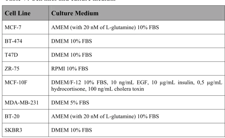
3.2 Cell Line Selection………49
3.3 RNA extraction……….50-53
Cell Lines
Fresh Tissue
Formalin-fixed Paraffin Embedded Tissue
3.4 Quality Assessment of RNA……….53-55
NanoDropAgilent BioAnalyzer
3.5 Reverse Transcription……….55
Cell LinesFresh Tissue and Formalin-fixed Paraffin Embedded Tissue
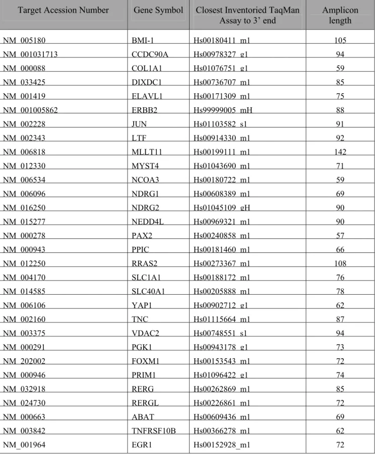
3.6 Q-PCR Probe Selection………56-57
3.7 Q-PCR………..58
3.8 Selection of Housekeeping genes……….58-59
3.9 Creation of a Low Density Array……….59
SECTION 4. RESULTS
4.1 Selection of Housekeeping genes……….60-64
4.2 Selection of Predictive genes………64-68
4.3 Assessment of expression levels in fresh tissue and FFPE samples…….69-80
4.4 Final selection of the gene signature……….81-82
SECTION 5. DISCUSSION AND CONCLUSION
5.1 Reproducibility between Q-PCR assays and microarray results………....83
5.2 Reproducibility of expression profiles between fresh and FFPE samples83-85
5.3 Tumour clustering based on gene signature expression patterns………...85
5.4 Rationale for an LDA platform for this tool……….85-86
5.5 Predictive tools in Breast Cancer………..86-90
Oncotype DxMammaprint Other Genomic tools
5.6 Clinical Utility of our tool………90-91
5.7
Limitations………....92
5.8 Future Perspectives………...92-94
Patient Section and HistoryCreation of Recurrence Score
SECTION 6. REFERENCES………..
95-105
SECTION 7. APPENDICES………
i-xxvi
Appendix A:
Isolation of RNA using Trizol Reagent
Appendix B:
ABI Protocol – RNA Extraction from Fresh tissue
Appendix C:
RNA Isolation from Fresh Tissue using QIAzol
Appendix D:
RNA Isolation from FFPE – ABI Protocol
RecoverAll Total Nucleic Acid Isolation
Appendix E:
RNA Isolation from FFPE – Roche Protocol
High Pure RNA Paraffin Kit
Appendix F:
RT-PCR used for cell lines
Appendix G:
RT-PCR used for Fresh and FFPE tissue
High Capacity cDNA RT Kit (ABI)
Appendix H:
Q-PCR ABI Protocol
Appendix I:
Analysis of Results from Q-PCR
Appendix J:
Q-PCR on LDA Protocol
Appendix K:
Consent Form and Research Protocol
Appendix L:
Approval of Scientific Committee
List of Tables
Table I: Germ Line Mutations in Breast Cancer
Table II: Genes that predict a good response to Tamoxifen
Table III: Genes that predict a poor response to Tamoxifen
Table IV: Identified Predictive Genes
Table V: Cell lines and culture medium
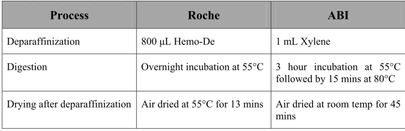
Table VI: Difference between Roche and ABI Protocol for RNA extraction from
FFPE tissues
Table VII: TaqMan Assays used for the Q-PCR experiments
Table VIII: Quantity and Quality of RNA extraction from Cell Lines (N1, N2)
Table IX: Housekeeping genes
Table X: Quantity and Quality of RNA extraction from Fresh Tissue (ABI vs.
CHUM QIAzol Protocol)
Table XI: Quantity and Quality of RNA extraction from FFPE (ABI Protocol
before and after RNA clean-up)
Table XII: Quantity and Quality of RNA extraction from FFPE (Adapted ABI
Protocol)
Table XIII: Quantity and Quality of RNA extraction from FFPE (ABI vs. Roche
Protocol)
Table XIV: Quantity and Quality of RNA extraction from FFPE and Fresh
Tissue of the same tumour
List of Figures
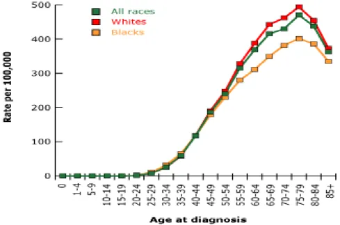
Figure 1: Incidence rate of breast cancer by age and race for 2000-2003.
Figure 2: The different modes of action of ER (Genomic and Non-genomic)
Figure 3: Prognostic versus Predictive tools
Figure 4: Heat map of the 30-gene signature applied to several published data
sets demonstrating its ability to predict the response to Tamoxifen
Figure 5: GeNorm Results (N1)
Figure 6: GeNorm Results (N2)
Figure 7: Heatmap results of the 30 Predictive genes in the 8 cell lines (N1)
Figure 8: Heatmap results of the 30 Predictive genes in the 8 cell lines (N2)
Figure 9: Correlation between ABI QPCR and Affymetrix Micro-Array of the
30 predictive genes on the 8 cell lines (N1 + N2)
Figure 10: Heatmap results of 24 Predictive genes in 8 different tumour samples
where RNA was extracted from FFPE tissues
Figure 11: The distribution of the average CT between fresh and paired FFPE
tumours. (A) Biological replicate 1 and (B) Biological replicate 2.
Figure 12: Heatmap correlation of the 25 predictive genes between Fresh and
paired FFPE tumour samples.
Figure 13: Pearson correlation between paired Fresh and FFPE tissues (N1 and
List of Abbreviations
ABI – Applied Biosystems Inc.
AF – Activating Functions
AI – Aromatase Inhibitor
BMI – Body Mass Index
BRCA1 – Breast Cancer Susceptibility gene 1
BRCA2 - Breast Cancer Susceptibility gene 2
cDNA – Complimentary DNA
CHUM – Centre Hospitalier de l’Université de Montréal
CSR – Core Serum Response
CYP2D6 - Cytochrome P450 2D6
DNA - Deoxyribonucleic acid
EBCTCG – Early Breast Cancer Trialists Collaborative Group
EGFR – Epidermal Growth Factor Receptor
EMT – Epithelial-Mesenchymal Transition
ER – Estrogen Receptor
ER+ - Estrogen Receptor Positive
ER- - Estrogen Receptor Negative
ER-α – Estrogen Receptor Alpha
ER-β – Estrogen Receptor Beta
ERBB2 - Erythroblastic leukemia viral oncogene homolog 2
ERE – Estrogen response elements
ESR1 – Estrogen Receptor 1
FFPE – Formalin-fixed paraffin embedded
HR – Hazard Ratio
IRIC – Institut de Recherche d’Immunologie et de Cancérologie
LDA – Low density array
mRNA – Messenger RNA
NSABP – National Surgical Adjuvant Breast and Bowel Project
PR – Progesterone Receptor
PR+ - Progesterone Receptor Positive
PR- - Progesterone Receptor Negative
Q-PCR – Quantitative Polymerase chain reaction
RIN – RNA Integrity Number
RNA - Ribonucleic acid
RR – Relative Risk
RS – Recurrence Score
RT – Reverse Transcription
RT-PCR – Reverse Transcription Polymerase chain reaction
SERM – Selective Estrogen Receptor Modulator
SERD – Selective Estrogen Receptor Downregulator
TNBC – Triple Negative Breast Cancer
Acknowledgments
I would like to thank my supervisor Sylvie Mader for her guidance and teachings. I would also like to thank my supervisor André Robidoux for his support and helping me initiate this project. I would like to acknowledge Slim Fourati for his significant contribution and help with this project, without whom this project would have not been possible. I would also like to thank the following members of the lab of Sylvie Mader for their support and teachings: Martine Bail, Edlie St-Hilaire, Maxime Parisotto, David Cotnoir-White, Laurence Fleury, Virginie Dupont and Marieke Rozendaal.
1. Introduction
1.1 Epidemiology
In Canada, one out of nine women will be diagnosed with breast cancer in their lifetime, by age 901. Breast Cancer is the most common cancer in women and the second most common cause of cancer death in women. It is the main cause of death in women aged 40-59. Fifty percent of the cases can be explained by risk factors and 10% are found to have a positive family history. Only 5% of all breast cancers have known genetic mutations and syndromes such as BRCA1 and BRCA2. The majority of breast cancers are thus considered sporadic cancers2.
In 2010, an estimated 23 200 women and 180 men in Canada were diagnosed with breast cancer. Approximately 445 Canadian women are diagnosed with breast cancer every week. In 2010, an estimated 5300 women and 50 men died of breast cancer in Canada. This means that, on average 100 women die of breast cancer in Canada, every week1.
Globally, breast cancer incidence rates are highest in North America and Northern Europe and lowest in Asia and Africa. The incidence in China and Japan has been rising in recent years3.
Since 1999, the incidence of breast cancer has remained quite stable, however, since 1986 the death rate from breast cancer has declined by more than 30%. This improvement in survival rate is most likely due to improvements in treatment strategies as well as better screening for breast cancer1. The decrease in mortality has been especially noted in women younger than 503 and women with ER/PR positive tumours4. At present, the five-year relative
1.2 Risk Factors
There are many risk factors that have been found to be associated with breast cancer. Some risk factors are stronger than others.
Gender and Age
Gender and age are among the strongest risk factors for breast cancer. Women are afflicted with breast cancer 100 times more frequently than men. In general, the older the person, the higher the risk. The incidence rises sharply with age until about the age of 45-50 and then the rise becomes less steep. At age 75-80, the incidence curve flattens and slightly decreases, as most women at this stage are menopausal and thus have less estrogen stimulation (Figure 1, p. 2)5.
Age-specific SEER incidence, rates of female breast cancer per 100,000, 2000-2003
Race and Ethnicity
Breast cancer is more common in whites and less common in Hispanic and African American women. However, African American women tend to have more aggressive, hormone receptor negative cancers at a younger age and thus a lower survival rate3.
Benign breast lesions
Proliferative benign diseases with cytological atypia increase the risk for breast cancer. Atypical lobular hyperplasia or atypical ductal hyperplasia, have a 4-6 fold relative risk (RR) of developing breast cancer and this becomes a 10-fold risk when the atypia is multifocal6. Personal History of Breast Cancer
A personal history of invasive breast cancer or a ductal carcinoma in situ also increases a person’s risk of having a cancer in the contralateral breast. With in situ lesions, the 10-year risk of developing an invasive cancer in the contralateral breast is 5%. In patients that have already had an invasive cancer the risk of developing a contralateral breast cancer is 1% in premenopausal women and 0.5% in postmenopausal women7.
Family history and genetic risk factors
A positive family history is an important risk factor, however, it is only reported by 15-20% of women diagnosed with breast cancer. In a pooled analysis done in 2001 by the Collaborative Group on Hormonal Factors in Breast Cancer, data was used from over 50 000 women with breast cancer and 100 000 controls. The results showed that the risk of breast cancer for a woman with one affected first-degree relative was increased 1.80 fold. With two affected first-degree relatives, the risk is increased 2.93 fold. The risk ratios were highest for women with young affected relatives. Thus, the risk was increased 2.9 fold for a woman
whose relative was diagnosed before age 30, but only 1.5 fold increased if the affected relative was diagnosed after age 60. Similarly, if one relative had breast cancer before age 40, the risk of breast cancer was increased 5.7-fold8.
Specific genetic mutations that predispose to breast cancer are very rare; only 5 to 6% of all breast cancers are directly attributable to inheritance of a breast cancer susceptibility gene (Table I, p. 5). Genetic mutations in these genes are often associated with various cancer syndromes, where patients may be afflicted with more than one type of cancer and various diseases. These germ-line mutations, which are often associated with triple-negative breast cancer (TNBC), are beyond the scope of this thesis, which focuses on the prognostic role of somatic mutations.
Table I: Germ Line Mutations in Breast Cancer
Mutation
Associated Cancers/Diseases
BRCA1
Breast Cancer
Ovarian Cancer (higher risk than BRCA2) Cervical Cancer
Uterine Cancer Pancreatic Cancer Colon Cancer
Male Breast Cancer (lower risk than BRCA2) Testicular Cancer Prostate Cancer
BRCA2
Breast Cancer Ovarian Cancer Pancreatic Cancer Gastric Cancer Gall bladder Cancer Bile duct Cancer Melanoma Male Breast Cancer (more common BRCA2) Prostate Cancer (more common BRCA2)
ATM
Breast Cancer Ataxia-telengectasia diseasep53
(Li Fraumeni Syndrome)
Breast Cancer
Soft tissue and Bone Sarcoma Leukemia
Brain tumours
CHEK2
(Li Fraumeni Sydrome)
Same as p53
PTEN
(Cowden Syndrome)
Breast Cancer
Benign breast diseases Digestive tract tumours Thyroid tumours Uterine tumours Ovarian tumours
CDH1
Breast Cancer (Invasive lobular carcinoma) Gastric CancerSTK11/LKB1
(Peutz-Jeghers Syndrome)
Harmartamous polyps in GI tract
Pigmented macules on lips, buccal mucosa, digits Digestive Cancers Cervical Cancer Lung Cancer Testicular Cancer Ovarian Cancer Uterine Cancer
Reproductive and Hormonal Risk Factors
Prolonged exposure to endogenous estrogen has also been shown as an important risk factor for breast cancer. For every two-year delay in the onset of menarche, there is a 10% reduction in cancer risk8. Moreover, the risk increases as menopause is delayed. The RR increases by 1.03% for each year older at menopause9. This was further emphasized when it was noted that women that had had a bilateral oophorectomy before the age of 40 had a 50% lifetime risk reduction of breast cancer10.
Data from the Nurses' Health Study suggest that the association is strongest for hormone receptor-positive breast cancers. Endogenous hormone levels were measured in 322 women who developed breast cancer and in 643 age-matched controls without breast cancer. When the highest and lowest quartiles of serum hormone concentration were compared, there was a significant direct association between breast cancer risk and levels of both estrogens and androgens. However, the association was strongest when the analysis was restricted to ER and PR-positive tumours, and in situ tumours11.
Breast density also seems to have an association with breast cancer risk. The denser breasts (greater than 75% density) compared to women of the same age with less or no dense tissues have five times greater risk of developing breast cancer12. Both endogenous and exogenous estrogen may influence mammographic density. Mammographic density decreases after menopause when ovarian function declines. Hormonal replacement therapy, with combination of estrogen and progesterone, increases mammographic density13, while tamoxifen, which has antiestrogenic effect, decreases mammographic density 14 . Mammographic density therefore can be regarded as a marker of the effect of estrogen on the
breast tissue. To what extent mammographic density is a predictor for both hormone receptor-positive and hormone receptor-negative tumors is still unclear.
Pregnancy related factors
Nulliparous women are at increased risk for breast cancer (RR 1.2-1.7). Moreover, the younger the woman at her first time full-term pregnancy the lower the risk. The risk is 20% lower if the first birth is at age 20, 10% lower for a first birth at age 25 and 5% higher if the first birth is at age 359.
Exogenous hormone factors
It is controversial whether or not long-term use of oral contraceptives increases the risk of breast cancer and data are conflicting. Long-term hormone therapy replacement with estrogen and progesterone has however been shown to increase the risk of breast cancer, especially hormone positive cancers. It must be noted, however, that women taking an unopposed estrogen therapy have a slightly lower risk15, 16.
Ionizing radiation
Exposure to ionizing radiation has been shown to greatly increase the risk of breast cancer. Patients who have received radiotherapy treatment for Hodgkin’s lymphoma to the chest wall, especially between the ages of 10-16 and up to the age of 45, are at increased risk of getting breast cancer17.
Lifestyle and Dietary Factors
Women of higher socioeconomic status are at a two-fold greater risk for breast cancer. This is thought to be due to differing educational, occupational and economic level reflecting
different reproductive patterns with respect to parity, age at first birth, age at menarche and utilization of screening mammography.
In postmenopausal women, it has been shown that a higher body mass index (BMI) is associated with a higher risk of breast cancer. Obese postmenopausal women have higher estrogen levels than non-obese postmenopausal women, due to the conversion of adrenal androgens to estrogens in fatty tissue. In a pooled analysis of seven prospective studies in the US, women who weighed at least 80 kg (176 lbs., BMI >33 kg/m2) had a 25% higher risk of breast cancer as compared to those weighing less than 60 kg (132 lbs., BMI<21 kg/m2), after
adjusting for height18. In the same-pooled analysis, the opposite association was found in premenopausal women. Those with a BMI≥31 kg/m2 were 46% less likely to have breast cancer compared to those women who’s BMI was <21 kg/m2 18.
Alcohol Intake
Many dietary risk factors have been evaluated but are quite difficult to interpret with regards to a direct causal relationship to breast cancer. Increased alcohol intake is the only dietary risk factor that has been consistently shown in several epidemiological studies, to increase the risk of breast cancer. More specifically, it increases the risk of hormone positive breast cancers and the use of hormone replacement therapy acts as an additive risk factor to increased alcohol intake. It is believed that it may be in part due to the increased estrogen and androgen levels in women who consume alcohol as well as increased mammary gland susceptibility to carcinogenesis and DNA damage in women who consume alcohol19. Moderate to increased use (≥ three drinks per day) as compared to those who abstain from drinking, has been shown to have a 12% increased risk of breast cancer20.
In conclusion, most of these risk factors for breast cancer are associated with increased exposure to estrogen, whether it be endogeneous or exogeneous. The higher or the longer breast tissue is exposed to estrogen, the higher the risk of breast cancer.
1.3 Molecular Subtypes of Breast Cancer
Breast cancer is a heterogeneous and phenotypically diverse disease. Classically, pathologic features of the cancer have been used to determine prognosis. These features include the histologic grade of the tumour, the presence of lymphovascular invasion, the presence of nodal disease as well the expression of various receptors, namely; ER, PR and Her-2-neu. These features have helped sub-categorize breast cancers into different groups with different tretment options.
However, more recently, due to the progress in molecular profiling and using gene expression arrays, Perou et al. further classified these 3 subtypes of breast cancer (ER/PR, Her-2 and TNBC) at a genetic level, and characterized other biologic subtypes21,22,23. As these different subtypes have distinct responses to therapy, this molecular portrait of breast cancer has further helped in determining prognosis, and may eventually aid in treatment strategies.
Five different intrinsic subtypes of breast cancer have been identified, each having their own distinct genetic profile.
Luminal Subtype
The most common subtype is the Luminal subtype which is further subdivided into luminal A and luminal B; making up two distinct intrinsic subtypes. They make up the majority of ER+ breast cancers. These tumours typically express luminal cytokeratin 8 and 18
and are characterized by their expression of ER, PR and other genes associated with ER activation. The subdivision is not only at a molecular level but also corresponds to different clinical outcomes prognostically.
Luminal A tumours, making up approximately 40% of all breast cancers, usually have a high expression of ER-related genes, low expression of HER2 genes and low expression of proliferation-related genes. As expected, they correspond to the best prognosis.
Luminal B tumours, making up approximately 20% of all breast cancers, have a lower to moderate expression of ER-related genes, variable expression of HER2 and a higher expression of proliferation genes. They relapse more frequently on antiestorgen/aromatase inhibitor therapy and thus have a worse prognosis than luminal A cancers.
HER-2 enriched Subtype
The second most common subtype is the ERBB2-positive or HER2-enriched subtype. These make up approximately 10-15% of all breast cancers and are characterized by high expression of the HER2 and proliferation gene clusters and a low expression of the luminal cluster. They are typically ER-PR-negative. Although this subtype has a poorer prognosis, with the advent of targeted therapy against HER2, Herceptin, the outcome of patients has greatly improved24.
Basal-like Subtype
The third subtype has been named the Basal-like subtype because of its similarity in expression to that of basal epithelial cells. They make up approximately 15-20% of all breast cancers. They have low expression of luminal and HER2 cluster genes. These tumours are usually ER/PR-negative and HER2-negative. Naming these breast cancers however,
“triple-negative” is a misnomer. Although most “triple-“triple-negative” tumours are basal-like and most basal-like tumours are triple-negative, there is a significant 30% discordance between the two types. Basal-like tumours have a high expression of the proliferation cluster of genes and are almost always histologically high-grade tumours. They demonstrate widespread genomic instability and have a high expression of the epidermal growth factor, and basal epithelial cytokeratin 5, 14 and 17. Eighty percent of BRCA1 mutation carriers have basal-like tumours25. These tumours have a poor prognosis, as they do not benefit from established targeted therapies, being mostly receptor negative. However, they do respond to chemotherapy, with a complete pathologic response rate of up to 45%, which is promising. Claudin-low Subtype
The fourth subtype is the non-basal TNBC. This subtype is more uncommon however clinically quite significant. They have an extremely low to absent expression of the luminal cluster genes and high expression of the epithelial-mesenchymal transition (EMT) genes, immune response genes and characteristics reminiscent of stem cells. Many studies have been performed regarding the EMT process and suggest its implication in tumour progression and spread of metastasis26, which may account for the poor prognosis of TNBCs. These tumours tend to respond to chemotherapy at an intermediate level between the basal-like and luminal tumours.
Normal-like Subtype
This final subtype is the hardest to characterize clinically, however is always present in gene expression arrays. It is difficult to know whether or not this is a true subtype or a technical artefact, due to low tumour cell composition of those specifically sampled
specimens. It has a similar gene expression pattern as normal breast tissue.
Understanding these different intrinsic subtypes has greatly aided in the understanding of the biology of these tumours, which can lead to better evaluation of treatment strategies for breast cancer.
1.4 ER/PR-positive Breast Cancer
Hormone receptor positive breast cancers are the most common type, making up approximately two thirds of all breast cancers. Hormone receptor-positive breast cancer was characterized over 40 years ago by Elwood Jensen. He noted that radiolabeled estrogens concentrated preferentially in some human breast tumors as well as in estrogen target organs. These findings then led to the discovery of the estrogen receptor (ER), as well as the progesterone receptor (PR), which were found in high abundance in a large fraction of malignant breast tumours. It has since become clear that human breast cancers are dependent upon estrogen and/or progesterone for growth and that this effect is mediated through ERs and PRs. As mentioned previously, these tumours are now classified as the luminal subtype of breast cancers. It is these luminal tumours that will be the focus of this thesis.
Molecular biology and physiology of the estrogen receptor
Estrogens have multiple actions on various sites including the cardiovascular, skeletal, immune, gastrointestinal and neural systems; however, their most important action is on the reproductive organs27. They reprogram gene expression via the activation of nuclear estrogen receptors. These receptors bind to estrogen with high affinity and specificity and function as ligand-modulated nuclear transcription factors28,29.
Two ER molecules have been identified, namely ER-alpha α) and ER-beta (ER-β)30. The key functional domains in these receptors are the C or DNA-binding domain, which binds with high affinity and specificity to specific DNA sequences (estrogen response elements (ERE)) in the promoter regions of target genes, and the E or ligand-binding domain, which bind estrogens and estrogen analogues31. The ERE in the target genes is a 15-base pair inverted-repeat DNA sequence (RGGTCAnnnTGACCY), to which ER dimers bind with high affinity and specificity32, with one receptor molecule in contact with each five base-pair segment of the response element33. The estrogen receptors contain two regions, termed activation functions (AF) that mediate the increase in transcriptional activity induced by the receptors in the presence of ligand. AF-1, located near the amino-terminal end of the receptor, acts independent of ligand, whereas AF-2, located in the ligand-binding domain, is ligand dependent34. There are also numerous co-regulator molecules, including RNA cofactors that interact with the receptors in a ligand-dependent manner, modulating receptor-mediated transcription by interacting with both AF regions and transcription factors associated with RNA polymerase II35.
When an estrogen or its analogue reaches the cell nucleus and binds to ER, the conformation of the ligand-binding domain of the receptor changes, either allowing or preventing interaction with the co-activators, depending on whether the ligand is an agonist or an antagonist, respectively. The estrogen receptor dimers bind to the ERE in target genes, and via agonist-dependent association with co-activators, increases the rate of transcription by interacting with and activating necessary components of the transcriptional apparatus. Moreover, the ability of steroid hormone receptors to activate transcription of endogenous genes likely depends upon their ability to affect chromatin structure. Many steroid hormone
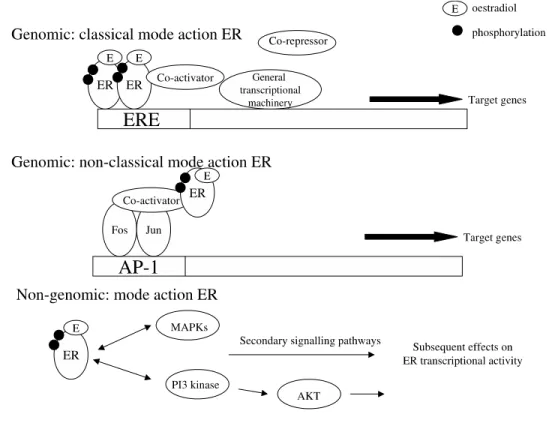
receptors interact with coregulator proteins that are implicated in the remodeling of local chromatin structure and the acetylation of histones36,37. In fact, enhancement of transcription by adding ligand to ER was observed using chromatinized template DNA but not when using naked DNA lacking histones38. This is referred to as the genomic classical mode of action (Figure 2, p. 15).
In the genomic non-classical mode of action, ER regulates gene expression without interacting with DNA directly. It acts via other transcription factors such as Fos/Jun activating protein-1 (AP-1) complex (Figure 2, p.15)39.
ERs can also function independently of estrogen. Both epidermal growth factor and insulin-like growth factor-1, acting via their extracellular membrane bound receptors, can stimulate transcription of ER target genes in the absence of estrogen40,41. Therefore, cross-talk and signal amplification occurs between growth factor signaling pathways and nuclear receptors42.
Estrogens also have non-genomic actions. They bind with high affinity to other cell components, including plasma membranes. Some effects of estrogen, such as rapid induction of MAP kinase and Erk pathways, appear to involve direct action of estrogen receptors at the plasma membrane rather than genomic modulation (Figure 2, p.15). As these rapid effects occur without ER-gene interaction, they are called "non-genomic," although the signals initiated by these mechanisms ultimately result in regulation of genes. These responses are observed in diverse tissues, including the cardiovascular system, central nervous system, and in breast cancer cells.
Figure 2: The different modes of action of ER (Genomic and Non-genomic)
Estrogens and the mammary gland
As female mice mature, the rudimentary ductal tree of the mammary gland elongates in response to estrogens and branches in response to progesterone to fill the stroma. In ER-α deficient mice, the ducts fail to elongate43. If ER-β is deficient the gland develops normally and the mice can nurse their young with a normal lactation function. If both ER-α and ER-β are deficient, the phenotype is similar to those mice with ER-α deficiency only, emphasizing the importance of ER-α in male and female reproduction44.
1.5 Hormone Therapy for ER/PR positive breast cancer
Currently there are three main anti-estrogen therapies that can be used in the treatment of ER+ breast cancer: Tamoxifen, Aromatase Inhibitors (AI) and Fulvestrant. Clinically,
Tamoxifen and AIs are the treatments most often used, while fulvestrant is mainly used as second line therapy in patients with metastatic disease that have not responded or are progressing on standard therapy with Tamoxifen or AIs.
Tamoxifen
Tamoxifen is a competitive inhibitor of estrogen binding to ERs, and has a mixed agonist and antagonist activity, depending on the target tissue. It is therefore called a selective estrogen receptor modulator (SERM).
Its physiological effects in postmenopausal women can illustrate this mixed antagonist/agonist effect. Tamoxifen is known to provide some protection against menopausal bone loss45 and lowers serum total and low-density lipoprotein-cholesterol concentrations46,47 via its agonistic properties. This has been shown to lead to less osteoporotic fractures. Whether it is protective against cardiovascular disease is still controversial. Other agonistic features include the induction of endometrial hyperplasia and the increased risk of endometrial cancer. Moreover, Tamoxifen increases the risk of thromboembolic events such as deep vein thrombosis and pulmonary embolism as well as increases the risk of stroke and cataracts47.
The most salient feature, however, of Tamoxifen is its antagonistic properties with respect to breast cancer. Among women with ER+ breast cancer, Tamoxifen reduces the risk of recurrence and death and prevents the development of contralateral breast cancer when given as adjuvant therapy for early stage disease and can provide palliation in patients with metastatic disease47,48,49. Other side effects due to is antagonistic properties include hot flashes and vaginal dryness.
The molecular mechanisms by which SERMs can act as ER agonists in one tissue and as antagonists are just starting to be better understood. The change in receptor conformation that follows the binding of Tamoxifen to the ER modulates interactions with co-repressors and co-activators that are required for ER-mediated gene regulation. Co-activators increase transcriptional activity by promoting the interaction between the receptor and the transcriptional apparatus and chromatin remodeling50, whereas co-repressors restrain ER
activity, maintaining the receptor in protein/DNA complexes that do not promote transcription and favor an inactive conformation of chromatin51. Thus, the main mechanism of action of Tamoxifen is the induction of an abnormal receptor conformation and altered recruitment of cofactors. The complement of co-activators/co-repressors expressed in different tissues may therefore dictate whether the receptor is active or inactive.
Aromatase Inhibitors
The aromatase inhibitors decrease circulating levels of estrogen in postmenopausal women by blocking the action of the enzyme, aromatase, which converts androgens to estrogens in peripheral tissues52. There are three different AIs used in the clinic, namely, Anastrazole, Letrozole and Exemastane. These agents are used in the treatment of postmenopausal patients with breast cancer in the adjuvant and metastatic setting. AIs are contraindicated in premenopausal women, since they may increase estrogen levels via a reduced feedback of estrogen to the hypothalamus and pituitary. This would lead to an increase in gonadotropin secretion and stimulation of the ovary, and ultimately to an increased concentration of the precursors of estrogens, androgen and increased expression of aromatase53. Contrary to Tamoxifen, AI’s are not associated with an increased risk of thromboembolic events or endometrial cancer, however they do have a similar profile with
regards to hot flashes and vaginal dryness and have additional musculoskeletal side effects including osteoporosis and arthralgia.
Fulvestrant
Fulvestrant is a selective estrogen receptor down-regulator (SERD). It has a steroidal structure that binds competitively to the estrogen receptor (ER), with high affinity, and downregulates ER by functional blockade and increased turnover. After binding to fulvestrant, degradation of the ER is accelerated, ultimately resulting in a reduction in cellular ER. The downregulation of cellular levels of the ER protein results in complete abrogation of estrogen-sensitive gene transcription. As a pure estrogen antagonist, fulvestrant avoids the risk of detrimental side effects of selective ER modulators such as tamoxifen, which has partial agonist activity. Due to its unique mode of action, fulvestrant lacks cross-resistance with existing agents. Fulvestrant, an antiestrogen classified as an estrogen receptor antagonist without known agonist effects is mainly used for the treatment of postmenopausal, hormone receptor-positive women with progressive metastatic breast cancer after antiestrogen therapy. The main adverse effects associated with therapy are nausea, asthenia, pain, vasodilation and headache54,55,56.
Efficacy of Hormone Therapy
Multiple randomized clinical trials have been performed to evaluate the efficacy of hormone therapy in breast cancer. One of the first trials to demonstrate a benefit for Tamoxifen therapy was the NSABP B-14 trial, which compared 5 years of adjuvant Tamoxifen to placebo. Since that trial multiple others were performed. In 2011, the Early Breast Cancer Trialists Collaborative Group (EBCTCG) performed a meta-analysis of
randomized trials, which compared tamoxifen versus no endocrine treatment in premenopausal and postmenopausal women49. With a median follow-up of 13 years, tamoxifen resulted in a reduction in breast cancer recurrence by 39% compared to placebo, which translated into a 15-year absolute reduction of 13% (33% versus 46%). This was seen in patients with both node-negative and node-positive ER+ breast cancer. There was no effect on recurrence for patients with ER-negative breast cancer. It also showed a reduction in risk of breast cancer mortality by 30%, which translated into a 15-year absolute reduction of 9% (24% versus 33%). The magnitude of benefit was similar in women less than 45 years of age and in women between the ages of 55 and 69 years.
With regards to AI’s, multiple trials have also been performed. In 2010, the EBCTCG performed a meta-analysis of these trials demonstrating the benefit of AI’s compared to Tamoxifen57. This meta-analysis showed that, with a mean follow-up of 6 years, treatment with an AI as a single agent therapy had a reduction in the risk of recurrence compared to Tamoxifen (3% absolute reduction in 5-year risk of recurrence, 12% versus 15%). There was no difference between an AI and Tamoxifen with regards to overall survival. A secondary analysis, with a mean follow-up of 4 years, evaluated the use of Tamoxifen for 2-3 years and then switching to an AI for the last 2-3years to complete 5 years versus staying on Tamoxifen for the entire 5 years of treatment. This showed that switching to an AI reduced the risk of recurrence by 3% and reduced 5-year breast cancer mortality by 2%. Finally, with regards to sequencing therapy, in the National Cancer Institute of Canada Clinical Trials Group MA17 study, 5 years of AI versus placebo was given to patients who had completed 5 years of Tamoxifen. With a median follow-up of 64 months, treatment with letrozole improved disease free and overall survival compared to placebo58.
As already mentioned, Fulvestrant has been shown to be an effective and well tolerated treatment for patients with metastatic breast cancer when compared to Tamoxifen and AI’s and is especially useful in patients with Tamoxifen resistance55. It, however, has not been shown to be superior to Tamoxifen or AIs and is much more costly, explaining its limited use clinically. Futher studies are necessary to evaluate its efficacy as adjuvant therapy.
Thus, in summary patients with ER/PR positive breast cancer greatly benefit from hormone therapy after surgery and in the metastatic setting. Unfortunately, however, despite the benefits of these therapies, 40% of patients still recur and eventually succumb to their disease. To date, we have an inability to identify which patients will respond and which will not. Multiple studies have been performed to understand the resistance mechanisms involved with regards to hormone therapy, most of these studying the resistance to Tamoxifen.
1.6 Resistance to Tamoxifen
Research over the last two decades has identified two forms of resistance to Tamoxifen therapy: Intrinsic (de novo) resistance, in which ER-negative and many ER+ tumours do not respond to Tamoxifen at the outset of therapy, and acquired resistance, where ER+ tumours that initially responded to therapy stop responding and may actually exploit the Tamoxifen-ER complex as a stimulator as opposed to an inhibitory signal59. As this is quite a complex and
exhaustive topic this will simply be summarized here. Intrinsic Resistance
Understandably, it has been noted that ER/PR-negative breast cancers do not respond to Tamoxifen therapy. However, it has also been noted that approximately 25% of ER+/PR+ tumours, 66% of ER+/PR- and 50% of ER-/PR+ tumours fail to respond to or develop early
resistance to Tamoxifen60. A number of factors have been identified that may contribute to the
intrinsic resistance of Tamoxifen. Loss of ER-α expression/function
Lack or loss of ER expression could confer resistance. This is the dominant mechanism of intrinsic resistance to Tamoxifen, with the majority of ER-/PR- breast cancers not responding to Tamoxifen or AIs. Although quite rare (<1%), mutations in coding of the ER gene alter the effects of bound anti-estrogens, leading to a hypersensitive receptor, with enhanced binding of co-activators in the presence of low estrogen levels. These somatic mutations alters the crosstalk between ER-α and various ER-α pathways that normally down-regulate ER signalling. Such loss of regulation could theoretically enhance ER-mediated cell growth and contribute to the development of resistance. Also, epigenetic changes have been identified that cause transcriptional inactivation of the ER gene39.
Altered expression of ER-β
Although the role of ER-β in Tamoxifen resistance remains unclear, it has been shown that relative changes in the expression of the ER isoforms that occur during tumorigenesis parallel the marked changes in estrogen action. Interestingly, in an RT-PCR study, the median ER-β mRNA levels were approximately 2-fold higher than ER-α levels in tamoxifen-resistant tumours compared with tamoxifen-sensitive tumours39.
Tissue-specific availability of co-activators and co-repressors
As already mentioned, when Tamoxifen is bound to an ER it changes the conformation of the ligand-binding domain, generating an abnormal receptor conformation, recruiting co-repressors, and thus leaving ER in an inactive state. In some cells, tamoxifen-induced AF2
inhibition may be bypassed when enough co-activator function is recruited to the ligand-independent domain, AF161. In other cell types, available co-activator proteins might bind to and activate AF2 despite the presence of tamoxifen62.
Modulation of ER expression through second messengers
As discussed before, ER can be activated independent of estrogen via growth factor signalling. Both ER expression and function correlate with distinct patterns of growth factor receptor overexpression. It appears likely that an appropriate growth factor environment is necessary for efficient mitogenesis in breast cancer cells, with steroid hormone and growth factor signalling pathways "cross talking" to reinforce each others' signalling. One proposed model for both primary and secondary hormone resistance in breast cancer is that phenotypic changes in growth factor signalling pathways may perturb this balance of steroid hormone and growth factor interaction, providing a selective advantage for tumour cell proliferation63, potentially explaining the resistance to endocrine therapy in breast cancer.
As an example, ER- and ER+ but PR- tumours overexpress proteins of the epidermal growth factor receptor (EGFR) family, particularly EGFR and the HER2 protein63. Studies have shown that ER expression is suppressed when HER2 or EGFR receptor is activated, leading to resistance to Tamoxifen64. Others suggest that the antagonist activity of tamoxifen on the ER may be diminished via an interaction between HER2 and AIB1, an ER co-activator65.
As another example, when ER is activated by tyrosine kinase receptors in response to growth factor stimulation, PI3K (phosphatidyl-inositol-3-OH kinase) catalyses the formation of PIP3. One of the downstream targets of this pathway is AKT, whose activation promotes
cellular proliferation and anti-apoptotic responses. There is evidence that ERα can bind in a ligand-dependent manner with a regulatory subunit of PI3K, leading to the activation of AKT and subsequent downstream effects. However the relationship with ER is reciprocal, in that PI3K activates AKT, which phosphorylates the ER at serine-167 resulting in ligand-independent activation. Interestingly, in vitro, elevated levels of AKT confer Tamoxifen resistance39.
Finally, in the presence of Tamoxifen, ER may interact with the stress-activated protein kinase/c-junNH2 terminal kinase pathway (SAPK/JNK) by binding with the AP-1 transcription complex. Tamoxifen-resistant tumours, compared with estrogen-treated tumours, have increased AP-1 dependent transcription and phosphorylated c-Jun and JNK levels. In addition, the conversion to a resistant phenotype has been associated with an increase in oxidative stress (as measured by increases in superoxide dismutases and glutathione-S-transferase). It has been shown that tamoxifen resistant tumours have high AP-1 DNA binding. This is due to the fact that tamoxifen can induce intracellular oxidative stress, which leads to activation of JNK and SAPK, which in turn increase the transcriptional activity of AP-1. This chain of events may explain the potentiation of the agonistic effects of tamoxifen at AP-1 sites in resistant tumours39.
Modulation of ER-α expression by BRCA1
Tamoxifen resistance in patients with the BRCA1 mutation may be due to the fact that most of these patients are ER-negative. BRCA1-mutant tumors fail to express ERα due to the loss of BRCA1-mediated transcriptional activation of ESR1. Loss of the wild-type BRCA1 allele, which occurs during neoplastic development in BRCA1 mutation carriers, has a direct effect on ESR1 transactivation, resulting in the loss of ERα mRNA and protein expression66.
Altered Tamoxifen metabolism
Tamoxifen is converted to its active metabolites, endoxifen and 4-hydroxytamoxifen, by two rate-limiting enzymes, cytochrome P450 2D6 (CYP2D6) and UDP-glucuronyltransferase-2B7 (UGT2B7)67,68. Although it was initially thought that CYP2D6
polymorphisms may confer a relative resistance to Tamoxifen, multiple sub-analyses of several clinical trials (IBIS-1, NCCTG, BIG 1-98, ATAC)69,70,71,72 have not shown a difference in outcomes with regards to survival or recurrence for patients that were poor versus good metabolizers of the drug.
Acquired Resistance
Loss of ER-α expression/function
Approximately 20-30% of patients, that initially have ER+ tumours, treated with tamoxifen, acquire a resistance via loss of ER-α in the recurrent tumours. It is however important to note that even in those patients that relapse under Tamoxifen treatment, 20% of them will still respond to an AI or to the full antiestrogen Fulvestrant, suggesting that ER continues to regulate tumour growth even in tamoxifen-resistant patients39.
Co-repressor and co-activator expression levels
Co-repressor and co-activator expression levels may influence the development of secondary resistance to tamoxifen. In animal models, prolonged tamoxifen exposure alters the balance between co-activators and co-repressors in favour of the agonist, growth-promoting properties of tamoxifen; the net effect is stimulation of growth despite the continued presence of tamoxifen73. This is accompanied by suppression of co-repressor N-CoR levels in the
Growth factor pathways
As already mentioned in de novo tamoxifen resistance, growth factor pathway “cross talk” also plays a role in acquired tamoxifen resistance. Signalling through EGFR and the HER2 receptor appears to bypass the estrogen requirement for breast cancer cell growth and may drive initially ER+ cells into an endocrine therapy-resistant state65,75. It is postulated that activation of growth factor pathways such as these modulates ER activity via phosphorylation, which alters its function, especially its ability to interact with tamoxifen76,77. The net result is that an ER+ cell becomes "hormone-independent" and therefore resistant to tamoxifen.
1.7 DNA Microarray versus RT-QPCR
Important goals of cancer research include the discovery of novel cellular targets to exploit for novel targeted treatments, new biomarkers for early cancer detection, and to provide a better classification of cancers for prognostication and treatment selection.
Toward this end, a significant effort has been devoted to understanding the molecular basis of carcinogenesis and the biologic behavior of human cancers. Carcinogenesis is a multistep process involving genetic and epigenetic events that result in altered expression of numerous genes78. Confounding this complexity, many of the so-called oncogenes and tumor suppressor genes are signaling molecules, which control the expression of a subset of downstream genes. Cells respond to environmental signals by modulating the expression of genes contained within the nucleus. When genes are activated, they are transcribed to generate messenger RNA, which is transported from the nucleus to the cytoplasm and translated into protein by the ribosomes79.
Approximately 3 to 5 percent of genes are active in a particular cell, even though all cells have the same information contained in their DNA. Most of the genome is selectively
repressed, a property that is governed by the regulation of gene expression, mostly at the level of transcription (ie, the production of messenger RNA from the DNA). In response to a cellular perturbation, changes in gene expression take place that result in the expression of hundreds of gene products and the suppression of others. This molecular heterogeneity is thought to underlie, at least in part, the variability in outcome and response to therapy that characterizes tumors of different histology. Significant variability also exists for tumors of a specific histologic type. In general, clinical management decisions and prognostic estimates are based solely upon histopathologic analysis of tumor tissue. However, tumor behavior cannot be adequately understood through the analysis of one or a small numbers of genes, particularly for the common solid tumors79.
DNA Microarray
The examination of multiple expressed genes and/or proteins provides more useful information for both classification and prognostication of individual tumors. The development of microarray methodology, which permits the expression of thousands of genes to be assayed simultaneously, represents a powerful technique to read the "molecular signature" of an individual patient's tumor. This process is termed gene expression profiling. Analyzing gene expression patterns across individual patients with the "same" disease may reveal molecular differences. Such classification may allow better treatment selection and prognostication.
The biggest advantage microarray technology has to offer is the large number of transcripts that can be quantified in a single experiment. DNA microarrays are capable of making tens of thousands of gene expression measurements simultaneously. Major commercial suppliers of DNA microarrays have recently released products in which the entire complement of known expressed human genes (the “transcriptome”; approximately 40,000
expressed sequences) can be measured on a single microarray. The unprecedented ability to monitor the expression of entire genomes has led to biological discoveries that would not have been possible by other methods. Nevertheless, microarray technology has limitations including its relatively high cost and inability to analyze more than one sample per array experiment80. Moreover, analysis of data is quite challenging and based on calculating the ratio of signal intensity between tissues based on signaling from fluorescent detectors (eg. tumour vs. normal, treated vs. untreated). This tool is best used for the discovery of candidate genes, as it analyses thousands of genes at once. Once these genes are discovered however, this smaller group of genes is now best suited to be studied using Real-time Reverse Transcription-Quantitative Polymerase Chain Reaction (RT-QPCR), as this is better suited to analyzing multiple samples at once.
RT-QPCR
Real time RT-QPCR is the gold standard by which other methods are compared. This technology not only provides a tissue’s genetic profile but it does so in a very quantitative method, requiring very small amounts of cDNA. RT-QPCR measures the accumulation of PCR product, with each PCR cycle. The main advantage of this method is its relative simplicity of experiments as well as the ability to obtain a quantitative result in a single reaction. Moreover, hundreds of samples (or genes) can be analysed simultaneously. The analytical precision of QPCR is superior to other methods of genetic profiling. Very small amounts of cDNA are required to run experiments which is especially useful when extracting RNA from formalin-fixed paraffin embedded (FFPE) tissues which often provides a low yield of degraded RNA.
Interestingly, the capabilities of microarray technologies and RT-QPCR are starting to overlap with companies offering high-density microarrays that are designed for the analysis of relatively smaller numbers of genes and for high throughput analysis platforms allowing the analysis of multiple samples at once. Conversely, companies are making advances in RT-PCR technology enabling simultaneous analysis of larger numbers of genes or samples.
1.8 Predictive versus Prognostic Tools in Breast Cancer
As previously mentioned, breast cancer is a heterogeneous and phenotypically diverse disease. There are multiple biologic subtypes of breast cancer and they each have a distinct behaviour and response to therapy. Classical pathologic and clinical indicators have been identified as factors that predict the prognosis of a patient with breast cancer. Such factors include age, patient co-morbidity, tumour size, the presence of lymphovascular invasion, high grade and poor-differentiation of a tumour as well as nodal involvement.
However, recently gene expression arrays have been used to identify profiles associated not only with good and poor outcomes in breast cancer patients, but also with response to specific therapies, such as chemotherapy or anti-estrogen treatment. In our current therapeutic model, treatments are not tailored specifically to the individual. There are general guidelines for a specific kind of tumour, however, we are unable to predict before treatment if patients will benefit or not from the treatments they receive. This leads to some patients being over-treated and incurring toxicities needlessly while others are undertreated. Predictive tools based on genetic expression arrays, can therefore provide more successful tailored treatments for patients.
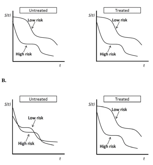
Predictive tools are based on the actual tumours’ molecular make up to determine whether or not they would benefit from a particular therapy. On the contrary, general prognostic tools will classify patients based on their gene expression profile into groups of good or poor prognosis irrespective of whether they respond to treatment. Importantly, characterization of the prognostic or predictive value of a biomarker identified in patients undergoing a given treatment requires a control group of non-treated patients, or treated with an alternative drug acting via an unrelated mechanism. A pictoral representation of the distinction between a predictive versus a prognostic tool can be seen in Figure 3, p.30.
A.
B.
Figure 3: Prognostic versus Predictive tools
A= Prognostic tool (whether untreated or treated, patients are categorized into a high risk versus low risk group; B = Predictive tool (will show the likelihood of response to a specific treatment).
1.9 Predictive Tools in Breast Cancer
The two most common gene signatures used clinically today are the 21-gene recurrence score (Oncotype DX) 81 and the Amsterdam 70-gene prognostic profile (Mammaprint)82. Oncotype DX is a predictive tool whereas Mammaprint is a prognostic tool. Multiple studies have shown that although some patients with ER+ tumours derive a benefit from adjuvant chemotherapy, the majority do not. Oncotype DX was created to identify the subset or ER+, node negative patients who would derive benefit and thus avoid over-treating patients who did not. Based on the genetic profile of a patient’s tumour, a recurrence score can be calculated and thus guide treatment. Mammaprint aids as a guide for decision-making with regards to adjuvant therapy. This tool also based on the genetic profile of a tumour, will categorize it as either being a good prognosis or poor prognosis tumour, once again guiding clinical decision-making.
1.10 Development of a gene signature that can predict the response to Tamoxifen
As already mentioned, two thirds of breast cancers are ER+ and their growth is stimulated by estrogens. Adjuvant therapy with anti-estrogens such as Tamoxifen and AIs has been shown to increase survival in breast cancer patients. This treatment is, however, not successful in all ER-positive tumours, with up to 40% of patients recuring despite completed treatment. Tumours can present intrinsic or acquired resistance to Tamoxifen, the mechanisms of which were described earlier. However, it is currently impossible to predict which patient will benefit from Tamoxifen therapy and which will not.
Preliminary studies in Dr. Mader’s lab have identified 20 genes whose expression levels in tumours are able to predict the response to Tamoxifen therapy (disease-free survival
including local-regional recurrence and metastatic recurrence). These markers, identified using bioinformatics analysis of published gene expression datasets, were able to discriminate patients that would respond best to Tamoxifen from those that did not.
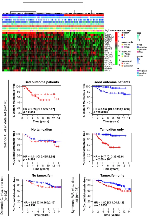
DNA microarray assays performed on the ER+ MCF-7 breast cancer cell line, allowed Dr. Mader’s laboratory to identify 170 primary estrogen receptor target genes 83 . Bioinformatics tools were thereafter used to demonstrate, using several published datasets of breast tumour expression profiles, that levels of expression of these genes in patients’ tumours predict outcome for Tamoxifen-treated breast cancer patients. A 20-gene signature was then derived from the best estrogen primary target genes combined with genes from unrelated signalling pathways found to have individual predictive value and was found to have a superior predictive value for Tamoxifen efficacy when tested against the 170-gene model84,85,86. This signature’s predictive value was found to be robust in two tumour gene expression datasets (Desmedt C et al. 200787, Sortiriou et al. 200688) and independent from
traditional predictors such as ER/PR and lymph node status (Figure 4, p. 33). This figure demonstrates the heatmap and Kaplan-Meier plots for the 20-gene signature.
Figure 4: Heat map of the 20-gene signature applied to several published data
The overall purpose of this study is to develop a PCR kit (gene signature) to monitor expression levels of these 20 genes and ultimately to test this 20-gene signature in a retrospective study using paraffin-embedded breast cancer tissues of patients with a known medical history. This tool will thus have a direct impact on clinical practice through the development of markers of therapeutic success for treatment with Tamoxifen and possibly AIs. Futile treatments would be avoided, preventing needless side effects, and improved identification of ER+ tumours with a low chance of success to anti-estrogen therapy will facilitate research into more appropriate treatments for hormone resistant tumours.
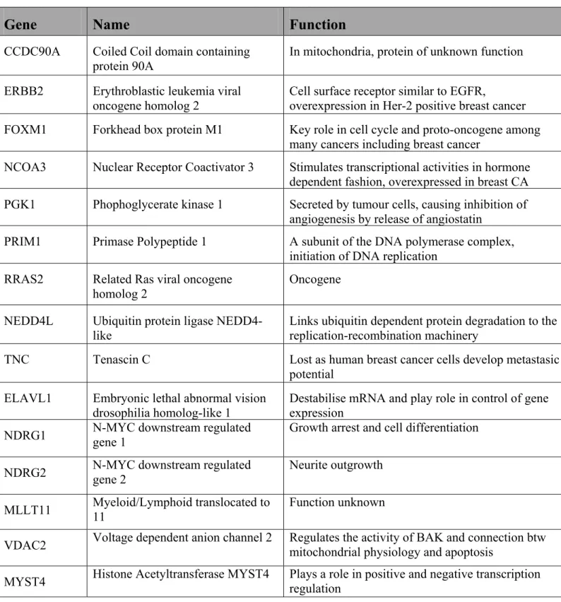
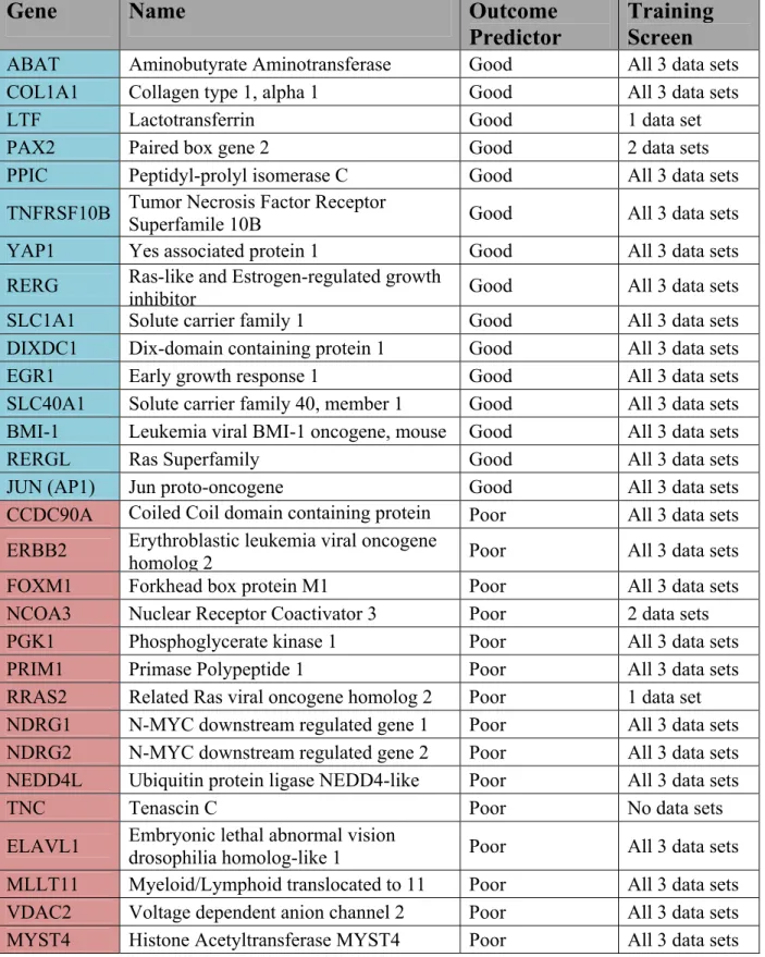
1.11 Identified Predictive Genes
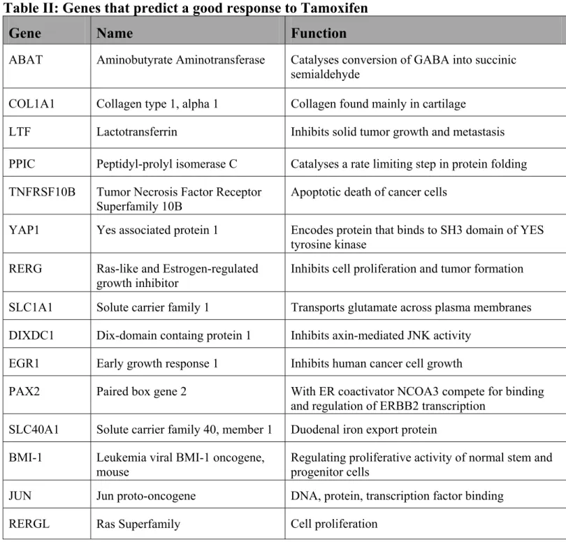
The ultimate goal of our study was to develop a 20-gene signature as seen in the preliminary results. However, these genes were identified using data sets of Affymetrix arrays. Knowing that Q-PCR assays may not always reproduce results obtained with micro-array probes, 10 additional robust predictive genes were identified for validation, so as to ensure a final 20-gene signature in the instance where genes could not be validated in Q-PCR. Good predictors
There were 15 genes that were selected as predictors of a good prognosis and good response to Tamoxifen therapy. A list of these genes as well as a summary of their function can be found in Table II, p. 35.
Table II: Genes that predict a good response to Tamoxifen
Gene
Name
Function
ABAT Aminobutyrate Aminotransferase Catalyses conversion of GABA into succinic semialdehyde
COL1A1 Collagen type 1, alpha 1 Collagen found mainly in cartilage
LTF Lactotransferrin Inhibits solid tumor growth and metastasis PPIC Peptidyl-prolyl isomerase C Catalyses a rate limiting step in protein folding TNFRSF10B Tumor Necrosis Factor Receptor
Superfamily 10B
Apoptotic death of cancer cells
YAP1 Yes associated protein 1 Encodes protein that binds to SH3 domain of YES tyrosine kinase
RERG Ras-like and Estrogen-regulated growth inhibitor
Inhibits cell proliferation and tumor formation SLC1A1 Solute carrier family 1 Transports glutamate across plasma membranes DIXDC1 Dix-domain containg protein 1 Inhibits axin-mediated JNK activity
EGR1 Early growth response 1 Inhibits human cancer cell growth
PAX2 Paired box gene 2 With ER coactivator NCOA3 compete for binding and regulation of ERBB2 transcription
SLC40A1 Solute carrier family 40, member 1 Duodenal iron export protein BMI-1 Leukemia viral BMI-1 oncogene,
mouse
Regulating proliferative activity of normal stem and progenitor cells
JUN Jun proto-oncogene DNA, protein, transcription factor binding
RERGL Ras Superfamily Cell proliferation
Some genes in this group are of particular interest. Many of them have interesting functions in breast cancer, potentially explaining why overexpression of these genes in an ER+ breast cancer may predict a better outcome with Tamoxifen treatment.
LTF has been shown to inhibit the growth of solid tumours and the development of experimental metastases in mice89. This was then further illustrated when evaluating primary breast tumours and their metastases, showing that LTF significantly decreased the metastatic potential in breast cancer by inhibiting and thus decreasing cellular motility90.
TNFRSF10B is among the tumour-necrosis factor receptor superfamily, which is associated with its TNF-related apoptosis-inducing ligand (TRAIL) R2. This ligand induces the process of cancer cell death/apoptosis. Although one study showed that TRAIL R2 was associated with higher-grade tumours, when compared to TRAIL R1, both are involved in cancer cell apoptosis and mammary carcinoma could be sensitised to TRAIL-R2-induced apoptosis, suggesting that TRAIL-R2 might therefore be used to therapeutically target such tumours91.
There is some suggestion in the literature that YAP1 is a tumour suppressor gene for breast cancer and thus if lost, may lead to more progression of breast cancer cells. In corollary therefore, if highly expressed, may lead to cancers with a better prognosis92.
In a five-gene model predicting the outcome of patients with early ER+ breast cancer, RERG overexpression was associated with increased survival and better outcome in patients with ER+ cancers treated with Tamoxifen93. This positive correlation has been noted in other studies, associating RERG with a better prognosis in breast cancer patients. One study showed that high RERG expression correlated with the expression of a set of genes that defined the ER+ subtype and was associated with a slow rate of tumour cell proliferation and growth inhibition and thus a favourable prognosis for these cancer patients94. Moreover, RERG expression was inversely associated with the proliferation marker MIB1. Strong RERG