HAL Id: hal-00508367
https://hal-mines-paristech.archives-ouvertes.fr/hal-00508367
Submitted on 10 Dec 2012HAL is a multi-disciplinary open access archive for the deposit and dissemination of sci-entific research documents, whether they are pub-lished or not. The documents may come from teaching and research institutions in France or abroad, or from public or private research centers.
L’archive ouverte pluridisciplinaire HAL, est destinée au dépôt et à la diffusion de documents scientifiques de niveau recherche, publiés ou non, émanant des établissements d’enseignement et de recherche français ou étrangers, des laboratoires publics ou privés.
Morphology of cellulose objects regenerated from
cellulose-N-methylmorpholine N-oxide-water solutions
Olga Biganska, Patrick Navard
To cite this version:
Olga Biganska, Patrick Navard. Morphology of cellulose objects regenerated from cellulose-N-methylmorpholine N-oxide-water solutions. Cellulose, Springer Verlag, 2009, 16 (2), pp.Pages 179-188. �10.1007/s10570-008-9256-y�. �hal-00508367�
Morphology of cellulose objects regenerated from cellulose-
N-methylmorpholine N-oxide-water solutions
Olga Biganska* and Patrick Navard**
Mines ParisTech, CEMEF- Centre de Mise en Forme des Matériaux, CNRS UMR
7635, BP 207 1 rue Claude Daunesse 06904 Sophia Antipolis Cedex, France.
Member of the European Polysaccharide Network of Excellence (EPNOE),
www.epnoe.eu
* Present address: L’Oréal, 188-200, rue Paul Hochart 94550 Chevilly-Larue, France
** To whom correspondence should be addressed
Tel.: +33 (0)4 93 95 74 66; fax: +33 (0)4 92 38 97 52
ABSTRACT
The precipitation in aqueous media of cellulose from solutions in
N-methylmorpholine N-oxide (NMMO) hydrates is an important stage in the process
of manufacturing of fibres, films and other cellulose objects. It is responsible for the
formation of the structure of the regenerated object and their morphological
characteristics significantly influence the properties of the final products.
Regeneration of rather large cellulose objects was observed in situ by optical
microscopy. It was found that all regenerated objects present an asymmetric
structure composed of a dense skin surrounding a sub-layer characterised by the
presence of finger-like voids. The porous texture of the cellulose parts between these
voids is typical of the one obtained by spinodal decomposition. The morphologies of
regenerated cellulose samples are described as a function of various parameters,
initial cellulose solutions and composition and temperature of the aqueous
regeneration bath. A mechanism of the structure formation during regeneration is
proposed.
KEY-WORDS
INTRODUCTION
N-methylmorpholine-N-oxide (NMMO) hydrates are direct solvents for cellulose,
used commercially in the preparation of homogenous cellulose-NMMO-water
solutions (dope) for making mainly fibres (Lyocell process). The production of the
cellulose objects by the NMMO process passes through the step of regeneration of
the spun or extruded dope in a coagulation bath (Krüger 1994). In order to
regenerate cellulose-NMMO-water solution, the liquid coagulating agent must be a
solvent for NMMO and a non-solvent for cellulose. Polar liquids, like water or
alcohol, are used as coagulation bath because they are miscible with NMMO and
cause NMMO removal from the cellulose solution. It is expected that the
regeneration of cellulose-NMMO-water solutions follows the well known principles
of phase separation in polymer solutions valid in membrane formation, Koenhen et
al (1977), Broens et al (1980), Shen and Cabasso (1982), Radovanovic et al (1992),
Pereira Nunes and Inoue (1996), Tsay and McHugh(1992), Barton et al (1997).
When a polymer solution is regenerated in a non-solvent coagulation bath, the
resultant objects have an asymmetric structure, i.e. a more or less dense skin is
supported by a porous sub-layer. The formation of the skin results from the increase
in polymer concentration stimulated by an extremely rapid solvent depletion from
the top layer of the solution. Two predominant morphologies – finger-like and
sponge-like – are usually observed in the sublayer (Broens et al 1980, Shen and
Cabasso 1982). The way these morphologies are forming depends on the rate of
precipitation of the polymer that is determined by the rate of solvent diffusing out
of, and the rate of non-solvent diffusing into, the polymer solution at the interface.
solution faster than the solvent diffuses out. Sponge-like structures are formed when
the solvent diffuses out faster than the non-solvent diffuses in (Shen and Cabasso
1982, Barton et al 1997).
The relations between the mechanisms of phase separation and the process of
structure formation are still a matter of active research, owing to their importance in
the processing of polymer membranes. Changes in composition bring a ternary
polymer/solvent/non-solvent system to a condition which favours liquid-liquid or
solid-liquid phase separation. Solid-liquid phase separation is considered in structure
formation of systems containing crystallisable polymers. Liquid-liquid phase
separation is considered for systems containing either amorphous or crystallisable
polymers. In the case of liquid-liquid demixing of polymer solutions, two different
mechanisms can happen: nucleation and growth or spinodal decomposition. Most of
the earlier papers ascribe the formation of the structure in membranes produced by
regeneration of a polymer solution in a non-solvent coagulation bath to the
nucleation and growth mechanism. The importance of spinodal decomposition, that
is very rapid and difficult to quantify, has been recognised more recently
(Radovanovic et al 1992, Pereira Nunes and Inoue 1996).
As far as the regeneration of cellulose-NMMO-water solutions is concerned, several
publications describing the morphological features of fibres and films are available
(Romanov et al 1988, Bang et al 1999, Fink et al 2001, Laity et al 2002). Romanov
et al. (1988) noted that the regeneration of fibres in a bath of isopropyl alcohol
increases their microporosity (0.1µm) and creates vacuoles in the structure in
comparison with a water bath. Fink et al. (2001) reported that fibres precipitated in
water show a dense cellulose network structure with small finely distributed voids
throughout the cross-section, except for a small boundary layer with highly densified
material. The authors also investigated the regeneration in various alcohols and
observed that the increase of their molecular mass leads to the formation of a distinct
skin-core structure. Laity et al. (2002) studied the composition and phase changes
during the regeneration of cellulose-NMMO-water solutions in water. They reported
that the cross section of regenerated solution observed by SEM appeared uniformly
dense. TEM observation revealed porosity on the scale of a few nanometres.
Crawshaw and Cameron (2000) show that there is a network of voids elongated in
the fibre direction. In the untreated wet state, there are many small voids (mean
length 36 nm, mean thickness 0.3 nm) while in the dry state after drying at 160°C,
there is a much lower overall volume fraction of much larger pores voids (mean
length 270 nm, mean thickness 5 nm). Jianchin et al. (1999) have reported the
presence of a skin-core structure in Lyocell fibres. Swelling experiments performed
in aqueous NaOH solutions and swollen fibres investigation with an environmental
scanning electron microscope allowed the authors to suggest that Lyocell fibres are
made of a composite skin and of a crystalline core formed by parallel fibrils. The
skin is amorphous and very elastic. Its thickness is estimated to be between 57 and
177 nm (average value is equal to 73 nm). The skin is made up of two layers: the
outer layer is very thin while the inner one is thicker. Cellulose chains in the outer
layer are less oriented than chains in the inner layer. The core of the fibre is made up
of highly oriented parallel fibrils and amorphous regions connecting these fibrils.
Some pores and defects could be also present in the amorphous regions.
Abu-Rous et al. (2006) showed that Lyocell fibres contain only nanopores in the
core of the fibre and a very porous skin layer. Schurz et al. (1995) proposed a
orientation of both crystalline and amorphous zones was reported to be very high.
Crystalline regions have rather isolated fibrils that can be easily separated by a
mechanical treatment, leading to fibrillation (Ducos et al 2006). A recent work
Abu-Rous et al. (2007) based on dye penetration proposes to distinguish three regions
inside Lyocell fibres, a skin layer with a compact structure, a porous middle zone
and a compact fibre centre.
The understanding of the structure formation during coagulation and the
morphology of regenerated cellulose-NMMO-water solutions, depending on the
regeneration kinetics, is essential for the properties of cellulose objects produced by
NMMO process. The main objective of this work is the investigation of the
morphologies of regenerated cellulose-NMMO-water solutions as a function of
various parameters of both cellulose solutions and coagulation baths. These
observations will be linked to the knowledge of the regeneration kinetic parameter
obtained before (Biganska and Navard 2005).
EXPERIMENTAL PART
Cellulose-NMMO-water solutions were prepared in the R&D department of the
Austrian company Lenzing AG using the method described previously (Biganska
and Navard 2005). Several cellulose pulp samples and different cellulose
concentrations were used in this study in order to describe the influence of these
parameters on the morphology of regenerated solutions. Four different pulp families
called Krafts 1-4 were used in this work. Each Kraft family has the same cellulose
origin, the difference being the molecular weight distribution (Table 1). The Location of
properties of the cellulose samples as well those of the cellulose solutions were
described elsewhere (Biganska and Navard 2005).
NMMO-water mixtures with initial NMMO concentration varying from 0% (pure
water) to 50% were used for coagulation baths. The required mixtures were prepared
by the dilution of the solution containing 50% of NMMO provided by Aldrich.
The regeneration of the cellulose solutions was performed with two types of
cellulose solution physical states. First, with solid (crystallised)
cellulose-NMMO-water solutions shaped by a press to obtain discs of definite thickness (1mm and
3mm) and diameter (20mm). Second, with molten, 2 to 3mm thick
cellulose-NMMO-water solutions. Samples were molten at 90°C.
Each cellulose solution sample was immersed into a coagulation bath to regenerate
it, i.e. to transform the polymer solution into a pure, highly swollen cellulose object.
Then, the regenerated cellulose sample was cut with a razor blade in order to obtain
a thin slice. These slices were then observed with an optical microscope (Leitz
Metallux 3) and an environmental scanning electron microscope (Philips XL
ESEM). In order to observe the regenerated solutions in a wet state, the scanning
electron microscope was equipped with a Peltier stage. This device allows keeping
the sample in the electron microscope chamber in an atmosphere of a desired
humidity ratio fixed by two parameters, the pressure and the temperature.
RESULTS AND DISCUSSION
Influence of the state of the cellulose solution before regeneration on the morphology of the regenerated objects
If before being immersed into the regenerating bath, the cellulose solution is solid
(crystallized) or liquid (in a molten state), the morphologies of the regenerated
cellulose objects are very different. In the case of a solid solution obtained by
cooling the solution, it was shown (Biganska et al 2002) that it is the solvent which
is crystallizing. This is leading a variety of morphologies like large spherulites. After
sublimation of NMMO and water, cellulose chains retain the general morphology of
the crystallised solution (Chanzy et al 1979). The same phenomenon is observed if
such a crystallized solution is regenerated in an aqueous bath. Figure 1 illustrates
this phenomenon for the regeneration of a 3wt% cellulose solution in a water bath.
The right (regenerated) and left (solution) sides of the picture show that they have
the same morphology. This is due to the fact that the solvent is crystallizing, pushing
cellulose chains out of the crystals but keeping them very close to the tiny crystals,
keeping thus the image of the crystal arrangements. When the crystals are dissolving
in the regeneration liquid, cellulose chains keeps the same organisation they had
around the solvent crystals. There is no phase separation involving cellulose during
the regeneration of a solid crystallized cellulose solution since the phase separation
between the solvent and cellulose had already occurred during the crystallisation of
the solvent. The observation of the cross-section of the solutions regenerated in a
water bath after crystallisation reveals a uniform and compact structure with few
voids. As we will see, voids are originating from liquid-liquid phase separation, a
case not occurring here.
The situation is completely different after the regeneration of a molten solution. In
this case the observation of the cross-section reveals a skin-core structure as
illustrated in Figure 2. Location of
fig 1
Location of fig 2
The thickness of the dense skin is not uniform along the perimeter of the sample.
The core contains finger-like voids that can occupy either the whole surface of the
cross-section or form a crown. The texture of the cellulose material that forms the
walls of the finger-like cavities has micrometer-scale porosity. In the following we
will describe in details the structure obtained after the regeneration of molten
cellulose-NMMO-water solutions and the parameters that influence it.
Fingering in regenerated objects obtained from a molten solution
Formation of the finger-like patterns is a common feature in liquid-liquid demixing.
The classic example is the Staffman-Taylor fingers (Saffman and Taylor 1958,
Kessler et al 1988), observed when air drives water from a Hele-Shaw cell (two
parallel plates with a narrow gap of constant thickness between them). A moving
air-liquid interface is driven by the gradient of a diffusive field. Planar or circular
interfaces are morphologically unstable and tend to finger, along directions favoured
by the boundary conditions and/or anisotropy. The difference between the
morphologies of the cellulose solutions regenerated in the molten or solid
crystallised states are thus clear. Finger-like voids are formed when a low viscosity
fluid is displacing a more viscous one. So, finger-like voids are formed in the molten
solutions in contact with water of the coagulation bath (viscosity of water is much
lower than the viscosity of any cellulose solution). In contrast, there is no moving
liquid-liquid interface during the regeneration of a crystallised solution. The
dissolution of the crystallised NMNO is only due to the fact that NMMO is highly
hygroscopic (Navard and Haudin 1981). The few observed voids in the regenerated
On the basis of what is known from the theory of membranes formation and taking
into account our own observations, the following mechanism of the formation of
structure during the regeneration of molten cellulose-NMMO-water solutions can be
proposed. The contact between the solution and the liquid from the coagulation bath
leads to the rapid outflow of the solvent from the top layer of the solution. The
polymer profile at the point of precipitation exhibits a very high interfacial
concentration, thus favouring the formation of a dense skin immediately after
immersion of the sample into the coagulation bath. The bulk of the sample is at near
the initial concentration and is in a fluid state. Thus, a rapid inflow of the coagulant
can take place through the weak spots at the skin interface. Rapid growth of
finger-like voids in the fluid region under the skin occurs due to the moving interface
created by the coagulant (less viscous) and the solution (more viscous).
Measurements of the coefficients of diffusion of NMMO solvent from the solution
into the bath and of the coagulant from the bath into the solution (Biganska and
Navard 2005) show that the inflow of the coagulant is one order of magnitude higher
than the outflow of the solvent. This result is in agreement with the fact that the
formation of a finger-like structure is favoured when the non-solvent enters the
sample more rapidly than the solvent exits from it (Shen and Cabasso 1982).
Influence of the concentration of cellulose, the nature and the temperature of coagulation bath on the morphology of regenerated solutions
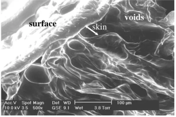
Cellulose concentration
Molten solutions with 3wt% of cellulose, 82wt% of NMMO and 15wt% of water
were regenerated in a water bath at room temperature. The morphology was very
similar, independent on the characteristics of the Kraft samples. The observation of Location of
the cross-sections reveals the presence of a skin surrounding finger-like voids. The
thickness of the skin is about 100µm. A regular ordering of voids is seen near the
skin and a more disordered distribution is found in the centre of the samples. The
length of voids can reach 500µm and the width 50µm. To illustrate this, Figure 3
presents a scanning electron microscopy image of the cross-section of the
regenerated solution prepared with the Kraft 3 pulp. Molten solutions with 6wt%,
8wt% and 10wt% of cellulose were regenerated in water bath at room temperature in
order to test the influence of cellulose concentration on the morphology of
regenerated solutions. Figure 4 shows the scanning electron micrographs, taken at
various magnifications, of the cross-section of the 6wt% cellulose solution
regenerated in a water bath. Figure 4a shows a general view of the cross-section
where one can distinguish the skin and the voids. Figure 4b, taken in a place close to
the sample centre, shows some smaller voids. Figures 4c and 4d are showing the
aspect of the walls between voids. The analysis of numerous samples reveals that the
ordering of voids observed for the samples with low cellulose concentration
decreases when the cellulose concentration increases. The length of voids decreases
while their width increases when the concentration of cellulose increases. Both
effect are most probably due to the viscosity of the highly concentrated, cooled
solution. The difficulty to displace large amounts of cellulose chains restricts the
propagation of the fingering instability, and disrupts its regular formation. In fact,
the lengths of the voids observed in the 6wt% cellulose samples are ranging between
400 and 500µm and their width is of about 30µm. The corresponding dimensions are
of 300µm and 50µm for the samples with 8wt% of cellulose. In the case of solutions
with 10wt% of cellulose, these dimensions are of 200µm and 50µm. It seems that
the thickness of the skin also increases with cellulose concentration. Location of
NMMO concentration in regenerated bath
The effect of the concentration of NMMO in the initial aqueous bath used for
regeneration was tested on 6wt% cellulose solutions. The observation of the
cross-sections of the samples regenerated in the baths with 0% (pure water), 10%, 20%,
30%, 40% and 50% of NMMO reveals that finger-like voids become less numerous
when the concentration of NMMO increases (Fig 5). This is due to the fact that we have a very “strong” precipitation in pure water while the regeneration is “weak” in
the presence of NMMO. The higher the NMMO concentration in the regenerating
bath is, the less rapidly this fluid is penetrating in the cellulose solution. The
fingering mechanism is thus less favoured.
Regenerating bath temperature
The effect of the temperature of water bath on the morphology of regenerated
solutions can be illustrated on the example of 8wt% cellulose solutions. Figure 6
gives the scanning electron micrographs of the cross-sections of the solutions
regenerated at 20°C, 50°C and 80°C. The left column shows the evolution of general
morphology while the right column presents the evolution of the material texture
between the voids. It can be seen that the thickness of the skin decreases, the number
of voids increases and their width decreases when the bath temperature increases.
Another effect of the temperature increase is the appearance of a dense part in the
middle of the samples and, consequently, the confinement of the void region in a
corona.
Mechanisms of cellulose-regenerating medium phase separation Location of fig 4 Location of fig 6 Location of fig 5
The cross-sections of molten solutions with 8wt% (a) and 12wt% cellulose (b)
regenerated in water bath at 50°C have a microporous globular texture (Fig 7) made
by the juxtaposition of small spheres of 1-2µm (Fig 6a). Such a texture is present in
almost all the investigated samples obtained from a molten initial state. This texture
is due to the phase separation mechanism. Two mechanisms of phase separation can
take place during liquid-liquid demixing of polymer solutions, either nucleation and
growth where the nuclei of one phase grow in the mixture or spinodal
decomposition where a periodic variation of concentration leads to the final phase
separation. If there is a clear difference between these two mechanisms in their first
stages (nucleation and growth shows isolated entities while spinodal decomposition
has a 3D network-like morphology), both tends to the same spherical morphology at
the end, due to surface tension effects. If there is no external nucleation agent in the
mixture, nucleation and growth gives a morphology composed of spheres of
different diameters (due to the sporadic nucleation), positioned randomly in the
regenerated material while spinodal decomposition gives rather monodisperse
spheres, usually ordered along lines (this is due to the 3D-network like structure of
the cellulose-rich phase). The micrographs shown on Figure 6 clearly suggest that
the phase separation mechanism is spinodal decomposition in the case of the
regeneration of a molten cellulose-NMMO solution in a water bath. Picture 6b,
seldom observed, represents a frozen periodic structure while the picture 6a,
frequently seen, corresponds to the periodic structure after it breaks down. This
conclusion is supported by independent observations made on cellulose objects
regenerated from NMMO solutions (Zhang and Shao 2001), (Mortimer and Peguy
1996) or during regeneration by NMR (Laity et al. 2002). Location of
CONCLUSION
The type of morphology of a regenerated cellulose object from a cellulose solution
in N-methylmorpholine N-oxide-water is not very much dependent on the cellulose
concentration or the bath composition. It is strongly dependent on the state of the
solution prior to regeneration. If the solution has crystallised, a dense morphology is
observed while a dense skin surrounding a core made of large voids with cellulose
walls is obtained from a molten solution. In this latter case, the cellulose wall
structure (small ordered spherical objects) is due to a spinodal decomposition.
Further investigations on the details of the fast spinodal decomposition by light
scattering and on the formation of the very thin skin during regeneration would help
the understanding of the morphology development of objects regenerated from
REFERENCES
Abu-Rous M, Ingolic E, Schuster KC (2006) Visualisation of the fibrillar and pore
morphology of cellulosic fibres applying transmission electron microscopy,
Cellulose 13:411-419
Abu-Rous M, Varga K, Bechtold T, Schuster KC (2007) A new method to visualize
and characterize the pore structure of Tencel (Lyocell) and other man-made
cellulosic fibres using a fluorescent dye molecular probe, J Appl Polym Sci 106:
2083-2091
Bang YH, Lee S, Park JB, Cho HH (1999) Effect of coagulation conditions on fine
structure of regenerated cellulosic films made from
cellulose/N-methylmorpholine-N-oxide/H2O systems. J Appl Polym Sci 73: 2681-2690
Barton BF, Reeve JL, McHugh AJ (1997) Observations on the dynamics of
nonsolvent-induced phase inversion. J Polym Sci Part B: Polym Phys 35:569-585
Biganska O, Navard P, Bedue O (2002) Crystallisation of
cellulose/N-methylmorpholine N-oxyde hydrates solutions. Polymer 43:6139-6145
Biganska O, Navard P (2005) Kinetics of precipitation of cellulose from
Broens L, Altena FW, Smolders CA, Koenhen DM (1980) Asymmetric membrane
structures as a result of phase separation phenomena. Desalination 32:33-45
Chanzy H, Dubé M, Marchessault RH (1979) Crystallization of cellulose with
N-methylmorpholine N-oxide: A new method of texturing cellulose. J Polymer Sci
Polym Lett Ed 17:219-226
Crawshaw J, Cameron RE (2000) A small angle X-ray scattering study of pore
structure in Tencel® cellulose fibres and the effects of physical treatments. Polymer,
41:-4698
Ducos F, Biganska O, Schuster KC, Navard P (2006)Influence of the Lyocell fibre
structure on their fibrillation. Cell. Chem. Techn. 40(5):299-311
Fink H-P, Weigel P, Purz HJ, Ganster J (2001) Structure formation of regenerated
cellulose materials from NMMO-solutions. Progr Polym Sci 26:1473-1524)
Jianchin Z, Meiwu S, Zhu H, Kan L (1999) Study of the skin-core structure of
lyocell staple fibers. Chemical Fibers International, 49:496-500
Kessler DA, Koplik J, Levine H (1988) Pattern selection in fingered growth
phenomena. Adv Phys 37:255-339
Koenhen DM, Mulder MHV, Smolders CA (1977) Phase separation phenomena
Krüger R (1994) Cellulosic filament yarn from the NMMO Process. Lenzinger
Berichte 9:49-52
Laity PR, Glover PM, Hay JN (2002) Composition and phase changes observed by
magnetic resonance imaging during non-solvent induced coagulation of cellulose.
Polymer 43:5827-5837
Mortimer SA, Péguy A (1996) Methods for reducing the tendency of lyocell fibers
to fibrillate, Journal of Applied Polymer Science. 60:305-316
Navard P, Haudin JM (1981) Etude thermique de la N-méthylmorpholine N-oxyde et
de sa complexation avec l'eau. J Thermal Anal 22:107-118
Pereira Nunes S, Inoue T (1996) Evidence for spinodal decomposition and
nucleation and growth mechanisms during membrane formation. J Membrane Sci
111:93-103)
Radovanovic P, Thiel SW, Hwang S-T (1992) Formation of asymmetric polysulfone
membranes by immersion precipitation. Part II. The effects of casting solution and
gelation bath compositions on membrane structure and skin formation. J Membrane
Romanov VV, Sokira AN, Lunina OB, Iovleva MM (1988) Morphological features
of the structure of fibres prepared from solutions of cellulose in methylmorpholine
oxide Fibre Chemistry 20:38-39; Khimicheskie Volokna 1:27–28
Saffman PG, Taylor GI (1958) The penetration of fluid into a porous medium or
Hele-Shaw cell containing a more viscous liquid. Proc Royal Soc London A
245:312-329
Schurz J, Lenz J, Wrentschur E (1995) Inner surface and void system of regenerated
cellulose fibers. Die Angewandte Makromoleculare Chemie, 229:175-184
Shen TC, Cabasso I (1982) Macromolecular Solutions, solvent-property relationship
in polymers, R.B. Seymour and G.A. Stahl, Eds., Pergamon Press, New-York 108
Tsay CS, McHugh AJ (1992) A rationale for structure formation during phase
inversion. J Polym Sci: Part B: Polym Phys 30:309-313
Zhang Y, Shao H, Wu C, Hu (2001) Formation and Characterization of Cellulose
Membranes from N-Methylmorpholine-N-oxide Solution Macromolecular
Captions for Tables and Figures.
Table 1 Characteristics of the cellulose used for the preparation of
cellulose/NMMO/water solutions. Molar mass distribution was measured by size
exclusion chromatography (SEC/GPC) and crystallinity by Fourier transform
infrared spectrometry (FT-IR). From Biganska and Navard 2005.
Figure 1 Optical micrograph of the moving interface created during the
regeneration of a solid cellulose-NMMO-water solution in a water bath.
Figure 2 Schematic representation of the cross-section of the regenerated
cellulose samples : solution regenerated after crystallisation (left) and solution
regenerated in molten state (right).
Figure 3 Scanning electron micrograph of the cross-section of a 3wt%
cellulose solution regenerated in a water bath at room temperature.
Figure 4 Scanning electron micrographs of the cross-sections of a 6wt%
cellulose solution regenerated in a water bath at room temperature : a) general view,
Figure 5 Scanning electron micrographs of the cross-sections of a molten
6wt% cellulose solution regenerated in a NMMO water bath at room temperature
with varying NMMO content in the bath.
Figure 6 Scanning electron micrographs of the cross-sections of a 8wt%
cellulose solution regenerated in a water bath at different temperatures.
Figure 7 Scanning electron micrographs of the fine structure observed in
regenerated cellulose-NMMO-water solutions with initial concentration of cellulose
Table 1
Kraft 1/1 Kraft 1/2 Kraft 2/1 Kraft 2/2 Kraft 3 Kraft 4
Mn (x1000) 77.4 44.2 53.0 40.4 50 51.7 Mw (x1000) 226.3 90.2 155.0 98.2 207 105.2 Mz (x1000) 468.9 158.0 410.5 206.9 619.9 186.7 wt % (DP<50) 1.1 1.6 1.5 2.3 2.6 1 wt % (DP<200) 7.8 18.5 14.3 21.8 15.8 14.6 wt % (DP>2000) 21 2 10.8 4 18 3.5 Crystallinity [%] 53 55 51 47 46 43
Figure 1
Figure 2
crystallised solution regenerated solution regeneration front 0.1mmcrystallised
voids skin microporesmolten
crystallised
voids skin microporesmolten
Figure 3
Figure 4
surface skin voids
a) b)
c) d)
skin
Figure 5
0% NMMO 10% NMMO 20% NMMO
Figure 6
20°C
50°C
80°C
Figure 7
a)