Université de Montréal
Inflammation dans les bronches du modèle d’asthme chez le rat Brown Norway
Inflammation in the Airways of the Brown Norway Rat Model of Asthma
par
Samobrata Samuel Nag
Département de Sciences Biomédicales Faculté de Médecine
Thèse présentée à la Faculté des études supérieures en vue de l’obtention du grade de
Philosophiae Doctor (Ph.D.) en sciences biomédicales
Juillet, 2003
•1
Université
dI
de Montréal
Direction des bibliothèques
AVIS
L’auteur a autorisé l’Université de Montréal à reproduire et diffuser, en totalité
ou en partie, par quelque moyen que ce soit et sur quelque support que ce
soit, et exclusivement à des fins non lucratives d’enseignement et de
recherche, des copies de ce mémoire ou de cette thèse.
L’auteur et les coauteurs le cas échéant conservent la propriété du droit
d’auteur et des droits moraux qui protègent ce document. Ni la thèse ou le
mémoire, ni des extraits substantiels de ce document, ne doivent être
imprimés ou autrement reproduits sans l’autorisation de l’auteur.
Afin
de
se
conformer
à
la
Loi
canadienne
sur
la
protection
des
renseignements personnels, quelques formulaires secondaires, coordonnées
ou signatures intégrées au texte ont pu être enlevés de ce document. Bien
que cela ait pu affecter la pagination, il n’y a aucun contenu manquant.
NOTICE
The author of this thesis or dissertation has granted a nonexciusive ticense
allowing Université de Montréal to reproduce and publish the document, in
part or in whole, and in any format, solely for noncommercial educational and
research purposes.
The author and co-authors if applicable retain copyright ownership and moral
rights in this document. Neither the whole thesis or dissertation, nor
substantial extracts from it, may be printed or otherwise reproduced without
the author’s permission.
In compliance with the Canadian Privacy Act some supporting forms, contact
information or signatures may have been removed from the document. While
this may affect the document page count, it does not represent any loss of
content from the document.
Cette thèse intitulée
Inflammation dans les bronches du modèle d’asthme chez le rat Brown Norway
Inflammation in the Airways 0f the Brown Norway Rat Mode) of Asthma
présentée par: Samobrata Samuel Nag
a été évalué par un jury composé des personnnes suivantes:
Dr. Guy Delespesse président-rapporteur Dr. Paolo M Renzi directeur de recherche Dt. Richard Bertrand membre dujury Dt. Sabbah Hussein examinateur externe Représentant du doyen de ia
t-Table of Contents Title Page
Table of Contents ii
Index of Figures y
Index ofAbbreviations vii
Summary 1
Résumé 3
Introduction
1.1 Introduction 5
1.1.1. Epidemiology 5
1.1.2. Risk Factors/Asthma Mechanisms 5
1.1.3. Hygiene Hypothesis 8
1.1.4. Genetics 9
1.2 Asthma Characteristics 10
1 .2.1. Early and Late Response 11
1.2.2. Airway Hyperresponsiveness 12
1.2.2.1. Airway Hyperresponsiveness in Humans 14
1.2.2.2. Airway Hyperresponsiveness in Animais 14
1.2.3. Airway Remodelling/Obstruction 15
1.2.3.1.AirwayWall Thickening 15
1.2.3.2. Basement Membrane Thickening 16
1.2.3.3. Smooth Muscle Hypertrophy/Hyperplasia 16
1.2.3.4. Mucus Metaplasia 18 1.2.3.5. Airway Vascularity 18 1.3. Airway Inflammation 18 1.3.1. Immunoglobulin E 16 1.3.2. Eosinophils 20 1 .3.3. Neutrophils 25 1.3.4. Mast cells 27 1.3.5. Macrophages 29 1 .3.6. Dendritic cells 31 1.3.7. Lymphocytes 30 1.3.8. Leukotrienes 34 1.3.9. Cytokines 35 1.3.9.1. Interleukin-4 36 1.3.9.2. lnterleukin-5 38 1.3.9.3. Interferon-gamma 39 1.3.9.4. Interleukin-2 40 1.3.10. Chemokines 41 1 .4. Asthma therapy 43 1.4.1.Drugs 43
1.4.1.3. Leukotriene Modifiers 45
1.4.1.4. Phosphodiesterase inhibitors 45
1.4.1.5. Steroid-sparing strategies 45
1.4.1 .Therapeutic guidelines 46
1.4.2.1. Inhaled corticosteroids vs Antileukotriene drugs 47
1 .4.2.Alternative therapies 48 1.4.3.1. Anti-Immunoglobulin-E 48 1.4.3.2. Anti-histamine 49 1.4.3.3. Cytokines 49 1.4.3.4. lmmunotherapy 49 1.5. Animal Models 50
1.5.1. Brown Norway rat model of aergic asthma 50
1.6. Active Immunization Models 53
1.6.1. Sensitization and challenge 53
1.6.2. Adjuvant 53
1 .6.3. Dose 54
Avant-Propos 55
Materials and Methods 57
2.1. Cysteinyl Leukotrienes, Cellular lmmunity and the Airway Response to Antigen 57
2.1.1. AnimaIs and sensitization 57
2.1.2. Measurement of lung resistance 57
2.1.3. Experimental protocol 59
2.1.3.1. Airway responsiveness to LTD4 59
2.1.3.2. IL-2 induced airway response vs. MK-0476 59
2.1.4. Bronchoalveolar lavage 61
2.1.5. Lung retrieval and preparation 61
2.1.6. RNA prepatation and reverse transcription 61
2.1.7. Semi-Quantitative Polymerase Chain Reaction 62
2.1.8. In Situ Hybridization 62
2.1.9. Data Analysis (2.1 to 2.1.8) 63
2.2. lnterleukin-5 and the Airway Response of Brown Norway rats 64
2.2.1. AnimaIs and sensitization 64
2.2.2. Eosinophil colony proliferation 64
2.2.3. Immunoglobulin E determination 64
2.2.4. Endotoxin determination and challenge 65
2.2.5. Bronchoalveolar lavage 65
2.2.5.1. Major basic protein staining 65
2.2.6. Measurement of lung mechanics 66
2.2.7. Airway responsiveness to methacholine 66
2.2.8. Airway response to ovalbumin 66
2.2.9. Lung mincing and digestion 68
2.2.10. Isolation and staining of blood lymphocyte subsets 68
2.2.11. Measurement of cytokine mRNA expression 69
2.2.12. Data Analysis (2.2 to 2.2.11) 69
Results .71
Resuits (Legends and Figures) .76
Discussion 93
Acknowledgements 106
Bibliography 107
Figure 1 (Hyperresponsiveness) 6
Figure 2 (Airway Response) 13
Figure 3 (Airway Inflammation) 17
Figure 4 (Airway Smooth Muscle) 19
Figure 5 (Eosinophil) 21
Figure 6 fEosinophil Cationic Protein) 22
Figure 7 (Mast celi, T lymphocytes and Eosinophils) 24
Figure 8 (Mast ceil Tryptase) 28
Figure 9 (Macrophage) 30
Figure 10 (Thi and Th2 Lymphocytes) 33
Figure 11 (Cells and Mediators in Asthma) 37
Figure 12 (Animal airway response measurement setup) 58
Figure 13 (Experimental Animal Groups) 60
Figure 14 (IL-5 Experimental Animal Groups) 67
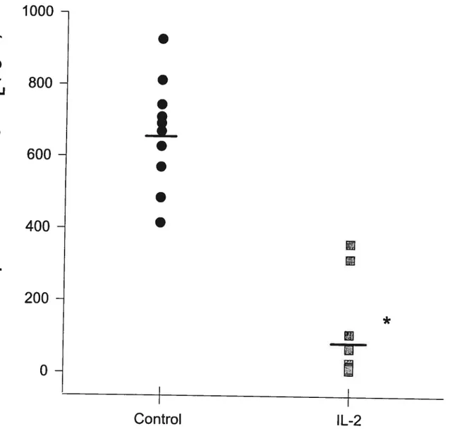
Figure 15 (Effect of IL-2 on airway responsiveness to LTD4) 76
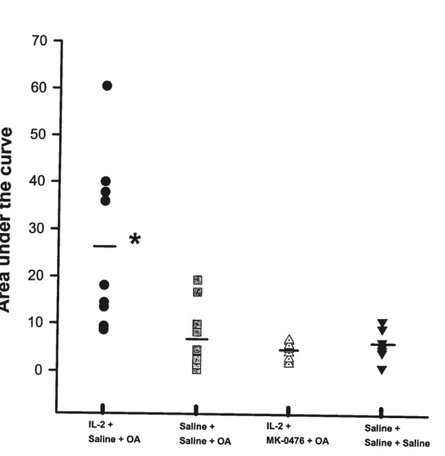
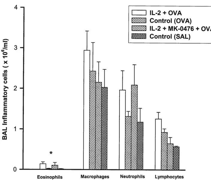
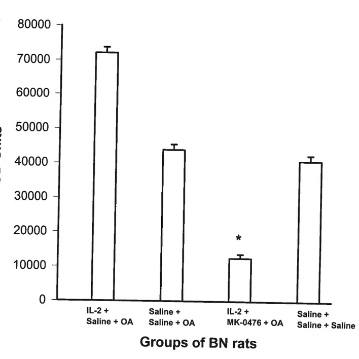
Figure 16 (Effect of Montelukast on IL-2-induced Late Airway Response) 77 Figure 17 (Effect of IL-2 and Montelukast on BAL Inflammatory celis) 78 Figure 18A
f
Effect of IL-2 and Montelukast on lung IL-4 mRNA expression) 79 Figure 18B (Effect of IL-2 and Montelukast on lung IL-5 mRNA expression) 80 Figure 18C (Effect of IL-2 and Montelukast on lung IFN-y mRNA expression) 81 Figure 19A (Effect of IL-2 and Montelukast on IL-4 mRNA positive cells in the airways) 82 Figure 198 (Effect of IL-2 and Montelukast on IFN-y mRNA positive cells in the airways) 83 Figure 20 (Effect of rhIL-5 on eosinophil progenitor colony formation from rat PBMC’s) 84 Figure 21 (Effect of ovalbumin sensitization on OA-specific serum IgE levels) 85 Figure 22f
Effect of IL-5 on airway responsiveness to Mch 20 hours after administration) 86 Figure 23 (Effect of lOpg rhIL-5 on AHR to Mch 30 min and 72 hrs after administration) 87 Figure 24Af
Effect of pre-treatment with rhIL-5 on Early Airway Response) 88Figure 24B (Effect of pre-treatment with rhlL-5 on the Late Airway Response) 89 Figure 25(Effect of rhlL-5 on lung resistance 20 hours after antigen challenge) 90 Figure 26 (Effect of rhIL-5 on AHR to Mch 20 hours after antigen challenge) 91 Figure 27 (Effect of rhIL-5 on CD4/CD8 Lymphocyte ratio
in
the blood) 92AHR : Airway Hyperresponsiveness Alum : Aluminum
AM : Airway Macrophages BAL: Bronchoalveolar Lavage BCG : Bacillus Calmette-Guerin BSA: Bovine Serum Album in
CCR : Cysteine-Cysteine Chemokine Receptor CFC : Chlorofluorocarbons
CFU-Eo : Colony forming units of eosinophils CyA: Cyclosporin A
Cys-LT1R: Cysteinyl-leukotriene receptor 1 DC : Dendritic ceils
DEP : Diesel Exhaust Particle DNA: Deoxyribonucleic Acid ECP : Eosinophil Cationic Ptotein
EC200RL: Concentration of Agonist causing 200% increase in Lung Resistance
EDN: Eosinophil-derived neurotoxin
ELISA: Enzyme-Linked Immunosorbent Assay ER: Early Response
EPO : Eosinophil peroxidase EU : Endotoxin Units
FEV1 : Forced Expiratory Volume pet second
GM-CSF : Granulocyte/Macrophage Colony Stimulating Factor Hz: Hertz
IgE: Immunoglobulin E IgG2: Immunoglobulin G2a IgM: Immunoglobulin M
ICS: Inhaled Corticosteroids
ICAM-1: Intercellular Adhesion Molecule-1 IFN-y: Interferon-gamma
IFN-yR: Interferon-gamma receptor IP1 O: Interferon-gamma inducible protein IL-2: lnterleukin-2
IL-3: lnterleukin-3 IL-4: Interleukin-4
IL-4R: Interleukin-4 receptor IL-5: Interleukin-5
IL-8: lnterleukin-8 IL-13: Interleukin-13 kD: kilodalton
LAL: Limulus Amebocyte Lysate LR: Late Airway Response LT: Leu kotrienes
LTC4: Leukotriene C4 LID4: Leukotriene D4 LIE4: Leukotriene E4 LPS: Lipopolysaccharides MBP : Major Basic Protein MCP-4: Major cationic protein-4
MHC : Major Histocomptabiîity Complex MMP-9: Matrix Metalloproteinase-9 MDI: Metered dose inhaler
Mch: Methacholine
M-MLV: Moloney Murine Leukemia Virus MDC: Monocyte derived chemokine
MUC-5: Mucin NK: Natural Killer NO: Nitric oxide
ODN: Oligodeoxynucleotide OCS: Oral Corticosteroids QA: Ovalbumin
PenH: Enhanced pause
PBMNC: Peripheral blood mononuclear celis PBS: Phosphate-buffered saline
PAF: Platelet-activating factor Ptp: Transpulmonary pressure PGE2: Prostaglandin 2
RS: Radioactive signal
RL: Lung Resistance
SQ-PCR: Semi-quantitative polymerase chain reaction STAT: Signal transducers and activators of transcription SRS-A: Slow reacting substance of anaphylaxis
SD: Sprague-Dawley ThO: Type O T lymphocyte Thi : Type 1 T Lymphocyte Th2 : Type 2 T Lymphocyte Th3: Type 3 T lymphocyte
Thp: Precursor T helper lymphocyte TRFK-5 : Anti-IL-5 Monoclonal Antibody TXA2: Thromboxane 2
TGF-f3: Transforming growth factor beta TBS: Tris-Borate Solution
TNF-a: Tumor Necrosis Factor Alpha UK: United Kingdom
pi: microlitre
VCAM-1 : Vascular celi adhesion moiecule 1 VLA-4: Vascular leukocyte antigen-4
Asthma is the most commoniy reported respiratory disease in clinical practice, affecting both children and aduits (1). This disease is characterized by reversible airflow obstruction, increased bronchial responsiveness, and airway inflammation (2). lt has been shown that persistent inflammation of the airways can lead to chronic and possibly irreversible changes that affect the airway physiological response to different stimuli (3). Over the last twenty years, cysteinyl leukotrienes (cys-LT) have been shown to be potent bronchoconstrictor agents and important inflammatory mediators in the pathophysiology 0f both acute and chronic asthma (1). Leukotrienes (LI) can induce smooth muscle contraction, increase vascular permeabiiity, stimulate mucus secretion and recruit eosinophils into the lungs (4). A number of activated inflammatory cells (i.e. mast cells and eosinophiis) can release cys-LTs (4) and it has been shown that cys-LTs can collaborate with cytokines, namely Interieukin (lL)-5, in the recruitment of eosinophils into the asthmatic airways (5), suggesting an autocrine cys-LT pathway mediating the asthma phenotype. IL-5, alone, is a very important cytokine in asthma. IL-5 s increased in the lungs of allergic and nonallergic asthmatics (6). Intra-tracheal administration of IL-5 ta human asthmatics or to sensitized animais with the characteristics of atopic asthmatics increases the airway response (7,8).
It s clear that enhanced cell-mediated immunity is an important char.açteristic of asthmatics. This immunity s centralized around the activity of activated T lymphocytes. Activated lymphocytes are present and increased in the airways of patients with asthma (9). The first part of my project studied the reiationship between LIs and ceil-mediated immunity. By pre-treating sensitized BN rats with interleukin (IL)-2, a T celI growth factor, these animais wiii have upreguiated cellular immunity with increased inflammatory cells in the lung lavage and increased airway response ta antigen (10), but bile LI production after antigen challenge in these animais has been previously shown ta be comparable ta contrais (11). My hypothesis is that upreguiated cellular immunity creates a state of heightened sensitivity to leukotrienes, which can be measured by assessing the response 0f the airways ta ieukotriene D4. As weII, I hypothesize that blocking the cys-LT1 receptor with Monteiukast wiil alter the effects of IL-2 on the late airway response and possibly affect cytokine production in the lungs.
2
The second part of my thesis examined whether IL-5 can cause the characteristics of asthma in animais that are considered normai. For these experiments I studied rats that do not develop the early or late airway response after sensitization and antigen challenge. I hypothesize that intra-tracheal administration of IL-5 can affect airway cholinergic responsiveness, the early and late airway response, lung resistance, and airway inflammation in these rats. At the same time, it wouid be important to see if these effects caused by IL-5 could be reversed by the anti-IL-5 monoclonal antibody (TRFK5).
My results show that there is a iink between ceII-mediated immunity and the leukotriene pathway.
Upregulation of the immune response with IL-2 increases the sensitivity of the airways to LTs. As well, Montelukast inhibits the IL-2-mediated late airway response and modulates cytokine mRNA production after antigen challenge, namely a decrease in Th2 cytokines (IL-4 and IL-5) and an increase in Thi cytokine (IFN-y). However, IL-5 administered to normal controis is unable to cause many of the physiologic and inflammatory characteristics of asthma (i.e early and late airway response and lung eosinophilia after challnge, respectively), but the cytokine can trigger airway hyperresponsiveness to methacholine, as welI as increase baseline lung resistance 20 hours after antigen challenge. Moreover, Th2 cytokine mRNA expression is increased in the Iungs of IL-5 treated animais. In conclusion, the information that is presented in this thesis establishes a link between cell mediated immunity and the leukotriene pathway. I also show that one mediator is not sufficient to induce ail the physiological changes that are present in asthma. (Keywords: Asthma, Cysteinyl-Leukotrienes, Interleukin-5)
Résumé
L’asthme est la maladie respiratoire la plus rencontrée en pratique médicale clinique aussi bien chez l’enfant que chez l’adulte (1). Cette maladie est associée à une obstruction réversible du flux d’air, à une augmentation de la réactivité bronchique, ainsi qu’à une inflammation des voies respiratoires (2). Il a été démontré qu’une inflammation persistante des voies respiratoires peut devenir chronique et entraîner des changements irréversibles affectant la réponse physiologique des voies respiratoires face à différents stimuli (3). Durant les deux dernières décennies, il a été démontré que les leukotriènes cysteinyleês (cys-LT) sont des agents broncho-constricteurs ainsi que d’importants médiateurs inflammatoires impliqués dans la physiopathologie de l’asthme chronique et de l’asthme aigu (1). Les leucotriènes (LT) peuvent induire la contraction des fibres musculaires lisses, l’augmentation de la perméabilité vasculaire, la stimulation de la sécrétion de mucus, ainsi que le recrutement des éosinophiles dans les poumons (4). Plusieurs cellules inflammatoires activées (ex. mastocytes et éosinophiles) peuvent libérer les cys-LT (4). lI a d’ailleurs été démontré que les cys-LT peuvent interagir avec les cytokines, notamment l’interleukine (lL)-5, pour recruter les éosinophiles aux voies respiratoires des asthmatiques (5). Il existe donc potentiellement une voie autocrine de la cys-LT promouvant le phénotype de l’asthme. A elle seule, IL-5 est une cytokine très importante dans l’asthme. Ainsi, la concentration d’IL-5 est augmentée dans les poumons des asthmatiques allergiques et non-allergiques
(6). L’administration intra-trachéale d’IL-5 augmente la réactivité des voies respiratoires et ce tant chez
les humains asthmatiques que chez les animaux sensibilisés servant de modèles de l’asthme atopique (7,8).
Il est maintenant clair que l’augmentation de l’immunité cellulaire est une caractéristique importante chez les patients asthmatiques. Cette immunité est centralisée autour de l’activité des lymphocytes T activés. En effet, les lymphocytes T activés sont présents et augmentés dans les voies respiratoires des patients asthmatiques (9). La première partie de mon projet étudie la relation entre les LT et l’immunité cellulaire. L’interleukine(lL)-2, un facteur de croissance des cellules T, administrée en pré-traitement à des rats Brown Norway sensibilisés, à pour effet de stimuler l’immunité cellulaire et augmenter les cellules inflammatoires du lavage broncho-alvéolaire. Par conséquence, il a été démontré que la réponse des voies respiratoires â l’antigène est augmentée (10), mais que la production du LT
4
chez ces animaux demeure comparable aux groupes contrôles (11). La première partie de ma thèse étudie l’hypothèse que lorsque l’immunité cellulaire est amplifiée, cela crée une augmentation de la sensibilité des voies respiratoires aux LT, qui peut être mesurée en testant la réactivité des voies respiratoires à la LTD4. En plus, mon hypothèse est que le blocage du récepteur de la cys-LT1 avec le Montelukast (MK-0476) altérera l’effet de ‘IL-2 sur la réponse tardive des voies respiratoires et affectera possiblement la production de cytokines dans les poumons.
La deuxième partie de ma thèse vérifie si l’administration de l’lL-5 peut causer des changements
caractéristiques de l’asthme, chez les animaux considérés comme normaux. Pour ces expériences, j’ai étudié des rats qui ne développent pas de réponse aigu ou tardive dans les poumons, suite à la sensibilisation et à la provocation antigénique. Mon hypothèse est que l’administration intra-trachéale
d’IL-5 peut affecter la réponse cholinergique des voies respiratoires, la réponse aigu ou tardive des voies
respiratoires, la résistance pulmonaire, ainsi que l’inflammation des voies respiratoires, chez ces rats. En même temps, il serait important de voir si ces effets causés par l’IL-5 peuvent être bloqués par l’anticorps monoclonal anti-IL-5fTRFK5).
Mes résultats montrent qu’il existe un lien entre l’immunité cellulaire et la voie des LT. L’augmentation de l’immunité cellulaire avec l’IL-2 augmente la sensibilité des voies respiratoires aux LT. De plus, le Montelukast inhibe la réponse tardive causée par l’i L-2 et réUuj d’une part la production
d’ARN messagers particulièrement celles de l’lL-4 et l’lL-5 libérés par les cellules Th2 et d’autre part
augmente la production de l’IFN-y libéré par les cellules Thi. Par contre, l’administration d’IL-5 chez les rats contrôles ne permet pas de reproduite plusieurs des caractéristiques physiologiques et inflammatoires de l’asthme (i.e. réponse aigu et tardive des voies respiratoires et éosinophilie des poumons respectivement), mais cette cytokine peut déclencher une hyper-réactivité des voies respiratoires à la méthacholine, ainsi qu’augmenter la résistance pulmonaire de base, 20 heures après la stimulation antigénique. De plus, l’expression des ARN messagers des cytokines de type Th2 est augmentée dans les poumons des animaux traités à l’IL-5. En conclusion, les résultats présentés dans cette thèse démontrent bien le lien entre l’immunité cellulaire et la voie de synthese des LT. De plus ces résultats montrent qu’un seul médiateur n’est pas suffisant pour induire tous les changements physiologiques présents dans l’asthme. (Mots Clés : Asthma, Leukotriènes-cysteinyleês, interleukine-5)
Introduction
1.1.1 Epidemiology
Allergic disease in general and asthma in particular have become an increasing problem for public health, especially in developed countries. Epidemiologic evidence suggests that the prevalence of asthma has increased significantly, especially among children (12,13). Asthma affects 5 to 10% of the population (14) and in the United States alone, asthma affects 14 to 15 million persons. It s the most common cause for hospitalisation of children and it continues to be fatal. In the United States, an estimated 5000 persons die of asthma per year (15).
Some scientists have questioned the validity of reports that suggest that the prevalence of asthma is increasing considerably in the population as a whole. Their main argument is that the reported increases are possibi y due to a greater awareness among physicians of the importance of asthma as a cause of chronic respiratory symptoms, especially among children (16). This theory is understandable because it is stiil true today that many chiidren with asthma receive other diagnoses (wheezy bronchitis, spastic bronchitis, etc.). However recent surveys that have included objective measures of risk factors for asthma, such as allergic sensitization and bronchial hyperresponsiveness (17) confirm that only a small proportion of the observed increases in the prevalence of asthma are due to a shift in diagnostic labelling. Most surveys of chiidren in Western countries have reported that the prevalence of asthma is higher in boys with some male/female ratios exceeding 2 to 1 (18).
1.1.2 Risk FactorsI Asthma Mechanisms
In recent decades, a number of studies have suggested that allergen exposure is the primary cause of asthma, and that the global increases in asthma prevalence could be the result of increases in exposure to aeroallergens (19). The hypothesized causal mechanism s that allergen exposure produces sensitization and continued exposure leads to clinical asthma through the development of airway responsiveness and inflammation (Figure 1). However, there is a distinction between factors that can precipitate attacks (secondary causation of asthma) and those that increase the risk of developing asthma (primary causation of asthma). It is welI established that allergen exposure is a secondary cause
D
Figure
1:
Hypothesized
deveÏopment
of
transient
and
persistent
hyperresponsiveness
Temporal
Influences
on
AHR
Genetic
Predisposition
Gene
Environrnent
Allergens
I
Viruses
‘5
OccupationaÏ
Sensitizers
Inflammation
Cellular
Inflammation
vvv
I
Transient
Airway
Hyperresponsiveness
I
Allergens
I
Occupational
Sensitizers
Viruses
Airway
Structure
Structural
Changes
vvv
I
Permanent
Airway
Hyperresponsiveness
I
Allergens
Exercise
-Inducers
{
Viruses
CoÏd
}
Inciters
Symptoms
and
Airftow
Obstruction
cf asthma in that it can trigger asthma attacks in sensitized asthmatic subjects and that prolonged exposure can lead to persistence cf symptoms (19). Other risk factors which precipitate attacks include
viral infections, cigarette smoke, atmospheric pollution, and stress (20). A factor that proiongs or
exacerbates asthma symptoms may thereby increase asthma prevalence even if it has no effect at ail on the incidence cf asthma. The evidence linking specific allergen exposure to asthma is weak and, if the association is causal, the population attributable risk appears te be small. Some researchers have theorised that the prevalence of asthma s related more te the total burden cf aercaliergens than to exposure to a particular allergen. An alternative explanation is that specific allergen exposure may net be a major primary cause cf sensitisation or of asthma itself, but may determine the specificity of the sensitisation in susceptible individuals.
Several studies have shown the adverse effects of ambient air pollution on respiratory health (20,21,22,23). A major causative agent could be cutdccr air pollution, derived from cars and other vehicles. Studies have demonstrated that urbanisation and high levels cf vehicle emissions 15 correlated
with increasing frequency cf pollen-induced respiratory allergy. Pollen grains or plant-derived paucimicronic components carry allergens that can prcduce allergic symptoms. There is evidence that air pollutants may promote airway sensitisaticn by increasing the allergenicity cf airbcrne allergens. By affecting plant growth, air pollutants can affect both the amount cf pollen produced and the amount cf allergenic proteins contained in pollen (24,25). Aercallergens released by pollen grains can be transferred te other, small, non-biclcgical particles cf air pollution such as diesel exhaust particulate (DEP) acting as biological aerocontaminants cf the inhaled air which can penetrate deep into the airways inducing allergic symptoms in sensitized subjects (26). DEP aIse exerts an adjuvant immunclcgical effect on Immunoglubulin(lg) E synthesis in atcpic subjects thereby influencing sensitization te airbcrne allergens (27). Airway muccsal damage and impaired muccciliary clearance induced by air pollution may facilitate the access cf inhaled allergens te the ceils cf the immune system. Despite evidence cf a correlation between the increasing frequency cf respiratcry allergy and the increasing trend in air pollution, the link and interaction is stili speculative.
Researchers have pcstulated that the recent increases in asthma prevalence cculd be due te an increase in the level cf exposure te certain indoor aeroallergens (17). They reason that changes in the
8
structure of modem homes have rendered them more impermeable to the outdoor environment, making the inhabitants of these homes significantly more exposed to indoor allergens. In addition, it is suggested that the use of certain materials for home construction and in furniture stimulates the gcowth of indoor allergen sources such as house-dust mites. Also the modem tendency to keep cats and dogs inside homes can also increase the risk of sensitization to allergens produced by these pets.
It is important to note than an individual’s response to allergen exposure depends on the source
and components, as wefl as climatic factors which can favour the accumulation of allergens at ground level. Therefore, the effects of allergens on lung function depends on the environmental concentration of the allergen, the duration of exposure and the total ventilation of exposed persons.
1.1.3. Hygiene Hypothesis
There has been a significant increase in the prevalence of allergic diseases over the past 2 ta 3 decades. There are also clear differences in the prevalence of allergy and asthma between rural and urban areas within one country. For example, in Ethiopia, asthme is more prevalent in urban areas than in rural villages (28), and asthma is more common in residents of urban Germany than in farmers living in rural Bavaria (29). b explain these observations, environmental factors associated with more industrialised and urban living have been studied intensively, but there is little consistent evidence ta suggest that obvious tisk factors, such as increased exposure ta indoor allergens (such as endotoxin and lipopolysaccharides (LPS)), pollution, or changes in diet and breastfeeding, could account for the rise in atopic diseases. lndeed, it may be that endotoxin prevents the development of asthma (30). Another category of environmental factors, childhood infections, shows an overwhelming and consistent negative association with atopy and allergic diseases. Allergic sensitization is overrepresented among first-born, but is less frequent in children from large families (31), and those attending day care (32), suggesting that a frequent exchange of infections may have a protective effect (31). It has been proposed that the lack of intense infections in industrialised countries due ta improved hygiene, vaccination, and use of antibiotics may alter the human immune system such that it responds inappropriately ta innocuous substances. This so-called “hygiene hypothesis” (31) has a particular immunological profile, specifically e unique balance between type 1 (Thi, associated with bacterial and viral infections and autoimmune diseases) and type 2
(Th2, associated with helminth infections and allergic diseases) immune responses (33). The theory is that limited exposure to bacterial and viral pathogens during eariy childhood results in an insufficient stimulation of Thi cells, which in turn cannot counterbalance the expansion of Th2 cells and results in a predisposition to allergy.
Th2 responses are characterised by increased IgE to allergens, mastocytosis, and eosinophilia. Helminths are prevalent in developing countries and lead to strong Th2 responses. Nevertheless, helminth-infected populations show little signs of aliergic disorders. This difference may be explained by the differences in exposure to pathogens. A high prevalence of chronic infections in developing countries resuits in persistent immune challenge, with cycles of infection and inflammation, which is followed by the triggering of anti-inflammatory molecuies to restrict immunopathology. This dynamic interaction educates the immune system to establish a robust regulatory network, possibly the key to controlling allergic diseases. Such a network would be weakly developed in industrialised countries with low pathogen load, allowing inappropriate immunopathologic reactions to develop more readily.
The immunological explanation for the hygiene hypothesis has been influential in directing strategies to prevent allergic diseases. Induction of allergen-specific Thi response by Bacille Calmette Guerin (BCG) or DNA vaccination is being advocated on the basis of the promising results obtained in experimental animaIs (34).
1.1.4 Genetics
The environmental modifications that are causing the increases in asthma are more likely to exert their influences in individuals who have certain genetic backgrounds. Genetic susceptibility is defined as the presence of polymorphisms in genes that determine that certain individuals are at increased risk of developing those diseases. The genetic variations that predispose those to asthma were present in the population long before the current epidemic started. However, in the absence of certain environmental influences, these variations did not lead to the development of clinical illnesses in the past. Complex diseases such as asthma and allergies are almost always the result of interactions between different sets of genes and environmentai influences that, acting together with these genetic variations make these complex diseases more likely.
10
It is very difficuit to measure simultaneously ail the environmental influences that can interact to
determine asthma risk, and particularly to measure them at the time in which they are active. Therefore several genome-wide screens for asthma and its associated or related traits have been carried out (35,36,37,38,39,40,41). Most of these studies lack sufficient statistical power, but some have been relatively consistent in reporting genetic linkage in the same chromosomal region. Therefore, these genetic loci may contain major genes influencing atopy and asthma (42). The latter include genes within the cytokine gene cluster on chromosome 5, 11, 12, and 16 as likely candidates that may contribute to asthma and allergy development. In addition, these data support the invoivement of genes involved in antigen presentation fMHC) and T ceil responses (43). Studies in identicai twins have convincingly demonstrated that at least 50% of the susceptibility to asthma is determined by inherited predisposition (44).
Some of these aforementioned candidate genes couid yieid new therapeutic approaches once they are fully validated. It remains to be established whether there are functional polymorphisms in these candidate genes that determine the severity of the disease phenotypes. These polymorphisms could be the target of several therapeutic scenarios, including a reduction in gene expression or an elimination or alteration in the protein of interest.
1.2. Asthma Characteristics
Asthma is often associated with atopy, a disorder characterized by sustained, inappropriate IgE responses to common environmentai antigens (allergens) encountered at mucosal surfaces (45). Cross
Iinking of surface Fc receptors to aliergen-specific IgE is assumed to play a role in the pathogenesis of
atopic asthma (46). However, a minority of asthmatic individuals are not demonstrably atopic according to conventional criteria, which has led to the suggestion that asthma may be divided clinically, and perhaps, mechanisticaiiy, into atopic (extrinsic) and nonatopic (intrinsic) variants (47).
Intrinsic asthma is considered to be a distinct pathogenetic variant of asthma since, uniike extrinsic asthma, patients with the disease are skin test-negative to common aero-allergens, and have total serum IgE concentrations within the normai healthy range. As well, the celiular pattern for eosinophils, neutrophils, T lymphocytes, and cytokines differs between atopic and non-atopic patients
with asthma. In atopic asthmatics, the airway inflammation is characterized by high numbers of eosinophils, mast celis, and T lymphocytes, whereas non-atopic asthmatics mainly display high number of neutrophils and mast cells (48). IL-4 and IL-5 positive cells are found in higher numbers in atopic than in non-atopic patients with asthme. There are also distinct structural alterations in the airway mucosa of patients with atopic asthme that are flot found in non-atopic asthmatics.
Intrinsic asthma occurs in an older age group of people without any history of ellergy to environmentaÏ factors (49). In contrast, extrinsic asthme usually develops in childhood, occurs often seasonally and remains present throughout life, with periods 0f remission (50,51). Intrinsic asthma has a preponderance to affect females and it is associated with nasal polyps and aspirin sensitivity (52,53). Whereas some authors suggest that around 10% of asthmatics are intrinsic, a Swiss study found that a third of total asthmatics are non-allergic (intrinsic)(53,54).
Ever since the first description of intrinsic asthma, there has been debate around the relationship of this variant of the disease to atopy (53). One suggestion is that intrinsic asthma represents e form of autoimmunity, or autoallergy, triggered by an infection since respiratory influenza-like illnesses often precedes the onset. Some researchers suggest that intrinsic asthmatics are allergic to an as yet undetected allergen and could therefore benefit from allergen avoidance, as previously demonstrated in atopic asthmatics (55).
1.2.1 Early Airway ResponseILate Airway Response
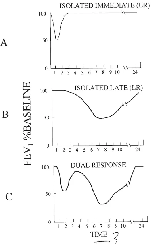
AI lergen challenge of sensitized atopic individuals results in an early response (ER), which in the skin is seen as the immediate wheal and flare reaction, and after inhalation as e decrease in airway caliber occurring within minutes (56). The eatly airway response (ER) occurs within minutes of inhalation of antigen, lasts up to an hour, and is followed by a prompt return to baseline lung resistance (57) (Figure 2A). The ER is mediated by IgE antibodies, which are present on mast ceils and basophils. These antibodies cross-link when in contact with allergen leading to degranulation of the cells and increased microvascular permeability (58). Bronchoconstriction of the airways during the ER is induced by the release of histamine, Ieukotrienes, eicosanoids, and possibly other bronchoconstrictive agents (59).
12
The late airway response (LR) occurs 4 to 12 hours after antigen exposure and may persist long after antigen exposure has ceased (60) (Figure 2E). In general, most atopic asthmatics develop both responses (Figure 2C), although occasionally, only an early or a late response is encountered (61). Airways of individuals experiencing the LR show increased edema and an infiltration with different inflammatory cells, in particular eosinophils and T lymphocytes (62,63).
1.2.2 Airway Hyperresponsivness
Airway hyperresponsiveness fAHR) is characterized by an increase in the sensitivity and reactivity of the airways to agonists, such as allergens, and the development of a lower threshold to spasmogens that will hinder airflow (64). AHR occurs before the symptoms of the LR arise and can remain after the symptoms have subsided (65,66). The type of physiological response s directly related to the degree and method by which various antigens are presented to the immune system in the airways of atopic asthmatics (67). Exposure to an antigen to which one is sensitized to may only cause an acute, transient increase in airway hyperresponsiveness, however continuous exposure to an allergen may cause an individual to reach a chronic state of
airway
inflammation and hyperresponsiveness (61).Inflammation is flot always necessary for AHR to occur. Bronchoconstrictive agents such as histamine, cholinergic agents and some -agonist blockers cause bronchospasm through smooth muscle contraction without airway inflammation (61).
AHR occurs through neuronal mechanisms and inflammatory mediator release which affects the sensitivity of the airways to stimuli and accordingly the amount of smooth muscle contraction. Consequently, increased AHR leads to a decrease in airway diameter (68). The relationship between inflammation and hyperresponsiveness in the airways is especially important during the LR. There s an increased presence of inflammatory cells and mediators in the airways during the LR (69).
Type 2 T helper lymphocytes (Th2) orchestrate the inflammation and are crucial for the development of AHR. Celis and molecules involved in T celI activation (dendritic cells, T celI receptor, major histocompatability complex molecule, and costimulatory molecules) are also vital. There are at least three pathways that lead to AHR. One is dependant on immunoglobulin E and mast celis, one on
B
I I I I12345678910
24
I I I I I I I I I]12345678910
24
T
Figure 2: Common patterns ofresponse afier pulmonary antigen challenge
ISOLATED IMMEDIATE (ER)
100
A
50
1
2 3 4
5
6
7
8
9 10
24
O
100
ISOLATED LATE (LR)
O
100
C
O
r
13
14
eosinophils and interleukin-5 (IL-5), and one on IL-13. Eosinophils are probably the most important effector ceils of AHR.
The nature of the relationship between AHR and atopy (specific lgE to aeroallergens) is unknown and one of the most important questions in asthma. It is clear from epidemiological studies that these two abnormalities are Iinked. However, flot ail allergic (atopic) people have AHR and flot ail individials with AHR are allergic. Furthermore, there is some suggestion that the risk factors for the two abnormalities are different.
12.2.1. Airway Hyperresponsiveness in Humans
Researchers have long used stimuli, such as histamine and methacholine, to measure the level
of AHR in patients with asthma and in animal models. By instilling or nebulizing incremental doses of
these mediators to the airway, they are able to assess and to quantify the threshold of tolerance toward non-specific irritant stimuli (70,71). AHR is often defined as a 20% fall in the FEV1 in response to a provoking agent, such as histamine, methacholine, or hypertonic saline, and sometimes to exercise or cold air hyperventitation. However a measure of AHR is not always considered to be a sign of asthma or even of airway inflammation (72). The amount of AHR to cholinergic agonists correlates only with the level of certain asthmatic symptoms such as wheezing and nocturnal cough and can give a limited prognosis of the severity of an asthmatic attack (72). Airway hyperresponsiveness is not restricted to non specific stimuli. AHR can occur in normal subjects following viral respiratory infection and can be present in atopic non-asthmatic individuals, in patients with chronic obstructive pulmonary disease or cystic fibrosis (73,74,75).
1.2.2.2. Airway Hyperresponsiveness in Animal Models
Airway responsiveness in the intact animal depends on tracheal smooth muscle airway contractiiity, chest wall compliance, bronchiolar mucus plugging, airway fibrosis, and other factors (76). AHR is a function of both airway hyper
reactivity
(an increased response to a given dose of bronchoconstrictor) and airway hypersensitivity
(the ability to respond to a smaller dose ofbronchoconstrictor) (77).
There are three ways to measure AHR (78). The
ex vivo
technique examines the contractility of dissected tracheal smooth muscle stimuiated by electricity or methacholine. The firstin vivo
methodmeasures the airway pressure and lung resistance of an anaesthetised animal with an intratracheal and intra-esophageal cannula (79). The second in vivo method uses unanaesthetised and unrestrained animaIs in a plethysmography box to produce a mathematically derived parameter called the enhanced pause (Penh) that reflects airway obstruction (78).
The ex vivo method differs from in vivo measurements because it neglects the effects of edema, mucus, or chest wall recoil on airway narrowing. The first in vivo method requires the technique of trachea and esophageal cannulations. The second in vivo method permits uninterrupted measurements, enabling the evaluation of early and late phase bronchial response and the discernment of airway hyperreactivity and airway hyperresponsiveness (80). Howevet, in some circumstances, the nasopharynx contributes significantly to the total airway resistance with this method.
1.2.3 Airway RemodellinglObstruction
Asthma has traditionally been thought of as an entirely reversible disorder. However, a number of studies have demonstrated that individuals with asthma experience an accelerated rate of respiratory function deterioration (81). Patients with asthma can develop a physiological state characterized by irreversible, or partially reversible, airway obstruction, and they manifest persistent AHR even after prolonged corticosteroid therapy. The pathogenic mechanisms responsible for these findings are poorly understood but they may be the resuit of structural changes, referred to as airway remodelling. Remodelling
in
chronic asthmatic airways is characterized by waii thickening, subepithelial fibrosis,mucous metapiasia, myofibroblast hyperplasia, vascular abnormalities, and myocyte hyperplasia and hypertrophy (82).
7.2.3.1 Aiway walI thickening
Ail components cf the airway wall (inner, outer and total) have been reported to be thickened in asthma. Many elements contribute to this response, including an increase in airway smooth muscle. edema, ïnftammatory celI infiltration (Figure 3), glandular hypertrophy, and connective tissue deposition. Compared to ncnasthmatic subjects,
airway
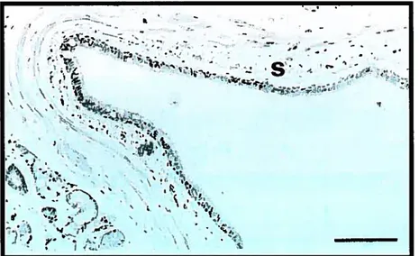
wall thickness is increased from 50% to 300% in cases cf fatal asthma, and from 10% to 100% in cases of non-fatal asthma (83). The exact physiologic andFigure 3: Photomicrographs of airway inflammation
(a) the airway submucosa (S) from a nonasthmatic
individual with few inflammatory celis, bar
=0.1 mm
(b) the submucosa from an individual with
fatal asthma with polymorphonuclear and
mononuclear celi infiltration, bar
=0.05 mm
Reprinted with permission from Holgate ST, Busse WW.,
Tnftammatory Mechanisrns in Asthma. Vol 117.
Marcel Dekker Inc.,NY, pg. 12, 2001.
• — f .• ..• ..•.. •• ‘•‘-..•_ s \ . -•: •15 j s. t t •55 ‘••‘s -u t ‘ sk’ 4 4,
k
I 4d%16
pathologic consequences of airway wall thickening are incompieteiy understood. Some models have demonstrated that the thickening response reduces the amount of smooth muscle shortening required to cause airway closure (84).
1.2.3.2 Basement Membrane Thickening
Collagen deposition beneath the basement membrane is described as “basement membrane thickening” (85). This has been recognized as increased coilagen types III and V, as weli as matrix components laminin and fibronectin along the basement membrane (composed of type IV collagen) (86). Multiple factors have been associated with subepitheliai coilagen thickening including increased frequency of asthma (87), longer duration of symptoms (88),
airway
hyperresponsiveness (89), increased T lymphocyte and fibrobiast activity (90,91), epithelial damage (92), as well as mast ceil and eosinophil infiltration (93). A possible mechanism is that growth factors reieased by cellular activity lead to increased myofibroblast numbers and activation observed below the basement membrane (94) where they appeat in relative abundance. Enhanced coilagen production by myofibroblasts and fibroblasts beyond the rate of collagen breakdown will lead to increased deposition. Potentiaily, the increased airway rigidity may contribute to mortality in asthma by reducing the maximal bronchodilator response. In addition, internai mucosai edema may cause significant internai luminai narrowing, without allowing outward radial expansion of the wall.1.2.3.3 Smooth Muscle Hypertrophy/Hyperplasia
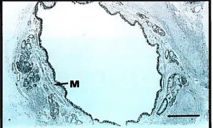
A 50-230% and 25-150% increase in the area of airway smooth muscle has been demonstrated in fatal and nonfatal asthma, respectiveiy (83) (Figure 4). These findings imply that the increased smooth muscle mass in some asthmatics may predispose to poorer iung function and a poorer response to a severe attack. Presumably, the factors known to be trophic for smooth muscle growth act unopposed in persistent airway inflammation. These inciude histamine, thrombin, thromboxane A2, and epidermal and platelet-derived growth factor (95). The modeliing studies of Wiggs and colleagues have indicated that shortening of inner airway walI smooth muscle by 40% may be sufficient to lead to airway closure (96).
Figure 4: Photomicrograph of airway smooth muscle
Reprinted with permission from Holgate ST, Busse WW.,
Inftammatory Mechanisrns in Asthma. Vol 117.
Marcel Dekker Inc.,NY, pg. 1$, 2001
(a) a nonasthmatic individual showing normal
airway smooth muscle (M), bar
=0.5
mm
(b) an individual with fatal asthma showing a
prominent layer of smooth muscle (M) surrounding
the folded airway mucosa, bar
=0.05 mm
Moreover, the added thickness may account for the loss of dynamic expansion of deadspace seen during inspiration in asthma (97).
1.2.3.4 Mucus Metaplasia
Mucus hypersecretion, epithelial mucus metaplasia, and airway obstruction due to bronchial mucus plugging are well-documented features of chronic asthma and status asthmaticus (98). Morphometric and immersion fixation studies demonstrate that the area of mucus glands is increased in fatal and nonfatal asthma. Liquid and mucus can also f111 in the interstices and folds of the airway surface. This could add to the forces tending to narrow the airway by amplifying the effect of muscle shortening and increasing the surface tension at the air-liquid interface.
1.2.3.5 Airway Vascularity
The bronchial mucosa of endobronchial biopsies from mild asthmatics has been shown to contain increased numbers of vessels per unit area (738 per mm2) (99). The increase in vascularity below the basement membrane and adjacent to the musculature is potentially capable of causing further luminal narrowing. Using anti-collagen type IV antibodies on biopsies from mild asthmatics, researchers have found significantly more vessels overali and more vessels larger than 300 pm2 in asthmatic airways, suggesting bronchial vasodilation in addition to angiogenesis. Canine models have demonstrated in vivo correlation between airway thickening and airflow obstruction (100). This engorgement might also contribute to the loss of airway distensibility seen in asthma (97).
1.3 Airway Inflammation 1.3.1 IgE
The secretion of IgE by B lymphocytes defines the allergic state, and the association between allergy and asthma is well established (101). The percentage of asthmatic subjects defined as allergic depends on whether subjects have a positive skin test to an aeroallergen in addition to elevated levels of serum IgE. IgE binds to high or low affinity receptors (FceR) and CD23, respectively) on the surface of a variety of effector celis, the most important of which are mast cells and basophils. The cognate antigen
20 crosslinks IgE bound to high affinity receptors on mast cells and on basophils causing the celis to release a variety of preformed and newly generated mediators that promote airway hyperresponsiveness, mucus hypersecretion, and increases in vascular permeability. The early asthmatic response is clearly dependant on IgE-mediated activation of mast cells through high affinity receptors (FceRI). Antigen specific IgE responses are regulated by HLA class Il and T-ceIl receptors and involve T-B cognate interaction while nonantigen-specific IgE response involve noncognate interaction of mast celis, basophils, and T and B cells (102). IL-4 is the most important cytokine mediating IgE synthesis and together with IL-13 plays a central role in the IgE-dependent allergic reaction.
The role of IgE in mediating an allergic airway reaction is confirmed by the finding that treatment of allergic asthmatic patients with monoclonal anti-IgE antibodies attenuated airway eosinophilia, increased the dose of allergen needed to provoke an early reaction, and reduced the mean maximum fali in FEV1 during the early and late responses to allergen challenge (103). Therefore, IgE has a direct role in mediating not only the ER, but also the LR.
While IgE-dependant inflammation plays a major part in allergic asthma, there are many inconsistencies. One study has demonstcated that total IgE s a poor diagnostic indicator of respiratory allergic disease (104) and in the African population serum levels of IgE have been reported to be higher in nonasthmatics than in asthmatics (105). Moreover, IgE knockout mice when challenged with allergen
can elicit an inflammatory tesponse in the airway as weIl as airway hyperresponsiveness (106). Even the syndrome of active anaphylaxis, with mast celi activation and mediator release can be displayed by both ovalbumin (OVA) sensitized lgE’ and FceRI-deficient mice after intravenous challenge with OVA (107,108). These studies cast a question mark on the precise role of IgE in allergic disease.
1.3.2 Eosinophils
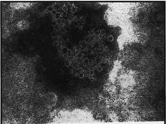
The current view of the eosinophil is that t is a proinflammatory celI with substantial tissue destructive potency. The biological activities exerted by the eosinophil are related to the products released from its granules, including the eosinophil cationic protein (ECP) and the major basic protein (MBP) (Figure 5) . These two potent cytotoxic proteins have the capacity to kiil both mammalian and non
,
ï’
Electron
photomicrograph
of
a
hurnan
peripheral
blood
eosinophil
with
the
crystalloid-containing
specific
granules
showing
electron-dense
cores
(C)
ofvarious
shapes
ernbedded
in
less
dense
matrix
(M).
Photo
courtesy
of
Dr.
Hirohito
Kita,
Mayo
Clinic,
Rochester,
MN
--Figure5
EosinophiÏ
gianules
—e -‘: C)
sf
I
:i-.
N
--ç--ç
Figure 6: Eosinophil Cationic Protein (ECP)
Electron microscopic demonstration ofthe effect of eosinophil
cationic protein on cellular membranes which can produce pores
with an approximate diarneter of 5nm.
Photo courtesy ofDr. Per Venge, Uppsala University,
Uppsala, $weeden
(Figure 6). The accumulation and activation of eosinophfls from the bone marrow to the lungs is governed by the upregulation of adhesion molecules on lung endothelial celis and the production of various cytokines and chemotactic molecules by mast cells and T cells. 0f these cytokines, IL-5 seems to play a central roTe, because it regulates most aspects of eosinophil behaviour, such as growth, apoptosis, adhesion, and secretion (109). Activation of the endothelium by cytokines such as IL-4 favours their migration to the lungs by upregulating the expression of vascular ce!! adhesion molecule 1 (VCAM-1) on endothelial cells (110).
Eosinophils differentiate within the bone marrow under the influence of GM-CSF, IL-3, and IL-5 (Figure 7). In response to allergic stimuli in the lung, the eosinophil pool in the bone marrow expands and the number of eosinophils residing in the blood and at the site of allergen provocation increase markedly. Circulating eosinophils are recruited into tissues following adhesion to endothelial celis expressing specific adhesion molecules, including VCAM-1, which recognizes vascular leukocyte antigen-4 (VLA-4) found on eosinophils, and ICAM-1 that binds CD11/CD18 molecules on a range of leukocytes types. Eosinophils migrate in response ta chemoattractants, including Iipid mediators, complement components, chemokines, and cytokines. Eosinophils express receptors for complement components C3a and C5a, for chemokines including 8, RANTES fCCR-1) and eotaxin (CCR-3), for cytokines including 1, 2, IL-3, IL-4, IL-5, IL-9, IL-16, GM-CSF, IFN-y, and TNF-Œ, and for immunoglobulins (1g) A, G, and E (high and 10w affinity) (111). Eosinophils are thus weII equipped ta respond ta a range of inflammatory stimuli.
Eosinophils are also an important source 0f inflammatory mediators. Among the lipid derivatives, eosinophils have the capacity to produce an amount of leukotrienes comparable with that of mast cells and basophils and higher than that of monocytes (Figure 7). Eosinophi!s also generate 15-HETE, lipoxins, platelet-activating factor (PAF), and small amounts of thromboxane2 (TXA2) and prostaglandin2 (PGE2). The eosinophil specific granule contains MBP in its core, and ECP, EPO, and EDN in its matrix. These granule-derived proteins have cytotoxic activity for helminths and are implicated in branchial epithelial damage (112).
Early studies on post-mortem Iungs obtained from patients who died of asthma showed significant eosinaphilia. Influx of eosinophilia into the branchoalveolar lavage (BAL) fluid was demonstrated during the late response aher allergen challenge of atopic subjects, at a time when
C
C
.,-4 CIDI)
(‘D.,-C
Qo
(‘DC
(f)C
o
I
.—
.—/
I
ç’
• —z
C?
—
—
• —
z
• —
(MrJ)
Ig—
C?
z
• —
I_
24
bronchial responsiveness is enhanced. In numerous clinical asthma studies, eosinophilia lias been demonstrated in bronchial biopsies, induced sputum, BAL fluid, and blood of allergic and nonallergic patients (113). Levels of MBP and ECP are elevated in lung tissue and BAL fluid obtained from asthmatics, suggesting eosinophil activation (112). Eosinophil numbers in biopsies and BAL fluid correlate with asthma symptom scores, Iung function (FEV1) and nonspecific AHR.
Although there is evidence of the eosinophil as a primary effector ceII leading to bronchoconstriction, epithelial damage and AHR in clinical asthma, its contribution in individual subjects may be highly variable (113). In several studies, a distinct proportion of patients with clinically significant asthma had negligible counts of eosinophils in bronchial biopsies or BAL fluid. Correlations between eosinophilia and AHR may be statistically significant, but individual patients may differ from this simple relationship, with activated T cells sometimes showing stronger correlations with measures of disease severity.
In contrast to wild-type litter mates, allergic IL-5 -I- mice do not generate eosinophilia in the blood
and bone marrow compartments in response to allergen provocation of the lung, and this greatly reduces the level of eosinophils recru ited to the airways (114). However, mature eosinophils stiil reside in the blood (albeit reduced numbers) and bone marrow compartments, indicating that baseline differentiation, maturation, and subsequent extramedullary migration persist in the absence of IL-5 (114). Results suggest that the primary role for IL-5 is in the promotion of peripheral eosinophilia in response to aHergic stimulation.
1.3.3 Neutrophils
The neutrophil has only recently boen the target of considerable interest regarding asthma pathogenesis. In recent years, the expansion of invasive studies to more severe forms of asthma, the advent of sputum analysis, concerns regarding the eosinophil as the most important effector celi and an appreciation of the properties intrinsic to the neutrophil, have Ied to an increased interest inthis celI type. The inflammatory products generated by the neutrophil range from cytokines, chemokines, and Iipid mediators to reactive oxygen species, various proteases and growth factors (115). The most important of
26 these are IL-8, LTB4, and matrix metalloproteinase (MMP)-9. IL-8, a potent chemoattractant for neutrophils, has been reported to be increased in asthmatic patients.
Neutrophils may predominate in the airways of patients with nocturnal asthma, sudden-onset fatal asthma, acute exacerbations or severe asthma poorly controlled by high dose glucocorticoids (116). Sputum analysis of asthmatics demonstrated that neutrophils were the prominent ceil in subjects with severe asthma, as weIl as mild asthmatics. Although the sputum reflects pathology
in
the larger airways, in both transbronchial biopsies and surgical evaluation of neutrophils in the small airways, there appears to be an increase in neutrophils, with specific localization to the small airway inner wall (117).It is not likely that the neutrophil plays any role in the acute bronchospasm associated with
asthma, given the profile of the mediators genetated by these celis. Neutrophils are more likely to be involved in chronic inflammation, wound repair, and remodelling processes of asthma. The neutrophil is
well-known to be part of the wound repair processes in the skin, eyes, and blood vessels (118). Studies indicate that the neutrophil may contribute to the fibrotic processes associated with asthma, particularly the basement membrane thickening, through its production of TGF-13 and MMP-9. TGF-f3 has been reported to be increased in bronchoalveolar fluid and in the celis of patients with asthma, with studies showing that at least 50% of these TGF-Ç3 ceils are neutrophils (117). MMP-9 has also been reported to be increased in asthma and in patients with status asthmaticus, but whether their source is the neutrophil or not is difficuitto prove. (119).
Neutrophils have also been suggested to play a role in mucus production and secretion, both prominent features in severe asthmatics. Several animal studies support a role for neutrophils and neutrophil elastase in both the upregulation of MUC-5 mRNA and protein, and in the degranulation of goblet cells (120,121). In animal models, removing the neutrophil appears to decrease mucus production (122). As well, data suggest that the neutrophil might be an important mediator of airway caliber in asthma but might not be an important mediator of bronchial hypersensitivity.
1.3.4 Mast Ceils
Mast ceils are key cellular participants in allergic disease (Figure 8). Their potential role in asthma was recognized early on with the identification of these ceils as major sources of the spasmogenic mediator histamine. The activation of mast cells is known to release a range of potent mediators of inflammation, including proteoglycans (heparin, chondroitin sulphate), proteases (tryptase, chymase), cytokines (IL-4, GM-CSF) and lipid mediators such as prostaglandins(PG) D2 and LIC4 which leads to bronchoconstriction. Ihese products may be stored
in
the prominent secretory granules of theseceils and are released following celI activation. Mast ceils can be stimulated to degranulate by cross linking allergen-specific IgE bound to high-affinity IgE receptors on the ceIl membrane. Mast cells express approximately 300 000 high affinity IgE receptorslcell, but cross-linking only 100 receptors will result in detectable responses (123). Mast cells may also be activated by diverse stimuli acting through other receptors (specific allergens, adenosine, neuropeptides, opiates). Histamine, the best studied of mast celi products, accounts for 5-10% of mast ceil granule content, and is stored in association with proteoglycans. Histamine receptor stimulation resuits in smooth muscle contraction, increased vascular permeability, and prostaglandin generation.
In the airways, mast cells are abundant in the mucosa. Ihey may also be ptesent in the submucosa, particularly in the vicinity of mucus glands, and small numbers are free in the lumen, where they are weil placed to respond to inhaled allergens. Mast celi derived mediators have been found in lavage fluid from patients with asthma, supporting the role of these cells in the immediate or early allergic reaction in asthma (123). When aHergen challenge preceded BAL, increases were documented for histamine, LTD4, PGE2, and tryptase (123). Ihe role of mast ceNs in the late allergic response has been more difficult to resolve. However, they are thought to play a key role in the development of the chronic inflammatory phase through their production of cytokines and chemotactic factors that lead to the recruitment 0f other celi types such as eosinophils (107).
o
E
E
C
7.3.5 Macrophages
Monocytes and macrophages are ceils of the mononuclear phagocyte lineage and are found in essentially every human tissue and body cavity. They derive from immature bone marrow precursors that are released into the blood, where they are termed monocytes, and are then recruited to tissues where they undergo tissue-specific terminal differentiation to macrophages. Macrophages exhibit a wide range of functions, ranging from the capacity to phagocytose and kili microorganisms, to presentation of antigen to T ceils and the release of large quantities of numerous soluble mediators (Figure 9).
Macrophages can be activated via the cross-linking of surface IgE bound to the high and low affinity receptors for IgE. Activation through the 10w affinity IgE receptor, FceRll, leads to release of soluble mediators, including proinflammatory cytokines and leukotriene B4 and C4 (124,125). Recent studies have shown greatly increased efficacy by monocytes of antigen uptake and presentation to T ceils via the high affinity IgE receptor, FceRl (126).
The pulmonary immune system represents a highly specialized and unique environment. Despite containing the greatest proportion of memory T ceNs of any compartment of the body and having continuaI exposure to foreign antigen in the air we breathe, littie or no immune activation is observed
in
the Iungs of normal healthy individuals. Such activation would be likely to result
in
damage to the fragile, permeable epithelial lining of the airways that permits gaseous exchange to occur, a primary and essential function of the lung. Macrophages play a central role in preventing such activation from occurring. Alveolar macrophages, located on the distal side of the epithelial lining of the ung, have a highly phagocytic and microbicidal nature. They are responsible for eliminating inhaled particulate antigens, such as microbes, allergens, and toxic substances, by physical means, namely ingestion followed by degraduation and elimination. In doing so, inadvertent and unnecessary immune activation is prevented.Figure
9:
Soluble
mediators
synthesized
by
mononuclear
phagocytes
and
their
potential
role
in
regulating
events
in
asthmatic
inflammation
IL-1,
TNF,
CSFs,
IL-10
chemokines,
prostaglandins
MEDIATORS:
Enzymes,
Complement
Prostaglandins
Leukotrienes
RESOLUTION
0f
INFLAMMATION.
REMO
VAL
0F
APOPTOTIC
CELLS
D
ACTIVATION,
MIGRATION
Smooth
muscle
celis
TGFs,
IL-l,
IL-ira,
IL-6,
chemokines,
PGE2
LTB4, LTC4, LTD4 TBX, PAF, PGD2, PGE.,ANTIGEN
PRESENTATION,
ACTIVATION
N
Th2
celis
FIBRO
SIS
Mucus-secreting
celis
Fibrob]asts
PGE-.
Epithelial
ceils
Fibrnectin
Endothelial
celis
Mast
celis
Basophuls
-Chemokines
LTB4.
PAF
Neutrophil
DIFFERENTIATION,
SURVI
VAL,
ADHESION,
TRANSMIGRATION,
PRIMING
1.3.6 Dendritic Ceils
Over the iast 25 years, it has become clear that dendritic celis (DC) are the major antigen presenting ceNs inducing the primary immune response in vivo (127). DCs capture antigens
in
the peripheral tissues and carry it into the T celi area of draining lymph nodes, where naive T lymphocytes are continuousiy re-circulating in search of specific antigens. Aithough many celi types such as macrophages and eosinophils have been shown to transport antigens into the draining nodes, the directed migration into the T celi area is a specialised function of DCs (128). The capacity to uptake antigens 15 a feature of immature DCs residing in peripheral tissues, and is largely lost during the migration of DCs into the draining lymph nodes. This way, immature DCs effectively make a “snapshot”of the antigens present in a peripheral inflammatory site. Interactions in the draining Iymph noUes are important for clonai expansion, differentiation, and avoidance of anergy in T ceNs.Lung DCs have an immature phenotype, specialized for uptake and recognition of inhaled antigen, but not yet capable of stimulating naive T celis, because they lack co-stimulatory molecuies (129,130,131,132). When antigen is encountered in an inflammatory context, there is a dramatic change in the behaviour of the DC5, a process called maturation. Upon recognition of foreign antigens, DCs have to migrate from the periphery to the draining Iymph nodes against the chemotactic gradient that attracts immature DCs. Therefore, upon recognition of antigen, DCs lose responsiveness to iung-expressed chemokines, e.g. by downregulation of the CCR6 receptor, but at the same time increase the expression of the CCR7 molecule, which directs the DCs towards the lymph noUes (133). The migration of airway DCs in response to an immunogenic stimulus is rapid and within 12h, lung derived DCs can be traced in the T ceil area of the mediastinai Iymph nodes of the iung (1 28,134,135).
The increased presence of DCs in the airways of atopic asthmatics and allergen exposed animais suggests that DC5 have a critical contribution to the disease pathogenesis (136,137,138). They have been associated with Th2-dependant sensitization Ieading to eosinophiihc airway inflammation (139). Reducing the number of DC5 eithet experimentaiiy or by inhaied corticosteroids is associated with a reduction in eosinophillic airway inflammation (140).
32
7.3.7 Lymphocytes
It is widely believed that T celis acting via the release of cytokines are central regulators 0f human
airway inflammation and in turn, the abnormalities in lung patho-physiology in asthma such as wide spontaneous fluctuations in airway caliber, bronchial hyperreactivity and airflow obstruction are the direct consequence of inflammation 0f the bronchial mucosa. Th2-based T cells (T ceil populations able to secrete IL-4, IL-5, IL-6, IL-9, and IL-13) are dominant effector ceils in the pathogenesis of asthma. Lymphocytes are activated by antigen-presenting celis and adopt functional phenotypes under instruction from soluble and physical signaIs that they receive during antigen presentation. They expand by proliferation, reaching sites of inflammation under the instruction of patterns of chemokines and adhesion molecules where, as armed effector cells, they affect other leukocytes. The abbreviations Thi (T helper
celi type 1) and Th2 (T helper celI type 2) have been classically referced to CD4 c3 TCR T ceil subsets
that are crucial to both the innate and adaptive immune systems. Other lymphocytes have been reported
to produce Thlrrh2-type subsets including y3 TCR T ceNs (141,142), and CD8 c43 TCR T cells (143).
Multiple Th forms have been described (e.g. Thi, Th2, Thp, ThO, and Th3) (141,144,145,146,147), and the terms naive, effector, and memory are often mentioned within the same context (141,144,148,149). Antigen-naive T cells are designated Thp for precursor of T helper celI (144). Antigen exposure to a Thp
ceil results in the selective maturation to either Thi or Th2 ceils. The Th phenotypes are characterized by
the cytokines they produce. The first Th ceil types characterized were mouse Thi and Th2 cells. Mouse Thi ceils secrete IFN-y, while Th2 ceils secrete IL-4 (150). In humans, Thi cells have been identified to secrete IFN-y, while Th2 ceils secrete IL-4 and IL-5 (151). Subsequent studies have established that Thi ceils produce IFN-y, TNF-f3, and IL-2, while Th2 cells produce IL-4, IL-5, IL-6, and IL-13 (144,147). Another Th ceil type with a unique cytokine secretion pattern is the Th3 ceIl (146,1 52,153) which appears to be a CD4 immune regulatory T celI that secretes TGF-3 (146). ThO ceNs have been described as producing both IL-4 and IFN-y (141,144), but their actual existence is controversial.
Thl and Th2 ceNs have been associated with specific immune responses due to the cytokines they secrete(Figure 10). For pathogens that require internalization, the presence of Thi cytokines (IFN-y and TNF-3) is consideted necessary. Conversely, for large extracellular parasites such as helminths, Th2-type cytokines (IL-4 and IL-5) have been consideted most protective (145,154,155,156). In the case of