ORIGINAL ARTICLE
Preserving the Morphology and Evaluating the
Quality of Liver Grafts by Hypothermic Machine
Perfusion: A Proof-of-Concept Study Using
Discarded Human Livers
Diethard Monbaliu,1*Qiang Liu,1*Louis Libbrecht,4Rita De Vos,2Katrien Vekemans,5
Charlotte Debbaut,6Olivier Detry,7Tania Roskams,2Jos van Pelt,3and Jacques Pirenne1
1Laboratory of Abdominal Transplant Surgery,2Department of Pathology, and3Liver Research Facility/
Hepatology Laboratory, Catholic University of Leuven, Leuven, Belgium;4Department of Pathology,
Ghent University Hospital, Ghent, Belgium;5Department of Health and Technology, Leuven University
College, Leuven, Belgium;6Biofluid, Tissue, and Solid Mechanics for Medical Applications, Institute of
Biomedical Technology, Ghent University, Ghent, Belgium; and 7Department of Surgery and
Transplantation, University Hospital of Lie`ge, Lie`ge, Belgium
The wider use of livers from expanded criteria donors and donation after circulatory death donors may help to improve access to liver transplantation. A prerequisite for safely using these higher risk livers is the development of objective crite-ria for assessing their condition before transplantation. Compared to simple cold storage, hypothermic machine perfusion (HMP) provides a unique window for evaluating liver grafts between procurement and transplantation. In this proof-of-con-cept study, we tested basic parameters during HMP that may reflect the condition of human liver grafts, and we assessed their morphology after prolonged HMP. Seventeen discarded human livers were machine-perfused. Eleven livers were nontransplantable (major absolute contraindications and severe macrovesicular steatosis in the majority of the cases). Six livers were found in retrospect to be transplantable but could not be allocated and served as controls. Meta-bolic parameters (pH, lactate, partial pressure of oxygen, and partial pressure of carbon dioxide), enzyme release in the perfusate [aspartate aminotransferase (AST) and lactate dehydrogenase (LDH)], and arterial/portal resistances were monitored during HMP. Nontransplantable livers released more AST and LDH than transplantable livers. In contrast, arte-rial/portal vascular resistances and metabolic profiles did not differ between the 2 groups. Morphologically, transplantable
Abbreviations:ALT, alanine aminotransferase; AST, aspartate aminotransferase; ATP, adenosine triphosphate; AUC, area under the curve; BSEP, bile salt export pump; CA-DCD, cardiac arrest during controlled donation after circulatory death; CCT, cranio-cerebral trauma; CV, central vein; DCD, donation after circulatory death; ECD, expanded criteria donor; END1, endothelin 1; HMP, hypothermic machine perfusion; ICAM1, intercellular adhesion molecule 1; ICB, intracranial bleeding; IL, interleukin; LDH, lactate dehydrogenase; LT, liver transplantation; MAPK, mitogen-activated protein kinase; MDR1, multidrug resistance 1; mRNA, messenger RNA; PBD, postanoxemic brain death; PCO2, partial pressure of carbon dioxide; PCR, polymerase chain reaction; PHA,
hepatic artery pressure; PO2, partial pressure of oxygen; PPV, portal vein pressure; PT, portal tract; QHA, hepatic artery flow; QPV,
portal vein flow; SCS, static cold storage; SEC, sinusoidal endothelial cell; TNF-a, tumor necrosis factor a; ZO-1, zona occludens 1.
Diethard Monbaliu and Jacques Pirenne are holders of a chair abdominal transplant surgery from the Centrale Afdeling voor Fractionering (Vilvoorde, Belgium).
This research was financially supported by grant IWT 040350 from the Institute for the Promotion of Innovation by Science and Technology in Flanders and by unrestricted educational grants from Roche and Astellas (Belgium).
*These authors contributed equally to this work.
Address reprint requests to Diethard Monbaliu, M.D., Ph.D., Laboratory of Abdominal Transplant Surgery, Catholic University of Leuven, Herestraat 49, B-3000 Leuven, Belgium. Telephone: þ32 16 348727; FAX: þ32 16 348743; E-mail: diethard.monbaliu@uz.kuleuven.ac.be DOI 10.1002/lt.23550
View this article online at wileyonlinelibrary.com.
LIVER TRANSPLANTATION.DOI 10.1002/lt. Published on behalf of the American Association for the Study of Liver Diseases
livers remained well preserved after 24 hours of HMP. In conclusion, HMP preserves the morphology of human livers for prolonged periods. A biochemical analysis of the perfusate provides information reflecting the extent of the injury endured. Liver Transpl 18:1495-1507, 2012.VC 2012 AASLD.
Received November 15, 2011; accepted September 3, 2012.
Expanded criteria donors (ECDs) and donation after circulatory death (DCD) donors represent an increas-ing proportion of the donor pool.1,2 However, livers from these donors remain underused because of the higher risk of poorer outcomes and the difficulty in assessing this risk.
To date, no objective tools are available for assisting clinicians in the evaluation of human liver grafts. The decision to accept or decline a particular liver offer is still based on the judgment of the surgeon, who takes into account the medical history of the donor, the liver biochemistry, the macroscopic appearance of the liver, and, if necessary, the microscopic appearance of the liver.
Compared to static cold storage (SCS), hypothermic machine perfusion (HMP) provides a window between procurement and transplantation during which the graft quality can be directly assessed ex vivo. Certain biomarkers in the perfusate and the vascular resist-ance during kidney HMP independently correlate with outcomes, and this additional information can help clinicians in decision making.3,4 In contrast, data on liver HMP are extremely scarce. Today, only 1 pilot trial has proven the feasibility of preserving standard livers with HMP and successfully transplanting them.5
The aim of this proof-of-concept study was to explore whether certain aspects of the quality of human livers could be assessed during their perfu-sion with standard biochemical, metabolic, and hydrodynamic parameters. For this purpose, we developed an HMP system for human livers and com-pared nontransplantable livers to control livers that could not be allocated, even though their quality was found to be good in retrospect. We also sought to determine whether the ultrastructural morphology of these livers thought to be transplantable could be pre-served after prolonged periods of HMP.
PATIENTS AND METHODS
Livers Discarded for Clinical Transplantation
From September 2003 to June 2009, 17 human livers initially accepted for liver transplantation (LT), which had been procured and allocated by Eurotransplant to particular recipients but eventually discarded, were machine-perfused. The reasons for discarding included unexpected macroscopic/microscopic find-ings during and/or after procurement, problems dur-ing transportation, and premature termination of the recipient operation (Fig. 1 and Table 1). According to Eurotransplant guidelines, all these declined grafts
were re-allocated to the next-in-line recipients at vari-ous centers but without success. The donor charac-teristics [age, sex, aspartate aminotransferase (AST), alanine aminotransferase (ALT), sodium, and total bil-irubin] are shown in Table 1. By design, these 17 dis-carded livers reached our laboratory after a prolonged period of SCS (mean ¼ 13 hours 48 minutes, range ¼ 3 hours 2 minutes to 24 hours 55 minutes).
Classification As Nontransplantable or
Transplantable Livers
Discarded livers were classified as nontransplantable or (in retrospect) transplantable according to generally accepted clinical criteria, medical data, surgical data, and macroscopic and microscopic appearances after their arrival at our laboratory and before the data analysis (Table 1).
The majority of the livers classified as nontrans-plantable (n ¼ 8) were massively steatotic (>60% mac-rovesicular steatosis). In 2 other nontransplantable livers, other contraindications were present in addi-tion to moderate (<30%) macrovesicular steatosis: high sodium levels and cholestasis/bilirubinostasis in one and diffuse Mallory bodies in another. Finally, 1 liver had severe histological signs of recent alcohol intake and acute hepatitis.
The livers that were potentially transplantable but could not be allocated included 2 livers that could not be re-allocated after the recipient operation was aborted because of the recipient’s death or the intra-operative discovery of extrahepatic metastases. In 1 case, the ice box containing the liver fell off the trolley when it was leaving the elevator, and the box was trapped and crushed between the closing doors. This liver was macroscopically and microscopically intact. In 3 cases, livers initially had been found to be non-transplantable by the procuring teams, and on the ba-sis of this first evaluation, they were successively declined by other centers and could not be allocated. Additional macroscopic and/or microscopic analyses showed only mild changes, and these livers were transplantable in retrospect. The reasons reported for discarding these livers included poor flush-out, mild steatosis, and subcapsular hematoma in one case and a suspicious cholestatic appearance of the liver along with a minimal elevation of the donor’s total bil-irubin level in another case. The third liver was ini-tially not offered for transplantation because of ele-vated transaminase levels after a short period of hemodynamic instability; however, at the time of kid-ney procurement, the liver appeared intact, and it was realized that although the transaminase levels were initially elevated, they were rapidly decreasing. This
liver was thus offered for transplantation but was declined by all centers, probably in part because of the short notice. Notably, these 6 discarded livers that were found in retrospect to be transplantable reached our laboratory after they were exposed to prolonged periods of SCS, which precluded their transplantation at our own center.
HMP Setup
A liver HMP device was derived from a LifePort kidney transporter (Organ Recovery System, Des Plaines, IL; Fig. 2). The livers were perfused by pressure-con-trolled continuous perfusion (20-30 mm Hg for the hepatic artery and 7 mm Hg or less for the portal vein). To prevent excessive shear stress on the endo-thelium, the portal flow was limited to 0.5 mL/g of liver/minute, and along with the arterial flow, this resulted in flows similar to those reported by Guar-rera et al.5 Experimental data on the oxygenation of the perfusate are conflicting. Both beneficial and det-rimental effects have been reported.6-10 In line with the only clinical experience available so far (reported by Guarrera et al.5), no active oxygenation of the per-fusate was used. All livers were machine-perfused for 24 hours with 2 L of University of Wisconsin machine perfusion solution. The temperature of the circulating perfusate was kept constant at approximately 4#C to 6#C.
Evolution of the Metabolic Profile During HMP
The pH, lactate level, partial pressure of oxygen (PO2), and partial pressure of carbon dioxide (PCO2) were measured with an ABL 625 analyzer (Radiometer, Co-penhagen, Denmark).
Evolution of Biochemical Parameters
During HMP
Perfusate samples were collected at the start of HMP and then after 30 minutes and 1, 6, and 24 hours. As a surrogate of hepatocellular injury, we measured AST and lactate dehydrogenase (LDH) in the perfusate with standard absorption techniques.
Evolution of Vascular Resistances During HMP
Arterial and portal vascular resistances were calcu-lated on the basis of perfusion pressures and flows. The perfusion flows were calculated by the multiplica-tion of the roller pump rotamultiplica-tions per minute by the volume delivered per revolution of the pump and were corrected for the size of the tubing used for the he-patic artery and portal vein. Vascular resistances were determined at the start of HMP and after 30 minutes and 1, 3, 6, 12, and 24 hours.
Assessment of Steatosis and Correlation
With AST in the Perfusate
The degree of steatosis according to baseline biopsy samples was quantified with (1) the stereological point counting method described by Franz!en et al.11and (2) semiquantitative scoring.
For the stereological point counting, 2 independent observers analyzed light microscopy images with mor-phometric imaging software (AnalySIS D, Olympus, Germany). Every observer scored 5 images of non-overlapping and randomly selected lobules (magnifica-tion $400). Within each image, 1 periportal zone and 1 centrilobular zone were selected and captured dis-tinctively. A point grid (108 crossings 35 lm apart) was superimposed onto each image. All vacuoles
Figure 1. Classification of livers as nontransplantable or trans-plantable according to the reasons reported to Eurotransplant for the livers being discarded. An asterisk indicates that the liver originated from a DCD donor.
T ABLE 1. Don or Ch aracte ristics for L ivers Pre served by HMP and Reas ons for D iscardin g Cas e Num ber HMP Date Sex Age (Y ears) AST (IU /L) AL T (IU/L) Sodiu m (mm ol/L) Bili rubi n (mg /dL) Cau se of Death SCS T ime (M in u te s) T ra n sp la n ta b le R ea so n s fo r D is ca rd in g * 1 Se ptemb er 2003 M a le 49 42 34 1 4 1 0 .3 C C T 6 1 1 N o S ev er e st ea to si s (> 60%) † 2 Nov embe r 2003 M a le 82 58 98 1 4 4 0 .6 8 IC B 1 2 0 7 Y es R ec ip ie n t n o t el ig ib le fo r tran splan tation bec ause of extr ahepati c hepatoc ellula r carcinoma 3 Nov embe r 2003 Mal e 19 46 36 156. 8 7 .78 CCT 663 No Hepa tome galy, b ilirubin ostasi s, †and mi cr o sc o p ic st ea to si s † 4 Nov embe r 2003 F em a le 42 35 34 1 4 4 0 .4 4 IC B 8 4 6 N o S ev er e st ea to si s (> 60%) † 5 Jan uary 2004 Femal e 58 46 22 151. 8 0 .35 CCT 182 No Pan lobular mediov esic ular stea tosis †with Mal lory bodies † 6 J u ly 2 0 0 4 M a le 43 23 28 1 4 9 0 .5 4 IC B 7 0 5 N o S ev er e st ea to si s (> 60%) † 7 J u ly 2 0 0 4 M a le 75 32 15 1 5 0 0 .4 C C T 1 0 6 5 N o S ev er e st ea to si s (> 60%) † 8 Octob er 2004 F em a le 55 40 31 1 3 9 0 .2 4 IC B 7 0 0 Y es P o o r fl u sh -o u t, su b ca p su la r bleed ing, and mild steat osis (< 30%) 9 Octob er 2004 F em a le 71 42 1 0 8 1 44 0 .3 7 IC B 8 5 1 N o S ev er e st ea to si s (> 60%) † 1 0 M a y 2 0 0 5 M a le 51 94 53 1 3 6 0 .7 IC B 4 6 4 N o S ev er e st ea to si s (> 60%) † 1 1 M a y 2 0 0 5 F em a le 64 36 65 1 4 2 0 .3 P B D 3 1 5 N o S ev er e st ea to si s (> 60%) † 12 June 2005 Mal e 58 50 23 1 28 0.7 ICB 1 394 No Ste atos is (> 60%) † 13 Jan uary 2006 M a le 46 19 54 1 4 1 2 .1 4 C C T 1 4 9 5 N o A lc o h o li c in to x ic a ti o n (alcoh olic steat ohepati tis †) 14 Jan uary 2006 Mal e 59 31 157 142. 6 1 .91 ICB 864 Y es Cho lestati c appear ance of donor liver † 1 5 M a rc h 2 0 0 6 F em a le 17 2 5 1 5 0 8 1 35 C er eb ra l isch emia 842 Y es Poor he pati c ci rculati on secon dar y to p reviou s isch emic trauma 16 March 2009 Mal e 39 13 12 1 47 0 .32 CA-DCD 882 Y es DCD and acc identa l trans por tation of icebox contai ning gr aft 17 June 2009 Femal e 47 62 17 1 43 0.3 CA-DCD 990 Y es D CD and d eath of recipi ent dur ing anhe patic pha se NOTE : The live rs are liste d in chro nolog ical or der . *A s re p o rt ed b y th e p ro cu ri n g or tr a n sp la n ti n g su rg eo n s to E u ro tr a n sp la n t. † Additi onal histo logi cal evide nce for disca rding live rs for trans plantat ion.
bonding to the grid crossings were counted and expressed as a percentage of the total number of crossings. Crossings outside the hepatocellular paren-chyma [eg, the portal tracts (PTs), central vein (CV), sinusoids, and artifacts] were excluded.
The semiquantitative scoring was performed by experienced liver pathologists. Macrovesicular steato-sis was defined as the presence of a single vacuole that was larger than the nucleus and displaced it in the vicinity of the cell membrane. Microvesicular stea-tosis was diagnosed when single vacuoles were smaller than half of the cytoplasm. Analogously to Spitzer et al.,12macrovesicular steatosis and
microve-sicular steatosis were expressed in 5% intervals with respect to the total liver parenchyma.
Analogously to a previous study that we conducted with a porcine model,13we examined whether a corre-lation between steatosis and AST release existed.
Correlation Between the Cold Storage Duration
and AST Release in the Perfusate
It was inherent to the experimental design that most of the studied livers reached our laboratory after a rel-atively prolonged period of SCS, during which all attempts were made by Eurotransplant to allocate
Figure 2. Schematic diagram of the HMP setup with 2 separate roller pumps integrated into 1 device. Livers were perfused with a pressure-controlled continuous perfusion of 20–30 mm Hg through the hepatic artery and 7 mm Hg or less through the portal vein. On the portal inflow side, the perfusion flow was limited to 0.5 mL/g of liver/minute, and this resulted in a total flow of 0.6 to 0.7 mL/ g of liver/minute. Two representative livers discarded for transplantation are shown. Case 2 involved a liver from an 82-year-old donor with a normal macroscopic appearance (classified as transplantable), and case 6 involved a liver with major (>60%) macrovesicular steatosis (classified as nontransplantable). The perfusate was chilled with a heater/cooler included in the circuit. Straight atraumatic cannulas (5-7 mm in diameter) with an appropriate conical tip were used for cannulation of the hepatic artery and the portal vein.
these livers. To investigate a potential bias due to the duration of SCS, we investigated whether there was a correlation between the duration of SCS and AST release [as assessed by the area under the curve (AUC)] in the perfusate.
Adenosine Triphosphate (ATP) Tissue Content
Snap-frozen liver samples taken before and after HMP were stored at %80#C. Subsequently, the samples were homogenized with 4.2% perchloric acid and 1 mM dieth-ylenetriamine pentaacetic acid and centrifuged at 14,000g, and the supernates were brought to pH 6 with 69% K2CO3. Then, ATP was measured with an ATPlite 1-step kit (PerkinElmer, Zaventem, Belgium) according to the manufacturer’s instructions with a Fluoroskan Ascent FL (Thermo LabSystems, Beverly, MA). ATP levels were expressed as nanomoles per gram of liver tissue.
Messenger RNA (mRNA) Level Quantification
We analyzed the mRNA levels of various cytokines [tu-mor necrosis factora (TNF-a) and interleukin-6 (IL-6)], adhesion molecules [intercellular adhesion molecule 1 (ICAM1)], transcription factors involved in inflamma-tion and apoptosis [jun proto-oncogene (JUN) and mitogen-activated protein kinase 1 (MAPK1)], proteins involved in signal transduction at tight junctions [zona occludens 1 (ZO-1)], endothelin 1 (END1), and bile salt transporters [multidrug resistance 1 (MDR1) and bile salt export pump (BSEP)]
Total RNA was isolated from snap-frozen liver tissue taken before and after HMP with the TRIzol reagent method (Invitrogen, Merelbeke, Belgium). The concen-tration and purity of the RNA were determined spec-trophotometrically at 260 and 280 nm. With 1 lg of total RNA, complementary DNA was synthesized with random primers (Invitrogen; 25 mM), a nucleotide mix (Promega, Leiden, the Netherlands; 0.5 mM), a recombinant RNase inhibitor (Invitrogen; 150 U), and Moloney murine leukemia virus reverse transcriptase (Invitrogen; 10,000 U). Primers and probes for beta-actin, BSEP, ICAM1, MDR1, TNF-a, and ZO-1 were designed with Primer3 version 0.4.0. For IL-6, JUN, MAPK1, and END1, we used gene expression assays (Applied Biosystems, Lennik, Belgium). Real-time po-lymerase chain reaction (PCR) amplification was per-formed with the ABI 7500 fast real-time PCR system (Applied Biosystems). The reaction was performed in duplicate in a total volume of 10 lL with TaqMan fast universal PCR master mix (Applied Biosystems). The thermocycling conditions consisted of 20 seconds at 95#C followed by 40 cycles of 3 seconds at 95#C and 30 seconds at 60#C. The fold change was calculated with theDDCtmethod.
Effect of Prolonged HMP on the Liver
Morphology and Ultrastructure
Standard and electron microscopy examinations were performed on biopsy samples taken after 24 hours of
HMP, and the results were compared to those for baseline biopsy samples. For standard microscopy, paraffin sections were stained with hematoxylin and eosin.
For electron microscopy, small liver biopsy frag-ments were trimmed into slices (thickness & 1 mm) and immediately fixed in 2.5% glutaraldehyde and a 0.1 mol/L phosphate buffer (pH 7.2) at 4#C. After 1 hour of postfixation in 1% osmium tetroxide and a 0.1 mol/L phosphate buffer (pH 7.2) at 4#C, the samples were dehydrated in a graded alcohol series and were embedded in an epoxy resin. Ultrathin sections (50-60 nm), which were cut and stained with uranyl acetate and lead citrate, were examined at 50 kV with a Zeiss EM 900 electron microscope. Images were recorded digitally with a Jenoptik Progress C14 cam-era system and Image-Pro Express software.
Ethical Approval
This study was approved by the ethics committee of University Hospitals Leuven (Catholic University of Leuven, Leuven, Belgium) and by the Belgian Liver and Intestine Committee, which represented all Bel-gian LT centers that provided livers for this study. The decisions to discard these livers were made by the procurement surgeons, transplant surgeons, physicians, and allocation organization (Eurotrans-plant) on the basis of the available information. These decisions were made completely independently of the possibility of the inclusion of these livers in a research protocol. After the termination of the experiment, the livers were sent to the pathology department.
Statistical Analysis
Data are expressed as means and standard devia-tions. The evolution of biochemical, metabolic, and hydrodynamics parameters was determined by the calculation of AUCs with Slide Write Plus (Advanced Graphics Software, Inc., Encinitas, CA). A Mann-Whit-ney test was used for comparisons at separate time points and for AUCs between groups. A sign test was used for comparisons between time points within groups. At every time point, there were sufficient data from both groups for adequate statistical analysis. The Pearson correlation coefficient was used to study the relationship between the degree of steatosis and AST release in the perfusate. Statistica 8.0 (StatSoft, Inc., Tulsa, OK) was used to perform the statistical analysis. A P value < 0.05 was considered significant.
RESULTS
Donor Data
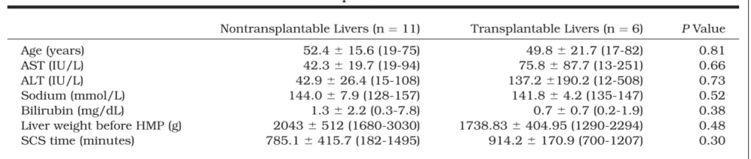
There were no differences between the livers of the 2 groups with respect to donor age, AST, ALT, sodium, total bilirubin, liver weight, or duration of SCS before HMP (Table 2).
Evolution of the Metabolic Profile During HMP
During HMP, the perfusate pH for all livers (nontrans-plantable and trans(nontrans-plantable) decreased from 7.28 6 0.13 at the baseline to 6.73 6 0.11 at 6 hours (P ¼ 0.003 for the baseline versus 6 hours) and to 6.40 6 0.19 at 24 hours (P ¼ 0.003 for 6 hours versus 24 hours). The lactate level increased from 0.9 6 0.22 mmol/L at the baseline to 7.94 6 3.69 mmol/L at 6 hours (P ¼ 0.004 for the baseline versus 6 hours) and to 14.47 6 4.92 mmol/L at 24 hours (P ¼ 0.004 for 6 hours versus 24 hours). PO2 decreased from 183.5 6 68.9 mm Hg at the baseline to 36.6 6 12.5 mm Hg at 6 hours (P ¼ 0.004 for the baseline versus 6 hours) and to 36.5 6 14.9 mm Hg at 24 hours (P ¼ 0.75 for 6 hours versus 24 hours). Finally, PCO2 increased from 8.0 6 7.1 mm Hg at the baseline to 23.4 6 8.0 mm Hg at 6 hours (P ¼ 0.008 for the baseline versus 6 hours) and to 20.1 6 6.3 mm Hg at 24 hours (P ¼ 0.75 for 6 hours versus 24 hours). There were no differences in the evolution of these metabolic parameters between the nontransplantable livers and the transplantable livers according to comparisons of their AUCs or their values at single time points (P > 0.05).
Evolution of Biochemical Parameters
During HMP
The AST release from the nontransplantable livers was remarkably higher than the release from the transplantable livers according to comparisons of the AUCs (9.39 6 6.31 $ 106versus 1.24 6 0.84 $ 106, P ¼ 0.006) and values at single time points: 30 minutes (4561 6 1853 versus 539 6 485 IU/L, P ¼ 0.009), 1 hour (4620 6 2062 versus 631 6 516 IU/L, P ¼ 0.003), 6 hours (7086 6 2928 versus 928 6 657 IU/ L, P ¼ 0.006), and 24 hours (8671 6 7294 versus 946 6 573 IU/L, P ¼ 0.002; Fig. 3A). Similarly, the LDH release from the nontransplantable livers was higher than the release from the transplantable livers accord-ing to comparisons of the AUCs (28.97 6 19.51 $ 106 versus 3.76 6 1.72 $ 106
, P ¼ 0.005) and values at single time points: 30 minutes (15,107 6 8164 versus 1564 6 1105 IU/L, P ¼ 0.008), 1 hour (14,442 6 8996 versus 1868 6 1167 IU/L, P ¼ 0.004), 6 hours (20,213 6 10,838 versus 2526 6 1348 IU/L, P ¼
0.009), and 24 hours (26,188 6 20,100 versus 3322 6 1311 IU/L, P ¼ 0.008; Fig. 3B).
Evolution of Vascular Resistances During HMP
During HMP, the AUC for arterial resistance (698.7 6 702.8) was higher than the AUC for portal resistance
Figure 3. (A) During HMP, AST release in the perfusate was higher for nontransplantable livers versus transplantable livers. (B) Similarly, nontransplantable livers released more LDH than transplantable livers.
TABLE 2. Representative Donor Characteristics and SCS Times for Nontransplantable and Transplantable Livers
Nontransplantable Livers (n ¼ 11) Transplantable Livers (n ¼ 6) PValue Age (years) 52.4 6 15.6 (19-75) 49.8 6 21.7 (17-82) 0.81 AST (IU/L) 42.3 6 19.7 (19-94) 75.8 6 87.7 (13-251) 0.66 ALT (IU/L) 42.9 6 26.4 (15-108) 137.2 6190.2 (12-508) 0.73 Sodium (mmol/L) 144.0 6 7.9 (128-157) 141.8 6 4.2 (135-147) 0.52 Bilirubin (mg/dL) 1.3 6 2.2 (0.3-7.8) 0.7 6 0.7 (0.2-1.9) 0.38 Liver weight before HMP (g) 2043 6 512 (1680-3030) 1738.83 6 404.95 (1290-2294) 0.48 SCS time (minutes) 785.1 6 415.7 (182-1495) 914.2 6 170.9 (700-1207) 0.30 NOTE: The data are presented as means and standard deviations with ranges in parentheses.
(16.2 6 20.8, P < 0.001). The arterial resistance grad-ually decreased during HMP from 1.29 6 0.67 to 0.52 6 0.47 mm Hg minute/mL. A similar phenomenon was observed, albeit to a lesser extent, for the portal resistance, which decreased from 0.25 6 0.30 to 0.13 6 0.13 mm Hg minute/mL. The AUCs for both portal and arterial resistances were similar for the nontrans-plantable and transnontrans-plantable livers (P ¼ 0.94 and P ¼ 0.48, respectively).
Correlation Between Steatosis and AST
in the Perfusate
Steatosis on baseline biopsy samples that was assessed by stereological point counting correlated with the AUC of AST release after 24 hours of HMP (P ¼ 0.006, correlation coefficient ¼ 0.9). In contrast, macrovesicular steatosis that was quantified semi-quantitatively did not correlate with the AUC of AST release after 24 hours of HMP (P ¼ 0.13, correlation coefficient ¼ 0.45). Microvesicular steatosis that was semiquantitatively quantified did not correlate with the AUC of AST release in the perfusate after 24 hours of HMP (P ¼ 0.12, correlation coefficient ¼ 0.47).
Correlation Between the Cold Storage Duration
and AST Release in the Perfusate
No correlation was observed between the duration of SCS before HMP and the AUC of AST release during HMP (P ¼ 0.44, correlation coefficient ¼ 0.25).
ATP Tissue Content
Before HMP, the ATP tissue content was 7.2 6 8.8 nmol/g in transplantable livers and 1.6 6 1.8 nmol/g in nontransplantable livers (P > 0.05). After HMP, the ATP tissue content was 12.7 6 22.7 nmol/g in trans-plantable livers and 2.5 6 2.3 nmol/g in nontrans-plantable livers (P ¼ 0.55).
mRNA Level Quantification
No changes in the mRNA expression of TNF-a, IL-6, ICAM1, JUN, MAPK, ZO-1, END1, MDR1, or BSEP were observed after HMP versus the start of HMP (P > 0.05).
Morphology and Ultrastructure After
24 Hours of HMP
According to standard microscopy, the parenchymal architecture of the livers deemed to be transplantable was maintained after 24 hours of HMP (Fig. 4A-D). All livers showed normal PTs without histological evi-dence of necrosis and/or apoptosis (Fig. 4C). A vari-able degree of sinusoidal dilatation was seen. Limited hepatocellular vacuolation was observed (Fig. 4D). Some anoxic vacuoles were observed in various zones extending from the CV into the PT but on other occa-sions remained mostly concentrated around the CV
(Fig. 4C,D). The distribution of this vacuolation was heterogeneous throughout the parenchyma, with some lobules containing numerous vacuoles and others containing no vacuoles.
According to electron microscopy, the ultrastructure of the hepatocytes and sinusoidal cells and the endo-thelial cell lining of the transplantable livers were well preserved after 24 hours of HMP (Fig. 5A-F). Some but not all hepatocytes contained anoxic vacuoles (Fig. 5D). The sinusoidal endothelial cells (SECs) and Kupffer cells showed no features of ischemic damage. The space of Disse and the sinusoidal lumina appeared slightly enlarged (Fig. 5D,E). Occasionally, slightly swollen mitochondria with a less electron-dense matrix (suggestive of some degree of autolytic yet reversible changes) were observed within certain hepatocytes (Fig. 5F). Mitochondria with flocculent densities (suggestive of more irreversible changes) were rarely seen. Other cytoplasm organelles and gly-cogen particles appeared normal (Fig. 5F).
In contrast to the transplantable livers, massive macrovesicular steatosis (>60%) was prominently present in 8 steatotic, nontransplantable livers according to standard and electron microscopy.
In 1 nontransplantable liver (case 13), there was evidence of alcoholic steatohepatitis at a septal stage. Another nontransplantable liver (case 3) displayed extensive coagulation necrosis (up to 40%) of the pa-renchyma together with hepatocellular ballooning and bilirubinostasis. After HMP, macrovesicular stea-totic lesions remained unchanged, and no obvious changes in the limited remaining hepatocellular pa-renchyma (including anoxic vacuolation) were observed. Similarly but to a lesser extent in compari-son with the transplantable livers, the space of Disse and sinusoidal lumina were enlarged. In these non-transplantable livers, more mitochondria tended to be irreversibly damaged in comparison with trans-plantable livers.
DISCUSSION
Because of the shortage of standard criteria donors, livers from ECDs (eg, donors with steatosis, older donors, and donors with hypernatremia) and DCD donors are increasingly being considered for trans-plantation. In the absence of objective criteria for transplantability, the final decision to accept or decline these higher risk livers remains subjective and is based solely on surgeons’ judgment. Although transplant surgeons are urged to consider higher risk grafts that may benefit the recipient population as a whole, they may be reluctant to expose individual patients to uncertain risks of poor outcomes. Conse-quently, a substantial number of grafts that may actually be transplantable are not used. The develop-ment of objective tools capable of assessing liver graft quality is a prerequisite for a real and safe expansion of the donor liver pool. In this proof-of-concept study, we have shown that a biochemical analysis of the per-fusate during HMP of human livers provides
information that may assist clinicians in decision making. We have also shown that the ultrastructural morphology of good-quality livers can be maintained for prolonged periods by HMP.
In comparison with SCS, HMP preservation has 2 potential advantages. First, continuously perfusing an organ with a cold preservation solution may provide better preservation than simple cold storage of the organ. A meta-analysis and randomized control trials have demonstrated that kidneys preserved by HMP function better and longer than those preserved by SCS.14,15Second, HMP allows us to directly evaluate preserved organs and may provide predictive informa-tion on the posttransplant outcome. The
concentra-tion of certain biomarkers reflecting cell necrosis (glu-tathione S-transferase, alanine aminopeptidase, N-acetyl-b-D-glucosaminidase, and heart-type fatty acid binding protein) in the perfusate of machine-perfused kidneys3and the vascular resistance during perfusion correlate with posttransplant function.4 The need to better preserve and assess ECD and DCD human liv-ers is also critical, and the revival of kidney HMP has also contributed to increased interest in liver HMP. A few animal studies have suggested a benefit for liver HMP versus SCS,6,16and a pilot clinical trial recently conducted by Guarrera et al.5 showed for the first time the feasibility (and potential superiority) of liver HMP. However, in contrast to kidney transplantation,
Figure 4. Representative histopathological findings (A,B) before and (C,D) 24 hours after HMP of a transplantable liver from a DCD donor that was rejected after the recipient was found to be unsuitable for transplantation (case 17). Panel A shows that the parenchyma was generally well preserved (magnification $100). Panel B shows that at a higher magnification ($400), the parenchyma next to the PT appeared normal. Panel C shows a variable degree of sinusoidal dilatation around the CV together with a variable degree of hepatocellular vacuolation in contrast to a rim of normal parenchyma around the PT after 24 hours of HMP (magnification $100). Panel D shows sinusoidal dilatation (arrows) and limited hepatocellular vacuolation after 24 hours of HMP, as occasionally observed, at a higher magnification ($400).
there are currently no data on the possibility of evalu-ating ECD or DCD liver grafts during HMP, and this was the main aim of this proof-of-concept study.
In analogy to kidneys, we hypothesized that the analysis of markers of cell necrosis in the perfusate of machine-perfused livers and their vascular resistance would provide valuable information on their baseline
condition. In a validated pig model of DCD LT, we pre-viously reported that AST in the perfusate correlated well with the length of the warm ischemia to which these porcine livers had been exposed and with the extent of damage and the associated risk of primary nonfunction after transplantation.17,18 However, in this porcine model (in contrast to what has been
Figure 5. Electron micrographs of liver parenchyma (A-C) before and (D-F) 24 hours after HMP of a transplantable liver (case 17). Panel A presents an overview of ultrastructurally well-preserved hepatocytes and sinusoids before HMP, whereas panel D shows that some hepatocytes with anoxic vacuoles and slightly dilated sinusoids were present after 24 hours of HMP. Panel B presents a detail of the sinusoidal lumen lined by a thin process of SECs before HMP; a Kupffer cell with heterogeneous lysosomes can be seen. Panel C shows a detail of a bile canaliculus formed by 2 adjacent hepatocytes before HMP; the mitochondria and other cytoplasmic organelles as well as glycogen particles (arrows) had a normal ultrastructure. Panel E shows that after 24 hours of HMP, SECs did not present signs of autolytic changes; a lymphocyte was lying in the sinusoidal lumen, and the endothelial cell lining appeared intact. Panel F shows that after 24 hours of HMP, only mitochondria were slightly swollen, with a less electron-dense matrix pointing to reversible changes; the other cytoplasmic organelles as well as the glycogen particles were normal. The following symbols are used in the figure: BC, bile canaliculus; D, space of Disse; glgn, glycogen; H, hepatocyte; K, Kupffer cell; L, lymphocyte; M, mitochondria; S, sinusoid; and V, vacuole. The scale bars represent (A) 7lm, (B) 1.5 lm, (C) 1 lm, (D) 1.5 lm, (E) 1.5 lm, and (F) 1 lm.
reported for kidneys), we observed no correlation between the severity of the ischemic damage endured and the arterial and portal vascular resistances dur-ing HMP.19
We have now developed and tested an HMP proto-type for human livers. In this proof-of-concept study, we adapted the perfusion protocol developed for renal HMP by performing dual (independent portal and ar-terial) perfusion and by using independent flows and pressure settings. Like Guarrera et al.,5no active oxy-genation of the perfusate was applied.
One of the first points of this study is that similarly to what we have observed for porcine livers, HMP is capable of maintaining liver morphology for long peri-ods.20Indeed, in good-quality livers not transplanted because of failed allocation, standard and electron mi-croscopy examinations showed well-preserved paren-chyma and an intact endothelial cell lining after 24 hours of HMP. The ultrastructure of hepatocytes and SECs was well maintained. Mitochondria displayed mostly mild and reversible changes. This is quite re-markable because these livers had been cold-stored for prolonged periods before they were machine-per-fused. Notably, vacuoles appeared in hepatocytes, although this was a limited phenomenon. We previ-ously described this vacuolization phenomenon in he-patocytes of pig livers exposed to warm ischemia.13 These vacuoles have also been called anoxic vacuoles because they seem to be the result of oxygen depriva-tion. In support of that, we found in another experi-ment that adding oxygen to the perfusate reduced the development of these vacuoles.7Importantly from the perspective of clinical applications, no technical prob-lems were encountered during prolonged HMP, and in particular, no vascular damage caused by the inser-tion of the cannulas was observed.
We then explored whether HMP of human livers could provide clinically relevant information on their condition. In accordance with our previous findings in pigs, differences were observed in the release of intra-cellular enzymes (AST and LDH) between massively steatotic, nontransplantable livers and control trans-plantable livers. These differences became apparent after only 30 minutes of HMP and persisted thereafter. Threshold values of 1500 and 4000 IU/L for AST and LDH, respectively, within the first hour of HMP differ-entiated nontransplantable livers from transplantable livers. This short time frame indicates that prolonged machine preservation is not necessarily required to provide clinically useful information. In our pig model, we found that liver-type fatty acid binding protein also correlated with the ischemic damage endured, and this is another potential marker of interest.18
Similarly to our earlier report on porcine HMP,20 PO2 in the perfusate decreased gradually, whereas PCO2increased in both groups. This indicates an ini-tially aerobic metabolism progressively replaced by some degree of anaerobic metabolism (as indicated by rising lactate levels and decreasing pH). However, no differences in metabolic profiles were observed between nontransplantable and transplantable livers.
It may be that the long period of cold ischemia that preceded HMP accounted for this absence of differen-ces in the metabolic profiles. Similarly, no significant differences in the tissue levels of ATP before and after HMP were observed when nontransplantable and transplantable livers were compared. In contrast to what has been reported for kidneys, no difference in vascular resistance was observed between nontrans-plantable and transnontrans-plantable livers. Similarly to our findings in pigs,20 the arterial resistance was con-stantly higher than the portal resistance and decreased during perfusion in both groups.
In this proof-of-concept study, only basic parame-ters similar to those routinely used to evaluate hypo-thermically perfused kidney grafts were measured. However, HMP technology provides additional possi-bilities, such as proteomic perfusate studies, more refined evaluations of ATP contents and energy stores, and examinations by magnetic resonance imaging and spectroscopy. Our group and others are studying the value of these more sophisticated methods.21,22
A potential intrinsic limitation of HMP, however, is that because of hypothermia, most aspects of cellular activity may remain quiescent. In support of this, we observed during HMP no changes in the expression of various mRNAs (TNF-a, IL-6, ICAM1, JUN, MAPK, END1, MDR1, BSEP, and ZO-1). Tulipan et al. com-pared mRNA changes in biopsy samples taken before and after preservation by HMP and SCS alone. No changes in monocyte chemoattractant protein 1 or IL-1 receptor antagonist were observed after HMP. In contrast, the monocyte chemoattractant protein 1 level was increased after SCS, and this suggested an active anti-inflammatory effect of HMP instead of met-abolic quiescent activity due to hypothermia itself.23 Furthermore, Guarrera et al.24 observed that the up-regulation of TNF-a, IL-8, and ICAM1, which is nor-mally seen after the transplantation of SCS-preserved livers, was reduced when livers had been preserved with HMP.
It is likely that the real-time assessment of metabo-lism, inflammation, and function will require more physiological midthermic or normothermic perfusion.25 An unexpected but interesting observation is that the concentration of AST in the perfusate had a linear correlation with the degree of steatosis assessed by stereological point counting. The underlying reason that steatotic livers release more AST during HMP is not clear. It may be that steatotic hepatocytes are more vulnerable to ischemic injury26,27and that this higher AST release simply reflects the suboptimal quality of these grafts. A correlate of this is that AST release from machine-perfused steatotic grafts may represent an additional and perhaps more reliable surrogate of the overall grade of steatosis in compari-son with conventional histology, which is subject to interobserver variation and sampling error as we and others have reported earlier.13,28
Our liver HMP setting (no active oxygenation and relatively low flow/pressure perfusion) was similar to the one used by Guarrera et al.,5who were the first to
successfully transplant human livers after 4 to 8 hours of machine perfusion. In contrast to our study, Guarrera et al. perfused predominantly standard crite-ria donor livers (eg, they excluded donors > 65 years of age, DCD donors, and donors with >25% macrovesicu-lar steatosis on biopsy). The primary aim of that trial was not to determine the value of HMP for assessing vi-ability but instead to prove the feasibility of preserving human livers by HMP in lieu of cold storage. Notably, AST in the perfusate of these predominantly standard criteria donor livers after 2 hours of HMP ranged from 307 to 609 IU/L5; these values are similar to those measured in the perfusates of the transplantable livers in our study and, therefore, corroborate our findings.
Our experimental design has intrinsic limitations, and our results need to be interpreted with caution. First, the reasons for discarding were multifactorial, and the classification of livers as nontransplantable or transplantable remains subjective. The group of nontransplantable livers was in fact homogeneous because most of them had massive steatosis (>60%). The reasons for discarding in the group of transplant-able livers were more heterogeneous. However, in the end, the quality of these livers was comparable and independent of the events leading to discarding. Importantly, the classification of livers as transplant-able or nontransplanttransplant-able was made before the data analysis. The fact that the release of AST and LDH in the perfusate was found a posteriori to be different between the nontransplantable and transplantable livers suggests that the original classification was accurate, and it supports ex post facto the concept that HMP can provide useful and objective data for assessing the degree of injury endured.
Second, HMP was preceded by a prolonged period of SCS that may have masked differences in metabolic profiles. This prolonged SCS may have exacerbated the release of AST and LDH, particularly in livers of poor quality, but no correlation was found between the duration of SCS and AST release. In addition, the prolonged SCS may have played a role in the clear and rapid decrease of PO2and the concomitant rise of the lactate level and PCO2. This decrease was not observed during HMP after shorter periods of SCS by Guarrera et al.,5 and this may point to the potential importance of active oxygenation during HMP when there is a long preceding period of SCS.
Third, the HMP prototype used in this study was derived from a kidney device, and certain adjustments more specific for the liver require further investigation. Lowering the perfusion pressure (to prevent excessive shear stress) results in better maintenance of ATP, less vacuolation, and eventually better preservation.7,8 Data on the value of oxygenation for the perfusate are conflicting. We used a setting similar to that of Guar-rera et al.,5who found that HMP of human livers had a favorable impact, despite the absence of active oxygen-ation of the perfusate. There are even data indicating that active oxygenation during perfusion may exacer-bate the production of radical oxygen species and eventually be detrimental.9 However, there is also
ex-perimental evidence from our group and others show-ing that oxygenation of the perfusate may be superior to nonoxygenated HMP,6,7,9probably because it main-tains a certain degree of aerobic metabolism and pre-vents at least partially a switch to an anaerobic metab-olism that we observed in this study. The presence of anoxic vacuoles on electron microscopy images reflects hypoxic trauma and suggests that supplemental oxy-genation of the perfusate may have been beneficial and will have to be tested. In support of this, oxygen persuf-flation of statically cold-stored pig livers has been shown by Minor et al.10to be protective.
Fourth, the ultimate test of viability that LT repre-sents could for obvious reasons not be done. To mimic LT ex vivo, we exposed an additional cohort of human livers to isolated and warm oxygenated reperfusion af-ter a period of HMP.29 In this latter experiment, we observed a linear correlation between AST in the per-fusate during HMP and AST release after warm oxy-genated reperfusion (data not shown). Notably, the maximum AST concentrations measured by Guarrera et al.5 in the perfusate of HMP-preserved human liv-ers correlated well with the AST peak that they observed after transplantation.
This proof-of-concept study is only a first step to-ward the development of new predictors of liver quality, and there is an urgent need for this. During the 6-year study period in Belgium, 60 procured livers were dis-carded (I. Tieken, MD, Eurotransplant, written commu-nication, January 2010), and high discard rates have also been reported in various European countries and in North America on the basis of subjective grounds alone. For massively steatotic livers, an assessment by HMP is probably superfluous. However, certain trans-plantable livers in our study had been discarded because of the overinterpretation of minimal changes or insufficient information at the time of procurement. It is likely that knowledge by the transplant surgeon of an objective indicator of minimal baseline damage (eg, low AST/LDH release as described in this study) would have encouraged their acceptance in the first place. Finally, in the future, it is likely that the availability of more objective and more sophisticated predictors of liver graft quality will stimulate LT surgeons to consider more ECD and DCD liver grafts for transplantation.
In conclusion, during HMP, nontransplantable, steatotic human livers release more AST and LDH in the perfusate, but they have similar vascular resistan-ces and metabolic profiles in comparison with trans-plantable human livers. HMP preserves the morphol-ogy of transplantable human livers for prolonged periods. Randomized control trials of the transplanta-tion of machine-perfused livers versus cold-stored liv-ers are now warranted to determine the added value of HMP not only for the preservation but also for the assessment of human liver grafts.
ACKNOWLEDGMENT
The authors thank Peter De Muylder, Bert Theunis, Jonathan Vercruysse, and John Brassil for their excellent assistance in the management of the
machine perfusion device; Veerle Heedfeld and Tine Wylin for their excellent assistance with this work; the members of the Belgian Liver and Intestine Committee and the different Belgian transplantation centers for their support; and Lydia Coolen and Lydia Vanden Wijngaert for their editorial assistance in the prepara-tion of this article.
REFERENCES
1. Wolfe RA, Merion RM, Roys EC, Port FK. Trends in organ donation and transplantation in the United States, 1998-2007. Am J Transplant 2009;9(pt 2):869-878. 2. Durand F, Renz JF, Alkofer B, Burra P, Clavien PA, Porte
RJ, et al. Report of the Paris consensus meeting on expanded criteria donors in liver transplantation. Liver Transpl 2008;14:1694-1707.
3. Moers C, Varnav OC, van Heurn E, Jochmans I, Kirste GR, Rahmel A, et al. The value of machine perfusion per-fusate biomarkers for predicting kidney transplant out-come. Transplantation 2010;90:966-973.
4. Jochmans I, Moers C, Smits JM, Leuvenink HG, Treck-mann J, Paul A, et al. The prognostic value of renal resistance during hypothermic machine perfusion of deceased donor kidneys. Am J Transplant 2011;11: 2214-2220.
5. Guarrera JV, Henry SD, Samstein B, Odeh-Ramadan R, Kinkhabwala M, Goldstein MJ, et al. Hypothermic machine preservation in human liver transplantation: the first clinical series. Am J Transplant 2010;10: 372-381.
6. de Rougemont O, Breitenstein S, Leskosek B, Weber A, Graf R, Clavien PA, Dutkowski P. One hour hypothermic oxygenated perfusion (HOPE) protects nonviable liver al-lografts donated after cardiac death. Ann Surg 2009; 250:674-683.
7. Vekemans K, Liu Q, Brassil J, Komuta M, Pirenne J, Monbaliu D. Influence of flow and addition of oxygen during porcine liver hypothermic machine perfusion. Transplant Proc 2007;39:2647-2651.
8. ’t Hart NA, der van Plaats A, Leuvenink HG, van Goor H, Wiersema-Buist J, Verkerke GJ, et al. Determination of an adequate perfusion pressure for continuous dual ves-sel hypothermic machine perfusion of the rat liver. Transpl Int 2007;20:343-352.
9. ’t Hart NA, van der Plaats A, Faber A, Leuvenink HG, Olinga P, Wiersema-Buist J, et al. Oxygenation during hypothermic rat liver preservation: an in vitro slice study to demonstrate beneficial or toxic oxygenation effects. Liver Transpl 2005;11:1403-1411.
10. Minor T, Koetting M, Koetting M, Kaiser G, Efferz P, Lu¨ er B, Paul A. Hypothermic reconditioning by gaseous oxy-gen improves survival after liver transplantation in the pig. Am J Transplant 2011;11:2627-2634.
11. Franz!en LE, Ekstedt M, Kechagias S, Bodin L. Semi-quantitative evaluation overestimates the degree of stea-tosis in liver biopsies: a comparison to stereological point counting. Mod Pathol 2005;18:912-916.
12. Spitzer AL, Lao OB, Dick AA, Bakthavatsalam R, Hall-dorson JB, Yeh MM, et al. The biopsied donor liver: incorporating macrosteatosis into high-risk donor assessment. Liver Transpl 2010;16:874-884.
13. Monbaliu D, Libbrecht L, De Vos R, Vekemans K, Walter H, Liu Q, et al. The extent of vacuolation in non-heart-beating porcine donor liver grafts prior to transplantation predicts their viability. Liver Transpl 2008;14:1256-1265. 14. Moers C, Smits JM, Maathuis MH, Treckmann J, van Gelder F, Napieralski BP, et al. Machine perfusion or
cold storage in deceased-donor kidney transplantation. N Engl J Med 2009;360:7-19.
15. Wight J, Chilcott J, Holmes M, Brewer N. The clinical and cost-effectiveness of pulsatile machine perfusion versus cold storage of kidneys for transplantation retrieved from heart-beating and non-heart-beating donors. Health Technol Assess 2003;7:1-94.
16. Pienaar BH, Lindell SL, Van Gulik T, Southard JH, Belzer FO. Seventy-two-hour preservation of the canine liver by machine perfusion. Transplantation 1990;49: 258-260.
17. Monbaliu D, Crabb!e T, Roskams T, Fevery J, Verwaest C, Pirenne J. Livers from non-heart-beating donors toler-ate short periods of warm ischemia. Transplantation 2005;79:1226-1230.
18. Liu Q, Monbaliu D, Vekemans K, Heedfeld V, Wylin T, Brassil J, Pirenne J, van Pelt J. Release of AST and LFABP from ischemically damaged livers during machine perfusion: a new tool to predict viability and primary-non-function [abstract]. Transpl Int 2009;22(Supplement 2):55 (O-210).
19. Derveaux K, Monbaliu D, Crabb!e T, Schein D, Brassil J, Kravitz D, et al. Does ex vivo vascular resistance reflect viability of non-heart-beating donor livers? Transplant Proc 2005;37:338-339.
20. Monbaliu D, Vekemans K, De Vos R, Brassil J, Heedfeld V, Qiang L, et al. Hemodynamic, biochemical, and mor-phological characteristics during preservation of normal porcine livers by hypothermic machine perfusion. Trans-plant Proc 2007;39:2652-2658.
21. Liu Q, Vekemans K, van Pelt J, Pirenne J, Himmelreich U, Heedfeld V, et al. Discriminate liver warm ischemic injury during hypothermic machine perfusion by proton magnetic resonance spectroscopy: a study in a porcine model. Transplant Proc 2009;41:3383-3386.
22. Liu Q, Monbaliu D, Vekemans K, Peeters R, De Keyzer F, Dresselaers T, et al. Can apparent diffusion coefficient dis-criminate ischemic from nonischemic livers? A pilot experi-mental study. Transplant Proc 2007;39:2643-2646. 23. Tulipan JE, Stone J, Samstein B, Kato T, Emond JC,
Henry SD, Guarrera JV. Molecular expression of acute phase mediators is attenuated by machine preservation in human liver transplantation: preliminary analysis of effluent, serum, and liver biopsies. Surgery 2011;150: 352-360.
24. Guarrera JV, Henry SD, Chen SW, Brown T, Nachber E, Arrington B, et al. Hypothermic machine preservation attenuates ischemia/reperfusion markers after liver transplantation: preliminary results. J Surg Res 2011; 167:e365-e373.
25. Vogel T, Brockmann JG, Friend PJ. Ex-vivo normother-mic liver perfusion: an update. Curr Opin Organ Trans-plant 2010;15:167-172.
26. De Gottardi A, Vinciguerra M, Sgroi A, Moukil M, Ravier-Dall’Antonia F, Pazienza V, et al. Microarray analyses and molecular profiling of steatosis induction in immor-talized human hepatocytes. Lab Invest 2007;87:792-806. 27. Angele MK, Rentsch M, Hartl WH, Wittmann B, Graeb C, Jauch KW, Loehe F. Effect of graft steatosis on liver function and organ survival after liver transplantation. Am J Surg 2008;195:214-220.
28. El-Badry AM, Breitenstein S, Jochum W, Washington K, Paradis V, Rubbia-Brandt L, et al. Assessment of hepatic steatosis by expert pathologists: the end of a gold stand-ard. Ann Surg 2009;250:691-697.
29. Vekemans K, van Pelt J, Komuta M, Wylin T, Heedfeld V, Detry O, et al. Attempt to rescue discarded human liver grafts by end ischemic hypothermic oxygenated machine perfusion. Transplant Proc 2011;43:3455-3459.