T
T
H
H
È
È
S
S
E
E
En vue de l'obtention du
D
D
O
O
C
C
T
T
O
O
R
R
A
A
T
T
D
D
E
E
L
L
’
’
U
U
N
N
I
I
V
V
E
E
R
R
S
S
I
I
T
T
É
É
D
D
E
E
T
T
O
O
U
U
L
L
O
O
U
U
S
S
E
E
Délivré par l'Université Toulouse III - Paul Sabatier Discipline ou spécialité : Immunologie
JURY
Pr. Joost van Meerwijk Président Pr. Peter van Endert Rapporteur Pr. Burkhard Becher Rapporteur
Pr. Roland Liblau Directeur de Thèse
Ecole doctorale : Biologie-Santé-Biotechnologies Unité de recherche : U563 INSERM Directeur(s) de Thèse : Pr. Roland Liblau
Rapporteurs : Pr. Peter van Endert
Présentée et soutenue par Amit SAXENA Le 9 April 2010
Titre :
Study of T cell pathogenicity in central nervous
inflammation of the central nervous system (CNS), axonal loss, and brain atrophy. The CD4+ and CD8+
T cells that are present in the demyelinating lesions are considered to mediate demyelination and axonal damage. An autoimmune process likely contributes to CNS tissue damage in MS, although direct evidence for this is still lacking. A major focus of my work is to understand the mechanisms by which autoreactive T cells contribute to CNS tissue damage.
Although CD8+
T cells mediate effector functions through production of cytokines or by direct cytotoxicity, the mechanisms by which they can cause CNS tissue damage are elusive. In order to assess whether CNS-infiltrating CD8+ T cells could directly induce oligodendrocyte death and demyelination, we developed an original mouse model combining selective expression of influenza hemagglutinin (HA) as a neo-self-Ag in oligodendrocytes with transgenic mice expressing a HA-specific TcR on CD8+ T cells. We demonstrate directly the potential of CD8+ T cells to induce oligodendrocyte death in-vivo, as a likely consequence of direct antigen-recognition.
Untill recently, CD4+
T helper cells have been held responsible for MS immunopathogenesis, partly because certain MHC class II alleles clearly predispose for developing MS. We investigated the pathogenic traits of the CD4+
T cell response targeting the immunodominant epitope of myelin oligodendrocyte protein (MOG). To this end we obtained 2D2 TcR-transgenic C57BL/6 mice, which harbor a large population of MOG-specific CD4+
T cells and spontaneously develop optic neuritis and at a lower prevalence, EAE. Strikingly when we crossed these 2D2 mice with MOG-deficient mice (MOG-/-), we discovered that the 2D2 TcR-transgenic mice developed spontaneous EAE regardless of the presence or absence of the target self-antigen MOG. Therefore we hypothesized, that the 2D2 TcR specific for MOG35-55 recognizes a second
CNS antigen. Furthermore, we were able to reveal, that MOG35-55 peptide shares sequence
homology with a stretch of neurofilament-medium (NF-M), a cytoskeletal protein expressed in neurons and axons. In addition, this epitope derived from NF-M shares 2D2 TcR contact residues with MOG35-55 when presented in the context of I-A
b
. Surprisingly, NF-M15-35 peptide showed
heteroclitic response when presented to 2D2 T cells. To test the in-vivo relevance of this observation, we transfer in vitro differentiated Th1 cells from 2D2-Rag2
mice in C57BL/6, MOG-/-, and MOG-/- x NF-M-/- mice. We observed no clinical sign of disease in MOG-/- x NF-M-/- mice, while MOG
mice developed slowly progressive disease with delayed onset, as compared to C57BL/6 mice, which developed early classical EAE, suggesting that recognition of MOG and NF-M by the 2D2 CD4+ T cells contribute to disease phenotype and severity.
We refer to this novel observation of self-mimicry, as ‘Cumulative autoimmunity’ where an autoimmune response target two self-autoantigens in the same tissue, resulting in enhanced CNS
It is an honor for me to thank to Prof. Joost van Meerwijk for his kind acceptance to be the president on my thesis defense. I would like to thank to Prof.
Peter van Endert and Prof. Burkhard Becher for their kind acceptance to be the
rapporteur for my defense.
I am sincerely grateful to my thesis Director, Prof. Roland Liblau, whose encouragement, supervision and support from initial to the final level enabled me to develop an understanding of the subject.
I owe my deepest gratitude to Dr. Lennart Mars, who was generously helpful and offered invaluable intellectual assistance, support and guidance.
I would like to thank to Dr. Daniel Dunia and Dr. Abdel Saoudi, who spent their time and shared their knowledge for helping me to complete my thesis. I would like to extend my thanks to all members of the lab and unit, who have helped me right from the beginning in accomplishing my pleasant and fruitful stay in the lab. I appreciate the help of Dr. Cassan during initial days.
I would like to thanks Dr. Kerschen, for his help and support during his stay in the lab. I would like to thank to Dr. Lazarczyk, Dr. Singh, Dr. Sharma, Dr. Mishra and Dr. Zein for their friendly support. I offer my regards to all of those who directly or indirectly become the part of my thesis.
I would like to express my gratitude to everyone from the University Paul Sabatier, animal house facility, FACS facility, administrative department, and ADR. I would also like to convey my thanks to French ministry, INSERM, ARSEP and FRM for providing the financial support and laboratory facilities.
Finally I would like to express my gratitude to my beloved family for their understanding and endless love, throughout the duration of my studies.
Acknowledgements
………... 2Table of content
………... 3List of figures
………... 6Abbreviations
………... 71. Introduction:
……….... 91.1. Generation of T-cell responses:
………... 111.1.1. Structure of the major components involved in antigen recognition 1.1.1.1. The major histocompatibility complex ……. ………….………….11
1.1.1.2. The T cell receptor ……….………….15
1.1.1.3. The CD3 complex ………..………….18
1.1.1.4. The CD4 & CD8 co-receptors ………..,………….20
1.1.2. Co-stimulation for naïve T cells and adhesion molecules 1.1.2.1. CD28 ………..………..22
1.1.2.2. Cytotoxic T lymphocyte-associated antigen-4 (CTLA-4) ………..23
1.1.2.3. CD80/CD86 ………..……….. 25
1.1.2.4. Inducible co-stimulatory molecule (ICOS) and its ligand (ICOSL).26 1.1.2.5. Programmed death-1 (PD-1) molecule ……….………..…….27
1.1.2.6. Adhesion molecules ……….………...29
1.1.3. T cell activation 1.1.3.1. Immunological synapse ………..……….32
1.1.3.2. TcR sensitivity ……….34
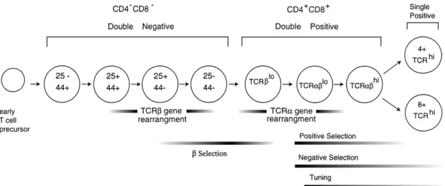
1.2.1.1. T cells ontogeny ……….. 40
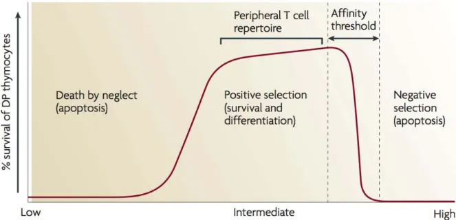
1.2.1.2. Positive selection of thymocytes ………..43
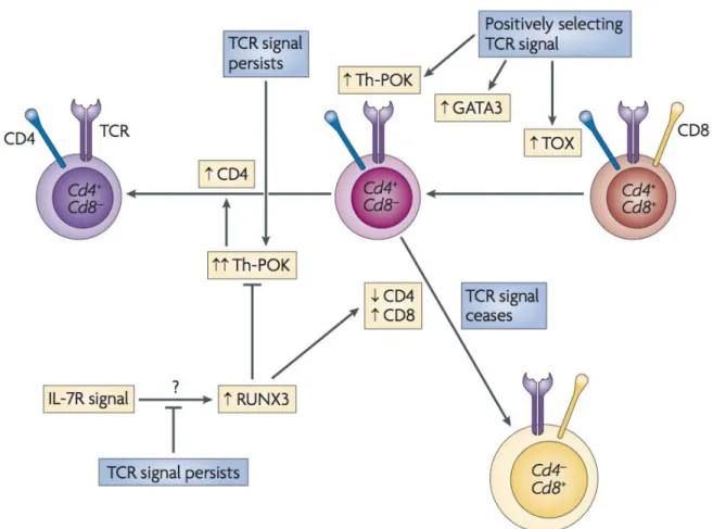
1.2.1.3. CD4 & CD8 lineage commitment ……….. 44
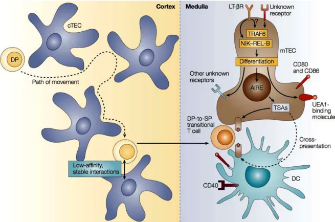
1.2.2. Central tolerance: 1.2.2.1. Role of autoimmune regulator (AIRE) ………48
1.2.2.2. Negative selection of thymocytes ………...51
1.2.3. Peripheral tolerance 1.2.3.1. T cell ignorance ………..52
1.2.3.2. T cell anergy ……….. 53
1.2.3.3. Apoptosis or Deletion ……… 55
1.2.4. Regulatory T cells 1.2.4.1. Naturally occurring FoxP3+ CD4+ CD25+ regulatory T (Treg) cells .58 1.2.4.2. T regulatory type1 (TR1) cells ………..67
1.3.
CNS inflammation and autoimmunity: ………681.3.1. Triggering auto-aggressive immune responses 1.3.1.1. Exposure of tissue-specific antigen ……… 68
1.3.1.2. Cryptic epitope hypothesis ……….. 71
1.3.1.3. Molecular mimicry ……….. 72
1.3.1.4. Dual TcR expression ………75
1.3.2. Multiple Sclerosis 1.3.2.1. Generalities ………..…78
1.3.2.2. Animal model for MS: EAE ………82
1.3.2.3. Pathophysiology ……….… 84
1.3.2.4. Effector mechanisms ………...91
2.1.1 Summary ………101
2.1.2 Publication ……….102
2.2. Neural self-mimicry: ‘Cumulative Autoimmunity’
2.2.1 Summary ………1072.2.2 Publication ……….108
3. Discussion & Perspective
……….………..1374. Bibliography
……….………150Figure 1 Architecture of MHC-like molecules ….. 13
Figure 2 Structural comparison of αβ and γδ TCRs ….. 17
Figure 3 The CD3 complex and T cell receptor signalling ….. 19 Figure 4 Dynamic processes of antigen recognition and T-cell activation ….. 33
by formation of microclusters and immunological synapse Figure 5 The cytokine milieu determines CD4+ T cell differentiation ….. 37
and conversion
Figure 6 Different stages of thymocyte development ….. 42
Figure 7 The affinity model of thymocyte selection ….. 43 Figure 8 Environmental cues and nuclear factors that influence CD4/CD8 ….. 45
lineage choice
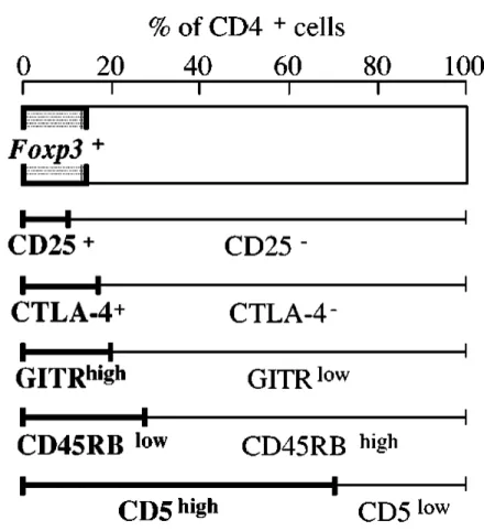
Figure 9 Aspects of the thymic medulla that are involved in central tolerance ….. 49 Figure 10 Cell surface markers for naturally arising CD4+ Treg cells ….. 60
Figure 11 Basic mechanisms of suppression used by Treg cells ….. 64 Figure 12 The evolution of the molecular mimicry concept and activation of ….. 73
autoreactive T cells via cross-reactivity with foreign antigens
Figure 13 Different scenarios for dual TcR T cells ….. 76
Figure 14 Geographical distribution of MS and migration ….. 79 Figure 15 Genes associated with risk of development of MS ….. 81
Figure 16 Immunopathogenesis of MS ….. 89
APL Altered peptide ligand
AIRE Autoimmune regulator
Bim Bcl-2-like protein
BBB Blood brain barrier
BAFF B-cell activating factor
C Constant
CD Cluster of differentiation
CDR Complementarity determining
region
CFA Complete freund’s adjuvant
CFSE 5,6-carboxy-succinimidyl-flurosceine-ester CL Clone CSF cerebrospinal fluid CTL Cytotoxic T lymphocyte CTLA-4 Cytotoxic-T-lymphocyte-associated Antigen-4 D Diversity DAG Diacylglycerol DC Dendritic cell DN Double negative
DNA Deoxyribonucleic acid
DP Double positive
EAE Experimental autoimmune
encephalomyelitis
ERK Extracellular-signal regulated
kinase
Fab Fragment antigen binding
FACS Fluorescent activated cell sorting
FCS Fetal calf serum
FoxP Forkhead box protein
GATA Trans-acting T-cell-specific
transcription factor
GAD Glutamic acid decarboxylase
G-CSF Granulocyte colony stimulating
factor
HA Hemagglutinin antigen
H&E Haemotoxylin and Eosin
HEL Hen egg lysozyme
HLA Human leukocyte antigen
GAD Glutamic acid decarboxylase
GM-CSF Granulocyte/Macrophage-colony
stimulating factor
ICAM Intercellular adhesion molecule
ICOS Inducible costimulator
IDDM Insulin dependent diabetes
mellitus
IDO Indoleamine 2,3-dioxygenase
IFN Interferon
Ig Immunoglobulin
IL Interleukin
IS Immunological synapse
ITAM Immunoreceptor tyrosine based
activation motifs
ITIM Immunoreceptor tyrosine based
inhibition motifs
ITSM Immunoreceptor tyrosine-based
switch motif
J Joining
JNK c-Jun-N-terminal kinase
kD Kilodalton
LAT Linker of activated T cells
LFA Lymphocyte function-associated
antigen
LN Lymph node
LPAM Lymphocyte peyer’s patch
adhesion molecule-1
MAPK Mitogen-activated
Protein-Kinase
MAdCAM Mucosal addressin cell adhesion
molecule
factor
MIP Macrophage inflammatory
protein
MHC Major histocompatibility
complex
MMP matrix metalloproteinases
MOG Myelin oligodendrocyte protein
MS Multiple sclerosis
NO Nitric oxide
NOD Non obese diabetic
NFAT Nuclear factor of activated T
cells
NK Natural killer
NOD Non-obese diabetic
ODC Oligodendrocyte
p Peptide
PBMC Peripheral blood mononuclear
cells
PBS Phosphate Buffer Saline
PCR Polymerase chain reaction
PKC Protein Kinase C
PLC Phosphoinositide-specific
phospholipase C
PLP Proteolipid protein
PMA Phorbol-12-myristate-13-acetate
PTK Protein tyrosine kinase
RAG Recombinase activating gene
RANTES Regulated upon activation,
Normal T expressed, and secreted
RNA Ribonucleic acid
ROR Retinoid-related orphan receptor
RPMI Roswell oak memorial institute
medium
RT Room temperature
Runx Runt-related transcription factor
SLP SH2 domain containing
leukocyte protein
SP Single positive
TSA Tissue specific antigen
TcR T cell receptor
TGF Transforming growth factor
Th T helper
ThPOK Zinc finger and BTB domain
containing 7B
TN Triple negative
TNF Tumor necrosis factor
Treg Regulatory T cell
V Variable
VCAM Vascular cell adhesion molecule
VLA Very late antigen
ZAP Zeta associated protein
1.
Introduction:
The immune system has two major components, an innate arm and an adaptive arm. The innate immune system is a universal and ancient form of host defense against infection. Innate immune system recognition relies on a limited number of germline-encoded receptors. These receptors evolved to recognize conserved metabolites produced by pathogens, but not by the host. Recognition of these molecular structures allows the immune system to distinguish infectious nonself from noninfectious self. Innate immunity covers many areas of host defense against pathogenic microbes, including the recognition of pathogen-associated molecular patterns (PAMPs) (Janeway, 1989). In contrast, the adaptive immune system involves great variability and rearrangement of receptor gene segments to generate receptors, which yield myriad of antibodies or T cell receptors (TcRs) of exquisite specificity for each of potential antigens, additionally the adaptive immune system is characterized by immunological memory. However, the adaptive immune response is also responsible for allergy, autoimmunity, and the rejection of allograft.
Because the innate immune responses do not depend on gene rearrangements, they occur much more rapidly than adaptive immune responses and do so in more stereotyped way, without tailoring a specific receptor to neutralize the danger signal in the form of antigen. Instead, in innate immunity, the danger signal (a motif from infectious microbes) can trigger toll-like receptors (TLRs), C-type lectin receptors (CLRs) to a membrane-based sensor of danger (Hemmi et al., 2000). In addition, the innate immune system has a cytosolic system to sense danger, the Nod-like receptors (NLRs), an intracellular system. The inflammosome is where signals from the TLR system and the NLR system are integrated. The major constituent of the inflammosome is the cytokine interleukin-1 (Church et al., 2008; Lamkanfi and Dixit, 2009; Martinon et al., 2009). The innate immune system is made of many cell types, such as macrophages, dendritic cells (DCs), mast cells, neutrophils, eosinophils and NK cells. These cells can become activated during an inflammatory response, which is a consistent sign of infection with a pathogenic microbe. Such cells rapidly differentiate into short-lived effector cells whose main role is to get rid of the infection. However, in certain cases, the innate immune system is unable to deal with the infection, and activation of an adaptive immune response becomes necessary. In these cases, the innate immune system can instruct the adaptive immune system about the nature
of the pathogenic challenge. It does so through cytokines and chemokines and the expression of costimulatory molecules, such as CD80 and CD86, on the surface of specialized antigen-presenting cells (APCs), with DCs as the most important ones that alarm from infection in virtually all tissues (Banchereau and Steinman, 1998; Fearon and Locksley, 1996; Janeway, 1989).
The central nervous system (CNS) provides special anatomic barriers to the movement of cells from the vascular compartment to the parenchyma. A group of autoimmune diseases of the brain and spinal cord that represents useful model for constitute a situation which involve antigen-specific T cells and an orchestrated antibody response to CNS components of the brain or spinal cord. My work is mainly focus on the autoimmune diseases where the brain and spinal cord itself are attacked.
1.1
Generation of T-cell responses:
1.1.1. Structure of the major components involved in antigen recognition 1.1.1.1 The major histocompatibility complex
The major histocompatibility locus has evolved to be polygenic and highly polymorphic, and is located on the short arm of chromosome 6 in humans, and on chromosome 17 in mice (Artzt, 1986; Klein et al., 1982; Ziegler, 1997). In humans the major histocompatibility complex (MHC) I locus contains 3 classical class I genes, named human leukocyte antigen-A (HLA-A), HLA-B, HLA-C and non-classical HLA-E and HLA-G, while the MHC II locus comprises HLA-DR, HLA-DQ and HLA-DP. In mice, the MHC I locus contains 3 main genes, H-2L, H-2D and H-2K, while the MHC II region comprises 2 main genes H-2IE and H-2IA, generally abbreviated to IE and IA (Bodmer et al., 1997; Trowsdale and Campbell, 1992).
Both classes of MHC molecules are heterodimers with similar architectures and are composed of three domains, one α-helix/β-sheet superdomain that forms the
peptide-binding site and two immunoglobulin (Ig)-like domains (Fig.1) (Bjorkman et al., 1987b; Brown et al., 1993; Fremont et al., 1996; Madden et al., 1993; Matsumura et al., 1992; Stern and Wiley, 1994). The overall architecture is the same in both MHC classes, where a
seven-stranded β-sheet represents the floor of the peptide-binding groove, and the sides are
formed by two long α-helices, that straddle the β-sheet.
In MHC class I molecules, the peptide-binding site is constructed from the heavy chain only, and an additional 12-kDa light chain subunit, β2-microglobulin (β2m), associates with the α3 domain of the heavy chain (Fig.1). β2m is a nonglycosylated protein of about 100 amino acids encoded on chromosome 15 in humans and chromosome 2 in mice. Only one allele is known in humans, while there are seven in mice. There appears to be little functional significance of the polymorphisms in β2m, although they may subtly affect MHC class I structure or peptide binding because different β2m alleles can alter cytotoxic T lymphocyte (CTL) recognition of some antigens (Perarnau et al., 1990). β2m has no transmembrane domain and is not covalently associated with the extracellular region of the heavy chain. Because of this, the conformation of the heavy chain is highly dependent on the presence or absence of β2m chain. The membrane-proximal α3 region of the heavy chain is an Ig-like domain that contains a binding site for the CD8 receptor on CTL. The α1 and
α2 domains, which are distal to the membrane and interact with the TcR on CTL, fold together to form a groove that binds and displays peptides. A β-pleated sheet forms the base
of the cleft, and the walls are made of two α helices. The allelic polymorphisms in heavy
chain primarily occur in those residues in and around this cleft, and in this manner, they alter the peptide-binding specificity of the class I molecules (Bjorkman et al., 1987a; Parham et al., 1995). The crystal structure of the MHC class I proteins revealed how a single type of molecule could bind and present so many different peptides. Many of the molecular interactions are with the peptide's main chain atoms and amino and carboxy termini, which are features common to all peptides. In addition a limited number of peptide side-chains extend into pockets along the groove thus imparting some specificity to peptide binding, although many different peptides can be accommodated (Wilson and Fremont, 1993), and determine general motifs of the peptides capable of binding to particular MHC class I molecules (Falk et al., 1991). The groove is generally long enough to accommodate 8 or 9 residues in an extended conformation (Madden et al., 1991) with the termini and the so-called anchor residues buried in specificity pockets that differ from allele to allele (Fremont et al., 1992; Madden et al., 1993). This binding mode leaves the upward-pointing peptide side chains available for direct interaction with the TcR. Longer peptides can either
bind by extension at the C terminus (Stern et al., 1994) or due to the fixing of their termini, bulge out of the binding groove, providing additional surface area for TcR recognition (Speir et al., 2001; Tynan et al., 2005).
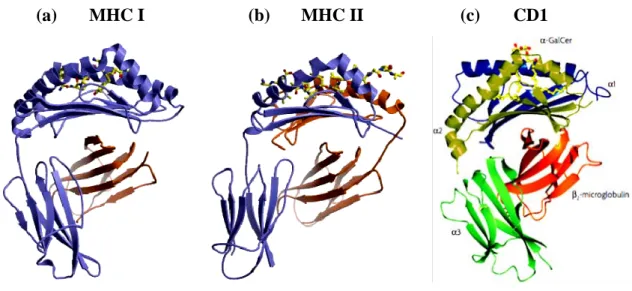
(a) MHC I (b) MHC II (c) CD1
Figure 1: Architecture of MHC-like molecules
(a) Class I molecules consist of a heavy chain (blue) and a light β2m chain (orange). The
peptide-binding site is formed exclusively by elements of the heavy chain (b) Class II molecules; the peptide-binding site is assembled of both subunits. (Rudolph et. al. 2006) (c) Nonclassical MHC molecule mouse CD1d crystal structure loaded with a synthetic variant of alpha-galactosylceramide (alpha-GalCer) (Barral et. al. 2007)
In contrast, the MHC class II molecule are assembled from two heavy chains (αβ) in
which the peptide-binding groove is open at either end, and the peptide termini are not fixed so that bound peptides are usually significantly longer than in MHC class I (Fig.1). The MHC class II allows presentation of peptides of 13-18 residues. The peptide backbone in MHC class II is confined mainly to a poly-proline type II conformation (Stern et al., 1994) and resides slightly deeper in the binding groove. Thus, the bound peptide is more accessible for TcR inspection in MHC class I due to its ability to bulge out of the groove, even for 9-mer peptides, however, in MHC class II the termini particularly the N-terminal extension, can play a major role in the TcR interaction.
T cells are sensitive to small numbers of antigenic pMHC ligands that are distributed among an excess of endogenous pMHC complexes on the surface of APCs. Recognition of
molecules by specific TcRs is central to T-cell activation. Two distinct pathways are used by MHC class I and class II molecules for the presentation of peptide antigens to CD8+
and CD4+
T cells, respectively (Ackerman and Cresswell, 2004; Kloetzel and Ossendorp, 2004; Rock et al., 2004; Watts, 2004). The MHC class I antigen presentation pathway is active in almost all cell types, providing a mechanism for displaying at the cell surface a sample of peptides derived from proteins that are being synthesized in the cell and are acquired during initial assembly in the endoplasmic reticulum (Cresswell et al., 2005; Kloetzel and Ossendorp, 2004; Rock et al., 2004). The MHC class II pathway is constitutively active only in professional APCs, including DCs, B cells, macrophages and thymic epithelial cells. The peptide presented by MHC class II molecules are derived from proteins that gain access to endosomal compartments, providing a way for CD4+ T cells to respond to exogenous antigens internalized by APCs. The α-chain and β-chain of MHC II molecules are
synthesized in the endoplasmic reticulum (ER) and associate with invariant chain (CD74) for proper folding, trafficking and protection of the antigen-binding groove (Bryant and Ploegh, 2004; Cresswell, 1996; Jensen et al., 1999; Watts, 2004).
A third lineage of antigen presenting molecules, distinct from MHC class I and MHC class II has been identified as CD1, which involves CD1a, CD1b, CD1c, CD1d and CD1e, and present both foreign and self-lipid antigens to T cells. The CD1 proteins share sequence homology and overall domain structure with MHC class I molecules, being constitute of a heavy chain with three extracellular domains that are non-covalently associated with β2m (Zeng et al., 1997) (Fig.1). On the basis of sequence analysis, the CD1 isoforms can be classified into three groups: group 1 constitutes of CD1a, CD1b and CD1c; group 2 constitutes CD1d; and group 3 constitutes CD1e. In humans all five CD1 isoforms (CD1a–CD1e) have been identified, whereas muroid rodents express only CD1d that include CD1d1 and CD1d2 (Porcelli, 1995). Group 1 CD1 molecules mainly present lipid antigens to clonally diverse T cells that mediate adaptive immunity to the vast range of microbial lipid antigens (Beckman et al., 1994; Sieling et al., 1995). By contrast, CD1d molecules present lipid antigens to natural killer T (NKT) cells (Bendelac et al., 1995).
1.1.1.2 The T cell receptor
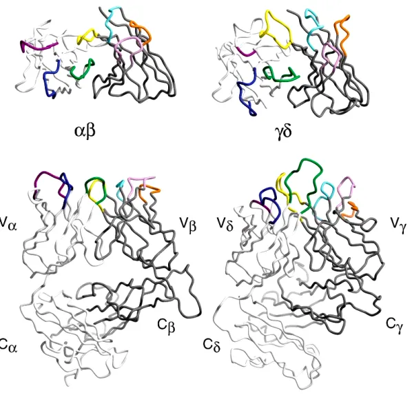
TcRs are cell surface heterodimers consisting of either disulfide-linked αand β or γand δ-chains (Brenner et al., 1986; Chien et al., 1987; Hedrick et al., 1984; Koning et al., 1987; Saito et al., 1984; Winoto and Baltimore, 1989; Yanagi et al., 1984). Sequence analyses correctly predicted that TcRs would share a domain organization and binding mode similar to those of antibody Fab fragments (Davis and Bjorkman, 1988).
The αβ TcR comprises 2 membrane proximal constant (C) regions and 2 membrane
distal variable (V) Ig-like domain regions (Garboczi et al., 1996; Garcia et al., 1996a). The pairing of the membrane distal α and β chain variable regions resembles the Ig variable
domain, such that the αβcombining site resembles the Ig-combining site (Fig.2). The loops connecting the βsheets located within the pMHC contact area consists of regions with highly variable amino acid sequences, called complementarity-determining regions (CDRs). The αβTcR binds pMHC with relatively low affinity (1–100µM) through CDRs presented in their variable domains.
Interaction between TcR and pMHC based on the three-dimensional structure of the pMHC moiety and X-ray crystallographic structure (Davis and Bjorkman, 1988; Machius et al., 2001) show that CDR1 and CDR2 of both the αand βchains of the TcR interact preferentially with the helices of the MHC molecule and that contacts to the bound peptide are made essentially by CDR3 alone (Garboczi et al., 1996; Garcia et al., 1996a; Teng et al., 1998; Wilson and Garcia, 1997).
The first crystal structures of TcRs with MHC class I molecules led to proposals that the TcR orientation is approximately diagonal with a mean around 35° (Rudolph and Wilson, 2002). By contrast, in the first MHC class II complexes, the orientation was described as being closer to 90° (Hennecke et al., 2000; Reinherz et al., 1999) suggesting a different binding mode between the MHC classes (Wang and Reinherz, 2002).
Insight into the structural changes that supplement TcR-pMHC engagement must include crystal structures of the same TcR in its free and bound forms or of the same TcR bound to different pMHCs. Until recently, only two well-studied systems, the 2C and A6 TcRs, fullfilled these requirements. The 2C system allowed comparison of the free 2C TcR (Garcia et al., 1996a) with an agonist (Garcia et al., 1998) and a superagonist peptide
functional hotspot between the CDR3 loops in the 2C TcR that finely discriminated between side chains and conformations of centrally located peptide residues through increased complementarity and additional hydrogen bonds. In the A6 system, altered peptide ligands (APLs) induced only subtle conformational changes in the TcR. In both the 2C and A6 systems, conformational changes are restricted mainly to the CDR3 loop regions, and the largest conformational differences were observed when comparing free versus bound TcRs (Rudolph and Wilson, 2002).
Many studies revealed substantial degeneracy in TcR recognition (Hemmer et al., 1998; Hunt et al., 1992; Loftus et al., 1999; Pinilla et al., 1999). Multiple structures comparing bound and unbound TcRs have shown, that the CDR 3 loops are flexible (Garcia et al., 1998; Reiser et al., 2003). Thus, the conformational flexibility of these regions may enable polyspecificity through induced-fit recognition of multiple peptides within the binding cleft of the same MHC molecule.
Figure 2: Structural comparison of αβ and γδ TCRs.
The top panel is a view down into the binding site, highlighted by the CDR loops. The bottom panel is rotated 90° around the horizontal axis. The light and heavy chains are shaded in light and dark gray, respectively. (Wilson et al. 2002)
Compared with αβTcRs, much less is known about γδTcRs (Fig.2). The biological function of the γδTcRs is also ill defined. γδ T cells appear to respond to bacterial and parasitic infections (Morita et al., 1995) and primarily recognize phosphate-containing antigens (phosphoantigens) from mycobacteria by an unknown mechanism (Belmant et al., 1999; Morita et al., 1995).
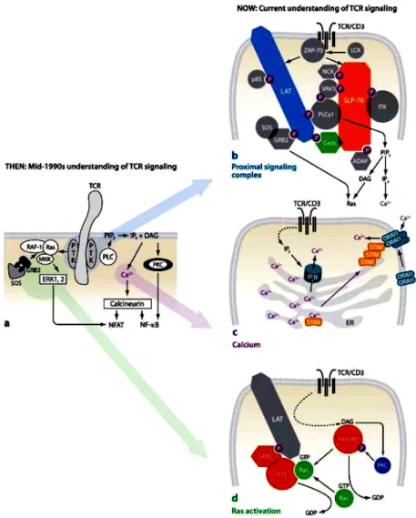
1.1.1.3 The CD3 complex
The TcR is a complex consisting of the variable αβchains noncovalently associated with the nonpolymorphic CD3 proteins. The CD3 proteins exist as a series of dimers including γε, δε, andζζ associated with a single αβheterodimer. These subunits lack enzymatic activity, but transduce signals via their immunoreceptor tyrosine-based activation motifs (ITAMs) (Reth, 1989). Tyrosine phosphorylation of the ITAMs serves as docking sites for interactions with other proteins. The earliest step in intracellular signaling following TcR ligation is the activation of src-family kinases p56lck and p59Fyn protein tyrosine kinases (PTKs), leading to phosphorylation of the CD3 ITAMs, followed by recruitment of Syk kinase family member ZAP-70 (ζ-associated phosphoprotein of 70 kDa)
(Fig.3).
The phosphorylated CD3ζ is a recruitment site of the ZAP-70 PTK (Chan et al.,
1992). The engagement of the TcR leads to Src family PTK activity resulting in ITAM phosphorylation and recruitment of ZAP-70. This converted the TcR: CD3 with no intrinsic enzymatic function into an active PTK associated molecular complex able to phosphorylate a spectrum of substrates leading to a myriad of downstream signals that, when integrated appropriately along with signals from other co-receptors, lead to T cell activation (Iwashima et al., 1994).
ZAP-70 targets are the transmembrane adapter protein linker for the activation of T cells (LAT) and the cytosolic adapter protein Src homology2 (SH2) domain–containing leukocyte phosphoprotein of 76 kDa (SLP-76) (Bubeck Wardenburg et al., 1996; Zhang et al., 1998). These two adapters form the backbone of the complex that organizes effector molecules in a way that allows the activation of multiple signaling pathways. The loss of either LAT or SLP-76 results in a nearly complete loss of TcR signal transduction reminiscent of Syk/ZAP-70 or Lck/Fyn double-deficient T cells (Koretzky et al., 2006; Sommers et al., 2004; Zhang et al., 1999). LAT contains nine tyrosines that are phosphorylated upon TcR engagement, which bind the C-terminal SH2 domain of PLCγ1,
the p85 subunit of phosphoinositide 3-kinase (PI3K), and the adapters growth factor receptor-bound protein 2 (GRB2) and GRB2-related adapter downstream of Shc (Gads) (Sommers et al., 2004). SLP-76 is then recruited to phosphorylated LAT via their mutual binding partner Gads (Liu et al., 1999). This proximal signaling complex results in the
activation of PLCγ1-dependent pathways including Ca2+
and diacylglycerol (DAG) induced responses, cytoskeletal rearrangements, and integrin activation pathways.
Following TcR ligation, PLCγ1 is found in the proximal signaling complex bound to
SLP-76, Vav1, and LAT, where it is phosphorylated and activated by Itk. Activated PLCγ1
then hydrolyzes the membrane lipid producing the second messengers IP3 (inositol 1,4,5-triphosphate) and DAG. These two messengers are essential for T cell function (Fig.3).
Figure 3: The CD3 complex and T cell receptor signalling
Ligation of the TcR/CD3 results in activation of Src and Syk family PTKs associated with the intracellular CD3 domains that then activate PLCγ1 and Ras-dependent pathways (a)
TcR signalling in mid 1990s (b) How the TcR couples to downstream pathways (c) The molecular basis for Ca2+
influx (d) The positive feedback loop responsible for Ras activation. (Smith-Garvin et.al. 2009)
Ca2+ ions are universal second messengers in eukaryotic cells. The IP3 generated by TcR-stimulated PLCγ1 activity stimulates Ca2+
permeable ion channel receptors (IP3R) on the ER membrane, leading to the release of ER Ca2+
stores into the cytoplasm. Depletion of ER Ca2+
triggers a sustained influx of extracellular Ca2+
through the activation of plasma membrane Ca2+
release-activated Ca2+
(CRAC) channels in a process known as store-operated Ca2+
entry (SOCE) (Oh-hora and Rao, 2008).
TcR-induced production of DAG results in the activation of two major pathways involving Ras and PKCθ. Ras is a guanine nucleotide–binding protein and is required for
the activation of the serine-threonine kinase Raf-1, which initiates a mitogen-associated protein kinase (MAPK) phosphorylation and activation cascade (Genot and Cantrell, 2000).
1.1.1.4 The CD4 and CD8 co-receptors
The two major subsets of T lymphocytes in the peripheral immune system, the helper and cytotoxic T cells, are defined by their expression of either the CD4 or the CD8 glycoprotein, respectively. Expression of these molecules, which serve as coreceptors by interacting with membrane-proximal domains of the MHC molecules (Garcia et al., 1996b; Genot and Cantrell, 2000), thus permitting simultaneous interaction of the TcR and either CD4 or CD8 with MHC.
The CD4 is an integral membrane glycoprotein of 55-60 kDa. The primary structure in humans was deduced from T cell cDNA libraries (Maddon et al., 1987), and indicated a protein of 458 amino acids. The first 25 amino acids include a cleavage signal, which after cleavage results in a mature protein of 433 amino acids. Aligning the cDNA sequence with X-ray crystallography indicated, that the 4 domains of CD4 are similar to the Ig superfamily (Brady and Barclay, 1996; Maddon et al., 1987). Domain 1 and 3 were classified as variable (V) domains due to their similarities with the Ig variable region, domain 2 and 4 were classified as constant (C) domains (Williams and Barclay, 1988). The two membrane distal domains (D1 and D2) are, like the two membrane proximal domains (D3 and D4), rigidly packed together, whereas the connection between domain 2 and 3 is flexible. Interestingly, crystallography revealed CD4 to dimerise via cross-binding of D4 (Wu et al., 1997).
CD8 is a transmembrane glycoprotein composed of a CD8 ααhomodimer or a CD8
extended O-glycosylated stalk region, a single-pass transmembrane domain, and a short cytoplasmic region. The Ig domains of CD8 bind MHC class I at a site distant from the TcR-binding site (Chang et al., 2005). CD8ααand CD8αβbind most but not all MHC class I and class I-like molecules and usually with a similar affinity/avidity (Kern et al., 1999). A notable exception is the tenfold higher affinity of CD8αα to the mouse MHC I-like protein,
thymus-leukemia, than CD8αβ(Leishman et al., 2001).
The co-receptor model proposed CD4 and CD8 to function as co-receptors able to migrate to and interact with the TcR, as well as transduce an independent signal necessary for complete TcR activation (Janeway et al., 1989). In accordance with this model, D3 of CD4 has been identified to interact with the TcR/CD3 complex (Mittler et al., 1989; Vignali et al., 1996; Vignali and Vignali, 1999), whereas D1 binds monomorphic regions on the MHC class II molecule (Doyle and Strominger, 1987; Fleury et al., 1996). Similarly, the CD8 co-receptor interacts with MHC class I (Gao et al., 1997).
The T-cell immune response is initiated upon engagement of the TcR and coreceptor CD4 or CD8, by cognate pMHC complexes presented by APCs. TcR/coreceptor engagement induces the activation of biochemical signaling pathways that, in combination with signals from costimulatory molecules and cytokine receptors, direct the outcome of the response. The intracellular region of CD4 and CD8 binds to Lck (Rudd et al., 1988; Veillette et al., 1988) and Fyn (zur Hausen et al., 1997). Activation of the these two kinases is central to the initiation of TcR signaling pathways, and their regulation is tightly controlled by conformational changes arising from binding of ligands to the SH3 and/or SH2 domains of the kinases (da Silva et al., 1997; Holdorf et al., 1999; Xu et al., 1999), and by the phosphorylation and dephosphorylation of two critical tyrosine residues (Palacios and Weiss, 2004).
A second intracellular signaling component, the LAT, was also found to co-immunoprecipitate with CD4 and CD8 (Bosselut et al., 1999; Kim et al., 2003).
1.1.2 Co-stimulation governing the activation of T cells
T-cell activation is mediated by antigen-specific signals from the TcRζ/CD3 and
CD4-CD8-p56lck complexes in combination with additional co-signals provided by coreceptors such as CD28, inducible costimulator (ICOS), cytotoxic T-lymphocyte antigen-4 (CTLA-antigen-4), programmed death (PD-1), and others. In general terms, CD28 and ICOS provide positive signals that promote and sustain T-cell responses, while CTLA-4 and PD-1 limit responses. The balance between stimulatory and inhibitory co-signals determines the ultimate nature of T-cell responses where response to foreign pathogen is achieved without excessive inflammation or autoimmunity.
1.1.2.1
CD28CD28 is the most characterized of the Ig family of coreceptors possessing an intracellular domain with several residues, which are critical for its effective signalling. In particular, the YMNM motif beginning at tyrosine 170 is critical for the recruitment of SH2-domain containing proteins, especially PI3K, GRB2 and Gads. CD28 is a 44 kDa homodimer expressed on both, naïve and activated T cells and binds the ligands B7-1 (CD80) and B7-2 (CD86), using a signature MYPPPY binding motif (Balzano et al., 1992). Its gene is located on human chromosome 2q33 (Naluai et al., 2000) and mouse chromosome 1 (Howard et al., 1991).
T-cell activation can occur with a potent TcR signal alone (i.e. high avidity TcR-pMHC interaction and/or high ligand density), however CD28 coligation is required in most responses to peptide antigens (Bluestone, 1995; Linsley, 1995; Noel et al., 1996). Without CD28 coligation, TcR-engagement often induces a non-responsive, anergic state or cell death (Linsley and Ledbetter, 1993). T cells from CD28-deficient mice show reduced proliferation in response to peptide antigens (Shahinian et al., 1993), although repeated antigen stimulation or long-term viral infection can bypass the requirement of CD28 (Kundig et al., 1996). CD28 co-signals can stabilize the mRNA of cytokines and amplify the activation of nuclear factor of activated T cells (NFAT) and nuclear factor kappa B
CD28 engagement enhances the production of various cytokines, including 1, IL-2, IL-4, IL-5, TNF, and IFN-γ (Linsley and Ledbetter, 1993; McArthur and Raulet, 1993;
Thompson et al., 1989). In addition it has been shown, that CD28 engagement plays a vital role in the early development and differentiation of both, Th1 and Th2 T cell subsets (King et al., 1995; Seder et al., 1994; Van der Pouw-Kraan et al., 1992). In the absence of CD28 signaling, naive T cells are biased towards a Th1 phenotype and no IL-4 is observed when CD28/B7 interactions are blocked with hCTLA4-Ig (Seder et al., 1994). Recently, it has been shown that CD28 costimulation downregulate Th17 development in-vitro (Bouguermouh et al., 2009). Furthermore, CD28 is also important in the homeostasis and function of regulatory T cells (Tregs), the importance of CD28 signaling in Treg proliferation is suggested by the effectiveness of anti-CD3 and anti-CD28 treatment to expand Tregs in-vitro. Accordingly, both mouse and human Tregs can be expanded using CD3 and CD28 antibodies in the presence of high concentrations of IL-2 (Earle et al., 2005; Tang et al., 2004). It is also established that mice lacking CD28 or its ligands have decreased numbers of CD4+
CD25+
regulatory T cells (Lohr et al., 2003; Salomon et al., 2000). In addition CD80/86-blockade experiments have suggested that continual expression of CD28 ligand in the periphery is necessary to maintain the peripheral Treg compartment (Salomon et al., 2000).
1.1.2.2
Cytotoxic T lymphocyte-associated antigen-4 (CTLA-4)CTLA-4, also known as CD152, is an activation-induced type 1 transmembrane glycoprotein of the Ig superfamily (Brunet et al., 1987). The CTLA-4 gene is located on chromosome 2 in humans and on chromosome 1 in mice (Dariavach et al., 1988; Ling et al., 1999). The CTLA-4 gene product contains 223 amino acids, with a 35 amino acid signal peptide (Brunet et al., 1987; Dariavach et al., 1988), and is found as a covalent homodimer of 41–43 kDa (Lindsten et al., 1993; Linsley et al., 1995).
The extracellular architecture of CTLA-4 is characterized by a single Ig V-like domain containing the CD80/CD86 binding site (Brunet et al., 1987; Dariavach et al., 1988; Linsley et al., 1995; Metzler et al., 1997; Ostrov et al., 2000). Homodimerization of CTLA-4 is mediated by cysteine-dependent binding at position 122 in the stalk region and by
N-glycosylation at positions 78 and 110 (Darlington et al., 2005; Linsley et al., 1995). The simplicity of its structure disguises its critical function as a major negative regulator of T cell–mediated immune responses (Alegre et al., 2001; Chambers et al., 2001; Rudd et al., 2002; Salomon and Bluestone, 2001).
The structural similarities and sequence homology between CTLA-4 and CD28 extend to share natural ligands: the B7 molecules CD80 and CD86 (Freeman et al., 1993a; Freeman et al., 1993b; Linsley et al., 1990). However, a key feature that may explain CTLA-4's mechanism of action is that CTLA-4 binds CD80 and CD86 with greater affinity and avidity compared to CD28 (Linsley et al., 1991; Linsley et al., 1994). In general, CTLA-4 is thought to raise the threshold for TCR signaling, thereby preventing responses to self-antigens (Chambers et al., 2001; Greenwald et al., 2005). Furthermore, cell extrinsic and intrinsic mechanisms influence the CTLA-4 function. Cell extrinsic mechanisms include the secretion of soluble CTLA-4 (sCTLA-4), the production of indoleamine 2,3-dioxygenase (IDO) and the involvement of Tregs (Rudd, 2008). Cell intrinsic mechanisms include CTLA-4 ectodomain competition with CD28 for the binding to CD80/86 (Masteller et al., 2000), the presence of associated phosphatases (Chuang et al., 2000; Lee et al., 1998; Marengere et al., 1996), blockade of lipid-raft expression (Chikuma et al., 2003; Martin et al., 2001) and microcluster formation (Schneider et al., 2008). It is likely that most of these factors contribute to the overall function of CTLA-4 in different contexts.
When engaged to B7, CTLA-4 plays a key role as a negative regulator of T cell activation (Krummel and Allison, 1995; Walunas et al., 1994), leading to downregulation of T cell responses and to the preservation of T cell homeostasis and peripheral tolerance (Tivol and Gorski, 2002). The mechanism of T cell inactivation by CTLA-4 involves antagonism of CD28-dependent costimulation and direct negative signaling through its cytoplasmic tail (Carreno et al., 2000). CTLA-4 can exert its inhibitory effects in both, CD80/CD86 dependent and independent fashions (Grohmann et al., 2002; Ise et al., 2010; Vijayakrishnan et al., 2004).
CTLA-4 has an intrinsic plasticity for signaling. It refers to the ability of CTLA-4 to actively deliver, by itself, a negative signal or a positive signal to a T cell, depending on the ligand it engages and its association/dissociation to protein phosphatase-2A (PP2A). When engaged to B7, it dissociates from PP2A and acts as an inactivator of T cells (Baroja et al.,
2002). In contrast, when CTLA-4 is engaged to recombinant ligands 24 and 26 Abs, its association to PP2A is stabilized and it acts as an activator of T cells (Madrenas et al., 2004).
1.1.2.3
B7-1 (CD80) and B7-2 (CD86)Co-signalling molecules are that, whose functions are entirely dependent on TcR signals (Schwartz, 2003), and they control the TcR signals itself. In the absence of sufficient TcR signalling, co-signalling molecules lose their function (Chen, 1998).
The two B7 family members, CD80 and CD86, bind to the same two receptors, CD28 and CTLA-4 (Chikuma and Bluestone, 2003; Coyle and Gutierrez-Ramos, 2001; Sharpe and Freeman, 2002). These receptors have distinct kinetics of expression and affinities for CD80 and CD86 (van der Merwe and Davis, 2003). Two additional B7 homologs, B7-H3 (Chapoval et al., 2001) and B7-H4 (Sica et al., 2003) [B7x (Zang et al., 2003), B7S1 (Prasad et al., 2003)], and another CD28 homolog, BTLA (B and T lymphocyte attenuator) (Watanabe et al., 2003), have been identified.
CD80 and CD86 have a restricted expression pattern, being expressed mainly by professional APCs and haematopoietic cells (Chen, 2004). CD80 and CD86 on APCs have well-recognized roles as T cell costimulatory molecules, but the functional significance of their expression on T cells is not well understood. CD86 is constitutively expressed on some resting T cells, whereas CD80 is not present on resting T cells (Taylor et al., 2004). CD80 and CD86 may influence immune responses through reverse signaling into B7-expressing APCs (Fallarino et al., 2003; Grohmann et al., 2002; Munn et al., 2004). CD86 on B cells can signal bidirectionally, it stimulates CD28 on T cells and transduces positive signals into B cells that increase IgG1 and IgE production (Podojil et al., 2004; Podojil and Sanders, 2003). Basal or constitutive expression of B7 appears to be key for maintaining regulatory T cell homeostasis and preventing immune responses against self-antigens.
In brief, CD28 first functions by costimulating T cells and preventing the induction of either anergy or apoptosis (Linsley and Ledbetter, 1993). Second, a CD28 homologue, CTLA-4, counterbalances CD28 by downregulating the immune response either by inducing apoptosis or competing with CD28 for its ligands, as both form homodimers and
bind the ligands CD80 and CD86, using a signature MYPPPY binding motif. CD80 binds CTLA-4 with much lower affinity than CD28 (van der Merwe et al., 1997) . Third, two distinct molecules, CD80 and CD86, function as costimulatory ligands for CD28 and CTLA-4 (Linsley and Ledbetter, 1993). While CD86 dominates in primary responses, the roles of CD80 and CD86 during an ongoing response depends on the relative expression of CD28/CTLA-4, the nature and concentration of cytokines in the surrounding milieu, and the characteristics of the APCs that are encountered. Finally, manipulation of the CD28/B7 pathway can alter the balance of the immune response by influencing the nature of the cytokines produced in response to antigen exposure.
1.1.2.4
Inducible co-stimulatory molecule (ICOS) and its ligand (ICOSL)ICOS is a glycosylated disulfide-linked homodimer (Rudd and Schneider, 2003) like CD28 and CTLA-4. The ICOS cytoplasmic tail contains a YMFM motif that binds the p85 subunit of PI3K, similar to the YMNM motif of CD28 (Coyle et al., 2000), and stimulates greater PI3K activity than CD28 costimulation (Okamoto et al., 2003; Parry et al., 2003). However, in contrast to CD28, the ICOS YMFM motif does not allow the binding of GRB2, which is critical for IL-2 production (Harada et al., 2001; Okamoto et al., 2003). ICOS signals lead to effector cytokine production and can be inhibited by CTLA-4 engagement (Riley et al., 2001).
ICOS is upregulated on CD4+ and CD8+ T cells following activation and is expressed on effector and memory T cells (Coyle et al., 2000; Yoshinaga et al., 1999). It is also upregulated on activated NK cells, and promotes NK cell function (Ogasawara et al., 2002). ICOS expression is stimulated on T cells by TcR and CD28 signals (Beier et al., 2000; Coyle et al., 2000; Mages et al., 2000; McAdam et al., 2000). ICOS engagement was shown initially to selectively produce high levels of IL-10 and IL-4, but in-vivo studies in several different model systems have demonstrated that ICOS can stimulate production of both, Th1 and Th2 cytokines during initial priming and during effector T cell responses (Hutloff et al., 1999; McAdam et al., 2000).
ICOSL has been detected on the surface of T cells, B cells, macrophages, DCs and other cell types including endothelial cells and some epithelial cells, and its mRNA is expressed constitutively in lymphoid and nonlymphoid tissues including kidney, liver, lung,
and testis (Richter et al., 2001; Swallow et al., 1999; Wang et al., 2000; Yoshinaga et al., 2000). Costimulatory signals through ICOS provide critical T cell help to B cells (Hutloff et al., 1999; McAdam et al., 2001; Tafuri et al., 2001).
ICOS and CD28 have unique and overlapping functions that synergize to regulate CD4+ T cell differentiation. Like CD28, ICOS augments T cell differentiation and cytokine production and provides signals for Ig production (Sharpe and Freeman, 2002; Sperling and Bluestone, 2001). It also regulates the outcome of autoimmunity in the murine model of multiple sclerosis (MS), experimental autoimmune encephalomyelitis (EAE) but plays distinct roles at different times during the pathogenesis of EAE (Chapoval et al., 2001; Rottman et al., 2001; Sporici et al., 2001). Blocking of ICOS during induction of EAE exacerbates disease, whereas ICOS blocking during the effector phase of EAE ameliorates disease (Dong and Nurieva, 2003; Rottman et al., 2001).
Both CD28 and ICOS signaling upregulate Th1 as well as Th2 cytokines; however, ICOS does not upregulate IL-2 production. Thus, ICOS stimulates T cell effector function but not T cell expansion. The role of ICOS in stimulating IL-10 production may contribute to its role in regulating Treg, T cell tolerance, and autoimmunity. The function of ICOS during in-vivo immune responses appears to depend on timing/stage of immune response as well as microenvironment, because ICOSL can be expressed on endothelial and epithelial cells as well as professional APCs.
1.1.2.5
Programmed death-1 (PD-1) moleculePD-1 is a monomeric member (Freeman et al., 2000; Latchman et al., 2001; Zhang et al., 2004) of the Ig superfamily (Ishida et al., 1992; Shinohara et al., 1994; Vibhakar et al., 1997) related to CD28 and CTLA-4, but it lacks the membrane proximal cysteine that allows these molecules to homodimerize. The PD-1 cytoplasmic domain has two tyrosines, one that constitutes an immunoreceptor tyrosine-based inhibition motif (ITIM), and the other an immunoreceptor tyrosine-based switch motif (ITSM) (Shlapatska et al., 2001). PD-1 is expressed during thymic development primarily on CD4-CD8- cells and double negative γδ thymocytes (Nishimura et al., 2000) and is induced on peripheral CD4+ and
CD8+ T cells, B cells, and monocytes upon activation (Agata et al., 1996). NK-T cells also express low levels of PD-1 (Nishimura et al., 2000).
PD-L1 is a 290 amino acid type I transmembrane protein encoded by the Cd274 gene on mouse chromosome 19 and human chromosome 9. The PD-1 ligands exhibit distinct patterns of expression; PD-L1 is expressed more broadly than PD-L2 (Dong et al., 1999; Freeman et al., 2000; Latchman et al., 2001; Tseng et al., 2001). PD-L1 is expressed on resting cells and upregulated on activated B, T, myeloid, and DCs (Liang et al., 2003). In contrast to CD80 and CD86, PD-L1 is expressed in nonhematopoietic cells including microvascular endothelial cells and in nonlymphoid organs including heart, lung, pancreas, muscle, and placenta (Ishida et al., 2002; Liang et al., 2003; Petroff et al., 2003; Rodig et al., 2003; Wiendl et al., 2003). The expression of PD-L1 within nonlymphoid tissues suggests, that PD-L1 may regulate self-reactive T or B cells in peripheral tissues and/or may regulate inflammatory responses in the target organs.
PD-L2 is a type I transmembrane protein encoded by the Pdcd1lg2 gene adjacent to Cd274 and separated by only 23 kb of intervening genomic DNA in mouse and 42 kb in human. There are three PD-L2 splice variants identified from activated human PBMCs (He et al., 2004; Latchman et al., 2001). PD-L2 is rarely expressed on resting cells and hardly induced on B cells and T cells. PD-L2 can be induced on macrophages by IL-4 and IFNγ
and on DCs by anti-CD40 antibody, GM-CSF, IL-4, IFNγ and IL-12. It has been reported
that IL-4 induces PD-L2 more strongly than IFNγ, while IFNγinduce PD-L1 more strongly than IL-4 on macrophages, suggesting that Th1 and Th2 responses mobilize L1 and PD-L2 differentially (Loke and Allison, 2003).
The lupus-like phenotype of PD-1
C57BL/6 mice first suggested a role for PD-1 in the regulation T cell tolerance and autoimmunity, as these mice exhibit hyperactivation of the immune system such as splenomegaly and in-vitro augmented proliferation of B cells (Nishimura et al., 1999; Nishimura et al., 2001). Administration of anti-PD-1 or anti-PD-L1 mAb to one to ten-weeks old prediabetic NOD female mice led to the rapid onset of diabetes, and it was associated with increased IFN-γ producing glutamate acid
decarboxylase (GAD) reactive splenocytes (Ansari et al., 2003). The expression of PD-L1 on islet cells suggests that it may critically control the responses of self-reactive T cells in the target organ (Ansari et al., 2003; Liang et al., 2003). PD-1 and its ligands have a
negative regulatory role in EAE (Latchman et al., 2004; Salama et al., 2003). PD-1, PD-L1, and PD-L2 are expressed on brain infiltrating inflammatory cells in mice with EAE (Liang et al., 2003; Salama et al., 2003). PD-L1 is also expressed on vascular endothelial cells, astrocytes, and microglia. A role for PD-L1 in controlling the responses of self-reactive T cells is further indicated by the severe clinical EAE that develops following immunization of PD-L1-/- mice with the myelin oligodendrocyte glycoprotein 35-55 peptide (MOG35-55) or
following the adoptive transfer of MOG35-55 specific T cells into PD-L1
recipients (Latchman et al., 2004).
The PD-1: PD-L1/PD-L2 pathway has critical roles in regulating T cell activation and tolerance. CTLA-4 and PD-1 have important nonredundant inhibitory functions. CTLA-4 appears to have a more central role in the lymphoid organs, whereas PD-1 has an important role in regulating inflammatory responses in peripheral tissues.
1.1.2.6
Adhesion moleculesThe continuous recirculation of naïve T cells and their subsequent migration to tissues following activation is crucial for maintaining protective immunity against invading pathogens. The preferential targeting of effector and memory T cells to tissues is instructed during priming and mediated by cell surface expressed adhesion receptors such as integrins. Integrins are αβ heterodimeric cell surface adhesion molecules that mediate
cell-extracellular matrix (ECM) and cell-cell interactions (Pribila et al., 2004), which are essential for T cell recirculation, migration into inflammatory sites and for a specific and effective immune response against foreign pathogens (Hynes, 2002; Shimizu et al., 1999). Integrin-dependent interactions of lymphocytes and APCs to endothelium regulate the efficiency and specificity of trafficking into secondary lymphoid organs and peripheral tissue.
Naïve T cells express a homogeneous array of cell surface molecules that promotes recirculation through the secondary lymphoid organs of the body, including the spleen, peripheral lymph nodes (pLN), mesenteric lymph nodes (mLN) and Peyer’s patches (PP) of the small intestine. Naïve T cells express low levels of the αLβ2 (LFA-1), α4β1 (VLA-4)
respectively (Springer, 1995). The interaction of αLβ2 with ICAM-1 is important for T cell
entry into pLN and for T cell interactions with APCs. The α4β1 ligand VCAM-1 is
expressed at low levels throughout the vasculature, but becomes upregulated on a wide array of tissue during inflammation (Henninger et al., 1997). MAdCAM-1, the major ligand for α4β7 is preferentially expressed at steady state in the mLN and Peyer’s patches, where
it promotes entry of naïve T cells into these sites through a high affinity interaction (Berlin et al., 1993; Erle et al., 1994; von Andrian and Mackay, 2000). Both VCAM-1 and ICAM-1 are also expressed on the vasculature of the inflamed brain. In particular, VCAM-1 is known to promotes T cell entry through its interaction with α4β1 expressed on activated T
cells (Engelhardt and Ransohoff, 2005).
In addition to mediating adhesion to cell surface and extracellular matrix ligands, integrins generate a diverse array of intracellular signals. In T cells, LFA-1 and several β1
integrins can provide costimulatory signals for TcR-induced T cell activation (Shiow et al., 2006; Sprent et al., 1971). Differences in the regulation of IL-2 production by integrins and CD28 following TcR stimulation demonstrate the unique costimulatory properties of these molecules. In CD4 T cells, LFA-1-mediated costimulation enhanced IL-2 transcription, which is not mediated by stabilization of IL-2 mRNA (Abraham and Miller, 2001), in contrast, CD28 costimulation alters IL-2 levels by stabilizing IL-2 mRNA (Arnold et al., 2004). Furthermore, LFA-1 and CD28 induce the activation of PI3-K in CD8 T cells however; PI3-K activity is required only for LFA-1-meditated costimulation (Ni et al., 2001).
In addition, osteopontin, also known as secreted phosphoprotein1 (SPP1), is a member of SIBLING (small integrin-binding ligand, N-linked glycoproteins) family of proteins (Fisher et al., 2001). It is highly expressed in MS lesions (Chabas et al., 2001). Studies in MS tissue and in models of relapsing-remitting (RR) and progressive EAE showed that osteopontin might have a role in the progression from RR disease to the more chronic form (Chabas et al., 2001). Osteopontin expression is induced on inflamed endothelium in the ECM of the perivascular cuff, and it serves as a ligand for two classes of adhesion molecules, CD44 and various integrins including VLA-4 (Ashkar et al., 2000). Furthermore, chemokine receptors served as adhesion molecules especially during
leukocyte migration, by acting on firm adhesion, locomotion, diapedesis, and chemotaxis (Cartier et al., 2005).
1.1.3 T cells activation
1.1.3.1
Immunological synapseActivation of T cells by APCs requires the formation of a specific and long-lasting interface between the two cells, known as the immunological synapse (IS). TcR interaction with pMHC complexes in the IS between the T cell and APC is required for T cell activation (Norcross, 1984; Paul and Seder, 1994; Valitutti et al., 1995). The required duration of signaling is on the order of hours, while the activating and inhibitory molecular interactions in the IS have half-lives on the order of seconds (Iezzi et al., 1998; Matsui et al., 1994). The mature IS has been defined by arrangement of supramolecular activation clusters (SMACs) that form within a few minutes of T cell–APC contact (Grakoui et al., 1999; Monks et al., 1998; Wulfing and Davis, 1998). Physiological T cell activation can be divided into a series of temporal stages: T cell polarization, initial adhesion, IS formation, and IS maturation (Grakoui et al., 1999) (Fig.4).
Circulating T cells are rounded and nonpolarized, with a uniform radial distribution of membrane domains, receptors, and microvilli on the cell surface (Kucik et al., 1996; Sanchez-Madrid and del Pozo, 1999). These cells are relatively nonmotile and integrin adhesion molecules are maintained in a low-activity state (Kucik et al., 1996). In order to extravasate into lymph nodes and inflamed tissues, these T cells must encounter chemokine gradients before they encounter APC, leading to the rapid polarization of the T lymphocyte. The close contact areas of IS between polarized T lymphocytes and APCs are established by adhesion molecules. These close contact areas are achieved indirectly through cytoskeletal protrusions anchored to larger adhesion mechanisms like LFA-1 and ICAM-1 (Grakoui et al., 1999) or more directly by smaller adhesion receptor pairs like CD2 and CD58 that would work beside the TcR. CD2 and its ligand CD48 are associated with rafts in the same fashion like TcR (Yashiro-Ohtani et al., 2000).
Figure 4: Dynamic processes of antigen recognition and T-cell activation by formation of microclusters and immunological synapse (Saito et.al. 2006)
Upon T-cell attachment, LFA-1/ICAM-1 complexes cluster in the center and the MHC complexes are located at the periphery of the synapse. Later, TcR/MHC complexes become clustered in the center, whereas the LFA-1/ICAM-1 complexes are segregated to the periphery to form a ring around the TcR/MHC clusters (Grakoui et al., 1999), which is facilitated by the CD2-CD48 interaction that provides the correct membrane alignment (Dustin et al., 1996; Dustin et al., 1997; Wild et al., 1999). The formation of the central cluster gives rise to the immunological synapse that will remain stable for over an hour.
Furthermore it has been shown by intravital two-photon laser scanning microscopy that T cells undergo rapid, random amoeboid locomotion in secondary lymphoid tissues (Miller et al., 2003; Miller et al., 2002b). This has been most intensively observed in mouse lymph nodes, which are subjected to either explanted or in-vivo imaging by two-photon laser scanning microscopy, which led to the model of stochastic repertoire scanning, in which T cells, in the T-cell zones move rapidly with average speeds of 10–15 µm/min and
peak speeds of over 30 µm/min. In another study, it has been shown by two-photon imaging, that T-cell priming by DCs occurs in three successive stages: transient serial encounters during the first activation phase are followed by a second phase of stable contacts culminating in cytokine production, which makes a transition into a third phase of high motility and rapid proliferation (Mempel et al., 2004).
1.1.3.2
TcR sensitivityThe sensitivity of the TcR for the pMHC complex plays a crucial role in T cell activation. The TcR discriminates between peptides by as little as a single amino acid, as was shown when a single amino acid mutation turned an agonistic peptide into an antagonistic peptide (Allen et al., 1987). The spectrum of peptides presented by the MHC is generally classified as full agonist, partial agonist APL, non-stimulatory, and antagonist peptides. Full agonist peptides induce full activation of the peripheral T cells or negative selection of thymocytes, partial agonist peptides induce incomplete peripheral T cell activation and positive selection of thymocytes (Kersh and Allen, 1996), and antagonistic peptides prevent activation of peripheral T cells though they do induce positive selection of thymocytes (Hogquist et al., 1994).
It has been shown that T cells are sensitive to minute amounts of stimulatory (agonist) pMHC complexes in a sea of endogenous ligands (Irvine et al., 2002; Krogsgaard et al., 2005; Purbhoo et al., 2004). T helper cells can stop rolling and transiently increase cytosolic calcium levels in response to even one agonist-pMHC complex. Ten agonist complexes appear to be sufficient for sustained calcium release and the formation of the immunological synapse (Grakoui et al., 1999; Huppa and Davis, 2003; Monks et al., 1998). Cytotoxic (CD8+) T cells also seem to be very sensitive (Brower et al., 1994; Purbhoo et al., 2004; Sykulev et al., 1996) and as few as three agonist-pMHC complexes have been demonstrated to be sufficient for the killing of target cells (Purbhoo et al., 2004). Signalling induced by agonist ligands can be severely impaired if antagonist-pMHC molecules are present. Antagonists can be obtained by mutating TcR contact residues of agonistic peptides (Madrenas et al., 1995; Sloan-Lancaster et al., 1994; Stefanova et al., 2003).