Cell Biomechanics of the Central Nervous System
byKristin Briana Bernick
B.S., Biomedical Engineering, University of California, Davis (2006)MASSACHUSE FTTS NSTITUTE
OF T!CHNO<oGY
JUL 14 2011
R IE1
L!~"
---
l~
Submitted to the Department of Biological Engineering in partial fulfillment of the requirementsfor the degree of
Doctor of Philosophy in Biological Engineering at the
MASSACHUSETTS INSTITUTE OF TECHNOLOGY June 2011
ARcmwaE
@Massachusetts Institute of Technology 2011. All rights reserved.
A uthor... . ...
Department of Biological Engineering
May 20, 2011
Certifiedby...
Simona Socrate, Ph.D. Principal Research Scientist, Harvard-MIT Division of Health Sciences and Technology and Institute for Soldier Nanotechnologies Thesis Supervisor
C ertified by ... (.. ... Roger Kamm, Ph.D. Singapore Research Professor of Mechanical and Biological Engineering Thesis Supervisor
Accepted by... ...
K. Dane Wittrup, Ph.D. Professor of Chemical and Biological Engineering Chairman, Course 20 Graduate Program Committee
Thesis Committee Simona Socrate, Ph.D. Thesis Co-advisor
Principal Research Scientist, Harvard-MIT Division of Health Sciences and Technology and Institute for Soldier Nanotechnologies
Massachusetts Institute of Technology
Roger Kamm, Ph.D. Thesis Co-advisor
Professor of Biological and Mechanical Engineering Massachusetts Institute of Technology
Ed Boyden, Ph.D.
Professor of Biological Engineering and Media Arts and Sciences Massachusetts Institute of Technology
Cell Biomechanics of the Central Nervous System
by
Kristin Briana Bernick
Submitted to the Department of Biological Engineering on May 20, 2011 in Partial Fulfillment of the Requirements for the Degree of Doctor of Philosophy in Biological Engineering
ABSTRACT
Traumatic brain injury (TBI) is a significant cause of death and morbidity in both the civilian and military populations. The major causes of TBI, such as motor vehicle accidents, falls, sports concussions, and ballistic and explosive blast threats for military personnel, are well established and extensively characterized; however, there remains much to be learned about the specific mechanisms of damage leading to brain injury, especially at the cellular level. In order to understand how cells of the central nervous system (CNS) respond to mechanical insults and stimuli, a combined modeling/experimental approach was adopted. A computational framework was developed to accurately model how cells deform under various macroscopically imposed loading conditions. In addition, in vitro (cell culture) models were established to investigate damage responses to biologically relevant mechanical insults. In order to develop computational models of cell response to mechanical loading, it is essential to have accurate material properties for all cells of interest. In this work, the mechanical responses of neurons and astrocytes were quantified using atomic force microscopy (AFM) at three different loading rates and under relaxation to enable characterization of both the elastic and viscous components of the cell response. AFM data were used to calibrate an eight-parameter rheological model implemented in the framework of a commercial finite element package (Abaqus). Model parameters fit to the measured responses of neurons and astrocytes provide a quantitative measure of homogenized nonlinear viscoelastic properties for each cell type. In order to ensure that the measured responses could be considered representative of cell populations in their physiological environment, cells were also grown and tested on substrates of various stiffness, with the softest substrate mimicking the stiffness of brain tissue. Results of this study showed both the morphology and measured force response of astrocytes to be significantly affected by the stiffness of their substrate, with cells becoming increasingly rounded on soft substrates. Results of simulations suggested that changes in cell morphology were able to account for the observed changes in AFM force response, without significant changes to the cell material properties. In contrast, no significant changes in cell morphology were observed for neurons. These results highlight the importance of growing cells in a biologically relevant environment when studying mechanically mediated responses, such as TBI. To address this requirement, we developed two model systems with CNS cells grown in soft, 3D gels to investigate damage arising from dynamic compressive loading and from a shock pressure wave. These damage protocols, coupled with the single cell computational models, provide a new tool set for characterizing damage mechanisms in CNS cells and for studying TBI in highly controllable in vitro conditions.
Thesis Supervisors: Simona Socrate, Ph.D.
Title: Principal Research Scientist, Harvard-MIT Division of Health Sciences and Technology and Institute for Soldier Nanotechnologies
Roger Kamm, Ph.D.
Acknowledgements
This thesis would not have been possible without the support of many people. First, I would like to thank Prof. Subra Suresh, Dr. Simona Socrate, and Prof. Roger Kamm for each providing guidance and advising throughout my graduate program. Prof. Subra Suresh served as my advisor for the first 4 years of my graduate studies, and during that time, he guided my development as a scientist by serving as an excellent role model with high scientific standards, professionalism, and a dedication to scientific research. Dr. Simona Socrate has been an excellent advisor, sharing her passion for research and continually inspiring me with new ideas and collaborations. Prof. Roger Kamm has served as both my committee chair, and more recently, as a co-advisor, providing insightful discussions and support throughout my time at MIT. In addition, I am grateful to Prof. Ed Boyden for serving on my thesis committee and providing key insight and discussions, especially in his area of expertise of neuroscience. I am
grateful to have had formative interactions with each of these researchers throughout my Ph.D. I would like to thank Prof. Alan Grodzinsky for discussions and encouragement throughout my Ph.D, and for making my time spent in Tech Square enjoyable. In addition, he provided excellent mentoring when I served as his teaching assistant in his graduate course on Molecular, Cellular, and Tissue Biomechanics.
In addition, I had help from fellow students and collaborators throughout my time at MIT. I extend immense thanks to Thibault Prevost for his significant contributions to this thesis especially in the areas of model development, AFM program coding, and set-up and design of the damage devices. I would like to thank Cathal Kearney for helping significantly with the blast system set-up and device calibration and Prof. Myron Spector for allowing me to use the Dolorclast shockwave device. I am also grateful for the help of the laboratory of Professor
Sebastian Seung, especially Heather Sullivan and Jeannine Foley, who provided me with all of the cortical tissue samples necessary for the work of this thesis. In addition, I would like to thank the laboratory of Dr. Michael Whalen at Massachusetts General Hospital, especially Dr. Jianhua Qiu, for helping to characterize our damage devices and for useful discussions on cell signaling and biochemistry.
I also had the opportunity to meet many wonderful people while at MIT who provided both insightful discussions and made my 5 years here fun and memorable. My class, BE 2006, helped me get through my first year here successfully and resulted in lifelong friendships. The members of the Suresh lab and other students in Technology Square made my time spent in lab enjoyable.
I also acknowledge the support of my funding sources, the National Science Foundation Graduation Research Fellowship and the National Institutes of Health Molecular, Cell, and Tissue Biomechanics Training Grant.
The help and support I had to successfully complete this thesis, did not just come from my time spent at MIT. I had many wonderful science teachers and professors who inspired me to pursue this path and showed me how rewarding and interesting a career in the sciences could be. I am especially grateful to Prof. Yin Yeh for letting me work in his lab as an undergraduate at the University of California, Davis and for giving me a positive first experience with academic research. His mentoring and encouragement was a key factor in my decision to attend MIT.
I was also lucky enough to meet my boyfriend, Bryan, during my time at MIT. He was always supportive after a long day in the lab and put up with the ups and downs that come along with graduate school. I am extremely grateful to have him in my life and value the encouragement and support he gives to all aspects of my life.
Most importantly, I extend tremendous gratitude to my family for continuous support over the years. My parents, Debbie and Dave, and my sister, Kim, have supported me in everything that I have done and always stood behind me 100%, encouraging me to pursue my dreams. Knowing that they believed in me helped me through even the toughest situations. I
Contents
1 Introduction 25
2 Background 29
2.1 Traumatic Brain Injury ... 29
2.2 Biomechanics of Single Neurons and Glia... 38
2.3 Existing Cellular Level Models of Traumatic Brain Injury... 44
3 Methods for Determining Cell Material Properties 49 3.1 O verview ... 49
3.2 Atomic Force Microscopy Experiments... 50
3.2.1 C ell Culture... 50
3.2.2 Measurements of Neuron Somata on Glass... 52
3.2.3 Measurements of Neuron Processes on Glass ... 56
3.2.4 Measurements of Astrocytes on Glass ... 57
3.2.5 Measurements of Neurons and Astrocytes on Gels ... 60
3.3 Finite Element Simulations... 62
4 Characterization of Neuron and Astrocyte Response on Glass 69 4.1 Introduction ... 69
4.2 Neuron Results ... 70
4.2.1 Response of the Neuron Som a... 70
4.2.2 Response of the N euron Processes... 75
4.3 Astrocyte Results... 78
4.4 Discussion ... 81
4.4 Sum m ary... 84
5 Effect of Substrate Stiffness on Neuron and Astrocyte Response 85 5.1 Introduction... 85
5.2 Results... 86
5.3 Discussion ... 102
5.4 Sum mary ... 106
6 Design of Damage Devices 107 6.1 Introduction... 107
6.2 3D Cell Cultures... 108
6.3 Shockwave device ... 111
6.4 Compression Device... 124
6.5 Sum mary ... 128
7 Conclusions and Thesis Contributions 131
List of Figures
2-1: Characteristic blast wave pressure profile and resulting changes in atmospheric pressure. Before the explosion (1) atmospheric pressure is normal. After the explosion and upon passage of the shockwave (2), the blast wind flows away from the explosion. When a drop in atmospheric pressure occurs below normal, the direction of the blast wind reverses (3). Reproduced with permission from Taber et al [1]... 30
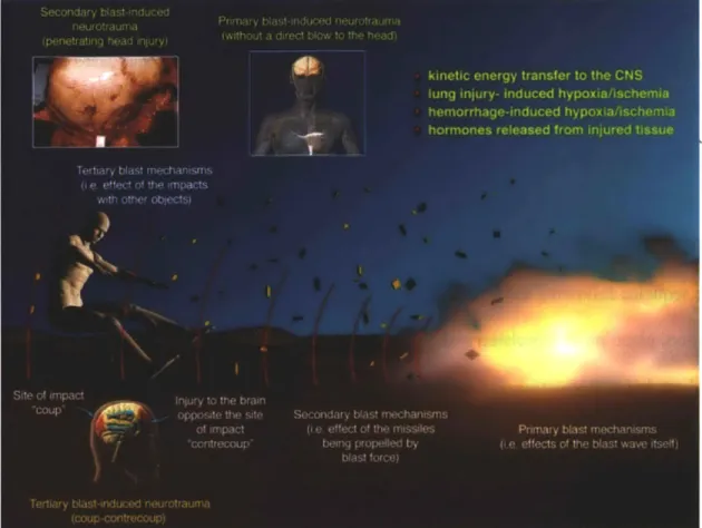
2-2: Schematic of the primary, secondary, and tertiary blast mechanisms and the characteristic brain injuries. Reprinted with permission from Macmillan Publishers Ltd: Journal of Cerebral Blood Flow and Metabolism, copyright 2010 [2]... 31
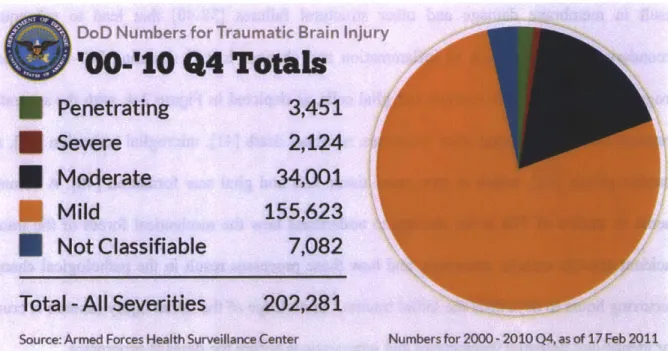
2-3: Official Department of Defense numbers for different severities of Traumatic Brain Injury (from http://www.dvbic.org/TBI-Numbers.aspx)... 33
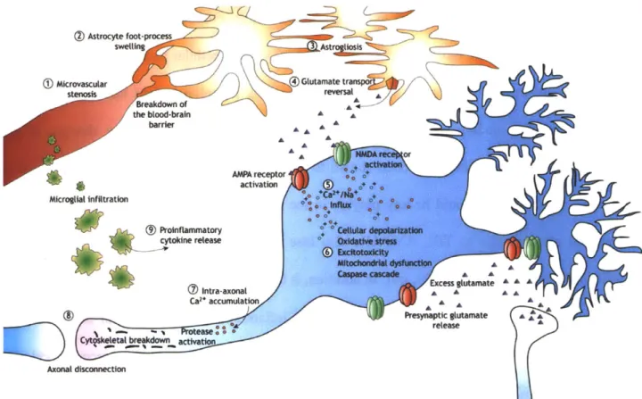
2-4: Schematic depicting some of the key pathways associated with secondary damage after TBI, involving neurons (blue), astrocytes (orange), and microglia (green). Reproduced with permission from Park et al. [3] © Canadian Medical Association. Copied under license
from the Canadian Medical Association and Access Copyright. Further reproduction prohibited ... 35
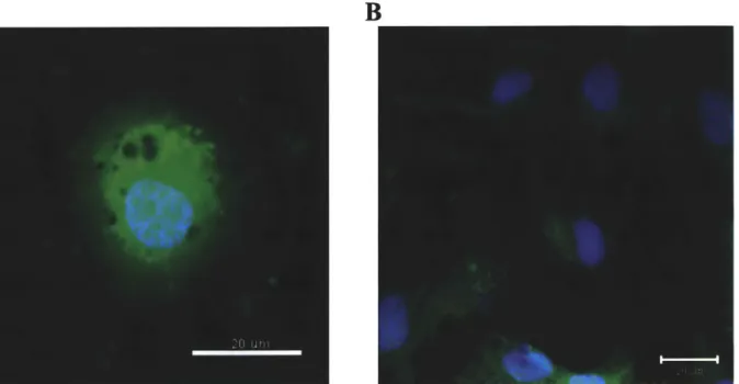
3-1: Results of immunocytochemistry to verify cell types. Cells grown in serum containing media were found to be primarily astrocytes (A) and those grown in serum free media primarily neurons (B). Scale Bars 20 m ... 51
3-2: (A) SEM image of tipless cantilever with attached 45 ptm polystyrene sphere (dimensions in
sm).
(B) Bright field image of AFM tip with bead adjacent to neuron to be indented; 20 pm scale bar. (C) Schematic of AFM experimental setup - Polystyrene bead compressing the cell body of a neuron plated on glass. (D) AFM testing procedure: sample approach, pre-load (black solid), sequences of load-unload segments at 10 [tm/s (red dot), 1 [tm/s (blue solid), 0.1 ptm/s (green dash-dot), followed by stress-relaxation (black dash). ... 543-3: Cell height determination procedure: cell (black solid) and glass substrate (red, blue, green dash) indentation curves were used to retrieve relative piezo positions associated with contact events between cell/glass and cantilever, thereby providing an estimate for the cell h eight...55
3-4: Representative neuron and AFM cantilever with pyramidal tip adjacent to process to be indented. Processes were indented close to the neuron soma... 57
3-5: Confocal images of astrocytes cultured for 1 day after passage (A) and 3 days (B), highlighting the choice to test cells after 1 day when cells remain isolated and measurements on single cells are possible; nucleus (Hoechst, blue) and cytoplasm (Calcein-A M , green); scale bars 20 tm ... 58
3-6: Astrocyte loading routine consisting of an initial approach to the cell to a pre-load of 0.3 nN to establish contact (green dash), a 15 s pause for equilibration (green solid), 3 sets of load-unload cycles to a 1 tm indentation depth at 3 different loading rates (10 pm/s (black solid), 1
sm/s
(cyan solid), and 0.1 pm/s (blue dash)), and a 2 minute stress relaxation test (red dash)... 593-7: Top (A) and side (B) views of an astrocyte to be indented with polystyrene sphere (darker blue in side view); cytoplasm (calcein-AM; green); nucleus (Hoechst 33342; cyan)... 59
3-8: (A) Schematic of cell material model, including 8 material parameters. (B) Representative finite element model geometry for an astrocyte tested on glass. (C) Representative geometry and meshing for a neuron grown on gel, with the gel layer included in the model. ... 6 8
4-1: AFM data for a representative neuron of diameter 14.2 jim and height 7.6 pm. Force versus displacement response at the 3 consecutive loading rates of 10
sim/s
(red dash-dot), 1 jm/s (green dot), and 0.1 jim/s (blue solid) ... 714-2: Force versus time responses measured for one representative neuron of diameter 14.2
sm
and height 7.6 pm (black) and simulated in Abaqus with actual cell geometry (red dash). Material parameters for this cell were found to be: o = 13 Pa, XL = 1.06, Go = 85 Pa, G. =80 Pa, rl = 3000 Pa.s, ao = 0.005 Pa, and n = 1. Error measure for the model fit was:
1.09 x 10⁻⁴...7 1
4-3: (A) Average force versus time response for 87 cells (black line) with plus and minus standard deviations (grey line); model fit (red dashes) to average response. The pictured model fit corresponds to an error measure of 1.1 x 10⁻⁴. (B, C, and D) Average force versus displacement response at 10, 1 and 0.1 .tm/s respectively. Error bars represent standard deviations and red dashes correspond to model fit. Material parameters obtained by fitting the force-indentation response to the average experimental response were found to be: t0 =
16 Pa, XL = 1.05, Go = 75 Pa, G. = 40 Pa, rj = 3000 Pa.s, ao = 0.005 Pa, and n = 1. K was
held constant at 10,000 Pa. (E) Distribution in maximum force level at the end of the first loading ramp for each displacement rate. Outliers are displayed with a red + sign. Rate effects were found to be statistically significant (P < 0.0001, one-way ANOVA)... 73
4-4: (A) Mean (black) and standard deviation (grey) for 10 neurons indented with loading rates in reverse order (0.1, 1, 10 pm/s). Model predictions (red dash) using the mean set of parameters obtained for indenting a neuron in 10, 1, 0.1 pm/s loading rate order ; (B) Peak forces reached at the end of the first loading ramp for each displacement rate. Rate effects were found to be significant (P < 0.006, one-way ANOVA)... 74
4-5: (A, B) Force versus displacement response for processes at 10 and 1 Rm/s, respectively
(Black=mean; Grey=standard deviation, N=25). (C, D) Sample Hertz fits for a single process at 10 and 1 [tm/s, respectively (AFM data= black dots; Hertz Fit= Red Line). (E) Box plot showing pseudo-elastic Young's Modulus dependency on loading rate. Differences are statistically significant with P<0.01... 76
4-6: AFM data (black) and elastic model fit (red dashes) for neuron soma indented at 10 tm/s (A) and 1 tm/s (B). Young's Modulus values used for fitting were 725 Pa at 10 Rm/s and 350 Pa at 1 Rm/s with the cells assumed to be incompressible with a Poisson's ratio of 0.5. ... 7 8
4-7: AFM data for a representative astrocyte of diameter 36
stm
and height 5 Rm. (A) Force versus displacement response at the 3 consecutive loading rates of 10 tm/s (blue dash-dot), 1 [tm/s (cyan dash), and 0.1stm/s
(red solid). (B) Force versus time response... 794-8: Average force versus time response for 37 astrocytes (black line) with plus and minus standard deviations (grey line); model fit (red dashes) to average response. Material parameters obtained by fitting the force-indentation response to the average experimental response were found to be:
sto=
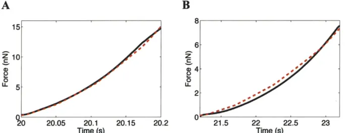
13 Pa, X = 1.08, Go= 275 Pa, G.= 80 Pa, q = 300 Pa.s, ao5-1: A. Sample Hertz fit for a representative Gel 1 (AFM data- black dots; Hertz model fit- red solid). B. Representative Gel 3 tested using cell loading routine at 10, 1, and 0.1 pm/s and in relaxation (to a depth of 0.8 pm )... 87
5-2: Dye versus no dye AFM force vs. time data for astrocytes response for cells with no dye is shown in red with +/-(Astrocytes N=9; Neuron N=9). Mean response for cells with with +/- standard deviation in blue (Astrocytes N=37; Neuron
(A) and neurons (B). Mean standard deviation in grey dye is shown in black dashes N=15)... 88
5-3: Force versus time response for astrocytes (A) and neurons (B) grown on gels of 4 different stiffnesses and glass (glass > Gel 4 > Gel 3 > Gel 2 > Gel 1) show astrocyte response to be more dependent on changing substrate stiffness with two groups emerging (Glass and Gel 4) and (Gel 1, Gel 2, and Gel 3)... 89
5-4: Maximum force reached for first 10
sm/s
loading ramp for astrocytes (A) and neurons (B) Astrocytes grown on Gel 4 were statistically different from Gels 1, 2, and 3 (P<0.001) while no statistical significance was shown for neurons grown on different gels... 895-5: Scatter in height and radius measurements for neurons (A) and Astrocytes (B) grown on different substrates; values obtained from confocal images... 91
5-6: Sample morphology of astrocytes grown on A. Gel 1, B. Gel 2, C. Gel 3, D. Gel 4, and E. Glass, and neurons grown on F. Gel 1, G. Gel 2, H. Gel 3, I. Gel 4, and J. Glass; all scale bars 20 tm ... 92
5-7: AFM force response for astrocyes grown on Gel 4 classified as having a rounded (A) or spread (B) morphology based on height/radius ratio. (Mean=black line; standard deviation= grey line; rounded N=8; spread N=19). Sample astrocytes classified as spread (C.) and round (D). E. Box plot showing the maximum force reached during the first 10
sm/s
loading. Force for round and spread are statistically significant with P<0.001...
935-8: Neuron response and simulation results for cells on (A) Gel 1, (B) Gel 2, (C) Gel 3, and (D) Gel 4. Mean AFM data shown in black with +/- standard deviation in grey and simulation results with parameters obtained from fitting mean response on glass (Chapter 4).
so
= 16 Pa, L = 1.05, G,= 75 Pa, G.= 40 Pa, il = 3000 Pa.s, ao = 0.005 Pa, and n = 1. K was heldconstant at 10,000 Pa... 95
5-9: Neuron response and simulation results for cells on (A) Gel 1, (B) Gel 2, (C) Gel 3, and (D) Gel 4. Mean AFM data shown in black with +/- standard deviation in grey and simulation results with parameters calibrated to data for each gel and listed in Table 5-2... 96
5-10: Neuron response and simulation results for cells on (A) Gel 1, (B) Gel 2, (C) Gel 3, and (D) Gel 4. Mean AFM data shown in black with +/- standard deviation in grey and
simulation results with parameters obtained by taking the mean of optimized parameters obtained for all substrates (Listed as mean in Table 5-2)... 97
5-11: Astrocyte response and simulation results for cells on (A) Gel 1, (B) Gel 2, (C) Gel 3, and (D) Gel 4. Mean AFM data shown in black with +/- standard deviation in grey and simulation results with parameters obtained from fitting mean response on glass (Chapter
4).
so
= 13 Pa, XL = 1.08, G. = 275 Pa, G.= 80 Pa, r1 = 300 Pa.s, a. = 0.005 Pa, and n = 0.80. K was held constant at 10,000 Pa... 995-12: Astrocyte response and simulation results for cells on (A) Gel 1, (B) Gel 2, (C) Gel 3, (D) Gel 4. Mean AFM data shown in black with +/- standard deviation in grey and simulation results with parameters calibrated to data for each gel and listed in Table 5-3...100
5-13: Astrocyte response and simulation results for cells on (A) Gel 1, (B) Gel 2, (C) Gel 3, and (D) Gel 4. Mean AFM data shown in black with +/- standard deviation in grey and simulation results with parameters obtained by taking the mean of optimized parameters obtained for all substrates (Listed as mean in Table 5-3)...101
5-14: Astrocytes grown on Gel 4 classified as round (A) or spread (B) with simulation results obtained with mean parameters for astrocytes on all substrates (Table 5-3) and round (C) and spread (D) with simulation results obtained using parameters optimized for all astrocytes grown on Gel 4 (mean in table 5-3)...103
6-1: Collagen gel cell culture system consisting of a thin upper layer containing cells and a thick low er layer w ith no cells...109
6-2: Confocal image of representative gel, containing both neurons and glia, showing cells are in close proximity and able to contact and communicate with one another. The cytoplasm is stained with calcein-AM (green) and the nuclei with Hoechst (blue)...110
6-3: Shockwave system set-up: needle hydrophone, Flexcell plate with cell cultures in collagen gel, Dolorclast machine, and computer running data acquisition system. ... 112
6-4: Mechanism of shockwave generation of Dolorclast machine: a compressed-air impulse accelerates a projectile in the gun; the projectile contacts the tip of the applicator; the resulting shockwave is delivered to object in contact with the tip. (Image taken from http://www.dolorclast.com/pageproductinfodevicebrochure.php)...113
6-5: Onda HNS needle hydrophone used for measuring pressures in collagen gels. From http://www.ondacorp.com/images/brochures/OndaHNSDataSheet.pdf...114
6-6: Characterization of the pressure signal measured at different locations in the Flexcell well in PBS with respect to the tip of the Dolorclast gun: (A) Centered, (B) at edge of tip, (C) off edge of tip . ... 116
6-7: Characterization of pressure signal of 2 consecutive pulses (A) and 10 consecutive pulses (B) in a Flexcell well containing PBS showing the time between pulses and the pressures reached for pressure trains taken at 15 pulses per second...117
6-8: Characterization of the change in pressure signal between the first and tenth shock taken at a frequency of 15 shocks per second in a Flexcell well containing PBS. Zoomed in view of the first pulse (A) and the tenth pulse (B) in the series for 4 different sequences of 10 shocks showing the timing of the initial pulse is consistent but there is some variation in the timing of the tenth pulse, with the sample in blue arriving earlier than the other 3 samples. Zoomed in view of the first (C) and the tenth pulse (D) for a single pressure measurement showing the change in pressures reached between the initial and final shock...118
6-9: Comparison of the shockwave obtained with the standard Dolorclast tip (red) and with a tip with elongated shaft (black). The longer tip produced an elongated shockwave. Measurements were taken in Flexcell wells in PBS. ... 119
6-10: Comparison of the shockwave generated with the high-powered gun (black), the normal gun (green) and the normal gun at reduced output (red)...120
6-11: Characterization of the reproducibility of the shockwave when switching between 8 different wells containing unseeded collagen gel and culture media...121
6-12: Pressure profiles measured in 3 wells containing collagen gels seeded with astrocytes. The full signal including reflections is shown in (A) and a zoomed in view of the initial shockw ave is show n in (B)...122
6-13: Viability assay on astrocytes subjected to one shock (A, B) and Control samples (C, D). Live cells labeled with Calcein-AM (Green) and dead cell nuclei labeled with Ethidium H om odim er 1 (R ed)...123
6-14: Schematic of the key components of the custom voice coil device used to compress cell cultures. ... 124
6-15: Compression device and cell culture set up in the laminar floor hood (A) and being used to compress cell cultures in a 24 well plate (B)...125
6-16: Reproducible displacement and force profiles obtained for compressive loading of collagen gels at 1 Hz (A, C, E) and 10 Hz (B, D, F) for 6 wells tested at each rate...126
6-17: Percent viability for control neuron samples as well as those compressed once at 0.01 Hz and 1 Hz. Data taken from counts of live and dead cells in representative images taken throughout the collagen gel constructs...127
6-18: Viability assays for neurons deformed to approximately 20% strain at 1 Hz (A) and 10 Hz (B), and for control with no contact (C), and with contact with gel (D). Live cells labeled with Calcein-AM (Green) and dead nuclei with Ethidium Homodimer 1 (Red). ... 129
6-19: Viability assays for co-cultures of neurons and glia deformed to approximately 20% strain at 1 Hz (A) and 10 Hz (B), and for control with no contact (C), and control with contact with gel (D). Live cells labeled with Calcein-AM (Green) and dead nuclei with Ethidium H om odim er 1 (R ed). ... 130
List of Tables
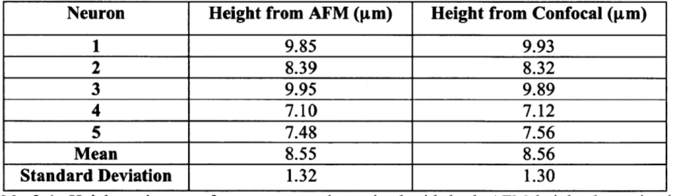
3-1: Height estimates of neuron soma determined with both AFM height determination methods and confocal im aging Z-stacks. ... 60
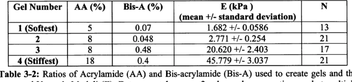
3-2. Ratios of Acrylamide (AA) and Bis-acrylamide (Bis-A) used to create gels and the measured Young's Moduli (E). Data taken over 2 gel sample preparations and at multiple locations on each sam ple ... 62
4-1: Distribution in parameters (mean and data range) obtained by fitting model response to experimental data for 33 cells, accounting for actual cell configuration by varying the model geometric parameters to match the measured height and cell radius. K was assumed to be constant for all cells and set at 10,000 Pa ... 75
5-1: Dimensions of neurons and astrocytes grown on substrates of varying stiffness (mean +/-standard deviation) obtained from confocal images. ... 91
5-2: Model parameters obtained by calibrating simulated response to mean AFM force data for neurons grown on different substrates. K was held constant at 10,000 Pa for all simulations. ... 9 4
5-3: Model parameters obtained by calibrating simulated response to mean AFM force data for astrocytes grown on different substrates. K was held constant at 10,000 Pa for all
sim ulations...98
Chapter 1
Introduction
A thorough understanding of the biomechanics of the central nervous system (CNS) is essential for understanding injury and pathology. While there are large bodies of research on the morphology, biochemistry, and electrophysiology of CNS cells, there is comparatively very little information on their biomechanical properties. Research dealing with the causes and disease progression of traumatic brain injury (TBI) is one area that would benefit greatly from a better characterization of the biomechanics of all cell types present in the CNS. CNS cells include neurons and glial cells, with glial cells consisting of astrocytes, microglia and oligodendrocytes. Glial cells were discovered in 1856 by a German pathologist, Rudolf Virchow [5] and named based on the idea that glial cells served as glue to hold the neurons together. Since their discovery, it has been shown that glial cells have many critical roles in brain function and the response to injury. Astrocytes perform a variety of functions in the CNS including release of neurotransmitters and synthesis of extracellular matrix (ECM) molecules [6-8]. They are also
involved in the process of synaptic neurotransmission and are capable of dynamically changing the coverage of synapases [9- 11]. In addition, unlike neurons, which are excited electrically via action potentials, astrocytes can be excited non-electrically. Oligodendrocytes play critical roles in myelination and microglia are involved in the host defense system. Microglia respond to chemokines released by astrocytes and localize to sites of injury during inflammation responses [11]. In addition, both neurons and glia are involved in the cell response after TBI. A better understanding of the biomechanics of neurons and glia will fill an important gap in characterizing the cellular response to mechanical trauma.
TBI is the result of various types of mechanical loading on the brain. A blow to the head, motor vehicle accident, sports injury, fall, or shock wave from an explosion are all examples of forces that elicit a mechanical response at both the cellular and tissue level in the brain [12]. This mechanical response ultimately leads to a pathophysiological response involving both neurons and glial cells, and eventually leads to brain damage and observed behavioral and cognitive changes. While it is well established that various mechanical loading scenarios can cause brain injury, there is still work to be done to fully connect the initial mechanical response with secondary signaling and eventual damage. A key component to filling in this knowledge gap is to have tools to study TBI at the cellular level in vitro. This includes having computational models to simulate how cells deform under various types of mechanical loading, as well as developing in
vitro damage devices to subject cells to a variety of mechanical insults. In addition, since the
initiator of the damage cascade is a mechanical load, it is critical to have measurements of cells grown in a mechanical environment that is biologically relevant. These experiments are critical for determining cellular injury thresholds and identifying potential therapeutics.
The work presented in this thesis provides a characterization of the biomechanics of neurons and glia in addition to developing important tools for studying and quantifying TBI at the cellular level. It builds on what is currently known about the mechanical response of single neurons and astrocytes, as well as on the existing in vitro cellular damage systems, as described in depth in Chapter 2. This work contributes significantly to the basic knowledge of the biomechanics of CNS cells.
Both experimental and computational approaches are utilized in this thesis to quantify the response of neurons and astrocytes to mechanical loading. Chapter 3 describes the methods used to measure single cell properties and create computational models for both neurons and astrocytes and contrasts these with previously published methods. This includes atomic force microscopy (AFM) indentation techniques to test cells at three different loading rates and in relaxation, as well as the formulation of a finite element model used to characterize the cell response. The AFM and modeling techniques are applied to test cells grown on substrates of different stiffness. Results and discussion of the single cell experiments and simulations are presented in Chapters 4 and 5. Chapter 4 quantifies and compares the mechanical response of neurons and astrocytes grown on glass, while Chapter 5 expands on this work and presents results of cells grown on gels of different stiffhess to address how the stiffness of the substrate influences the mechanical properties of CNS cells.
In addition, this thesis presents the design and preliminary characterization of two in vitro cell injury systems. Details on the systems are described in Chapter 6. One system compresses the cell cultures, simulating what might occur with an impact. The second system sends a shock wave through the cell culture, simulating loading associated with a blast wave from an explosion. Both systems utilize what was learned in Chapter 5 about the importance of substrate selection
on ensuring cells have morphology and force response similar to what would be expected in vivo, and damage the cells in a soft, 3-dimensional collagen gel. In addition, preliminary data and future plans for further characterizing the cellular damage and biochemical response are presented.
As summarized in Chapter 7, this thesis provides insight into how CNS cells respond to mechanical loading. The single cell models and measurements at a range of loading rates and on substrates of different stiffness provide a comprehensive characterization of the biomechanics of CNS cells in a variety of conditions and provide critical information for simulating TBI at the cellular level and determining damage thresholds. The quantification of cell properties, coupled with devices to damage cells in vitro, provides key tools to link well-defined stresses and strains to measured biochemical responses. These systems provide efficient ways to study TBI, identify new target molecules, and screen potential therapeutics.
Chapter 2
Background
2.1 Traumatic Brain Injury
TBI is a major cause of death and morbidity in the United States, affecting some 2 million civilians each year [13] and an estimated 20% of the 1.6 million veteran population returning from Iraq and Afghanistan [14, 15]. In addition, based on statistics collected from 2002 to 2006, the Centers for Disease Control and Prevention (CDC) estimated that TBI results in 1.365 million emergency room visits and 52,000 deaths each year in the United States [12]. They also found that TBI is a contributing factor in one third of the injury related deaths in the United States [12]. Cases of TBI can range from mild to severe, however, mild cases are much harder to diagnose, and as a result, accurate incidence estimates are hard to obtain. Mild TBI is characterized by a brief loss of consciousness (although not in all cases) and ongoing symptoms of headaches, dizziness, memory and concentration problems, and other behavioral and cognitive
changes [3]. Civilian TBI is often due to falls, sports injuries, and motor vehicle accidents, with falls being the leading cause of TBI and motor vehicle accidents being the leading cause of TBI-related death [12]. In the case of the veteran population, an additional cause of TBI is blast injury, which has become so prevalent in recent years it is often referred to as the signature injury of military troops today [16, 17].
peak overpressure
as the blast wave passes, pUrsure oscillates
0 - - - - -
-Y vacuum J
(A peak dynamic pressure
air flows from hilgh to lowv pressure
0 - - - - -- - - - -
---Figure 2-1: Characteristic blast wave pressure profile and resulting changes in atmospheric
pressure. Before the explosion (1) atmospheric pressure is normal. After the explosion and upon passage of the shockwave (2), the blast wind flows away from the explosion. When a drop in atmospheric pressure occurs below normal, the direction of the blast wind reverses (3). Reproduced with permission from Taber et al [1]
Blast TBI (bTBI) is caused by the interaction of a shockwave with the brain. After an explosion, the blast wave rapidly expands and propagates through the air and tissues. The blast wave is characterized by a near instantaneous rise in pressure followed by a decay profile reaching negative pressures (Figure 2-1). This quick rise in pressure results in a blast wind due to the kinetic energy transmitted to the air particles. bTBI is classified into four main categories, depicted in Figure 2-2 [14, 16-18]. Primary blast injury results from the shock wave itself. The
Figure 2-2: Schematic of the primary, secondary, and tertiary blast mechanisms and the characteristic brain injuries. Reprinted with permission from Macmillan Publishers Ltd: Journal of Cerebral Blood Flow and Metabolism, copyright 2010 [2]
shock wave consists of both over and under-pressures that travel through the brain tissue and act to compress and stretch the tissue. In addition, shockwaves can be concentrated and reflected at air-fluid interfaces, rendering certain regions of the body especially vulnerable. Typical shockwaves take place over millisecond timescales, but the exact pressure profile changes with the type and size of the explosive [16]. In secondary bTBI, shrapnel penetrates the head and causes damage. Tertiary injury occurs when the head is accelerated by the blast wind, potentially causing impact with surrounding objects. Finally, quaternary injury includes all other factors not accounted for in the first three phases. This could include chemical or thermal burns or breathing problems from smoke inhalation. Often, primary blast injury can be classified as mild TBI, or
concussion, and results in a brief loss of consciousness or altered mental status [17]. Mild bTBI is especially hard to diagnose but the consequences are still significant and cases are prevalent. In two studies of sample populations of U.S. soldiers, 15-16 % [17, 18] were found to have symptoms consistent with mild TBI. In addition, based on the most up-to-date numbers for military TBI in Iraq and Afghanistan (shown in Figure 2-3), it is clear that the vast majority of the TBI occurrences fall in the group of mild TBI, a category based on the characteristics of the acute sequelae following the injury. Memory and executive dysfunction (e.g. deficits in selective attention, planning and decision making, cognitive flexibility) are among the most common and disabling, but non-specific, features of mild bTBI. The prevalence of bTBI highlights the need for a better understanding of damage thresholds, injury progression, and potential therapeutics and preventative measures for both mild and more severe forms of bTBI.
Blast TBI has been studied in various animal models, showing that the primary blast wave alone is sufficient to cause TBI [19-29]. The animal studies indicated the involvement of both neurons and glial cells in the injury response. For example, Kaur and colleagues found both microglia and astrocytes to be activated in rats after being subjected to an explosive-generated blast [21, 25, 26]. Changes in neurons after blast injury have also been detected in animal models. For instance, Kato et al. studied the effects of a blast on rats and showed the
characteristics of neuronal damage varied with the magnitude of the overpressure. Pressures greater than 10 MPa resulted in an increase in TUNEL positive neurons, indicating involvement of apoptotic signaling pathways. For lower overpressures (1 MPa), morphological changes in neurons were identified, with neurons taking on a spindle shape and their nuclei becoming elongated [29]. Despite recent advances from what has been learned in animal models of blast TBI, there remains a need for a better understanding of the damage progression and injury
thresholds, especially at the cellular level, to enable development of preventative measures and potential therapeutics.
DoD Numbers for Traumatic Brain Injury
IF
0-'10 Q4
Totals
Penetrating
3,451
* Severe214
* Moderate
34,001
Mild
155,623
Not Classifiable
7,082
Total -All Severities
202,281
Source: Armed Forces Health Surveillance Center Numbers for 2000 -2010 Q4, as of 17 Feb2011
Figure 2-3: Official Department of Defense numbers for different severities of Traumatic Brain Injury (from http://www.dvbic.org/TBI-Numbers.aspx)
While the most common damage occurrences leading to mild or moderate forms of TBI (e.g. motor vehicle accidents or falls [30-32], sports concussions [33-35], and blast exposures [1,
32, 36, 37]) have been widely acknowledged and thoroughly reviewed, the etiology of the
ensuing cognitive, behavioral or neuropsychological disorders/impairments (e.g. memory loss, language difficulties, concentration deficiencies, behavioral abnormalities and/or depression) remains poorly understood. In particular, little is known about the multiple damage mechanisms suspected to unfold at the CNS cell level in the seconds to hours (and probably days) following initial mechanical insult(s) to the brain, and likely to result in cell/tissue alteration.
Cellular damage following TBI is a complex process involving both the initial damage caused from deforming the cells and tissue and secondary cellular responses involving diverse
damage pathways. Due to the time lag between the initial primary damage and secondary injury, there is hope that a better understanding of secondary damage mechanisms could lead to meaningful therapeutics and minimization of lasting damage [3]. The initial deformation can result in membrane damage and other structural failures [38-40] that lead to subsequent secondary mechanisms such as inflammation and changes in cell signaling [41-44]. Damage progression involves both neurons and glial cells, as depicted in Figure 2-4, with the secondary response leading to, among other processes, neuronal death [41], microglial activation [45], and reactive gliosis [46], which in turn cause tissue loss and glial scar formation [46]. A common theme in studies of TBI is the attempt to understand how the mechanical forces of the trauma incident activate cellular processes, and how these processes result in the pathological changes occurring hours to days after the initial trauma. Knowledge of the initial injury cascade is crucial for identifying potential therapeutics and interventions before the damage progresses.
One of the immediate consequences of mechanical trauma is an increase in plasma membrane permeability or mechanoporation [38, 39, 47, 48]. This can be a result of mechanically activated ion channels as well as tearing of the membrane. While mechanoporation may not kill the cells, especially in mild TBI, it does result in changes in cell function, ion homeostasis, electrical activity, and cell signaling [49]. In addition, it causes conduction block [50], neurofilament compaction [42], and impaired axonal transport [51], suggesting a role in connecting initial mechanical damage with cell biochemical response. Increased membrane permeability after mechanical trauma has been shown in both animal models [38] and in vitro cellular models [52-54]. In addition, work at the cellular level has shown that P-188, a nontoxic, nonionic, tri-block amphiphilic co-polymer that has been shown to reseal membranes, is able to block the mechanically induced increase in membrane permeability [53, 54].
()Astrocyte foot-process
-nlm
swelling A_ st Hosis
s
Microvascular B k Gdutaoate tra
the blood-brain barrier A AMPA receptor activation Microglial infiltration Proinfammatory hw cytokine release intra-axonal C-a2+ accumulation @ resynaptic glutamnate AA releaseA Axonal disconnection
Figure 2-4: Schematic depicting some of the key pathways associated with secondary damage
after TBI, involving neurons (blue), astrocytes (orange), and microglia (green). Reproduced with permission from Park et al. [3] © Canadian Medical Association. Copied under license from the Canadian Medical Association and Access Copyright. Further reproduction prohibited.
Excitotoxicity also occurs after TBI, as a result of an increase in extracellular concentration of glutamate, an excitatory neurotransmitter (for a detailed review see Yi and Hazell [55]). This increase in glutamate levels is thought to also be involved in the toxic increase in intracellular calcium levels following TBI [3]. In neurons, abnormal calcium homeostasis is involved in cell death processes. Changes in the glutamate receptors are among the causes for the observed increase in glutamate levels. There are two main classes of glutamate receptors, N-methyl D-aspartate receptors (NMDARs) and a-Amino-3-hydroxy-5-methyl-4-isoxazolepropionic acid receptors (AMPARs). NMDARs have been shown to be especially critical in the loss of ionic homeostasis after TBI [56]. For instance, they have been shown to be involved in the increase in cytosolic calcium levels [57]. NMDARs are also coupled to the
35
generation of reactive oxygen and nitrogen species. These products, coupled with other free radicals produced by the mitochondria eventually lead to fatal cellular processes [3]. In addition, NMDARs are linked to the actin cytoskeleton, providing potential insight into how stretch or other mechanical loading results in activation of NMDARs and the resulting changes in ion levels [58]. AMPARs have also been shown to be involved in TBI. AMPARs are involved in synaptic plasticity and could be key players in the immediate and long-term alterations of the neuronal network due to TBI. AMPARs also lose their desensitization property for 24 hours following mechanical injury [59, 60]. In addition, it has been shown that tumor necrosis factor a (TNFa), which is released from injured glia and inflammatory cells, leads to an increase in expressed AMPARs lacking the GluR2 subunit [61], a subunit involved in receptor calcium permeability [62]. As a result, this change in AMPAR subunit content could be a major contributor to the post-injury increase in calcium levels [3]. For a detailed review of the glutamate receptors and their role in TBI see Yi and Hazell [55] and Whalen et al [43].
The changes in levels of excitatory amino acids, free radicals, and intracellular calcium, along with many other complex pathways can ultimately result in loss of function and cell death. Cell death can occur via either a necrosis or apoptosis pathway. For detailed reviews of the cell death pathways see Raghupathi [41], Yuan et al [63], and Whalen et al [43]. Apoptosis, or programmed cell death, can occur as the result of multiple pathways, one of which involves the activation of death-inducing proteases. One class of proteases, the caspases, has been found to be activated in TBI [41, 43, 64]. It is believed that caspases cleave multiple proteins and the additive effects lead to cell death [65]. Cytoskeletal proteins, cell survival enzymes, and inhibitors of DNA endonucleases are all substrates on which the caspases act [43]. Work by Yakovlev and colleagues showed that cell death following fluid-percussion brain injury was
reduced when a caspase inhibitor was administered to the rats [66]. There are also both intrinsic and extrinsic pathways of apoptosis. The intrinsic pathway is triggered by the injured mitochondria. In the extrinsic pathway of apoptosis, membrane bound receptors such as Fas and TNFR1 are activated. This leads to recruitment of cytosolic adapter proteins. In turn, activated adapter proteins bind procaspases and form a death-inducing signaling complex (DISC). TNFRs are also involved in intracellular signaling pathways that can lead to cell survival. Activation of NFKB has been shown to be antiapoptotic and offers protection against TNF and Fas induced apoptosis by promoting increased transcription of antioxidant and antiapoptotic genes [43]. In addition, it has been shown that inhibition of Fas receptor and TNF-a improves neurologic function after brain injury in both adult and immature mice [43, 67].
Cell death following TBI can also occur via the necrosis pathway. Severe insults to the brain result in increased membrane permeability, organelle swelling, changes in both cellular and nuclear size and decreases in cell energy levels that lead to cell death [43]. This type of cell death is characterized as necrosis and occurs early in the damage progression and results in an inflammatory response [63]. There is also some work that suggests that necrosis can occur as a type of programmed cell death initiated by members of the tumor necrosis factor receptor family [68]. Since most of the damage leading to necrosis occurs early after injury, potential treatments targeting necrosis would need to be administered very soon after injury to be successful interventions.
Glial cells also play a unique and significant role in responding to CNS damage via a process known as reactive gliosis. Reactive gliosis involves hypertrophy and proliferation of microglia, astrocytes, and NG2 cells. Microglia are activated first [69], followed by increased proliferation of NG2 cells [70], and finally astrogliosis occurs [71]. Astrogliosis involves
astrocytes in the region surrounding the lesion responding to damage by proliferating, increasing process length, producing extracellular matrix, and increasing intermediate filaments and glial fibrillary acidic protein (GFAP) [7, 72, 73]. Initially, reactive gliosis has seemingly positive effects, minimizing tissue damage and helping to re-establish the blood-brain barrier [74]. Within days after injury, astrocytes "wall off' the damaged area and the resulting glial scar replaces the damaged neuronal cells. However, this glial scar is ultimately detrimental to neuron regeneration, as it has been shown that the neurites cannot penetrate the scarred region [46, 75].
The processes described in this section are just some of the complex cellular processes occurring after mechanical trauma that ultimately result in observed phenotypes associated with TBI. As described, these are complex processes involving both neurons and glial cells. Better understanding of these events requires not only knowledge of the activated biochemical pathways, but also a quantitative analysis of cell biomechanics, to truly connect the initial mechanical damage with injury thresholds and downstream cellular responses.
2.2 Biomechanics of Single Neurons and Glia'
One line of approach towards better elucidating some of the key damage mechanisms involved in TBI relies on addressing two distinct, yet interrelated, questions: (1) how mechanical transients applied to the organ boundary (head) translate into local stress-strain (force-displacement) distribution maps at the mesoscopic tissue level and microscopic cell level, and (2) how the cell machinery responds to these mechanical stimuli. An improved quantitative knowledge of material properties at the CNS cell level is necessary to understand the former on a quantitative
1 Section 2.2 is adapted from [76] Bernick K.B., Prevost T.P., Suresh S., Socrate S. Biomechanics of Single Cortical Neurons. Acta Biomaterialia 2011;7:1210. with permission from Elsevier
basis and to better characterize the latter in a controlled environment. Such characterization inevitably calls for measurable external mechanical inputs (e.g. pressure waves and imposed deformation profiles) to be applied to the boundary of in vitro cell systems (e.g. two-dimensional (2D)/ three-dimensional (3D) cell culture constructs or organotypic tissue slices) in a reproducible manner so that the latter inputs may systematically be associated with reliable estimates of force and deformation magnitudes at the single cell level.
Probing mechanical properties of individual cells has been made possible in recent years through the advent of novel testing techniques (for a review, see e.g. [77-79]) including magnetic twisting cytometry [80-83], particle-tracking microrheology [84-86], atomic force microscopy (AFM) [87-92], micropipette aspiration [93-98], optical tweezing and stretching [99-102], and microplate rheometry [103-105]. The last three techniques, which have been successfully employed to characterize the deformability of certain cell types in suspension (e.g. red blood cells [95, 101], white blood cells [93, 96], Mller glial cells [102], chondrocytes [98], myofibroblasts [94], and pancreatic cancer cells [105]), may not be easily applied to CNS neuronal cultures because neurons in vitro form intricate networks of adherent cells interconnected via multiple processes whose continuous growth and viable maintenance require the support of a substrate. Magnetic twisting cytometry is a powerful measurement technique providing local material properties at the membrane level but is not suited to examine global properties at the cell body level. AFM, originally developed to image surfaces of inorganic materials with atomic resolution [106], has proven to be a highly versatile testing tool in mechanobiology, enabling the measurement of material properties at the cell/subcellular level over a large range of forces (from pico- to nanonewton levels), speeds (from quasistatic to dynamic load levels), and length scales (from nano- to micrometers) via a variety of tip
geometries [107]. The diversity in available AFM tip geometries allows for a range of experiments to be performed under various loading conditions. Sharp tips may probe local properties at the cytoskeletal level or fine cellular structures such as neuronal processes, while large spheres may provide global "homogenized" properties at the whole cell level. Although widely used to characterize the mechanical response of numerous cell types including fibroblasts [87], leukocytes [91], cardiac myocytes [90] and blood cells [89, 92], AFM has been infrequently utilized to examine the response of CNS cells.
Limited information on the material properties of neurons and glia is currently available in the literature. To our knowledge, only Lu et al [102] have reported dynamic mechanical measurements on single CNS neurons - with measurements conducted in the linear infinitesimal strain regime only. Elastic storage and viscous loss moduli were extracted from the force-displacement output of oscillating 3 tm spherical AFM probes actuated to small indentation depths at the cell surface. These measurements, aimed at characterizing some of the local viscoelastic properties of neural cells, could not provide significant insights into the global mechanical response of single neural cell bodies, nor were they directed at investigating the mechanical nonlinearities observed at finite deformation typical of anticipated cell response in TBI cases, for which strains larger than 15-20% may be expected [108-111]. Although astrocytes have been studied more readily than neurons, there is still limited information on the global mechanical response of astrocytes. In addition, there is considerable variation in reported moduli values. Shiga et al [112] used both force-mapping and contact mode AFM techniques to measure the elasticity and topography of membranes of astrocytes cultured on glass and found the cell membrane (and immediate underlying areas) above the nucleus to be relatively soft with an elastic modulus between 1 and 5 kPa when compared to the membrane above the cytosol, which
they measured to have an elastic modulus between 1 and 35 kPa. Vergara and coworkers also measured the mechanical properties above the nucleus of astrocytes grown on tissue culture plastic using force mapping methods, finding the Young's modulus to range from roughly 30 to 60 kPa [113], values considerably stiffer than those measured by Shiga et al. In contrast, using the same methods used for measuring properties of neurons, Lu et al found the elastic moduli of astrocytes to range between 300 Pa and 520 Pa [102]. While there are many experimental differences between these three astrocyte studies, one of interest is that Shiga et al and Vergara
et al kept astrocytes in culture on stiff substrates, either on glass or on tissue culture plastic, for
weeks before testing. In contrast, Lu et al acutely isolated astrocytes and measured their properties after the cells had only been exposed to glass for a short time period. This suggests that the culture conditions, especially substrate properties, could be one variable responsible for measured differences in astrocyte properties.
The influence of substrate properties on measured cell mechanical response is a very important issue when connecting in vitro measurements to those expected in vivo, especially when developing multi-scale mechanical models. It is well established that cells are able to sense their mechanical environment and respond in a variety of ways [114-121]. In the case of cells found in the CNS, astrocytes have been shown to change morphology and cytoskeletal content on substrates of different stiffness, appearing small and rounded on softer substrates and more spread on stiffer substrates [122-124]. In addition, astrocytes grown on soft gels have been shown to have less F-actin stress fibers running through the cell, while astrocytes grown on hard substrates have very distinct stress fibers [122]. In contrast, neurons have been shown to have increased neurite branching on soft substrates [122, 125] and a corresponding increased F-actin content [122], while their process length remains unaffected by substrate stiffness [126, 127].
Soft substrates also promote the maturation of neural stem cell derived neurons, supporting growth of long neurites and synaptotagmin positive presynaptic terminals [128]. Further influence of substrate stiffness on neural stem cell behavior was shown by Saha and colleagues [129]. They found that in mixed differentiation conditions, that should favor both neuron and glial cell development, the proportion of neurons versus glial cells was dependent on the modulus of the substrate. In addition, soft substrates have been shown to select for neuronal growth over glial cell growth, with stiff materials allowing for more astrocyte spreading and adherence when compared with soft substrates [122]. In some cases, soft gels have been shown to result in glial cell death, with only neurons surviving [125]. In 3D culture systems, stiffness of the gel matrix has been shown to alter morphology as well. The mechanical stiffness of agarose gels has been shown to correlate inversely with the rate of neurite extension [130]. Sundararaghavan et al generated stable ID gradients of mechanical properties in 3D collagen gels and showed that neurites favor growth down the gel stiffness gradient [131]. These observations show that the mechanical environment influences both neurons and astrocytes and is an important factor to consider when studying processes in vitro at the cellular level, especially mechanical phenomena such as TBI.
In order to develop accurate single cell mechanical models as well as multi-scale models, encompassing cell, tissue, and organism level data, it is very important to have accurate predictions of cellular properties in vivo. One major difference between cells in vivo and cells typically grown in tissue culture is the mechanical environment surrounding the cell. Brain tissue
is one of the softest tissues in the body with a measured modulus reported on the order of a few kPa [132]. This is significantly different from traditionally used tissue culture plastic or glass, which are essentially rigid with moduli on the order of GPa. One way to begin to investigate
potential differences between in vivo and in vitro cellular mechanical properties, stemming from the mechanical environment surrounding the cell, is to grow cells on substrates of varying stiffness. While this cannot completely encompass the intricacies of an in vivo environment and all the extra cellular matrix interactions present in a 3D tissue environment, it does provide a measure of how sensitive cells are to changes in their mechanical environment and how much deviation from true in vivo properties may be observed in cells grown and tested on hard substrates such as glass or tissue culture plastic.
A critical component to understanding AFM data and creating models capable of accurately capturing the single cell response is the development of material models. AFM mechanical measurements conducted at the (whole) cell level on cell types other than neurons and astrocytes have been interpreted quantitatively with the aid of various continuum models. The modeling approaches most commonly used borrow their formulation from the contact theory developed by Hertz for linear elastic materials [87, 102, 133-135], many of which typically incorporate time-dependencies inherent in the cell response [87, 102]. The Hertz contact theory, however, relies on highly reductive assumptions including linearity, homogeneity, infinitesimal deformation, and infinite substrate dimensions - all of which are unlikely to hold for biological cell systems submitted to mechanical transients. In order to address some of these limitations, investigators have proposed alternative continuum approaches integrating part of the complexities observed in the mechanical response of biological cells. These approaches include piecewise linear elastic variations [136], linear hyper-elastic/viscoelastic composite material formulations [92, 137, 138], and biphasic linear elastic constitutive relations [139, 140]. More complex variations borrow elements from continuum and piecewise continuum models [141]. While successful at capturing specific quantitative features of the cell response under selected