HAL Id: hal-02985756
https://hal.sorbonne-universite.fr/hal-02985756
Submitted on 2 Nov 2020HAL is a multi-disciplinary open access archive for the deposit and dissemination of sci-entific research documents, whether they are pub-lished or not. The documents may come from teaching and research institutions in France or abroad, or from public or private research centers.
L’archive ouverte pluridisciplinaire HAL, est destinée au dépôt et à la diffusion de documents scientifiques de niveau recherche, publiés ou non, émanant des établissements d’enseignement et de recherche français ou étrangers, des laboratoires publics ou privés.
neuroinflammation in ALS
Aude Chiot, Christian S Lobsiger, Séverine Boillée
To cite this version:
Aude Chiot, Christian S Lobsiger, Séverine Boillée. New insights on the disease contribution of neuroinflammation in ALS. Current Opinion in Neurology, Lippincott, Williams & Wilkins, 2019, 32 (5), pp.764-770. �10.1097/WCO.0000000000000729�. �hal-02985756�
New insights on the disease contribution of neuroinflammation in ALS
Aude Chiota, Christian S. Lobsigera, Séverine Boilléea,b
a: Institut du Cerveau et de la Moelle épinière, ICM, Inserm U 1127, CNRS UMR 7225, Sorbonne Université, F-75013, Paris, France.
b: Correspondance to Séverine Boillée: Séverine Boillée, Institut du Cerveau et de la moelle épinière, ICM, Inserm U 1127, 47 boulevard de l’hopital, 75013 Paris, France. e-mail: severine.boillee@upmc.fr
Abstract:
Purpose of review: Amyotrophic Lateral Sclerosis (ALS) is a degenerative motor neuron
(MN) disease with a strong neuroinflammatory component. This review summarizes how the connection between neurodegeneration and the immune system is strengthened by new discoveries from ALS genetics and the analysis of subpopulations of immune cells in ALS.
Recent findings: Recent genes identified in ALS encode for proteins with direct immune
roles, that when mutated lead to deregulation of immune functions, potentially influencing the disease. Although, neuroinflammation in the CNS of ALS has been well documented, new evidence suggests also direct malfunctions of immune cells in the CNS and at the periphery: While CD4+ T regulatory lymphocytes are protective in ALS, their number and function are altered over the disease course. CD8+ T cells are detrimental for MNs in the CNS, but show some protective roles at the periphery. Likewise, presence of mast cells in muscles of ALS models and patients, and impairments of monocyte functions reveal potential new players in ALS disease progression.
Summary: Although MN degeneration is considered the prime event in ALS, dysfunctions
in immune processes can impact the disease, highlighting that targeting specific immune components is a strategy for developing biomarkers and ultimately new drugs.
Keywords: Amyotrophic Lateral Sclerosis (ALS), neuroinflammation, lymphocytes,
Introduction
Amyotrophic Lateral Sclerosis (ALS) is the most common adult onset motor neuron (MN) disease, characterized by the degeneration of upper and lower MNs. ALS is a heterogeneous disease, multifactorial, with some known genetic causes, but for the majority of cases, multigenetic and probable environmental causes are likely present [1]. To date, mutations in more than 25 genes, mainly autosomal dominant, have been linked to ALS, the major genes being C9orf72, SOD1, TARDBP and FUS/TLS (http://alsod.iop.kcl.ac.uk) [2,3]. A hallmark of both familial and sporadic ALS, is the presence of reactive immune cells in postmortem tissues, with microglial cells, the macrophages of the CNS, found in the vicinity of degenerating MN cell bodies and axonal tracts [4]. Microglial activation (as part of the neuroinflammatory response) has also been correlated to MN damage in ALS patients [5,6]. To understand the role of these immune cells during the disease course, ALS mouse models have been extensively used, mostly mice expressing mutant Cu/Zn Super oxide Dismutase (SOD1), as they remained for the longest time the only model with clear recapitulation of progressive MN degeneration, and only in most recent time, alternative mouse models start to appear, although with variable efficiency and usability. Importantly, these studies with mutant SOD1 mice strongly suggested that ALS is a non-cell autonomous disease, where certain, but not all (non-neuronal) cell-types surrounding MNs contribute to disease toxicity, in particular and the focus of this review, microglial cells [7–9]. Interestingly, while downregulating mutant SOD1 in MNs delayed disease onset, in microglial cells, it influenced disease
progression [7–9]. This specificity is of prime importance and led to three insights: (i)
consistent with mutant SOD1 expressing microglial cells not being able, on their own, to kill control MNs in mice [9], ALS remains primarily a MN disease, and microglial cells act by
reacting to neurodegeneration; (ii) but it also indicates that reactive microglial cells can
contribute to MN degeneration; and thus, (iii) it suggests that targeting microglial cells to slow disease progression could be effective, even in the sporadic ALS population. Microglial cells are part of the myeloid lineage, but they have a different developmental origin than most other tissue macrophages and blood monocytes. Other immune cells coming from the lymphoid lineage take also part in the disease. While depletion of B cells did not impact ALS disease in mice, specific deletion of CD4+ or CD8+ T lymphocytes has emphasized their opposite impact, detailed thereafter [10–14**].
When ALS genes show immune functions
C9orf72 is the major ALS-linked gene in the western world, characterized by a
hexanucleotide expansion in its intronic region. Following this discovery, several mouse lines deleted for the C9orf72 ortholog were produced (C9orf72 KO mice), to understand the unknown function of C9orf72 and assess a possible loss-of-function mechanism [15–21]. While only one study reported mild locomotor abnormalities [15], five described C9orf72 KO mice suffering from strong immune system defects, including splenomegaly, massive myeloid and lymphoid cell infiltration in the spleen and the lymph nodes, an elevated rate of autoantibodies, and elevated levels of several pro-inflammatory cytokines in the blood - phenotypes resembling an autoimmune syndrome [15–19]. These defects were reported in homozygous but not heterozygous mice. Importantly, ALS patients carry the mutant expansion in only one C9orf72 allele, which leads to some decrease of C9orf72 mRNA and protein levels [20,22*]. In C9orf72 ALS patients, the link between their MN disease and possible immune system dysfunctions is not obvious. Although, an increased number of autoimmune diseases in ALS/FTD patients has been reported, a specific link with C9orf72 remains to be determined [23]. Microglial cells isolated from C9orf72 KO (but not from C9orf72+/-) mice displayed a lysosomal transport defect, also noticed in microglial cells of motor cortex and spinal cord postmortem tissues of mutant C9orf72 ALS patients [18]. Removing murine C9orf72 from neuronal and glial cell precursors only (but not targeting immune cells, including microglial cells), did not lead to any pathological neuronal signs of ALS or neuroinflammatory reaction [24]. Therefore, the current hypothesis is a mechanism of gain-of-toxic function in neurons (and glial cells of neural origin), possibly potentiated by a downregulation of C9orf72 and a loss-of-function haplo-insufficiency effect in immune cells, which, however, remains to be further characterized in mutant C9orf72 ALS patients.
TBK1 (Tank-Binding Kinase-1) discovered in 2015 as a new ALS-linked gene, was
previously known for its functions in autophagy but also immune system regulation (originally described as a mediator of NF-κB signalling pathway) [25–27]. Full deletion of
Tbk1 in mice on a C57Bl6 genetic background, is embryonic lethal (due to liver failure), but
some Tbk1 KO mice on a 129S5 genetic background get born and, interestingly, develop a phenotype very similar to the C9orf72 KO mice, including massive infiltrations of mononuclear cells in different organs, higher rate of circulating monocytes and greater amounts of pro-inflammatory cytokines in the blood [28]. Mutations in TBK1, discovered in familial ALS cases, result (in most cases) in a loss-of-function of the mutant protein, strongly
suggesting a mechanism of haploinsufficiency [25,26]. In mice, heterozygous deletion of
Tbk1, did not lead to any obvious MN degeneration (at least, not until 6 months of age) [29*].
However, and surprisingly, in the neurodegenerative and inflammatory context of mutant SOD1G93A ALS mice, heterozygous deletion of Tbk1 induced, first, an accelerated MN denervation, followed, second, by decreased microglial activation at late disease stages and, remarkably, an increased survival. This highlights a possible dual role of Tbk1 in MNs and microglial cells, that both can influence the ALS disease course separately and distinctly.
In this context, another gene previously reported for carrying rare (most likely loss-of-function) mutations in ALS, OPTN (encoding for Optineurin), implicated in autophagy and regulation of NF-κB, interacts and gets phosphorylated by TBK1 [30–32]. While Optn KO mice did not show a clear MN degeneration, they did display signs of distal axonopathy [33].
Optn KO mice also produced elevated amounts of pro-inflammatory cytokines, and
transcriptional analysis of Optn KO microglial cells revealed a rather pro-inflammatory phenotype. Therefore, both mutations in Optn and Tbk1 induce inflammatory dysregulations, potentially contributing to ALS.
Other genes expressed by immune cells have been suspected as causative or modifiers of ALS, including CX3CR1, encoding the receptor for the chemokine CX3CL1 (fractalkine), constitutively expressed by neurons including MNs. Microglia are the only CNS cells expressing CX3CR1, making CX3CL1/CX3CR1 a candidate pathway for a MN-microglia cross talk [34,35]. Membrane-bound fractalkine is considered a main actor to keep microglia in a homeostatic, non-activated state [36]. Deletion of Cx3cr1 in SOD1G93A ALS mice revealed increased MN loss and a tendency to higher microglial activation. SOD1G93A/Cx3cr1-/- males (but not females) displayed a faster decline in motor functions and a reduced survival compared to SOD1G93A/Cx3cr1+/- males [36,37*]. In ALS patients, specific variants in the CX3CR1 gene have been associated with a faster progression of the disease, although there was no association with ALS disease risk [38,39*].
TREM2, well-known for its mutations causing rare forms of Alzheimer’s disease, encodes for
Triggering Receptor Expressed Myeloid cell 2, expressed by myeloid cells and in particular microglial cells [40]. A specific variant in TREM2 (p.R47H) was identified as a risk factor for ALS without, however, being correlated with the age of onset, the site of the first symptoms or disease progression [41]. TREM2 encodes for an extracellular receptor involved in immune
functions, especially phagocytosis. The p.R47H variant was associated with maturation defects of the receptor causing impaired phagocytosis [42]. Increased expression of Trem2 mRNA was measured in the spinal cord of symptomatic mutant SOD1G93A mice, but also sporadic ALS patients [41]. Very recently, upregulation of Trem2 mRNA was measured, at early disease stages in ALS mice, with a new technique of spatial transcriptomics, allowing full transcriptome analysis directly on tissue sections [43**]. Interestingly, this study revealed that Tyrobp, which forms a complex with Trem2 that triggers phagocytosis, was up-regulated on the mRNA level, even before Trem2. In addition, the Trem2-ApoE pathway (Trem2 is also known for inducing ApoE signalling) was previously shown to trigger a common molecular signature of microglial cells in several neurodegenerative conditions, including ALS [44**]. Deletion of Trem2 in mutant SOD1-expressing microglial cells, induced downregulation of inflammatory genes and restored microglial homeostatic gene expression [44**]. The example of Trem2 is an interesting indicator of microglial implications in ALS, suggesting that such implication could be triggered both by internal defects, e.g. caused by genetic ALS-linked mutations (as SOD1), but also by (perturbed) external signalling, mediated, through the neurodegenerative/neuroinflammatory context.
Lymphocytes implicated in ALS neurodegeneration and a possible biomarker in ALS
Originally, CD4+ cells were the main T cell population thought to be implicated in ALS, with removal of CD4+ cells (CD4 KO mice) showing reduced survival of SOD1G93A ALS mice [10,11]. Simplified, CD4+ cells can be subdivided in regulatory (Treg) and effector (Teff) T cells, with Tregs regulating the proliferation of Teff cells. In SOD1G93A ALS mice, passive transfer of T cell populations enriched in Tregs compared to whole CD4+ T cells, led to a more pronounced increase in survival [45,46]. Treg expansion in SOD1G93A mice (using peripheral injection of Interleukin 2 (IL-2)/IL2 antibody complex), decreased microglial activation and increased MN and mouse survival [47**]. In ALS patients, a higher proportion of Treg in the blood was correlated with a slower rate of disease progression. [46–48]. Functional assays with Tregs of ALS patients showed their reduced ability to suppress the Teff proliferation, compared to control Tregs [49**]. Therefore, both reduced numbers and impaired suppressive function could influence ALS progression. A phase II clinical study assessing the potential of IL-2 as a therapeutic agent is currently in progress in ALS (ClinicalTrials.gov NCT03039673). FoxP3, a marker of Treg was found downregulated in ALS patients’ Tregs compared to controls. FoxP3 levels were also inversely correlated with the rate of disease progression, with FoxP3 expression being reduced in rapidly progressing
patients [48,49**]. Importantly, a prospective study pointed out low levels of FoxP3, not only reflecting a rapid disease progression at the time of blood collection, but also able to predict future rapid disease progression rate. Therefore, FoxP3 appears as a promising biomarker for ALS disease progression [48].
More recent studies have focused on the, so far in ALS understudied, CD8+ lymphocytes. While previous reports mentioned a very late infiltration of CD8+ T cells (compared to CD4+ cells) in the spinal cord of SOD1G93A ALS mice (arguing in favor of no possible impact of these cells), two recent studies measured the presence of infiltrating CD8+ T cells as early as disease onset [10,13**,14**]. To study CD8+ T cell involvement, ALS mice either lacking functional CD8+ T cells (CD8a-/- /SOD1G93A) or defective for CD8+ T cell actions (ß2-microglobulin (ß2M)-/- /SOD1G93A, displaying no MHC-I expression) were generated
[13**,14**] or were injected with anti-CD8 antibodies [50**]. Although survival of CD8a
-/-/SOD1G93A mice or of SOD1 mice injected with anti-CD8, was not modified, survival of ß2M
-/- /SOD1G93A mice showed a different outcome in two independent studies with either, for the
earlier study, no modified onset but a decreased survival [51], or in the more recent study, an earlier onset (measured by muscle weakness) followed by a prolonged survival [13**]. Interestingly, in CD8a-/- /SOD1G93A mice, MNs survived better in the lumbar spinal cord, while in ß2M-/- /SOD1G93A mice, it was not the case, however MNs were protected in the
cervical spinal cord [13**,14**,51]. Since CD8+ T cell toxicity towards MNs was dependent on MHC-I recognition by CD8+ T cells in vitro, a compensatory mechanism could explain the difference obtained in ß2M-/- and CD8a-/- mice and the heterogeneity between cervical and lumbar MNs in ß2M-/- mice [13**,14**]. As MHC-I is expressed also by myeloid cells, an additional effect of deleting MHC-I could also come from deregulated microglia/macrophage reactivity. In addition, CD8+ T cells seem to have a dual role in ALS, with a protective effect at the periphery (accelerated muscle atrophy and axonal defects in ß2M-/- /SOD1G93A mice), and a toxic role on MN survival when infiltrating the CNS [13**,14**] (Fig1). Of note, for both CD4+ and CD8+ cells, it remains to be determined if their role in ALS disease is linked to mutant gene expression (e.g. mutant SOD1) or their neuroinflammatory responses.
Another subpopulation of lymphocytes (which are part, however, of the innate immunity, as they do not recognize specific antigens), the Natural killer (NK) cells with their cytotoxic activity, have recently also been studied in ALS. A longitudinal study showed an increased number of NK cells in the blood of ALS patients compared to controls, but also in ALS patients along the disease [52*]. Interestingly, infiltrating NK cells have been observed in the
spinal cord of ALS mice, however, decreasing the number of NK cells in SOD1G93A mice had no impact on disease [11,50**].
Mast cell involvement in ALS
Mast cells, granulated leucocytes involved in allergic reactions, have recently gained interest as the target cells of Masitinib (AB1010), a Tyrosine Kinase inhibitor, tested in clinical trials on ALS patients (ClinicalTrials.gov NCT02588677). Mast cells were observed in muscles (extensor digitorum longus) and sciatic nerves of symptomatic SOD1G93A ALS rats but not in the spinal cord. Attention has therefore been focused on their impact at the periphery [53**,54*]. Presence of mast cells (in variable amounts compared to controls), was also described in ALS patient quadriceps muscles with frequent degranulation, suggesting mast cell reaction and a potential role in ALS [54*]. In SOD1G93A rats, Masitinib treatment led to a
decreased number of mast cells in muscles and sciatic nerves and delayed neuromuscular junction denervation, suggesting a deleterious role for mast cells towards axonal integrity in ALS [54*,55] (Fig. 1). However, Masitinib is also an inhibitor of Csf1-R, required for macrophage and microglia survival. Since selective inhibition of the Csf1-R (with GW2580) was also potent at slowing the disease course in SOD1G93A mice [56], the impact of Masitinib in ALS could be through mast cells and macrophage/microglial cells, and specific depletion or inhibition of mast cells would be needed to definitely conclude on mast cell contribution in ALS.
Deregulation of monocytes in ALS
For their easy accessibility in the blood, and since, as part of the myeloid lineage, they share common functions with microglial cells, monocytes have gained recent interest in ALS. While analysis of overall proportions of monocytes among all the leucocytes was not sensitive enough to show differences between ALS patients and healthy controls, a recent longitudinal study, performed over 3 years, highlighted an increased monocyte number (and also of the whole leucocyte content in the blood) during the course of ALS, although it was not correlated with the functional ALSFRS-R score [52*,57]. Expression of the specific marker, CX3CR1, in this longitudinal study, correlated, however, with the ALS Functional Rating Scale Revised (ALSFRS-R) score [52*]. Monocytes can be divided into two major populations, classical monocytes, considered to be inflammatory and recruited into the tissues upon inflammation, and non-classical or patrolling monocytes, in charge of tissue homeostasis. A higher proportion of classical to non-classical monocytes has been described
in ALS patients compared to healthy controls [58*]. One hypothesis would therefore be, that deregulated proportions of monocyte subtypes could lead to dysfunctions of immune responses, contributing to ALS disease. However, possible intrinsic dysfunctions in ALS monocytes have also been reported, and included alterations in phagocytosis, cytokine secretions upon external stimuli, and of their transcriptome [57,58*,59**] (Fig. 1). Upregulation of genes involved in inflammatory responses such as Interleukine-1ß, Interleukine-8 or NLRP3, and downregulation of genes involved in Tumor Necrosis Factor or Toll Like Receptor signaling (two important pathways of the innate inflammatory response), were revealed with RNAseq analyses in monocytes from ALS patients compared to controls, suggesting a possible dysfunction of myeloid cells in ALS [57,59**].
Conclusion
Although, MNs are the primary cells degenerating in ALS, rising evidence show that ALS has an inflammatory component, with some ALS related genes linked to different immune functions. Defects caused by mutations in those genes are likely to drive defects in their immune functions, potentially impacting ALS disease course. Modifying immune cells through bone marrow transplantation has been envisaged and targeting specific myeloid or lymphoid populations is the focus of ongoing clinical trials. However, better knowledge of the specific deregulations of the different subpopulations of immune cells (including CD4+ Tregs, CD8+ T cells, mast cells and monocytes) and this, over the course of the disease, between different ALS patient cohorts, are now necessary and the essential next step required for the development of immune-related therapies and biomarkers in ALS.
Key points:
• Recent genes discovered in ALS reinforce the link between the disease and alteration of the immune system.
• Blood lymphocytes and in particular regulatory T lymphocytes appear as promising predictors of the rate of disease progression.
• Presence of mast cells in muscles in ALS models and patients and deregulated monocytes in ALS patients highlight new immune cell players at the periphery to consider for future therapy.
• Failure of fundamental functions of monocytes calls on a possible systemic immune deregulation in ALS
Financial support and sponsorship
This work was supported by Thierry Latran Foundation, EraNet Neuron, ALSA, NRJ-Institut de France, ARMC, SLAFR, La Longue route, AC was financed by the French ministry of higher education, research and innovation, and ARSLA.
Conflicts of interest
The authors declare no conflict of interest
References
[1] Phukan J, Elamin M, Bede P, et al. The syndrome of cognitive impairment in amyotrophic lateral sclerosis: A population-based study. J Neurol Neurosurg Psychiatry 2012;83:102–8.
[2] Millecamps S, Salachas F, Cazeneuve C, et al. SOD1, ANG, VAPB, TARDBP, and FUS mutations in familial amyotrophic lateral sclerosis: genotype-phenotype correlations. J Med Genet 2010;47:554–60.
[3] Brown RH, Al-Chalabi A. Amyotrophic Lateral Sclerosis. N Engl J Med 2017;377:162–72.
[4] Engelhardt JI, Appel SH. IgG reactivity in the spinal cord and motor cortex in amyotrophic lateral sclerosis. Arch Neurol 1990;47:1210–6.
[5] Turner MR, Cagnin A., Turkheimer FE, et al. Evidence of widespread cerebral microglial activation in amyotrophic lateral sclerosis: An [11C](R)-PK11195 positron emission tomography study. Neurobiol Dis 2004;15:601–9.
[6] Brettschneider J, Toledo JB, Van Deerlin VM, et al. Microglial activation correlates with disease progression and upper motor neuron clinical symptoms in amyotrophic lateral sclerosis. PLoS One 2012;7:13–5.
[7] Boillée S, Yamanaka K, Lobsiger CS, et al. Onset and progression in inherited ALS determined by motor neurons and microglia. Science 2006;312:1389–92.
[8] Wang L, Sharma K, Grisotti G, Roos RP. The effect of mutant SOD1 dismutase activity on non-cell autonomous degeneration in familial amyotrophic lateral sclerosis. Neurobiol Dis 2009;35:234–40.
knockout mice with familial amyotrophic lateral sclerosis 2006;103:16021–6.
[10] Beers DR, Henkel JS, Zhao W, et al. CD4+ T cells support glial neuroprotection, slow disease progression, and modify glial morphology in an animal model of inherited ALS. Proc Natl Acad Sci U S A 2008;105:15558–63.
[11] Chiu IM, Chen A, Zheng Y, et al. T lymphocytes potentiate endogenous neuroprotective inflammation in a mouse model of ALS. Proc Natl Acad Sci U S A 2008;105:17913–8.
[12] Naor S, Keren Z, Bronshtein T, et al. Development of ALS-like disease in SOD-1 mice deficient of B lymphocytes. J Neurol 2009;256:1228–35.
[13] Nardo G, Trolese MC, Verderio M, et al. Counteracting roles of MHCI and CD8 + T cells in the peripheral and central nervous system of ALS SOD1 G93A mice. Mol Neurodegener 2018;13:1–24.
** First evidence of CD8+ T cell involvement in ALS pathology in mutant SOD1 mice, with an interesting comparison between the effect in the CNS and in the PNS.
[14] Coque E, Salsac C, Espinosa-carrasco G, et al. Cytotoxic CD8 + T lymphocytes expressing ALS-causing SOD1 mutant selectively trigger death of spinal motoneurons. PNAS 2019;10.
** This study shows evidence of direct toxicity of ALS mouse CD8+T cells towards motor neurons in culture and of infiltrated CD8+ T cell on motor neuron survival in ALS mice
[15] Atanasio A, Decman V, White D, et al. C9orf72 ablation causes immune dysregulation characterized by leukocyte expansion, autoantibody production, and glomerulonephropathy in mice. Sci Rep 2016;6:23204.
[16] Burberry A, Suzuki N, Wang J, et al. Loss-of-function mutations in the C9ORF72 mouse ortholog cause fatal autoimmune disease. Sci Transl Med 2016;8.
[17] Jiang J, Zhu Q, Gendron TF, et al. Gain of Toxicity from ALS/FTD-Linked Repeat Expansions in C9ORF72 Is Alleviated by Antisense Oligonucleotides Targeting GGGGCC-Containing RNAs. Neuron 2016;90:535–50.
[18] O’Rourke JG, Bogdanik L, Yanez A, et al. C9orf72 is required for proper macrophage and microglial function in mice. Science 2016;351:1324–9.
immune system-related pathology and neoplastic events but no motor neuron defects. Acta Neuropathol 2016:3–5.
[20] DeJesus-hernandez M, Mackenzie IR, Boeve BF, et al. Expanded GGGGCC hexanucleotide repeat in non-coding region of C9ORF72 causes chromosome 9p-linked frontotemporal dementia and amyotrophic lateral sclerosis. Neuron 2011;72:245–56.
[21] Renton AE, Majounie E, Waite A, et al. A hexanucleotide repeat expansion in C9ORF72 is the cause of chromosome 9p21-linked ALS-FTD. Neuron 2011;72:257– 68.
[22] Frick P, Sellier C, Mackenzie IRA, et al. Novel antibodies reveal presynaptic localization of C9orf72 protein and reduced protein levels in C9orf72 mutation carriers. Acta Neuropathol Commun 2018;6:1–17.
* In this study, a novel antibody againt C9orf72 has been generated and characterized allowing the detection of C9orf72 protein in ALS patient brains, giving further evidence of a haploinsufficiency mechanisms in C9orf72-linked ALS.
[23] Miller ZA, Sturm VE, Camsari GB, et al. Increased prevalence of autoimmune disease within C9 and FTD / MND cohorts Completing the picture. Neurol Neuroimmunol NeuroInflammation 2016:1–9.
[24] Koppers M, Blokhuis AM, Westeneng HJ, et al. C9orf72 ablation in mice does not cause motor neuron degeneration or motor deficits. Ann Neurol 2015;78:426–38. [25] Cirulli ET, Lasseigne BN, Petrovski S, et al. Exome sequencing in amyotrophic lateral
sclerosis identifies risk genes and pathways. Science 2015;347:1436–41.
[26] Freischmidt A, Wieland T, Richter B, et al. Haploinsufficiency of TBK1 causes familial ALS and fronto-temporal dementia. Nat Neurosci 2015;18:631–6.
[27] Pomerantz JL, Baltimore D. NF-kappaB activation by a signaling complex containing TRAF2, TANK and TBK1, a novel IKK-related kinase. EMBO J 1999;18:6694–704. [28] Marchlik E, Thakker P, Carlson T, et al. Mice lacking Tbk1 activity exhibit immune
cell infiltrates in multiple tissues and increased susceptibility to LPS-induced lethality. J Leukoc Biol 2010;88:1171–80.
[29] Brenner D, Sieverding K, Bruno C, Lüningschrör P, Buck E, Mungwa S, et al. Heterozygous Tbk1 loss has opposing effects in early and late stages of ALS in mice. J
Exp Med 2019;216:267–78.
* This study indicates that heterozygous Tbk1 deletion in mice does not induce a motor neuron phenotype (at least not up to 6 months of age). However hemizygous deletion of Tbk1 in SOD1G93A ALS mice leads to a dual effect of acceleration of onset followed by increased survival, suggesting a deleterious effect of reducing Tbk1 in motor neurons (likely through deregulated autophagy) and a protective effect of reducing Tbk1 in immune cells.
[30] Maruyama H, Morino H, Ito H, et al. Mutations of optineurin in amyotrophic lateral sclerosis. Nature 2010;465:223–6.
[31] Millecamps S, Boillée S, Chabrol E, et al. Screening of OPTN in French familial amyotrophic lateral sclerosis. Neurobiol Aging 2011;32:557.e11-557.e13.
[32] Zhu G, Wu CJ, Zhao Y, Ashwell JD. Optineurin Negatively Regulates TNFa- Induced NF-kB Activation by Competing with NEMO for Ubiquitinated RIP. Curr Biol 2007;17:1438–43.
[33] Ito Y, Ofengeim D, Najafov A, Das S, Saberi S, Li Y, et al. RIPK1 mediates axonal degeneration by promoting inflammation and necroptosis in ALS. Science 2016;353:603–8.
[34] Harrison JK, Jiang Y, Chen S, et al. Role for neuronally derived fractalkine in mediating interactions between neurons and CX3CR1-expressing microglia. Proc Natl Acad Sci U S A 1998;95:10896–901.
[35] Jung S, Aliberti J, Graemmel P, et al. Analysis of Fractalkine Receptor CX3CR1 Function by Targeted Deletion and Green Fluorescent Protein Reporter Gene Insertion. Mol Cell Biol 2000;20:4106–14.
[36] Cardona AE, Pioro EP, Sasse ME, et al. Control of microglial neurotoxicity by the fractalkine receptor. Nat Neurosci 2006;9:917–24.
[37] Liu C, Hong K, Chen H, et al. Evidence for a protective role of CX3CL1 / CX3CR1 axis in a model of amyotrophic lateral sclerosis. Biol Chem 2018;400:651–61.
* This study reports the deregulation of the CX3CR1/CX3CL1 signalling during the course of the disease in SOD1G93A ALS mice.
[38] Lopez-Lopez A, Gamez J, Syriani E, et al. CX3CR1 is a modifying gene of survival and progression in amyotrophic lateral sclerosis. PLoS One 2014;9:e96528.
[39] Calvo A, Moglia C, Canosa A, et al. Common polymorphisms of chemokine (C-X3-C motif) receptor 1 gene modify amyotrophic lateral sclerosis outcome: A population-based study. Muscle and Nerve 2018;57:212–6.
* This study indicates that a specific variant in CX3CR1 is associated with a faster disease progression in ALS patients.
[40] Sims R, Van der Lee SJ, Naj AC, et al. Rare coding variants in PLCG2, ABI3, and TREM2 implicate microglial-mediated innate immunity in Alzheimer’s disease. Nat Genet 2017;49:1373–87.
[41] Cady J, Koval ED, Benitez BA, et al. The TREM2 variant p.R47H is a risk factor for sporadic amyotrophic lateral sclerosis Janet. JAMA Neurol 2014;71:449–53.
[42] Kleinberger G, Yamanishi Y, Suárez-calvet M, et al. TREM2 mutations implicated in neurodegeneration impair cell surface transport and phagocytosis. Sci Transl Med 2014;6.
[43] Maniatis S, Vickovic S, Braine C, et al. Spatiotemporal dynamics of molecular pathology in amyotrophic lateral sclerosis. Science 2019;93:89–93.
** This study provides a spatial transcriptomic analysis of SOD1G93A mouse spinal cord over the course of the disease, and identifies dynamic regulations of several immune signalling pathways.
[44] Krasemann S, Madore C, Cialic R, et al. The TREM2-APOE pathway drives the transcriptional phenotype of dysfunctional microglia in neurodegenerative diseases. Immunity 2017;47:566–81.
** This study identifies a molecular signature of microglial cells common to several neurological disorders and shows how the Trem2-ApoE pathway mediates a switch towards this reactive phenotype, which then could be targetted to reduce deletrious microglial cell reactivity.
[45] Banerjee R, Mosley RL, Reynolds AD, et al. Adaptive immune neuroprotection in G93A-SOD1 amyotrophic lateral sclerosis mice. PLoS One 2008;3:e2740.
[46] Beers DR, Henkel JS, Zhao W, et al. Endogenous regulatory T lymphocytes ameliorate amyotrophic lateral sclerosis in mice and correlate with disease progression in patients with amyotrophic lateral sclerosis. Brain 2011;134:1293–314.
with progression of amyotrophic lateral sclerosis a study of humans and a transgenic mouse model. JAMA Neurol 2018;75:681–9.
** This study shows a correlation between regulatory T cell (Treg) levels and rate of disease progression in ALS patients and reports a protective effect of IL2 treatment inducing Treg expansion in SOD1G93A ALS mice.
[48] Henkel JS, Beers DR, Wen S, et al. Regulatory T-lymphocytes mediate amyotrophic lateral sclerosis progression and survival. EMBO Mol Med 2013;5:64–79.
[49] Beers DR, Zhao W, Wang J, et al. ALS patients’ regulatory T lymphocytes are dysfunctional, and correlate with disease progression rate and severity. Jci 2017;2:e89530.
** This study demonstrates how ALS patient regulatory T cells have defective suppressive functions (on the effector T cell proliferation) and shows an association between the level of failure and the rate of disease progression. This indicates that restoring their function could be effective in slowing disease progression.
[50] Komine O, Yamashita H, Fujimori-Tonou N, et al. Innate immune adaptor TRIF deficiency accelerates disease progression of ALS mice with accumulation of aberrantly activated astrocytes. Cell Death Differ 2018:1–17.
** First evaluation of the possible involvement of Natural Killer cells in ALS.
[51] Staats KA, Schönefeldt S, Van Rillaer M, et al. Beta-2 microglobulin is important for disease progression in a murine model for amyotrophic lateral sclerosis. Front Cell Neurosci 2013;7:6–10.
[52] Murdock BJ, Zhou T, Kashlan SR, et al. Correlation of peripheral immunity with rapid Amyotrophic lateral sclerosis progression. JAMA Neurol 2017;74:1446–54.
* A longitudinal study aiming to correlate disease progression and long-term fluctuations of blood cell numbers and markers in the blood of ALS patients.
[53] Trias E, Ibarburu S, Barreto-Núñez R, et al. Evidence for mast cells contributing to neuromuscular pathology in an inherited model of ALS. JCI Insight 2017;2:1–14. ** This study documents how mast cells could contribute to the deregulation of the
neuromuscular unit in the mutant SOD1 ALS rat model and proposes mast cells as a novel potential target to improve ALS disease.
pathway degeneration in ALS Graphical abstract Find the latest version : Mast cells and neutrophils mediate peripheral motor pathway degeneration in ALS. J Clin Investig Insight 2018;3.
* Further evidence of the peripheral effect of mast cells and neutrophils in ALS, with a focus on ALS patient tissues
[55] Trias E, Ibarburu S, Barreto-Núñez R, et al. Post-paralysis tyrosine kinase inhibition with masitinib abrogates neuroinflammation and slows disease progression in inherited amyotrophic lateral sclerosis. J Neuroinflammation 2016;13:177.
[56] Martínez-Muriana A, Mancuso R, Francos-Quijorna I, et al. CSF1R blockade slows the progression of amyotrophic lateral sclerosis by reducing microgliosis and invasion of macrophages into peripheral nerves. Sci Rep 2016;6:1–13.
[57] Zondler L, Müller K, Khalaji S, et al. Peripheral monocytes are functionally altered and invade the CNS in ALS patients. Acta Neuropathol 2016;132:391–411.
[58] Zondler L, Feiler MS, Freischmidt A, et al. Impaired activation of ALS monocytes by exosomes. Immunol Cell Biol 2017;95:207–14.
* This study provides a functional analysis of ALS patient monocytes showing defects in essential functions and explores if exosomes are important mediators of immune dysregulations of monocytes in ALS.
[59] Zhao W, Beers DR, Hooten KG, et al. Characterization of gene expression phenotype in amyotrophic lateral sclerosis monocytes. JAMA Neurol 2017;74:677–85.
** This transcriptomic study shows that monocytes from ALS patients have a more "pro-inflammatory" profile, particularly in fast progressing ALS patients.
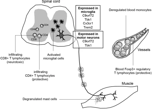
Figure 1. Evidence of involvement of neuroinflammatory processes in amyotrophic lateral sclerosis (ALS). New ALS-linked genes with important immune functions are shown
in boxes. Different subpopulations of immune cells that have been linked to ALS: CD4+ (including Foxp3+ regulatory cells) and CD8+ T lymphocytes, mast cells, monocytes and microglial cells are depicted with their described functions in the different tissues.