Manuscript received on June 8, 2010. Revised version arrived on August 20, 2010. Manuscript accepted on September 13, 2010. Acknowledgments: we would like to thank Mrs Vanessa Brennan and Valerie Webb for their help in obtaining patients’ samples. Funding: this work was supported by funds from Leukaemia & Lymphoma Research, the Julian-Starmer Smith Lymphoma Fund and the National Institute for Health Research Oxford Biomedical Research Center Program. Correspondence:
Karen Pulford, PhD, FRCPath, Level 4, Academic Block, John Radcliffe Hospital, Oxford, Oxon, OX3 9DU, UK. Phone: +44.01865220246. Fax: +44.01865220078. E-mail:
karen.pulford@ndcls.ox.ac.uk
Background
Vaccine development targeting the novel immunogenic Per ARNT Sim Domain containing 1 (PASD1) cancer testis antigen represents an attractive therapeutic approach for the significant number of patients with diffuse large B-cell lymphoma who are refractory to conventional treatment. Since CD4-positive T helper cells have crucial roles in promoting and maintaining immune responses to tumor antigens, the presence of a CD4-positive T-helper immune response to the PASD1 antigen in patients with diffuse large B-cell lymphoma was investigated in the current study.
Design and Methods
Thirty-one patients with diffuse large B-cell lymphoma (25 with de novo, five with transformed and one with T-cell-rich B-cell lymphoma) were studied. Five immunogenic PASD1 peptides predicted to bind to several major histocompatibiliy complex, class II DR beta 1 alleles were identified using web-based algorithms. Peripheral blood mononuclear cells from patients were used to investigate the immunogenicity of these DR beta 1-restricted peptides in vitro using both gamma-interferon release enzyme-linked immunospot and cytolytic assays.
Results
Two of the five PASD1 peptides, PASD1(6) and PASD1(7), were shown to be immunogenic in 14 out of 32 patients studied in a gamma-interferon release assay. CD4-positive T-helper cell lines from two patients raised against PASD1 peptides were able to lyse cell lines derived from hematologic malignancies expressing endogenous PASD1 protein.
Conclusions
This is the first report of a CD4-positive T-helper response to the PASD1 protein in patients with lymphoma. The immunogenic peptides described here represent valuable additional can-didates for inclusion in a vaccine to treat patients with PASD1-positive diffuse large B-cell lym-phoma whose disease is refractory to conventional therapies.
Key words: diffuse large B-cell lymphoma, CD4 T-helper cells, immune response, PASD1, can-cer-testis antigen.
Citation: Ait-Tahar K, Liggins AP, Collins GP, Campbell A, Barnardo M, Cabes M, Lawrie CH, Moir D, Hatton C, Banham AH, and Pulford K. CD4-positive T-helper cell responses to the PASD1 protein in patients with diffuse large B-cell lymphoma. Haematologica 2011;96(1):78-86.
doi:10.3324/haematol.2010.028241
©2011 Ferrata Storti Foundation. This is an open-access paper.
CD4-positive T-helper cell responses to the PASD1 protein in patients
with diffuse large B-cell lymphoma
Kamel Ait-Tahar,1Amanda P. Liggins,1Graham P. Collins,1 Andrew Campbell,1 Martin Barnardo,2Maite Cabes,1 Charles H. Lawrie,1 Donald Moir,3 Chris Hatton,4Alison H. Banham,1and Karen Pulford1
1Nuffield Department of Clinical Laboratory Sciences, John Radcliffe Hospital, Oxford; 2Transplant Immunology & Immunogenetics, Oxford Transplant Centre, Churchill Hospital, Oxford; 3Department of Hematology, Milton Keynes General Hospital, Milton Keynes, and 4Department of Hematology, John Radcliffe Hospital, Oxford, UK
Introduction
Diffuse large B-cell lymphoma (DLBCL) is the most common form of adult non-Hodgkin’s lymphoma and is heterogeneous with respect to morphology, clinical fea-tures and immunophenotype.1 Despite advances in the definition of clinically relevant subtypes and treatment, a significant proportion of patients with DLBCL fail to achieve long-term remission.2 The development and use of cancer vaccines boosting the immune response of patients represents an attractive approach to improve ther-apeutic options for high-risk patients.
A prerequisite for immunotherapeutic approaches is the identification of tumor-associated antigens, of which the cancer-testis antigens are of particular interest.3-6 Cancer-testis antigens, especially the CT-X antigens, have a restricted distribution in normal tissues (being predomi-nantly expressed in testis, an immunoprivileged site with low or even absent HLA expression) but are present in a range of tumors.6,7Such an expression pattern raises the possibility of targeted treatment while minimizing any attendant potential autoimmune problems for patients.
Although cytotoxic T cells (CTL) are considered to be the major effector cells in tumor immunity, there is increasing evidence for the importance of the CD4+T helper (CD4 Th) cell population in maintaining tumor immunity.8 Possible mechanisms of these cells include roles in maintenance and support of the CTL response, the cross-presentation of tumor antigens, their interaction with other effector cells such as natural killer cells and the release of immunological-ly relevant cytokines involved in anti-tumor effects, such as interleukin-2 and interferon-γ.9-14Evidence from human and animal studies has indicated that optimal cancer vaccines require the participation of both CD4+and CD8+T cells.15-17 A recent report highlighted the successful use of tumor-associated antigen-specific CD4 T-cell clones as therapeutic agents in the treatment of melanoma.18
We initially identified PASD1 as a tumor-associated antigen in DLBCL19 and acute myeloid leukemia20while later studies showed PASD1 to be a novel cancer-testis antigen expressed in a wide range of hematologic malig-nancies.21,22 Our subsequent identification of a CTL response to PASD1 in DLBCL23supports this molecule as a candidate for vaccine development, not only for DLBCL, but also for use in a generic vaccine for the treatment of other PASD1-positive malignancies. The current study was performed to investigate the CD4 Th response in DLBCL patients to obtain further support for the inclusion of PASD1 in a lymphoma vaccine.
Design and Methods
Subjects
Peripheral blood was obtained from 31 patients with DLBCL attending the Haematology Departments of the John Radcliffe Hospital (n=28) and the Milton Keynes General Hospital (n=3). Of these 31 patients, 25 had de novo DLBCL, five had transformed DLBCL and one had T-cell rich B-cell lymphoma. The clinical details of these patients are given in Table 1. Peripheral blood was also obtained from five healthy subjects. Tissue typing was done by polymerase chain reaction as previously described.24Ethical
approval and written consent was obtained from the Oxfordshire Research Ethics Committee B for all blood samples collected and tissue sections used in the immunolabeling studies.
Peptides
The TEPITOPE prediction algorithm25and SYPETHI
(www.syf-peithi.de) programs were used to select PASD1 peptides predicted to be immunogenic in the context of HLA-DRB1 *0101, *0301, *0401, *0701,*1101 and *1501 (the most prevalent alleles among the Caucasian population).26The following peptides were
identi-fied: PASD1(6)31-50(DYFNQVTLQLLDGFMITLST); PASD1(7)42-61
(DGFMITLSTDGVIICVAENI); PASD1(8)58-77
(AENISSLL-GHLPAEIVGKKL); PASD1(9)170-189(VGNVCILRTQLLQQLYTSKA)
and PASD1(10)599-618 (NHPVRFLQAQPIVPVQRAAE). The
PASD1(6)31-50peptide encompasses the immunogenic PASD1(1)38-47
CTL epitope while PASD1(8)58-77encompasses another
immuno-genic PASD1(3)63-71CTL epitope.23The location of the PASD1
pep-tides in the PASD1 protein is illustrated in Figure 1. The irrelevant control peptide was HIV-1121-140 (DESFRKYTAFTIPSMNNETP)
(Invitrogen, UK). The PASD1 peptides were synthesized by stan-dard chemistry and were more than 90% pure (Invitrogen, Paisley, UK).
Preparation and culture of peripheral blood
mononuclear cells
Peripheral blood mononuclear cells (PBMC) in RPMI 1640 con-taining 10% fetal calf serum (RPMI 1640/FCS) were prepared as previously described.23PBMC (2¥105) in 200 μL were then added
to each well of a 96-well flat-bottomed plate and incubated for 10-14 days with 10 μM of one of following: one of the PASD1 pep-tides, the irrelevant HIV121–140peptide, 10 μg/mL
phytohemagglu-tinin (Sigma-Aldrich Co. Ltd., Dorset, UK) or RPMI 1640/FCS only. Recombinant interleukin-2 (20 IU/mL; Roche Diagnostics, Indianapolis, IN, USA) and recombinant human interleukin-7 (25 ng/mL; R&D Systems, Minneapolis, MN, USA) were added on days 2, 5, and 7. In some experiments, PBMC were co-cultured with the PASD1(1) CTL epitope for 8 days before being used in an enzyme-linked immunospot (ELISPOT) assay.
Enzyme-linked immunospot assay
After 10-14 days of culture, the cells were washed and incubat-ed for 18 h with RPMI 1640/FCS at 37°C in 5% CO2with one of
the PASD1 peptides (10 μM), HIV control peptide (10 μM), phyto-hemagglutinin (10 μg/mL) or medium only. Interferon-γ release assays were performed according to manufacturer’s instructions (Mabtech, Stockholm, Sweden). Results were considered positive if the number of spots in the test wells was at least twice the num-ber present in the control cultures (media only or in the presence of the HIV-1 irrelevant peptide) and assays were excluded if there were more than 25 spots per well in the absence of peptides.
Generation of CD4 T-helper cell lines, depletion
and blocking experiments
PBMC cultured at a density of 2¥106cells/mL were stimulated
with 10 μM of either the PASD1(6) or PASD1(7) peptides. After 72 h, an equal volume of RPMI 1640/ FCS containing 50 IU/mL of recombinant interleukin-2 was added to each culture well. Half of the medium was removed and replaced with fresh medium every 3 days thereafter for up to 6 weeks before being used in ELISPOT and cytolytic assays. Cells were also used to produce cytocen-trifuge preparations for immunophenotyping at 4 weeks. In some experiments, a CD4+T-cell purification step was performed using
magnetic beads coated with anti-human CD4 antibody according to the manufacturer’s instructions (Dynabeads, Dynal, Oslo, Norway) prior to being used in an interferon-γ release ELISPOT assay as described above. In other experiments, 2 μg/mL of the HLA-DR-specific WR18 antibody (Abcam, Cambridge, UK) were added to the CD4 Th cell population for 1 h prior to the interfer-on-γ release ELISPOT assay.
Antibodies
Monoclonal
Major histocompatibility complex (MHC) class II expression was studied using the monoclonal HLA-DP, DQ, DR anti-body (CR3/43) (DAKOCytomation, Glostrup, Denmark). Antibodies to CD4 (T4-10, IgG1 isotype), CD20 (DAKO-L26, IgG2a isotype), CD45RO (UCHL1, IgG2a isotype), CD56 (T199, IgG1 isotype) and CD68 (PGM1, IgG3 isotype) were purchased from Dako (Ely, Cambridgeshire, UK) while anti-CD8 (X-107, IgG1 isotype) and anti-CD45RA (4KB5, IgG1 isotype) were gener-ated in the authors’ laboratory.
Polyclonal
The rabbit/mouse Envision-HRP labeling system was obtained from DAKOCytomation. Isotype-specific goat anti-rabbit immunoglobulin (Ig) and anti-mouse Ig-isotype specific
antibod-Table 1.Clinical details of the DLBCL cases studied.
∞Patients Diagnosis Subtype# Stage IPI Sex Age Treatment Outcome
1 DLBCL(dn) NGC 1 1 F 23 CHOP-R + MTX (CNS prophylaxis) CR (40 months)
2 DLBCL(dn) GCB 3 3 M 67 CHOP-R + MTX+RX CR (44 months)
3 DLBCL(dn) GCB 3 3 M 81 VIN/PRED Died (22 months)
4 DLBCL(dn) NGC 1 2 F 76 CHOP-R PR (73 months)
8 DLBCL(dn) GCB 1 0 M 63 CHOP-R CRU (48 months)
9 DLBCL(dn) NGC 3 2 F 71 CHOP-R PR (47 months)
10* DLBCL(dn) GCB 1 0 F 60 CHOP-R + RICE + ESHAP CR (37 months)
+ BEAM + TX
11 DLBCL(dn) GCB 1 1 M 38 CODOX-M + RX PR (41 months)
12 DLBCL(dn) GCB 1 0 F 59 CHOP-R CR (46 months)
13 DLBCL(dn) GCB 3 3 M 67 CHOP-R + MTX PR (41 months)
14 DLBCL(dn) NGC 3 2 M 63 CHOP-R + MTX Died of disease (14 months)
15 DLBCL(dn) NGC 3 3 M 85 VIN/PRED Died of disease (6 months)
1 7 DLBCL(dn) GCB 3 4 M 60 CHOP-R Died –cause unknown (21 months)
18 DLBCL(dn) NGC 4 4 M 74 CNOP-R Died of buccal cancer (31 months)
19 DLBCL(dn) GCB 4 2 M 56 CHOP-R + RX Died of disease (19 months)
20 DLBCL(dn) NGC 2 2 F 70 NONE Died of disease (2 months)
21 DLBCL(dn) GCB 1 3 M 73 CHOP-R PR (47 months)
24 DLBCL(dn) NGC 1 1 F 62 CHOP-R CR (35 months)
25 DLBCL(dn) GCB 2 2 F 74 CHOP-R Died (6 months)
27 DLBCL(dn) GCB 1/2 2 M >60 CHOP-R CR (52 months)
28 DLBCL(dn) GCB 1 2 M >60 CHOP-R CR (44 months)
29 DLBCL(dn) GCB 3 4 F 71 CHOP-R Died of disease (7 months)
30 DLBCL(dn) NGC 3 3 M 62 CHOP-R CR (48 months)
31 DLBCL(dn) NGC 1 1 M 63 CHOP-R CR (47 months)
32 DLBCL(dn) GCB 1 3 M 75 CNOP-R CR (47 months)
37 DLBCL(t) ND 1 2 M 59 CHOP-R + RX CR (46 months)
39 DLBCL(t) ND 4 2 F 39 CHOP-R Died of disease (6 months)
40 DLBCL(t) ND M 64 CHOP-R Died of disease (4 months)
41 DLBCL(t) ND 4 4 F 60 CHOP-R + CNOP-R CR (53 months)
43 DLBCL(t) ND 4 2 F 60 CHOP-R + RX CR (48 months)
47 TCR ND 3 2 F 80 PMITCEBO-R CR (36 months)
Patients previously reported in the description of a CTL response to PASD1.22DLBCL(dn): De novo diffuse large B-cell lymphoma; DLBCL(t): diffuse large B-cell lymphoma transformed; TCR: Tcell rich Bcell lymphoma; # subtyped according to expression of CD10, BCL6 and MUM1; GCB: germinal centerderived; NGC: nongerminal centerderived; CHOP -R: cyclophosphamide, doxorubicin, vincristine, prednisolone, rituximab; MTX: intrathecal methotrexate; RX: radiotherapy; PRED: prednisolone; VIN: vinblastine; RICE: rituximab, ifos-famide, carboplatin, etoposide; ESHAP: etoposide, methyprednisolone, cytarabine, cisplatin; TX: autologous transplant; CODOX-M: cyclophosphamide, vincristine, doxorubicin, methotrexate; BEAM-BCNU: (bis-chloro-ethyl nitrosourea), etoposide, cytarabine, melphalan; CNOP-R: cyclophosphamide, mitoxantrone, vincristine, prednisolone, rituximab; PMITCE-BO-R: prednisolone, mitoxantrone, cyclophosphamide, etoposide, bleomycin, vincristine, rituximab; IVAC: ifosfamide, etoposide, cytarabine. *Sample at relapse; CR: complete response; PR: partial response: CRU: complete remission unconfirmed.
Figure 1. Diagram to show the positions of the CD4 PASD1 peptides investigated here across the PASD1a and PASD1b protein isoforms.
ies conjugated to either fluorescein isothiocyanate or Texas RedTM
(diluted 1:100) were obtained from Invitrogen (Paisley, UK).
Immunolabeling studies
Immunoperoxidase
Paraffin-embedded tissue sections were de-waxed and antigen retrieval was performed using 50 mM Tris:EDTA at pH 9.0. Immunolabeling for anti-MHC-class II was carried out using the Envision-HRP labeling kit (DAKO, Ely, UK)26 The staining for
PASD1 protein expression (using the monoclonal antibodies PASD1-1 and PASD1-2) and subtyping of these DLBCL cases as non-germinal or germinal center subtypes had been performed previously.23
Immunofluorescence
Cytocentrifuge preparations of CD4 Th cell lines cultured for 4 weeks were fixed in acetone for 10 min and air-dried. The slides were then incubated for 30 min with one of the following com-binations: (i) a mixture of rabbit anti-CD3 with one of the follow-ing mouse monoclonal antibodies to CD4, CD8, CD20, CD45RA, CD45RO, CD56 or CD68 or (ii) a combination of two mouse monoclonal antibodies: anti-CD4 or CD8 (both isotype IgG1) and anti-CD45RO (isotype IgG2a) or CD20 (isotype IgG2a) and CD45RA (isotype IgG1). After washing in phosphate-buffered saline, the slides were incubated for a further 30 min with a mixture of: (i) fluorescein isothiocyanate-conjugated goat anti-rabbit Ig and an isotype-specific anti-mouse Ig conjugated to TexasRedTMor two anti-mouse Ig isotype-specific antibodies
con-jugated to either fluorescein isothiocyanate or TexasRedTMwhere
relevant. A minimum of 300 cells were counted per sample. The slides were washed and mounted in an anti-fade mountant Vectorshield containing 4’,6-diamine-2’phenylindole dihy-drochloride (Vector Laboratories, Peterborough, UK). Results were then visualized as previously described.22
Cell lines
The PASD1-positive (HLA-DRB1*0301) OCI-Ly3 (DLBCL-derived), PASD1-positive (HLA-DRB1*0401,*1101) Thiel (myelo-ma-derived), and PASD1-negative (HLA-DRB1*0101, *0401) SUDHL-6 (DLBCL-derived) cell lines were obtained and cultured as described previously.27
Cytolytic assay
A 51Cr-labeling release assay was used to investigate the ability
of CD4 Th cell lines generated from DLBCL patients to lyse PASD1-positive tumor target cells. The OCI-Ly3, SUDHL-6 and Thiel target cell lines were radio-labeled for 4 h with 100 μCi 51Cr
and then added to the CD4+Th lines at effector:target ratios of
1:10, 1:20 and 1:40. The cells were incubated for 18 h (rather than the 4 h used for CTL assays) at 37°C in 5% CO2. 51Cr release was
determined following the addition of 10% Triton-X to the effec-tor:target cell cultures as previously described.23
Statistical analysis
Student’s t-test was used to analyze the results obtained in the ELISPOT and cytolytic assays.
Results
Detection of interferon-
γ release
As shown in Table 2, 15 patients displayed a significant response to at least one of the PASD1 peptides, with respect to the control peptide (P<0.05), after 10-14 days culture. Three patients (1, 41 and 47) responded to four
peptides while patients 8, 14 and 24 recognized only one peptide. These patients included eight of the 16 patients previously reported to have a CTL response to PASD1.23 Peptide PASD1(7) also elicited a CD4 Th response in patient 24, despite a previous observation that no CTL response was detected in this HLA-A*0201-positive patient.22Furthermore, an interferon-γ response was also detected in six other DLBCL patients who were HLA-A*0201-negative. While the number of patients studied here was relatively small, ten of the 15 patients with a CD4 Th response remain in complete remission, one is in partial remission and four have died (one of carcinoma). These results contrast with the outcome of those patients unable to immunologically recognize the PASD1 peptides of whom only five patients are in remission, four in partial remission and seven have died (one of a carcinoma). These results are summarized in Table 3.
It is noteworthy that the PASD1(6) peptide which encompasses the immunogenic 10-mer CTL epitope PASD1(1)23 was also immunogenic eliciting interferon-γ release from stimulated PBMC in the majority of patients studied here. Data from the ELISPOT assays enabled PASD1(6) and PASD1(7) to be identified as the most immunogenic of the five PASD1 peptides studied.
The interferon-γ responses were investigated in two high responder patients (patients 4 and 47) at the time of diagnosis and 1 year after diagnosis (Figure 2A). In both patients, a significant interferon-γ response to the two PASD1 peptides PASD1(6) and PASD1(7) was sustained 1 year after diagnosis. Both of these patients were consid-ered to be in complete remission at this time with the absence of obvious minimal residual disease (suggesting the absence of tumor cells expressing PASD1).
CD4-enriched cell immune response
CD4+enriched T-cell lines were generated following the weekly stimulation with PASD1(6) and PASD1(7) peptides of PBMC from patient 4 (with de novo DLBCL) and patient 47 (with T-cell rich B-cell lymphoma) in order to enable further study of their functional activity. The cell lines demonstrated a significant response to both PASD1 pep-tides (Figure 2B). These responses were abrogated after the removal of the CD4 Th cell population or the addition of anti-HLA-DR antibody (Figure 2B) thus demonstrating that the response is dependent on both CD4 and MHC class II.
Cytolytic activity
CD4 Th cell lines specific for PASD1(6) and PASD1(7) raised from patients 4 and 47 lysed PASD1-positive Thiel and OCI-Ly3 cell lines in a dose-dependent fashion (Figure 4). The lysis of these cells expressing different HLA-DRB1 alleles demonstrates the promiscuity of the PASD1 pep-tide epitopes. No lysis was detected in the PASD1-nega-tive cell line SUDHL-6 despite its expression of HLA-DRB1*0101. Since the HLA-A*0201-negative OCI-Ly3 cell line was shown to be resistant to lysis by CTL raised against the PASD1(1) and PASD1(2) peptides in our previ-ous study,23lysis of this cell line in the current study was not due to CTL raised against these peptides in the PASD1(6) and PASD1(7) peptides. This factor, combined with the immunophenotyping results of the cell lines, and the cell purification and blocking steps shown in Figure 2, provides additional evidence to support CD4 Th cell killing in the cell lines used here.
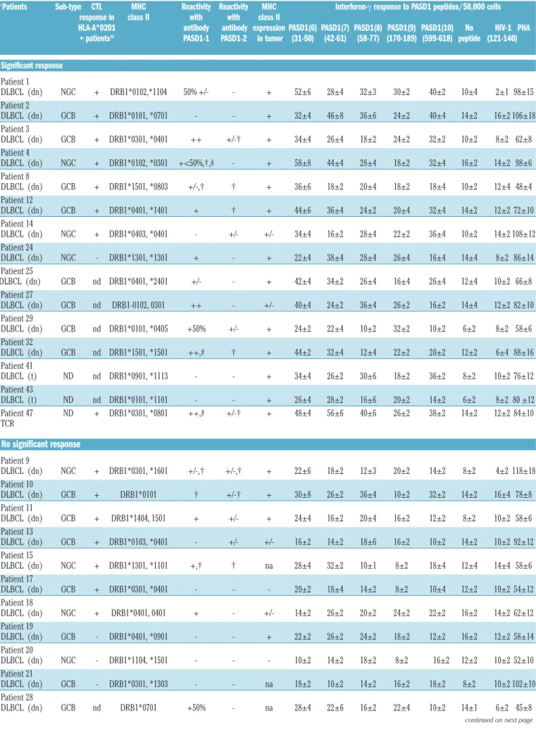
Table 2.Summary of the CD4 Th immune response to the PASD1 peptides by DLBCL patients and normal donors.
∞Patients Sub-type CTL MHC Reactivity Reactivity MHC Interferon-γ response to PASD1 peptides/50,000 cells
response in class II with with class II
HLA-A*0201 antibody antibody expression PASD1(6) PASD1(7) PASD1(8) PASD1(9) PASD1(10) No HIV-1 PHA
+ patients22 PASD1-1 PASD1-2 in tumor (31-50) (42-61) (58-77) (170-189) (599-618) peptide (121-140)
Significant response Patient 1 DLBCL (dn) NGC + DRB1*0102,*1104 50% +/- - + 52±6 28±4 32±3 30±2 40±2 10±4 2±1 98±15 Patient 2 DLBCL (dn) GCB + DRB1*0101, *0701 - - + 32±4 46±8 36±6 24±2 40±4 14±2 16±2 106±18 Patient 3 DLBCL (dn) GCB + DRB1*0301, *0401 ++ +/-† + 34±4 26±4 18±2 24±2 32±2 10±2 8±2 62±8 Patient 4 DLBCL (dn) NGC + DRB1*0102, *0301 +<50%,†,# - + 58±8 44±4 28±4 18±2 32±4 16±2 14±2 98±6 Patient 8 DLBCL (dn) GCB + DRB1*1501, *0803 +/-,† † + 36±6 18±2 20±4 18±2 18±4 10±2 12±4 48±4 Patient 12 DLBCL (dn) GCB + DRB1*0401, *1401 + † + 44±6 36±4 24±2 20±4 32±4 14±2 12±2 72±10 Patient 14 DLBCL (dn) NGC + DRB1*0403, *0401 - +/- +/- 34±4 16±2 28±4 22±2 36±4 10±2 14±2 108±12 Patient 24 DLBCL (dn) NGC - DRB1*1301, *1301 + - + 22±4 38±4 28±4 26±4 16±4 14±4 8±2 86±14 Patient 25 DLBCL (dn) GCB nd DRB1*0401, *2401 +/- - + 42±4 34±2 26±4 16±4 26±4 12±4 10±2 66±8 Patient 27 DLBCL (dn) GCB nd DRB1-0102, 0301 ++ - +/- 40±4 24±2 36±4 26±2 16±2 14±4 12±2 82±10 Patient 29 DLBCL (dn) GCB nd DRB1*0101, *0405 +50% +/- + 24±2 22±4 10±2 32±2 10±2 6±2 8±2 58±6 Patient 32 DLBCL (dn) GCB nd DRB1*1501, *1501 ++,# † + 44±2 32±4 12±4 22±2 20±2 12±2 6±4 88±16 Patient 41 DLBCL (t) ND nd DRB1*0901, *1113 - - + 34±4 26±2 30±6 18±2 36±2 8±2 10±2 76±12 Patient 43 DLBCL (t) ND nd DRB1*0101, *1101 - - + 26±4 28±2 16±6 20±2 14±2 6±2 8±2 80 ±12 Patient 47 ND + DRB1*0301, *0801 ++,# +/-† + 48±4 56±6 40±6 26±2 38±2 14±2 12±2 84±10 TCR No significant response Patient 9 DLBCL (dn) NGC + DRB1*0301, *1601 +/-,† +/-,† + 22±6 18±2 12±3 20±2 14±2 8±2 4±2 118±18 Patient 10 DLBCL (dn) GCB + DRB1*0101 † +/-† + 30±8 26±2 36±4 10±2 32±2 14±2 16±4 78±8 Patient 11 DLBCL (dn) GCB + DRB1*1404, 1501 + +/- + 24±4 16±2 20±4 16±2 12±2 8±2 10±2 58±6 Patient 13 DLBCL (dn) GCB + DRB1*0103, *0401 - +/- +/- 16±2 14±2 18±6 16±2 10±2 14±2 10±2 92±12 Patient 15 DLBCL (dn) NGC + DRB1*1301, *1101 +,† † na 28±4 32±2 10±1 8±2 18±4 12±4 14±4 58±6 Patient 17 DLBCL (dn) GCB + DRB1*0301, *0401 - - - 20±2 18±4 14±2 8±2 10±4 12±2 10±2 54±12 Patient 18 DLBCL (dn) NGC + DRB1*0401, 0401 + - +/- 14±2 26±2 20±2 24±2 22±2 16±2 14±2 62±12 Patient 19 DLBCL (dn) GCB - DRB1*0401, *0901 - - + 22±2 26±2 24±2 18±2 12±2 16±2 12±2 58±14 Patient 20 DLBCL (dn) NGC - DRB1*1104, *1501 - - - 10±2 14±2 18±2 8±2 16±2 12±2 10±2 52±10 Patient 21 DLBCL (dn) GCB - DRB1*0301, *1303 - - na 18±2 10±2 14±2 16±2 18±2 8±2 10±2 102±10 Patient 28 DLBCL (dn) GCB nd DRB1*0701 +50% - na 28±4 22±6 16±2 22±4 10±2 14±1 6±2 45±8
Immunolabeling studies
Table 2 summarizes the presence of MHC class II anti-gens in the DLBCL cases studied. Tumor cells of 25 of the 27 patients tested expressed MHC class II. No interferon-γ response to PASD1 was detected in those two patients whose tumors were MHC class II-negative. With the exception of three patients (patients 2, 41 and 43), PASD1 protein was detected in the tumor cells of those patients with an interferon-γ response (Table 2). PASD1 protein was undetectable in seven of those 12 patients who did not mount an interferon-γ response to any of the PASD1
Patient 30 DLBCL (dn) NGC nd DRB1*0401, *1104 - - + 8±2 12±2 14±2 18±4 28±2 16±1 12±2 76±10 Patient 31 DLBCL (dn) NGC nd DRB1-0701, 1301 - - na 22±2 20±1 18±2 12±4 24±2 12±1 4±2 48±4 Patient 37 DLBCL (t) ND + DRB1*0103, *1101 +/-,† † + 38±4 32±6 16±2 28±4 34±2 10±2 16±2 126±22 Patient 39 DLBCL (t) ND + DRB1*0701, *1301 - - +/- 24±4 22±4 26±2 26±4 16±2 12±1 10±2 92±12 Patient 40 DLBCL (t) ND nd DRB1*0401, *0401 + - + 12±2 20±4 20±2 14±2 16±2 10±3 8±2 64±8 Healthy Donors HD 1 - DRB1*0102, *0301 14±2 20±2 18±2 14±2 10±2 10±2 8±2 86±14 HD 2 - DRB1*0401, *0701 10±2 8±2 10±2 1±1 4±2 4±2 6±2 66±10 HD 3 - DRB1*0401, 0701 5±2 10±2 6±2 8±2 2±1 4±2 1±1 46±6 HD 4 - DRB1*10301, *0101 14±2 20±2 10±2 19±2 18±2 8±2 10±2 74±16 HD 5 DRB1*0301, *0701 24±2 22±2 10±2 18±2 16±2 12±2 8±2 63±10
GCB: germinal center-derived; NGC: non-germinal center-derived; +/-, + and ++ denotes intensity of cytoplasmic labeling; † denotes from 5-30%nuclear labeling # denotes labeling of some smaller lymphocytes and vessels in tumor; nd: not done; na: tissue not available. ∞ denotes the same patients previously reported in the description of a CTL response to PASD1.22The results +/- are from triplicate ELISPOT cultures. The SD was calculated using standard techniques. Shading of figures denotes significant interferon γ responses.
continued from previous page
Table 3. Comparison of the presence of a CD4 Th response to PASD1 in DLBCL patients with clinical outcome.
Clinical outcome of patients Complete Partial Died
remission remission Patients with 1, 2, 8, 12, 24, 27, 4 3, 14, 25, 29 a significant 32, 41, 43, 47 CD4 response Patients with 10, 28, 30, 31, 37 9, 11,13, 21 15, 17, 18, no significant 19, 20, 39, 40 response
Figure 2. Interferon-γ ELISPOT studies showing CD4 Th respons-es to PASD1 peptidrespons-es of patients 4 and 47. (A) A study monitoring the CD4 Th responses of these two patients at the time of diagno-sis and 1 year after-diagnodiagno-sis showed the presence of signifi-cant immune interferon-γ respons-es to PASD1(6) and PASD1(7) peptides even after 1 year in remission (P<0.05). In contrast, no significant responses to the control HIV peptide or medium only were observed. (B) CD4 Th cell lines were cultured from patient 4 and patient 47 and CD4+
cell populations were obtained using a magnetic purification step. After overnight incubation with either PASD1(6) or PASD1(7) peptides, the purified CD4+ cells
from both patient 4 and 47 responded to these PASD1 pep-tides. No significant response was detected to the irrelevant control peptide or to medium only. The removal of the CD4+ cells or the
addition of anti–HLA-DR antibody abrogated the interferon-γ response to both peptides. The results are the mean ± SD and were from triplicate cultures.
A B Patient 4 Patient 47 70 60 50 40 30 20 10 0 70 60 50 40 30 20 10 0 50 0 50 0 Patient 4
Purified CD-cell CD+cells and
CD+cells fraction anti-HLA-DR
Purified CD-cell CD+cells and
CD+cells fraction anti-HLA-DR
PASD1(6) PASD1(7) Medium HIV
At diagnosis After 1 year
γ -I F N r e s p o n s e /5 0 ,0 0 0 c e ll s γ -I F N r e s p o n s e /5 0 ,0 0 0 c e ll s γ -I F N r e s p o n s e /5 0 ,0 0 0 c e ll s γ -I F N r e s p o n s e /5 0 ,0 0 0 c e ll s
At diagnosis After 1 year
peptides. Of these, two of the five cases tested were neg-ative for MHC class II.
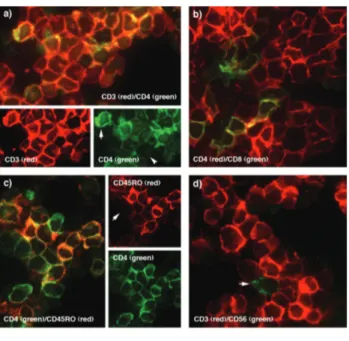
Immunophenotyping of the CD4 T-cell lines showed that 93.3±2.6 % of the cells were CD3+ T cells of which 86.6±2.4% expressed CD4 and 15.4±4.3% were CD8+: 88.0±2.1% of the CD4+T cells were CD45RO+with only 12.0±4.1% expressing CD45RA. Representative examples of the labeling results are shown in Figure 4.
Discussion
Previous reports have described limitations in using short peptide sequences from tumor-associated antigens to elicit long-lasting CTL responses to the target molecule. Repeated boosting with peptides may be necessary to achieve a sufficient immune response, a strategy potential-ly leading to the induction of immunotolerance.28,29 Indeed, Lehe et al. reported the undesirable recognition of a 15 amino-acid sequence of WT1 by immunosuppressive T regulatory cells.30There is, however, accumulating evi-dence that longer peptides containing CD4 Th epitopes are essential for obtaining and maintaining a long-lasting CTL response.31,32The inclusion of such peptides in a vac-cine should increase the efficacy of the resulting immune response. The current study was performed to identify longer immunogenic PASD1 peptides capable of eliciting CD4 Th cell responses. Such peptides could then be included in a lymphoma vaccine for the treatment of DLBCL as well as other hematologic malignancies expressing PASD1.
Five potential CD4 Th PASD1 peptides of 20 amino acids in length that would be recognized by a number of HLA-DRB1 subtypes were selected by a stringent method using two highly reliable search tools: the TEPITOPE and the SYPEITHI programs.17 The identification and use of promiscuous MHC class II epitopes has been previously
reported for the NY-ESO-1 cancer-testis antigen.33,34 The presence of such epitopes, recognizable in the context of a variety of different MHC class II molecules, expands the population of patients for whom the peptides could be immunogenic beyond that determined by their MHC class I allele. All five peptides in the current study were able to elicit interferon-γ responses in DLBCL patients, with the PASD1(6) and PASD1(7) peptides being identified as the most immunogenic peptides. It is notable that these pep-tides were able to expand the numbers of patients recog-nizing PASD1 since they elicited interferon-γ responses in a patient who, despite being HLA-A*0201-positive, was previously unable to respond to the PASD1 CTL peptides and also in seven HLA-A*0201-negative patients.23 The lack of significant homology of the PASD1 peptides with other molecules combined with the absence of any signif-icant response with the irrelevant peptide demonstrates the specificity of the response to PASD1.
Although interferon-γ responses to the PASD1 peptides were present in 12 of the 15 patients whose tumors expressed PASD1 protein, three patients recognizing the PASD1 peptides lacked detectable PASD1 protein. A dis-parity between PASD1 expression and CTL activity has been described previously23 and it is possible that the immunolabeling techniques used here may not constitute a sufficiently sensitive tool to identify low levels of PASD1 protein expression.35Importantly, high levels of target pro-tein expression may not be necessary for immune recogni-tion.36There is also evidence that additional isoforms of PASD1 may exist21and some of these proteins may lack the epitopes recognized by the PASD1-1 and PASD1-2 antibodies that are currently being used to study PASD1 protein expression. The development of more sensitive assays for PASD1 expression, such as western blotting and/or the production of additional antibodies, should assist in the identification of the maximum number of PASD1-positive patients in future clinical trials targeting Figure 3. Cytolytic activity of PASD1-specific CD4 Th cell lines derived from two DLBCL patients 4 and 47. The cytolytic activity of CD4 Th cell lines raised from patient 4 (DRB1*0102,*0301) and patient 47 (DRB1*0301,*0801) against PASD1(6) (A) and PASD1(7) (B) peptides was test-ed in an 18 h 51
Cr release assay on the PASD1-positive Thiel (DRB1*0401,*1101) and OCI-ly3 (DRB1-0301) cell lines. Significant lysis was observed only in those cultures containing the PASD1-positive Thiel or OCI-Ly3 cells. This was in contrast to the lack of lysis observed of the PASD1-negative SUDHL-6 (DRB1*0101,*0401) cell line (P<0.001). Similarly, significant lysis was also observed for CD4 Th cell lines raised from patient 47 against (C) the PASD1(6) and (D) PASD1(7) peptides. The results are the mean ± SD and were from triplicate cultures.
B A D C Patient 4 PASD1(6) Patient 47 PASD1(6) Patient 4 PASD1(7) Patient 47 PASD1(7) 35 30 25 20 15 10 5 0 35 30 25 20 15 10 5 0 40 35 30 25 20 15 10 5 0 35 30 25 20 15 10 5 0 % s p e c if ic l y s is % s p e c if ic l y s is % s p e c if ic l y s is % s p e c if ic l y s is 10:1 20:1 40:1
Effector:target ratio Effector:target ratio
Effector:target ratio Effector:target ratio
Thiel OCI-Ly3 SUDHL-6
10:1 20:1 40:1
10:1 20:1 40:1
this antigen. Another possible explanation for the lack of detection in the present study is that PASD1 expression may have been lost through immunoediting by the tumor cells.37
The tumor cells of only two patients in the current study lacked MHC class II expression and PBMC from neither patient were able to respond to the PASD1 peptides. Although cross-presentation of antigen by antigen-pre-senting cells can occur at the tumor site in the absence of MHC class II,12loss of MHC class II has been linked to poorer prognosis.38,39 and it will be of interest in future studies to determine whether PASD-positive MHC class II-negative patients have a significantly worse prognosis than PASD1-positive MHC class II-positive patients.
CD4 Th cell lines raised from two patients were able to lyse positive tumor cell lines (but not the PASD1-negative cell line) confirming that these T cells were able to recognize endogenously expressed PASD1. Of note is the ability of the CD4 Th cell lines to lyse both the PASD1-positive Thiel and OCI-Ly3 cells, despite the fact that these two cell lines express different HLA-DRB1 alle-les. This could be explained by the high degree of homol-ogy present between the DRB1 molecules25 and further demonstrates the promiscuity of the PASD1 peptides, which enables them to be recognized by closely related DRB1 molecules.
The persistence of PASD1-specific CD4 Th cell popula-tions in patients after 1 year in remission is an indication of the immunogenicity of the PASD1 protein and is sug-gestive of pre-existing memory T cells able to recognize PASD1. Such cells could play an important role not only in protective tumor immunity40but also in the maintenance of the CTL previously reported to be present in PASD1-positive DLBCL patients.23 In contrast to the previous study on the CTL immune response to PASD1 (in which 79% and 37% of the cultured cells were CD8+ and CD45RO+, respectively), the majority of the cell lines cul-tured from these patients were CD4+ and expressed CD45RO, an antigen present on memory cells. It will be of interest to look at the ex vivo phenotype (memory or naïve) of circulating T cells from patients in future experi-ments.
Previous studies have shown the potential of using sin-gle peptides containing epitopes predicted to bind to both MHC class I and class II to obtain more effective immune responses.41,42The PASD1(6) peptide, encompassing a CTL epitope previously shown to be immunogenic in PASD1-positive patients,22was highly immunogenic in the major-ity of patients and could, therefore, represent an attractive
candidate for inclusion in a vaccine formulation.
In conclusion, this study is the first report of a CD4 Th response to a cancer-testis antigen by patients with lym-phoma. These CD4 Th peptides hold future potential for use in conjunction with PASD1 peptides eliciting CTL in a vaccine for relapsing DLBCL patients or those who are refractory to conventional treatments.
Authorship and Disclosures
The information provided by the authors about contributions from persons listed as authors and in acknowledgments is avail-able with the full text of this paper at www.haematologica.org.
Financial and other disclosures provided by the authors using the ICMJE (www.icmje.org) Uniform Format for Disclosure of Competing Interests are also available at www.haematologica.org. Figure 4. Double immunofluorescent labeling of the cultured CD4+
T-cell lines. In a) the majority of cultured T-cells express both CD3 and CD4 antigens (arrow). Heterogeneity was observed in CD4 expres-sion. An example of a CD3+
cell CD4–
T cell is shown (arrowhead). b) Shows the presence of a small population of CD8+
cells (green) c) Co-expression of CD45R0 on the majority of CD4+
cells can be observed. An example of a CD4+
CD45RO–
cell is arrowed. d) Only the occasion-al cell was CD56+
(arrowhead) confirming the presence of only a small number (<1%) of natural killer cells in the cultured cells.
References
1. Stein H, Chan JKC, Warnke RA, Gatter KC, Chan WC, Campo EC, et al. Diffuse large B-cell lymphoma, not otherwise specified. WHO Classification of Tumours of Haematopietic and Lymphoid Tissues. 2008;4th edition, IARC Press, Lyon: 233-7. 2. Coiffier B. Rituximab therapy in malignant
lymphoma. Oncogene. 2007;26(25):3603-13.
3. Chen YT, Old LJ. Cancer-testis antigens: targets for cancer immunotherapy. Cancer J Sci Am. 1999;5(1):16-7.
4. Old LJ. Cancer/testis (CT) antigens - a new
link between gametogenesis and cancer. Cancer Immun. 2001;1:1.
5. Scanlan MJ, Simpson AJ, Old LJ. The can-cer/testis genes: review, standardization, and commentary. Cancer Immun. 2004;4:1. 6. Caballero OL, Chen YT. Cancer/testis (CT) antigens: potential targets for immunother-apy. Cancer Sci. 2009;100(11):2014-21. 7. Hofmann O, Caballero OL, Stevenson BJ,
Chen YT, Cohen T, Chua R, et al. Genome-wide analysis of cancer/testis gene expres-sion. Proc Natl Acad Sci USA. 2008;105 (51):20422-7.
8. Muranski P, Restifo NP. Adoptive immunotherapy of cancer using CD4(+) T cells. Curr Opin Immunol.
2009;21(2):200-8.
9. Ossendorp F, Mengede E, Camps M, Filius R, Melief CJ. Specific T helper cell require-ment for optimal induction of cytotoxic T lymphocytes against major histocompati-bility complex class II negative tumors. J Exp Med. 1998;187(5):693-702.
10. Janssen EM, Lemmens EE, Wolfe T, Christen U, von Herrath MG, Schoenberger SP. CD4+ T cells are required for secondary expansion and memory in CD8+ T lymphocytes. Nature. 2003;421 (6925):852-6.
11. Janssen EM, Droin NM, Lemmens EE, Pinkoski MJ, Bensinger SJ, Ehst BD, et al. CD4+ T-cell help controls CD8+ T-cell
memory via TRAIL-mediated activation-induced cell death. Nature. 2005;434 (7029):88-93.
12. Perez-Diez A, Joncker NT, Choi K, Chan WF, Anderson CC, Lantz O, et al. CD4 cells can be more efficient at tumor rejection than CD8 cells. Blood. 2007;109(12):5346-54.
13. Smith CM, Wilson NS, Waithman J, Villadangos JA, Carbone FR, Heath WR, et al. Cognate CD4(+) T cell licensing of den-dritic cells in CD8(+) T cell immunity. Nat Immunol. 2004;5(11):1143-8.
14. Zhang S, Zhang H, Zhao J. The role of CD4 T cell help for CD8 CTL activation. Biochem Biophys Res Commun. 2009;384 (4):405-8.
15. Hung K, Hayashi R, Lafond-Walker A, Lowenstein C, Pardoll D, Levitsky H. The central role of CD4(+) T cells in the antitu-mor immune response. J Exp Med. 1998; 188(12):2357-68.
16. Toes RE, Ossendorp F, Offringa R, Melief CJ. CD4 T cells and their role in antitumor immune responses. J Exp Med. 1999;189 (5):753-6.
17. Rosa DS, Ribeiro SP, Cunha-Neto E. CD4+ T cell epitope discovery and rational vac-cine design. Arch Immunol Ther Exp (Warsz). 2010;58(2):121-30.
18. Hunder NN, Wallen H, Cao J, Hendricks DW, Reilly JZ, Rodmyre R, et al. Treatment of metastatic melanoma with autologous CD4+ T cells against NY-ESO-1. N Engl J Med. 2008;358(25):2698-703.
19. Liggins AP, Brown PL, Asker K, Pulford K, Banham AH. A novel diffuse large B-cell lymphoma-associated cancer testis antigen encoding a PAS domain protein. Br J Cancer. 2004;91:141-9.
20. Guinn BA, Bland EA, Lodi U, Liggins AP, Tobal K, Petters S, et al. Humoral detection of leukaemia-associated antigens in presen-tation acute myeloid leukaemia. Biochem Biophys Res Commun. 2005;335(4):1293-304.
21. Cooper CD, Liggins AP, Ait-Tahar K, Roncador G, Banham AH, Pulford K. PASD1, a DLBCL-associated cancer testis antigen and candidate for lymphoma immunotherapy. Leukemia. 2006;20(12): 2172-4.
22. Ait-Tahar K, Liggins AP, Collins GP, Campbell A, Barnardo M, Lawrie C, et al. Cytolytic T-cell response to the PASD1 can-cer testis antigen in patients with diffuse large B-cell lymphoma. Br J Haematol. 2009;146(4):396-407.
23. Sahota SS, Goonewardena CM, Cooper CD, Liggins AP, Ait-Tahar K, Zojer N, et al.
PASD1 is a potential multiple myeloma-associated antigen. Blood. 2006;108(12): 3953-5.
24. Bunce M, O'Neill CM, Barnardo MC, Krausa P, Browning MJ, Morris PJ, et al. Phototyping: comprehensive DNA typing for HLA-A, B, C, DRB1, DRB3, DRB4, DRB5 & DQB1 by PCR with 144 primer mixes utilizing sequence-specific primers (PCR-SSP). Tissue Antigens. 1995;46(5): 355-67.
25. Sturniolo T, Bono E, Ding J, Raddrizzani L, Tuereci O, Sahin U, et al. Generation of tis-sue-specific and promiscuous HLA ligand databases using DNA microarrays and vir-tual HLA class II matrices. Nat Biotechnol. 1999;17(6):555-61.
26. Southwood S, Sidney J, Kondo A, del Guercio MF, Appella E, Hoffman S, et al. Several common HLA-DR types share largely overlapping peptide binding reper-toires. J Immunol. 1998;160(7):3363-73. 27. Pulford K, Banham AH, Lyne L, Jones M,
Ippolito GC, Liu H, et al. The BCL11AXL transcription factor: its distribution in nor-mal and nor-malignant tissues and use as a marker for plasmacytoid dendritic cells. Leukemia. 2006;20(8):1439-41.
28. Aichele P, Brduscha-Riem K, Zinkernagel RM, Hengartner H, Pircher H. T cell prim-ing versus T cell tolerance induced by syn-thetic peptides. J Exp Med. 1995;182(1): 261-6.
29. Toes RE, Offringa R, Blom RJ, Melief CJ, Kast WM. Peptide vaccination can lead to enhanced tumor growth through specific T-cell tolerance induction. Proc Natl Acad Sci USA. 1996;93(15):7855-60.
30. Lehe C, Ghebeh H, Al-Sulaiman A, Al Qudaihi G, Al-Hussein K, Almohareb F, et al. The Wilms' tumor antigen is a novel tar-get for human CD4+ regulatory T cells: implications for immunotherapy. Cancer Res. 2008;68(15):6350-9.
31. Bijker MS, van den Eeden SJ, Franken KL, Melief CJ, Offringa R, van der Burg SH. CD8+ CTL priming by exact peptide epi-topes in incomplete Freund's adjuvant induces a vanishing CTL response, whereas long peptides induce sustained CTL reac-tivity. J Immunol. 2007;179(8):5033-40. 32. Bijker MS, van den Eeden SJ, Franken KL,
Melief CJ, van der Burg SH, Offringa R. Superior induction of anti-tumor CTL immunity by extended peptide vaccines involves prolonged, DC-focused antigen presentation. Eur J Immunol. 2008;38(4): 1033-42.
33. Mandic M, Almunia C, Vicel S, Gillet D, Janjic B, Coval K, et al. The alternative open
reading frame of LAGE-1 gives rise to mul-tiple promiscuous HLA-DR-restricted epi-topes recognized by T-helper 1-type tumor-reactive CD4+ T cells. Cancer Res. 2003; 63(19):6506-15.
34. Ohkuri T, Sato M, Abe H, Tsuji K, Yamagishi Y, Ikeda H, et al. Identification of a novel NY-ESO-1 promiscuous helper epitope presented by multiple MHC class II molecules found frequently in the Japanese population. Cancer Sci. 2007;98(7):1092-8. 35. Sugita Y, Wada H, Fujita S, Nakata T, Sato
S, Noguchi Y, et al. NY-ESO-1 expression and immunogenicity in malignant and benign breast tumors. Cancer Res. 2004; 64(6):2199-204.
36. Vierboom MP, Zwaveling S, Bos GMJ, Ooms M, Krietemeijer GM, Melief CJ, et al. High steady-state levels of p53 are not a prerequisite for tumor eradication by wild-type p53-specific cytotoxic T lymphocytes. Cancer Res. 2000;60(19):5508-13. 37. Dunn GP, Old LJ, Schreiber RD. The three
Es of cancer immunoediting. Annu Rev Immunol. 2004;22:329-60.
38. Rimsza LM, Roberts RA, Miller TP, Unger JM, LeBlanc M, Braziel RM, et al. Loss of MHC class II gene and protein expression in diffuse large B-cell lymphoma is related to decreased tumor immunosurveillance and poor patient survival regardless of other prognostic factors: a follow-up study from the Leukemia and Lymphoma Molecular Profiling Project. Blood. 2004; 103(11):4251-8.
39. Rimsza LM, Farinha P, Fuchs DA, Masoudi H, Connors JM, Gascoyne RD. HLA-DR protein status predicts survival in patients with diffuse large B-cell lymphoma treated on the MACOP-B chemotherapy regimen. Leuk Lymphoma. 2007;48(3):542-6. 40. Broderick L, Yokota SJ, Reineke J,
Mathiowitz E, Stewart CC, Barcos M, et al. Human CD4+ effector memory T cells per-sisting in the microenvironment of lung cancer xenografts are activated by local delivery of IL-12 to proliferate, produce IFN-gamma, and eradicate tumor cells. J Immunol. 2005;174(2):898-906.
41. Zeng G. MHC class II-restricted tumor anti-gens recognized by CD4+ T cells: new strategies for cancer vaccine design. J Immunother. 2001;24(3):195-204. 42. Wagner WM, Ouyang Q, Pawelec G. The
abl/bcr gene product as a novel leukemia-specific antigen: peptides spanning the fusion region of abl/bcr can be recognized by both CD4+ and CD8+ T lymphocytes. Cancer Immunol Immunother. 2003;52(2): 89-96.