HAL Id: tel-03079113
https://tel.archives-ouvertes.fr/tel-03079113
Submitted on 17 Dec 2020HAL is a multi-disciplinary open access
archive for the deposit and dissemination of sci-entific research documents, whether they are pub-lished or not. The documents may come from teaching and research institutions in France or abroad, or from public or private research centers.
L’archive ouverte pluridisciplinaire HAL, est destinée au dépôt et à la diffusion de documents scientifiques de niveau recherche, publiés ou non, émanant des établissements d’enseignement et de recherche français ou étrangers, des laboratoires publics ou privés.
Study of cellular mechanisms allowing dendritic cell
migration in restricted spaces
Lucie Barbier
To cite this version:
Lucie Barbier. Study of cellular mechanisms allowing dendritic cell migration in restricted spaces. Subcellular Processes [q-bio.SC]. Université Paris-Saclay, 2020. English. �NNT : 2020UPASL028�. �tel-03079113�
Study of cellular mechanisms
allowing dendritic cell migration in
restricted spaces
Thèse de doctorat de l'université Paris-Saclay
École doctorale n°577 : Structure et Dynamique des Systèmes Vivants (SDSV)
Spécialité de doctorat : Sciences de la Vie et de la Santé Unité de recherche : UMR144Biologie Cellulaire et Cancer, Institut Curie, 75005, Paris
Référent : Faculté des sciences d’Orsay
Thèse présentée et soutenue à l’Institut Pierre-Gilles de
Gennes, Paris, le 15 Octobre 2020, par
Lucie BARBIER
Composition du Jury
Guillaume MONTAGNAC
CR, HDR, Institut Gustave Roussy, Université Paris-Saclay
Président
Marie-Emilie TERRET
DR, HDR, Collège de France Rapporteur & Examinatrice
Tim LÄMMERMANN
DR, Max Planck Institute of Immunobiology and Epigenetics
Rapporteur & Examinateur
Florence NIEDERGANG
DR, HDR, Institut Cochin Examinatrice
Pablo VARGAS
CR, HDR, Institut Curie Directeur de thèse
Matthieu PIEL
DR, HDR, Institut Curie Co-Directeur de thèse
T
h
è
se
d
e
d
o
ct
o
ra
t
N N T : 2 0 2 0 U PA S L0 2 8III
« Être docteur, ce n’est pas simplement un
manuscrit et une soutenance, c’est un état d’esprit.
»
V
ACKNOWLEDGMENTS
Je souhaiterais commencer ce manuscrit en remerciant toutes les personnes qui y ont participé, qui m’ont soutenue et sans qui ce travail n’aurait pas abouti.
À mon jury, Guillaume Montagnac, Florence Niedergang et particulièrement aux rapporteurs, Marie-Emilie Terret, Tim Lämmermann, merci d’avoir pris le temps d’évaluer mon travail.
À mon comité de thèse, Danijela Vignjevic, Stéphanie Descroix et Manuel Théry, merci pour vos conseils tout au long de ces quatre ans.
Au Ministère de l’Enseignement supérieur de la Recherche et de l’Innovation et aux donateurs de la Fondation pour la Recherche Médicale, merci d’avoir financé mes quatre années de recherches doctorales.
À Matthieu P., merci de m’avoir accueillie dans votre laboratoire, pour vos nombreuses idées et votre soutien scientifique tout en me laissant la liberté de mener mes projets au laboratoire et en dehors.
À Pablo V., merci de m’avoir encadrée pendant ces quatre ans, de m’avoir appris tous les secrets des DCs et à chuchoter à l’oreille des microscopes. Tu m’as accompagnée avec bienveillance vers l’indépendance, j’espère que tu pourras t’épanouir et mener à bien tes projets avec tes prochains doctorants.
À tous les membres actuels et passés de l’équipe Bio6, merci pour toute l’aide que vous m’avez apportée des premières manips jusqu’à la présentation de thèse et pour les nombreuses discussions pas toujours scientifiques mais à chaque fois passionnées et intenses ; Pablo S., Mathieu D., Aastha et Mathilde pour les longues discussions du lundi matin et tous les conseils scientifique et technique ; Olivier (Jian), Li et Rafaele pour la micro-fabrication, Juanma pour le partage de ton enthousiasme indéfectible pour les sciences, Larisa et Bianca pour être toujours prête à aller prendre un verre, Ghuuuuilerme pour ton expertise sur les lovely WB et dynamic confiner, Alice (pour l’orthographe de ces remerciements) et Zahraa pour les séances salon de thé dans le labo, Damien pour rester simplement toi-même, Nishit pour tes blagues que je comprendrai un jour, Ido et Valentin pour le regard différent que vous avez porté sur mon travail, et Nico pour l’analyse d’image et surtout pour permettre au labo de continuer à tourner avec tous ce petit monde.
VII apportée sur les DCs.
Aux personnels des plateformes de l‘IPGG et de Curie : Guillaume(s), Olivier, Nawel, Bertrand, Kevin, Nhung et Ludovic, merci pour votre disponibilité et votre aide au cours de mes longues séances en salle de microscopie.
Aux personnels administratifs : Caroline, Gabriela, Edwige N, Edwige, Sylvia et Rolie, merci pour votre support qui a facilité mon quotidien à l’IPGG et à Curie.
À Perrine et Manon, merci pour votre sourire et votre positivisme et merci de m’avoir accueilli dans votre bureau de la bonne humeur durant ma dernière année de thèse. En particulier, merci Perrine pour toute l’énergie que tu donnes pour faire vivre l’IPGG et pour m’avoir initiée à la Com’-Innovation.
Aux autres membres du 3ème étage de l’IPGG : l’équipe Micromégas, Jacques et son équipe,
Tommaso et sa futur équipe, merci pour l’atmosphère sympathique et ouverte que vous avez su créer lors de nos discussions de couloirs et des soirées de Noël.
À l’équipe du 4ème et en particulier à Steph, merci d’avoir été toujours présent pour papoter,
m’apporter votre soutien scientifique et personnel, et surtout pour partager vos gâteaux.
Un merci particulier à Larisa, Juanma, Léa et Elodie avec qui j’ai traversé cette période de thèse et qui l’ont égayée de Saint-Marc, jambon espagnol, séances de bad et goûters. Bonne chance à tous les quatre pour la suite et j’espère que nos chemins se recroiseront.
À mes amis d’enfance, de Bio, du BDE, du YRLS et du Palais, merci pour tous les bons moments et aventures partagés au cours de ces quatre ans, de vrais bols d’air qui m’ont fait relativiser les déboires de la thèse.
À ma famille et mes proches, merci de m’avoir soutenue tout au long de ces longues études qui ont pu vous paraître sans fin !
XI
Table of content
FIGURE LIST……….……….……….……….XVIII ABBREVIATIONS………..XXI
INTRODUCTION
CHAPTER 1: DENDRITIC CELL MIGRATION DURING IMMUNE RESPONSE
I. Principles and organization of the immune system ... 1
A. Two types of immune response ... 1
1. Innate immune response ... 1
2. Adaptive immune response ... 2
B. Steady-state organization of the immune system ... 2
C. Innate immune cells ... 3
D. Adaptive immune cells ... 3
1. T lymphocytes ... 4
2. B lymphocytes ... 4
E. Antigen-presenting cells ... 5
1. Macrophages ... 5
2. Dendritic cells ... 5
II. Local immune response: activation of the innate system ... 8
A. Inflammation ... 8
B. Leukocyte recruitment: from blood to the site of infection ... 8
1. Transendothelial migration ... 9
2. Interstitial migration ... 9
C. Dendritic cell maturation ... 10
1. Antigen uptake and processing ... 10
2. Cytokine and co-stimulatory molecules ... 11
3. Migration to the lymph nodes ... 14
III. Systemic immune response: activation of the adaptive system by dendritic cells ... 15
A. Antigen presentation to CD4+ T cells... 16
B. Differentiation of CD4+ T cells and activation of the specific immune cell effectors ... 16
CHAPTER 2: MECHANISMS OF CELL MIGRATION I. Intracellular forces for cell deformation ... 21
A. Acto-Myosin Cytoskeleton ... 21
1. Actin filament ... 21
a) Branched network – Arp2/3 ... 22
b) Non-Branched Network – Formins ... 22
3. Myosin ... 23
B. Different types of cell protrusions ... 25
1. Actin-rich protrusion ... 25
2. "Bleb" protrusion ... 26
C. Mechanisms of cell retraction ... 28
D. Generation of an actin flow ... 28
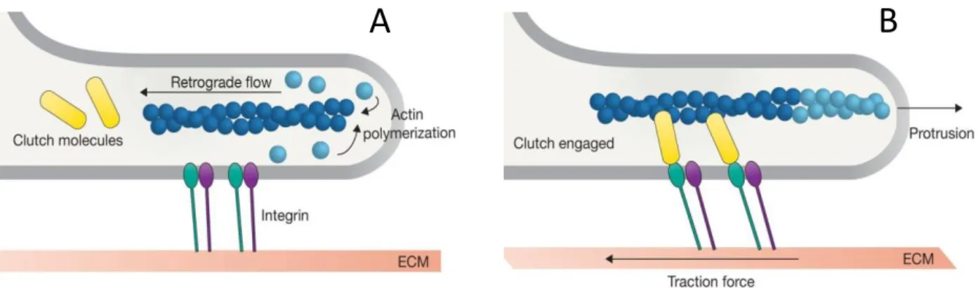
II. Force transmission to the substrate ... 29
A. Adhesion-dependent interaction ... 29
B. Anchorage-independent ... 30
1. Actin flow friction ... 30
2. Hydrostatic pressure and 'Chimneying.' ... 31
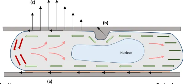
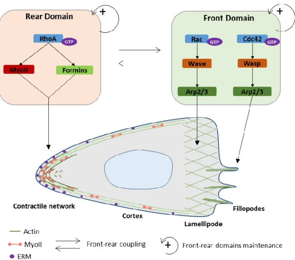
III. Maintenance of front-rear polarization ... 32
A. Definition of front and rear domains ... 32
1. Rho family of small GTPases paradigm ... 32
2. Cortical polarization ... 33
B. Front-to-rear coupling during migration ... 34
1. Membrane tension ... 34
2. Sustained actin flow ... 35
3. Long-range cytoskeleton structures: microtubule network ... 36
a) Organization of the microtubule network ... 36
b) The role of microtubules in cell polarization ... 36
CHAPTER 3: MECHANISMS OF RAPID DENDRITIC CELL MIGRATION IN THE INTERSTITIAL SPACE I. Physical and chemical properties of the interstitial space: challenges for cell migration ... 39
A. Chemical properties ... 39
1. The extracellular matrix ... 39
2. Chemokines ... 40
B. Physical properties ... 40
C. Challenges for DCs interstitial migration ... 41
1. In vitro models of the interstitial migration ... 41
2. Main characteristics of mDCs migration in 3D confined-environment and limiting factors 42 II. Maturation optimizes DCs migration machinery for rapid and directed migration ... 43
A. Remodelling of the actomyosin cytoskeleton ... 43
XIII
III. DCs strategies to overcome physical obstacles ... 45
A. Cell branching coordination in a porous environment ... 45
1. Rac and Cdc42 control front protrusions in mDCs ... 46
2. Microtubule network coordinate protrusion and retraction ... 47
B. mDCs migration in confined environments ... 48
1. The mechanical properties of the nucleus will limit cell migration ... 48
2. Selection of the path of least resistance ... 49
a) Hydraulic resistance ... 49
b) Pore size selection ... 50
3. Mechanism of Nuclear deformation ... 51
a) MyoII-based contractility ... 51
b) Arp2/3-dependent actin network ... 52
c) Different requirements for nuclear deformation ... 52
d) Specific mechanisms for mDCs entry into the lymphatic vessels ... 53
METHODS I. Bone marrow-derived dendritic cells as a cell model ... 57
A. Culture protocol ... 57
B. Characteristics of bone marrow-derived dendritic cells ... 57
C. Visualization of the cytoskeleton components and cellular compartments in live BMDCs .. 58
II. Microfabricated devices to study mDCs migration under different degrees of confinement. 58 A. Manufacturing protocol ... 59
B. MDCs migration in microchannels of different dimensions ... 59
1. Devices for live-cell imaging ... 60
2. Microscopic set-up and image analysis ... 63
... 66
3. Device developed for immunofluorescence staining of cells ... 67
C. Mature DCs migration in microchannels of varying dimensions ... 68
1. Design of the microfabricated devices ... 68
2. Microscopic set-up and image analysis ... 68
D. Arrays of pillars combining cell constraints and branching ... 71
III. From microfabricated devices to ex-vivo tissue explants ... 71
A. Mature DC migration assay in mouse ear explant ... 71 B. A simple method to image tissue collagen structures without second harmonic generation
RESULTS
I. Myosin II activity is selectively needed for migration in highly confined microenvironments
in mature dendritic cells ... 76
II. Confinement triggers ROCK-dependent actomyosin reorganization associated with mature dendritic cell speed in confined microchannels. ... 95
A. Characterization of actomyosin organization at different degrees of confinement ... 95
B. Dynamic cytoskeleton remodeling in microchannels of varying dimensions ... 98
C. ROCK activity controls actin remodeling during confined migration ... 101
III. Impact of confinement on the intracellular organization of mature dendritic cells. ... 106
A. Confined migration decreases actin fraction at the nucleus and triggers rupture of the perinuclear lamina ... 106
B. Confinement triggers actin accumulation around the lysosomes and Golgi apparatus clustered around the microtubule organizing center. ... 111
1. Actin and microtubule interplay during confined migration... 111
2. Golgi apparatus and lysosomes form a compact structure clustered on the MTOC surrounded by cortical actin ... 115
3. Confinement-induced fast actin accumulation around the lysosomal cluster ... 119
4. Confinement-induced lysosomal actin network is dependent on formin activity. ... 125
5. Microtubule depolymerization affects the confinement-induced actin network at the center of the cell ... 127
6. Fragmentation of the Golgi apparatus does not affect confinement-induced actin remodeling ... 131
DISCUSSION AND PERSPECTIVES I. Summary of the results ... 135
II. Mechanisms of cell plasticity ... 136
A. Does dendritic cell intra-cellular reorganization favor cell migration in confined areas? .. 136
B. Cytoskeleton reorganization and plasticity of the mode of migration ... 138
C. Potential mechano-responses and confinement sensors in mature dendritic cells ... 139
III. Microtubules as an integrator of the physical and chemical properties of the microenvironment. ... 140
IV. Impact of confine migration on mDCs immune functions ... 142
A. Dense cortical actin and antigen transport. ... 142
B. Actin and microtubules interplay with antigen processing in lysosomes. ... 143
C. Confined migration impacts the nuclear shell. ... 143
ANNEXES I. Publications list ... 145
XV
A. Préambule ... 148
B. Contexte ... 148
C. Méthodes ... 150
D. Résultats ... 152
1. Réorganisation du cytosquelette d’actine induit par le confinement des DCs ... 153
2. La structure d’actine à l’arrière de la cellule est dépendante de l’activité de la kinase ROCK et permet le maintien de la vitesse des DCs dans les espaces restreints. ... 155
3. La dépolymérisation des microtubules affecte l'actine induite par le confinement au centre de la cellule ... 157
E. Conclusions et perspectives ... 159
XVII
FIGURE LIST
Figure 1 Schematic summary of the organization of the immune system in a steady-state condition
... 6
Figure 2 Schematic representation of the integration of the inflammatory context during dendritic cell maturation ... 12
Figure 3 Leukocyte migration in the interstitial space ... 15
Figure 4 Information transfer during contact between mature dendritic cells (mDCs) and CD4+ T cells ... 17
Figure 5 Schematic summary of the immune cell movements in response to infection. ... 18
Figure 6 Different organization of actin networks polimerized by Arp2/3 or formins. ... 23
Figure 7 MyoII structure and organization in bipolar mini-filaments. ... 24
Figure 8 Platinum replica electron microscopy of the lamellipodia of B16F1 cell. ... 26
Figure 9 Schematic diagram of the different steps of bleb formation and retraction. ... 27
Figure 10 Walker 256 carcino-sarcoma sublines from different type of protrusion. ... 28
Figure 11 Force transmission in adherent migration ... 30
Figure 12 Schematic summary of force generation and transmission in a non-adherent migrating cell. ... 31
Figure 13 Simplified front and rear domain diagram in an actin-prutrusive cell ... 34
Figure 14 Collagen heterogeneity in mouse ear explant ... 41
Figure 15 Protrusions coordination and cell deformation are necessary for mDCs migration in 3D complex environments. ... 42
Figure 16 Schematic summary of the signaling pathway coupling microbial detection with reorganization of mDC migration machinery ... 45
Figure 17 The structure of the ECM limits mDCs migration in the interstitial space. ... 48
Figure 18 Schematic diagram of the mechanism used by DCs to overcome small pores in the interstitial space. ... 52
Figure 19 MDCS migration through the lymphatic endothelium causes a strong deformation of the nucleus ... 54
Figure 20 Physical constraints imposed by microchannels of different sizes ... 63
Figure 21 Overview of the image analysis workflow and an example of cell length quantification . ... 64
Figure 22 Quantitative method to analyze the organization of the LifeAct or MyoII-GFP signal in cells
... 66
Figure 23 Diagram of the microfabricated device used to perform immunofluorescence staining within microchannels ... 67
Figure 24 Example of devices with constrictions in the microchannels ... 68
Figure 25 Overview of the data processing workflow applied to analyze cell migration through constrictions ... 70
Figure 26 Examples of microfabricated devices with pillar networks ... 71
Figure 27 Overview of the mature dendritic cell (mDC) migration assay protocol in mouse ear explant... 72
Figure 28 Collagen staining in a mouse ear explant with fluorescently labelled SureCoat collagen 73 Figure 29 Figure 1. MyoII activity regulates mDCs migration in dense extracellular matrices ... 91
Figure 30:. Figure 2. MyoII motor activity is specifically required for mDCs migration in very confined microenvironments. ... 92
Figure 31. Sup. Figure 1. ... 93
Figure 32: Sup. Figure 2. ... 94
Figure 33 Actomyosin accumulation in the posterior end of the cell under confinement ... 96
Figure 34 Dynamic actomyosin reorganization upon cell confinement ... 101
Figure 35 Actin accumulation in the posterior end of the cell is dependent on ROCK activity ... 105
Figure 36 Confinement decreases actin recruitment at the nucleus and triggers lamina rupture . 111 Figure 37 Confinement reinforces microtubule network polarization and triggers cortical actin accumulation around the MTOC. ... 115
Figure 38 Lysosomes and the Golgi apparatus form a compact cluster around the MTOC. ... 119
Figure 39 Confinement of the lysosomal area triggers a rapid cortical actin accumulation ... 123
Figure 40 Lysosomal actin accumulation in collagen gels and upon uniaxial confinement ... 124
Figure 41 Investigation of the actin nucleator involved in the lysosomal network. ... 127
Figure 42 Microtubule depolymerization provokes cell oscillation and actin remodeling. ... 131
Figure 43 Fragmentation of the Golgi apparatus does not affect confinement-induced actin remodeling ... 133
Figure 44 ROCK inhibition reverted the Nucleus-lysosome axis... 137 Figure 45 Les DCs se déplaçant dans l’espace interstitiel doivent coordonner l’extension de multiples branches cellulaires et être capables de se déformer dans les pores de la matrice extra cellulaire.
XIX
... 149
Figure 46 Contraintes physiques imposées par des microcanaux de tailles différentes ... 152
Figure 47 Accumulation d'actine dans les cellules sous confinement ... 154
Figure 48 L'accumulation d'actine dans les cellules dépend de l'activité de ROCK... 157
Figure 49 La dépolymérisation des microtubules affecte l'actine induite par le confinement au centre de la cellule. ... 158
XXI
ABBREVIATIONS
2D: 2 dimensions 3D: three dimensional ABP: actin-binding protein ATP: Adenosine Triphosphate BCR: B Cell Receptor
BMDC: bone marrow-derived dendritic cell CD4+ LT: CD4-positive T lymphocyte CD8+ LT: CD8-positive T lymphocyte cDC: Conventional dendritic cell
DAMP: danger-associated molecular pattern DC: dendritic cell
ECM: extracellular matrix ELC: essentials light chain ER: endoplasmic reticulum ERM: Ezrin, radixin and Moesin
ESCRT: Endosomal Sorting for Transport F-actin: Filamentous actin
FMNL1: formin-like 1
FRC: Fibroblastic reticular cells
FRET: fluorescence resonance energy transfer G-Actin: monomeric actin
GAP: GTPase activator protein- GDI: GDP-dissociation inhibitor- GDP: Guanosine diphosphate GEF: GTP-exchange factor GFP: Green fluorescent protein
GM-CSF: granulocyte-macrophage colony-stimulating factor GPCR: G-protein coupled receptors
IDC: Immature dendritic cell IF: Immunofluorescence Ii: invariante chain KO: Knock out
LINC: Linker of Nucleoskeleton and cytoskeleton LN: lymph nodes
LPS: lipopolysaccharide
MAP: Microtubules associated portein MDC: Mature dendritic cell
MHC I: Major histocompatibility complex class I MHC II : Major histocompatibility complex class II MHC: Major histocompatibility complex
MLCK: myosin light chain kinase MTOC: microtubule organizing center mTOR: mechanistic target of rapamycin MyoII: Non-musle myosin Class II NLS: nuclear localization sequence
PAMP: pathogen-associated molecular pattern PDMS : Polydimethylsiloxane
PRR Pattern-recognition receptors RLC: regulatory light chain
ROCK: Rho-associated coiled-coil containing kinase ROI: regions of interest
SD: standard deviation
SEM: standard error of the mean Sema3: class 3 semaphorins TCR: T cell Receptor
TFEB: Transcription factor EB TLR: Toll-Like Receptor
TRPML1: Transient Receptor Potential cation channel, Mucolipin subfamily, Member 1 WASH: WASP and SCAR homolog
XXIII WASP: Wiskott–Aldrich Syndrome protein
WGA: Wheat Germ Agglutinin WT: Wild-type
1
INTRODUCTION
CHAPTER 1: DENDRITIC CELL MIGRATION
DURING IMMUNE RESPONSE
The immune system facilitates the elimination of pathogens and damaged cells. Its proper functioning is essential for maintaining the integrity of the organism. It is a molecular and cellular system that detects and eliminates pathogens. Unlike conventional organs, the immune system is made up of billions of cells scattered throughout the body. The coordination of their function involves a strict regulation of their movement within the body. The immune response is the main physiological process that involves cell migration at the adult stage. In this first introductory chapter, I present an overview of the immune system organization and the migration processes involved in the immune response and highlight the central role of dendritic cells (DCs)—the focus of my doctoral research.
I.
Principles and organization of the immune system
A. Two types of immune response
Two types of immune responses have been described on the basis of their activation process.
1.
Innate immune response
The innate immune response detects a limited set of conserved features associated with pathogens: the pathogen-associated molecular patterns (PAMPs) or, with tissue damage: the danger-associated molecular patterns (DAMPs). PAMPs are common to a family of pathogens—for example, the gram-negative bacterial cell wall component lipopolysaccharide (LPS) or viral nucleic acids—whereas DAMPs include mainly cytosolic components, such as adenosine triphosphate (ATP), released into the extracellular medium upon non-controlled cell death (Alberts et al., 2002a).
The innate immune cells detect PAMPs and DAMPs through pattern-recognition receptors (PRRs), which are cytosolic or present on the cell surface. The different classes of PRRs trigger different immune signals depending on the type of pathogens encountered. For example, the toll-like receptor (TLR) 4 binds to LPS on the plasma membrane and triggers an anti-bacterial immune response. TLR3, TLR7, TRL8, and TLR9 bind to different types of viral nucleic acids in endolysosomes and trigger an antiviral response (Mogensen, 2009). The combined expression of PRRs by a large number of cells facilitates the direct pathogen sensing upon infection. Thus, the innate immune response occurs almost immediately after pathogen entry into the body.
Pattern-recognition receptors are hereditary; effective pathogen recognition is based on host-pathogen co-evolution. Thus, the innate immune response evolves at the population level (Alberts et al., 2002a).
2.
Adaptive immune response
The adaptive immune response is based on the specific detection of each pathogen by a vast and diverse repertoire of receptors present on effector cells. A particular receptor recognizes a specific peptidic marker of a pathogen—the antigen—and is present in very small numbers in the body. Thus, the adaptive immune response requires a step of activation and expansion of pathogen-specific effector cells; this activation step is crucial for an effective immune response (Alberts et al., 2002b).
The receptor repertoire is formed by a random gene rearrangement and is not hereditary. Successive activation and clonal expansion events shape the repertoire over the lifetime of an individual (Alberts et al., 2002b).
B. Steady-state organization of the immune system
The immune system comprises more than a billion cells with specific functions. Immune cells are not all assembled in the same compartment as conventional organs. They are localized in specialized lymphoid organs, circulate in the blood or lymphatic system, or infiltrate tissues for immune surveillance (Mescher, 2016).
The primary lymphoid organs—bone marrow and thymus—produce immune cells, whereas the secondary lymphoid organs generate the immune response. They ensure the immune surveillance of extracellular fluids: the spleen for blood, lymph nodes for lymph, and unencapsulated
3
lymphoid organs for tissue fluid from the intestine or mucosa (Mescher, 2016). In the context of my doctoral research, I will elaborate on the immune response in the lymph nodes.
Immune cell scattering in different compartments involves cell movement throughout the body and communication between cells. The immune cells interact through direct cell-to-cell contact or by secretion of soluble factors—the cytokines (Xie et al., 2013). In the following section, I present the different cell types involved in the immune response.
C. Innate immune cells
Innate immune cells detect and respond directly to pathogen entry with their set PRRs. They form the first line of warning and defense (Alberts et al., 2002a). I will first describe the non-antigen-presenting innate cells.
Granulocytes are short-lived leukocytes circulating in the blood. They include eosinophils, basophils, and neutrophils, the most abundant white blood cells. Neutrophils are recruited within a few hours at the site of infection. They contain specialized granules and can engulf pathogens by phagocytosis. They are essential for the immune response against extracellular pathogens (bacteria, fungi, or parasites) (Luster et al., 2005).
Mast cells are tissue-resident sentinels. They are in close proximity to arterioles and venules. Upon detecting pathogens or sensing inflammatory signals, they release a substantial quantity of cytokines through degranulation. They are essential for leukocyte recruitment at the site of infection (Luster et al., 2005).
Natural killer cells are lymphocytes circulating in the blood vessels. They detect and eliminate infected or stressed cells. They play a central role in anti-viral and anti-cancer immunity (Luster et al., 2005).
D. Adaptive immune cells
Adaptive immunity is a pathogen-specific response mediated by B and T lymphocytes. A fraction circulate in the bloodstream. However, their activation occurs mainly in the secondary lymphoid organs. This section provides an overview of the specificity and effector functions of these two main types of lymphocytes (Alberts et al., 2002b).
immunoglobulin, the B-cell receptor (BCR). There is a wide variety of TCRs and BCRs, each specifically recognizing one antigen. Each T or B cell expresses only one type of TCR or BCR. Thus, they detect one antigen specific to one pathogen. Estimates suggest that each TCR or BCR is present on approximately only hundred cells in an adult. Because of this very restrictive number, they cannot directly detect and eliminate a pathogen. Their immune action relies on a critical step of activation and expansion (Alberts et al., 2002b).
1.
T lymphocytes
In T lymphocytes, TCRs form a complex with the transmembrane molecule CD4 or CD8. They interact with antigens bound to the major histocompatibility complex (MHC) of a presenting cell (Abbas et al., 2017a).
CD4-positive T lymphocytes (CD4+ TLs) recognize antigens presented on MHC class II (MHC II)
molecules. Upon activation by the cognate antigen, they differentiate into helper T cells. The helper T cells stimulate CD8-positive T lymphocytes (CD8+ TLs) or B lymphocytes and orchestrate the
adaptive immune response (Abbas et al., 2017b).
CD8+ TLs recognize antigens presented on MHC class I (MHC I) molecules. Their activation requires
an interaction with both the cognate antigen and helper T cells. They differentiate into cytotoxic T cells and migrate to the site of infection. They induce specific apoptosis of infected cells or tumor cells presenting the cognate antigen on their MHC I molecules (Abbas et al., 2017c).
2.
B lymphocytes
B cell activation requires two signaling events. First, the BCR interacts with the cognate antigen in soluble form or on a presenting cell. The BCR–antigen complex is internalized, and the cognate antigen is loaded onto MHC II molecules. The second activation step involves the interaction between the B cell and a helper T cell specific for the antigen presented on MHC II. Activation of B cells leads to their differentiation into plasma cells. They release a substantial amount of a soluble version of their BCR in the bloodstream. These antibodies bind the pathogens with high affinity and promote their recognition and elimination by other effector molecules or immune cells (Abbas et al., 2017d).
The adaptive immune response relies on the specific interaction between an antigen and its BCR or TCR counterpart. The probability of a random interaction throughout the body is low. Cellular
5
organization and communication in the lymph nodes optimize the rate of this encounter and facilitate effective activation of the adaptive response. CD8+ TL activation leads to a cellular immune
response, whereas B cell activation leads to a humoral immune response. During activation, B and T cells also detect co-stimulatory molecules and cytokines produced by antigen-presenting cells. They trigger the differentiation of effector cells into specific subtypes, which I have not presented in detail here. They facilitate the fine-tuning and adaptation of the immune response to the pathogen characteristics and the inflammatory context (Hilligan and Ronchese, 2020). Upon activation, a subset of cells differentiates into long-lived memory cells, which facilitate a faster response in the event of a second infection with the same pathogen. Finally, the integrity of the organism requires a tolerance mechanism that eliminates B or T cells reacting to self-antigens (Alberts et al., 2002b). This process will not be described in this thesis.
E. Antigen-presenting cells
Antigen-presenting cells link the innate immune response to the adaptive immune response. They have the ability to process external antigens on the MCHII and to detect infection through PRRs. Thus, they restrict the adaptive immune response to a pathogenic context. They are also migratory cells that transfer antigens from the site of infection to the lymph node. Most antigen-presenting cells are tissue resident; however, a fraction of these cells circulate in the blood. Macrophages and dendritic cells (DCs) are considered professional antigen-presenting cells (Kambayashi and Laufer, 2014). The present doctoral research focuses on DCs, which are the major antigen transporters from the periphery to the lymph nodes.
1.
Macrophages
Macrophages are tissue-resident cells. They originate from embryonic precursors or are differentiated from monocytes after entering tissues. They are highly phagocytic and can directly eliminate pathogens. They also play a critical role in resolving inflammation and tissue homeostasis by removing cellular debris or apoptotic vesicles (Perdiguero and Geissmann, 2016).
2.
Dendritic cells
characterized by their common ability to activate T lymphocytes (Ueno et al., 2007).
Langerhans cells—the first DC type described—are located in the dermis where they form long extensions scanning the tissue (Ueno et al., 2007).
Conventional DCs (cDCs) can be divided into two groups: resident cDCs populate lymphoid organs and participate in maintaining tolerance, whereas interstitial cDCs are located in tissues throughout the body. These are the main antigen transporters to the lymph nodes upon pathogen detection.
Plasmacytoid DCs circulate in the bloodstream and secrete large quantities of type 1 interferon, which activates the effectors of the anti-viral response (Ueno et al., 2007).
Monocyte-derived DCs differentiate from monocytes after they enter inflamed tissues. They ensure the renewal of the interstitial cDC population (Ueno et al., 2007).
In the present thesis, I focus on the interstitial cDCs, as they are the main migratory cells that activate the adaptive immune response. Henceforth, the general term DCs is used to refer to interstitial cDCs.
Figure 1 Schematic summary of the organization of the immune system in a steady-state condition
(Top panel) Presentation of the different cells of the innate (left: orange) and adaptive (right: green) immune systems. Antigen presenting cells (middle: blue) link the innate and adaptive immune systems. Image from the Servier Medical ART database.
(Bottom panel) Distribution of immune cells in the body. Innate immune cells circulate in the bloodstream and infiltrate tissues to ensure immune monitoring with antigen-presenting cells. Adaptive immune cells circulate in the bloodstream and colonize the secondary lymphoid organs (only lymph nodes and spleen represented here). At steady state, antigen-presenting cells residing in the secondary lymphoid organs ensure adaptive immune tolerance.
This first section provides a brief description of the main actors of the immune system and their location in a steady-state condition. Next, I describe how the immune response is orchestrated in case of infection and focus on the central role of DCs as a link between the innate and adaptive immune responses (Banchereau and Steinman, 1998).
II. Local immune response: activation of the innate system
The innate immune response involves tissue-resident and blood-circulating immune cells. It is fast and restricted to the site of pathogen entry. The main function of this first line of defense is to detect the pathogen and to contain the infection.A. Inflammation
Inflammation is a local and immediate response to an injury or pathogen entry. Tissue-resident immune cells—mainly DCs, macrophages, and mast cells—detect pathogens or damaged cells through their PRRs. The engagement of PRRs triggers signaling pathways, leading to the secretion of pro-inflammatory cytokines. The set of cytokines secreted depends on the type of PAMPS or DAMPs detected and is therefore specific to a family of pathogens. The release of pro-inflammatory cytokines corresponds to an alarm system that modifies the local environment to recruit effector immune cells (Mogensen, 2009).
Some cytokines can guide leukocytes in tissue to the site of infection; these chemoattractant molecules are named chemokines (Zlotnik and Yoshie, 2012). Another group of inflammatory cytokines acts on the blood vessel endothelium. They cause endothelial cells to increase blood flow, increase endothelial barrier permeability, and express adhesion molecules on the luminal surface of the blood vessels. This blood vessel endothelial activation promotes the entry of circulating leukocytes into the inflamed tissue (Pober and Sessa, 2007).
B. Leukocyte recruitment: from blood to the site of infection
The recruitment of blood leukocytes to the site of infection in tissues involves several steps of migration through the blood vessel endothelium and into the interstitial space. Neutrophils—the most abundant blood leukocytes—are essential for pathogen clearance, and their recruitment to
9
the site of infection is the most studied (Weninger et al., 2014).
1.
Transendothelial migration
The transendothelial migration of neutrophils in inflamed tissues involves a well-established multi-step adhesion cascade. Selectin expression on activated endothelial cells allows neutrophils to slow down and roll along the walls of inflamed blood vessels. Integrin engagement and chemokine sensing leads to firm adhesion of neutrophils, followed by their transmigration through the endothelial cell monolayer (Vestweber, 2015). After transmigration, the neutrophils crawl into the subendothelial space confined by the dense basement membrane and pericytes surrounding the blood vessels. The neutrophils extravasate into the tissue through low-density regions of extracellular proteins in the basement membrane. Chemokines secreted by the perivascular immune cells guide the neutrophil extravasation (Vestweber, 2015).
Blood leukocyte transmigration is a limiting step that requires specific signaling events triggered by inflammation. Thus, leukocyte infiltration is limited to the site of infection (Pober and Sessa, 2007).
2.
Interstitial migration
Once in the interstitial space, leukocytes navigate in a three-dimensional environment composed of a network of cross-linked extracellular matrix fibers (Lämmermann and Germain, 2014). To reach the site of infection, they integrate chemoattractant signals from tissue-resident immune cells, damaged cells, or pathogens. The neutrophil population exhibits coordinated chemotactic migration, leading to the formation of a dense cell cluster that contains the injury (Kienle and Lämmermann, 2016; Ng et al., 2011). This swarming behavior relies on the secretion of chemokines that act as a paracrine signal among the neutrophils. This process of self-amplification of a local inflammatory signal facilitates neutrophil recruitment throughout the tissue at a long distance from the site of injury (200–300 µm) and in a short time (~20 min) (Lämmermann et al., 2013).
Leukocyte migration into the interstitial space does not require high-affinity integrin signaling. Thus, immune cell recruitment is mainly orchestrated by chemokines secreted during the inflammation process and is independent of the composition and structure of the extracellular matrix. This migration mechanism, which will be described in detail later in this thesis, is a critical step in the immune response that requires leukocytes to be able to rapidly reach any type of tissues
upon detection of inflammatory signals (Lämmermann et al., 2008). Integrins transduce signals of arrest rather than migration during the immune response. They induce leukocyte adhesion on the inflamed blood vessels (Vestweber, 2015) and are necessary for neutrophil accumulation at the site of injury (Lämmermann et al., 2013).
At the site of infection, innate immune cells release molecules and enzymes that directly eliminate the pathogen or prevent its spread (Alberts et al., 2002a). The molecules are specific to the pathogen family. Phagocytic cells, mainly neutrophils and macrophages, engulf pathogens and remove cellular debris. This process is essential for the resolution of inflammation at a later stage of the immune response (Westman et al., 2020). While innate immune cells control the spread of pathogens, they release a second set of cytokines that orchestrate the activation of an adaptive immune response (Iwasaki and Medzhitov, 2015).
C. Dendritic cell maturation
Antigen transport from the injury site to the lymph nodes is essential for the initiation of an adaptive immune response. Microbial detection triggers DC maturation, a multi-step process that leads to mature DCs (mDCs) capable of activating T lymphocytes. This process involves antigen uptake and presentation on MHC, integration of the inflammatory context to activate appropriate adaptive immune cells, and acquisition of the ability to migrate to the lymph node (Cella et al., 1997a).
1.
Antigen uptake and processing
Before microbial detection, immature DCs (iDCs) scan the tissues for harmful particles. Tissue surveillance is ensured by projecting long cell protrusions in random directions or by active cell migration. In vitro studies suggest that the migration patterns of iDCs follow an intermittent random walk model. They alternate between rapid migration phases and arrest phases when sampling their surroundings; this behavior optimizes the pathogen search in the environment (Chabaud et al., 2015).
Direct pathogen detection through PRRs or inflammatory chemokine sensing transiently increases the ability of DCs to internalize extracellular components by endocytosis, phagocytosis, or macropinocytosis. Subsequently, internalized pathogenic particles are degraded into antigenic peptides and loaded onto MHC II for presentation to CD4+ TLs and onto MHC I for presentation to
11 CD8+ TLs (West et al., 2004).
MHC II binds to antigens degraded in the lysosomal pathway. In the absence of antigens, the invariant chain (Ii) stabilizes MHC II. After pathogen detection, the lysosomal protease cathepsin-S cleaves Ii and allows peptide loading onto MHC II. The complex is then transported to the plasma membrane to present the antigen to T lymphocytes (Neefjes et al., 2011).
MHC I binds to antigens degraded by cytosolic and nuclear proteasomes. Cytosolic peptides are transported to the endoplasmic reticulum (ER) where they are loaded onto MHC I molecules (Neefjes et al., 2011). Extracellular antigen presentation on MHC I requires a process called cross-presentation. There are two main pathways. In the vacuolar route, MHC I vesicles encounter the lysosomal pathway, and antigens degraded in the lysosomes are loaded onto MHC I. The second pathway is cytosolic: internalized particles escape from endosomes and are degraded by the cytosolic proteasome before being loaded onto MHC I in the ER (Alloatti et al., 2016).
Antigen processing is tightly regulated to avoid the presentation of self-antigens in an inflammatory context. Maturation of DCs increases MHC expression and translocation to the cell surface (Cella et al., 1997b) and modulates the lysosomal activity to process antigens properly (Trombetta et al., 2003). Increasing lysosomal acidification promotes antigen processing on MHC II, whereas maintaining a high pH promotes cross-presentation on MHC I (Samie and Cresswell, 2015). Cytokines and PRRs such as TLRs are known to signal from the endolysosomal compartments. PRRs directly modulate antigen presentation by co-trafficking with pathogenic endocytic particles (Watts et al., 2010) and locally regulate lysosomal activity to promote pathogen particle degradation into antigens (Roche and Furuta, 2015). Thus, he endolysosomal compartment plays a role as a signaling platform that integrates the inflammatory context to control antigen presentation.
2.
Cytokine and co-stimulatory molecules
Antigens are not the only type of information acquired by DCs at the site of infection; they also integrate PAMPs and DAMPs present in the microenvironment and cytokines secreted by other immune cells. These factors provide information on the pathogenicity and location of the microbe as well as the level of tissue damage. This inflammatory context shapes the cytokine profile secreted by the mDCs and the nature of the co-stimulatory molecules expressed on their surface (Iwasaki
and Medzhitov, 2015).
Figure 2 Schematic representation of the integration of the inflammatory context during dendritic cell maturation
Internalized pathogenic particles are degraded to form antigens and presented on MHC II in the endolysosomal pathway (right) or on MHC I by cross-presentation (left). Engagement of PRRs and cytokine sensing at the site of infection induce signaling events that control antigen processing and trigger the expression of a specific set of cytokines and co-stimulatory molecules
13 Cross-presentation Pathogenic Particle PRR Cytokine Receptor MHCI Endo-lysosomes Antigens Protease Ii MHC II Endosomes Proteasome ER Signaling Cluster Lysosomes Activity Cytokines MHC Cos-stimulatory Molecules Nucleus Inflammatory Context Cytokines secreted by other immune cells
Mature dendritic cell
M atu ra ti on: in te gr ati on of the i n fla m m at or y c on te xt
MHCII – pathogenic antigen MHCI– pathogenic antigen Specific set of cytokines and co-stimulatory molecules
MHC II Antigen processing
3.
Migration to the lymph nodes
The final stage of maturation leads to the migration of DCs to the draining lymph node via the lymphatic system. It is characterized by reduced internalization of extracellular components, loss of adhesion, and increased cell motility (Ueno et al., 2007). A key event is the expression of CCR7 chemokine receptors (Sallusto et al., 1998). It allows the detection of the chemokine CCL21, which is essential for guiding the mDCs in the lymphatic system (Ohl et al., 2004). Endothelial lymphatic cells secrete CCL21 and form a chemokine gradient that guides the mDCs into the interstitial space (Weber et al., 2013). CCL21 is additionally released at the site of transmigration and promotes the entry of mDCs into the lymphatic vessels (Vaahtomeri et al., 2017). Finally, the CCL21 gradient within the vessels promotes the migration of mDCs to the lymph node (Russo et al., 2016).
As for other leukocytes, mDCs migration into the interstitial space is independent of specific integrin engagement. This mode of motility promotes flexible migration across any tissue context (Lämmermann et al., 2008). However, mDCs move away from the site of infection in the opposite direction to other innate immune cells. Thus, within the same infected tissue, immune cells exhibit specific migration patterns depending on their function (Vargas et al., 2017). Therefore, coordination of immune function and migration requires the correct interpretation of signals from the pathogen, other immune cells, and tissue microenvironment.
15
In vitro, DC maturation requires 16 to 20 h before the co-stimulating molecules, MHC II and
CCR7, are expressed on the cell surface (Alloatti et al., 2016). During this process, DCs integrate both the pathogen nature through antigen presentation and the inflammatory context through the expression of cytokines and co-stimulatory molecules. Once DCs have assimilated this information, they acquire the ability to migrate to the lymph nodes. The mDCs carry the information necessary to instruct T lymphocytes and activate the appropriate effectors of the adaptive immune response
III. Systemic immune response: activation of the adaptive
system by dendritic cells
The adaptive immune response is specific to the pathogen encountered at the injury site. It allows the elimination of the infection and the establishment of memory cells. They facilitate the activation step and trigger a rapid response if the same pathogen is encountered a second time. In this section, I describe the key role of mDCs in triggering this immune response in the lymph nodes.
Figure 3 Leukocyte migration in the interstitial space
In infected tissue, neutrophils cross the blood vessel endothelium and migrate to the site of infection. They form a cell cluster that contains the infection. The inflammation also activates the DCs. After maturation and antigen uptake, they acquire the ability to migrate toward the lymphatic vessels and activate the adaptive immune response in the lymph nodes. In the same environment, the two cell types follow different migration patterns depending on their immune function. This coordination requires specific migration machinery and the correct interpretation of signals from the microenvironment. From Vargas et al., 2017
A. Antigen presentation to CD4+ T cells
The antigen-specific activation of CD4+ T cells is the first step in triggering the adaptive
immune response. Expansion of pathogen-specific CD4+ T cells and their differentiation into
appropriate helper T cells are essential to orchestrate the action of effector cells (Abbas et al., 2017b). CD4+ T cell antigen priming requires physical contact with mDCs presenting their cognate
antigen (Bousso, 2008). The probability of an encounter is very low (~1 in 1 million). The microarchitecture of the lymph nodes optimizes the contact between mDCs and their cognate T cells (Lämmermann and Sixt, 2008). Within the lymph nodes, B cells and T cells are compartmentalized and separated. B cells form follicles in the outer cortex, whereas T cells reside primarily in the inner cortex, below the B cell zone.
The mDCs enter the lymph nodes through the afferent lymph vessel and actively migrate to the T-cell-rich cortex. This area is a cell-rich environment, densely populated with T cells and fibroblastic reticular cells (FRCs), which form a three-dimensional scaffold (Lämmermann and Germain, 2014). The FRCs produce CCL21 and thus regulate the intranodal movement of T cells and mDCs, both expressing CCR7 (Worbs et al., 2007). The CD4+ T cells migrate randomly along the FRC tracks
(Bajénoff et al., 2006), while mDCs can adhere to the cellular scaffold (Lindquist et al., 2004). This organization optimizes the scanning of the antigen-presenting mDCs by the T cells. Other work suggests that the mDCs migrate to the lymph node entry site of the T cells from the bloodstream. Thus, mDCs effectively scan all incoming T cells and retain the T cells specific to their antigen (Bajénoff et al., 2003). The mDCs also have specific shape dynamics in the lymph nodes, forming long cell extensions (Bousso and Robey, 2003), which increase the area of contact with T cells. This behavior is essential for T cell priming (Benvenuti et al., 2004) and allows a single mDC to simultaneously present its antigens to more than 10 T cells simultaneously (Bousso and Robey, 2003).
B. Differentiation of CD4
+T cells and activation of the specific immune
cell effectors
Once the cognate mDCs encounter CD4+ T cells, they form a strong interaction (Bousso,
2008). This facilitates information transfer from the mDCs to the CD4+ T cells. This information
17
and cytokines, which summarize the inflammatory environment at the site of infection (Kambayashi and Laufer, 2014). The interaction with mDCs leads to the activation and proliferation of the antigen-specific CD4+ T cells and their differentiation into context-specific CD4+ helper T cells (Hilligan and
Ronchese, 2020).
The CD4+ helper T cells then orchestrate the pathogen-specific immune response. They
secrete a precise set of cytokines that activate the appropriate immune effectors (Iwasaki and Medzhitov, 2015).
Depending on their immune phenotype, CD4+ helper T cells can activate CD8+ T cells to
differentiate into cytotoxic T cells or B cells to differentiate into plasma cells. Both cell types also require antigen-specific priming. CD8+ T cell activation involves antigen presentation on the mDC
MHC I molecules. B lymphocytes can interact with soluble antigens or antigens presented on DC plasma membrane (Alberts et al., 2002b). The specific mDC localization in the lymph node cortex may result in preferential activation of CD8+ T cells or B cells and thus shape the immune response
(Gerner et al., 2017; Kissenpfennig et al., 2005).
Finally, pathogen elimination by effector T cells requires their subsequent migration from the lymph node to the site of infection. Effector T cells recirculate in the blood; as with innate
PAMPs DAMPS PRR MHCII CD4 TCR Antigen Mature Dendritic Cell CD4+ T Cell
Figure 4 Information transfer during contact between mature dendritic cells (mDCs) and CD4+ T cells
Mature DCs carry antigens to the lymph nodes that enable the activation and expansion of pathogen-specific T-lymphocytes. They also express a specific set of cytokines and co-stimulatory molecules that summarize the inflammatory context at the site of infection. They induce the differentiation of CD4+ T lymphocytes into helper T
immune cells, their transmigration through the blood vessel endothelium of infected tissues requires specific adhesion molecules and signaling pathways (Abbas et al., 2017b). Effector T lymphocytes can acquire some of these homing receptors when primed by mDCs (Pejoski et al., 2019). Thus, mDCs also carry the signature of the tissues in which they encountered the pathogenic antigen. The mechanism underlying the effector T cell tropism towards infected tissues is not completely understood and remains an obstacle to the development of effective vaccination against cancer or human immunodeficiency virus, which requires a T cell-based immune response (Fu et al., 2016).
Figure 5 Schematic summary of the immune cell movements in response to infection.
Tissue Infection results in coordinated steps of immune cell movement that ensure pathogen elimination. Pathogen detection induces a local infiltration of innate immune cells. They trigger a local inflammation and limit the pathogen spread. The inflammation activates dendritic cells, and they migrate to the lymph node. In the lymph node, mature dendritic cells trigger the activation, expansion, and differentiation of specific effectors of adaptive immunity (top panel - image from Servier Medical ART database). The adaptive effector cells recirculate in the bloodstream and migrate into the infected tissue where they mediate pathogen elimination. All these steps of immune cell migration are tightly regulated to limit the inflammation and cell infiltration to the infected tissue only and to avoid the damage of healthy tissue.
19 Spleen Lymph Node Heart Lymph Node Tissue 1 Tissue 2
0
0
0
0
0
0
0
0
0
0
Mature Den-dritic Cell Local infiltration of innate cellsMature dendritic cells migra-tion to the lymph node
Systemic circulation of adap-tive effector cells Infection
0
Effector cells
Adaptive immune cells
T Lymphocyte B Lymphocytes Plasma cell Memory B cell Memory T cell CD8+ cytotoxic T cells CD4+ Helper T cell Mature Dendritic Cell
Dendritic cells play an essential role in the immune system activation by linking the innate and adaptive response. They uptake information on the pathogen and inflammatory microenvironment and transport it to the lymph nodes, where they activate pathogen-specific immune effectors.
Their function involves multiple stages of migration from different tissues to the lymph nodes. They are optimized to migrate efficiently in different physical and chemical contexts. They are, therefore, an appropriate model to study cell migration. In my doctoral research, I focus mainly on the interstitial migration of mDCs. Indeed, the physical and chemical properties of this environment can vary among tissues and require an adaptation of mDC migration mechanisms. Moreover, neutrophils and T lymphocytes also migrate in the interstitial space to perform their immune function. Therefore, a comparison between different immune cells could highlight the migration mechanisms shared by rapidly moving leukocytes in the interstitial space and the specific mechanisms required for the immune function of antigen-transporting mDCs. Finally, understanding the mechanisms controlling mDC migration from the site of infection to the lymph nodes could provide molecular tools to modulate the activation of the adaptive response. This is particularly relevant in the context of vaccine and cancer immunotherapy development.
21
CHAPTER 2: MECHANISMS OF
CELL MIGRATION
As described in the previous chapter, cell migration is an essential process for the immune response. It is also involved in other physiological or pathological events such as embryonic development or metastasis progression. Thus, the mechanisms underlying cell migration have been studied in a variety of different contexts (Yamada and Sixt, 2019). They are dependent on the properties of both the cell and its environment. In this chapter, I will describe globally the different mechanisms involved in single-cell migration. Then, in the following section, I will put them in the context of DCs interstitial migration.Cell migration relies on internal forces that drive cell movement. Previous studies have highlighted different strategies developed by cells to meet the requirements for cell migration (Vicente-Manzanares et al., 2005). This chapter will focus on describing how internal forces are generated in cells, transmitted to the substrate and further polarized inside cells to result in single-cell displacement to a particular direction.
I.
Intracellular forces for cell deformation
In most animal cells, the actomyosin cytoskeleton generates the forces necessary for cell motion. Its expansion by polymerization is associated with pushing forces and membrane protrusion while its shrinkage causes pulling forces and membrane retraction (Svitkina, 2018). In this section, I will first describe the molecular organization of the actomyosin network and then discuss on how its remodeling leads to cell movement
A. Acto-Myosin Cytoskeleton
1.
Actin filament
Actin filament (F-actin) assembles from monomeric actin proteins (G-Actin). It forms a polar filament with a "barbed" end and a "pointed" end. F-actin diameter is ~7 nm, and its persistence
length is ~10 µm. As compared to other cytoskeleton elements, actin filaments are more flexible and dynamic, with structural changes in the time scale of minutes (Blanchoin et al., 2014).
In the cell, the concentration of G-actin allows spontaneous and asymmetric polymerization of F-actin. Single filaments elongate ten-times faster at the barbed end than at the pointed end. However, actin-binding proteins (ABPs) modulate this spontaneous elongation rate by interacting with G-actin and F-actin. APBs such as profilin bind directly to G-actin and promote the assembly reaction or sequester actin monomers. Capping proteins stabilize the barbed ends, preventing both polymerization and depolymerization. Cofilin interacts with F-actin, cleaves the filament and, increases its dynamics (Pruyne et al., 2002). The binding sites of the different ABPs partially overlap, resulting in competition for F- or G-actin interaction and tight regulation of filament elongation.
While the elongation of F-actin is spontaneous, the nucleation step of the first three monomers is thermodynamically unfavorable and prevented by the interaction with profilin. Thus, the formation of new F-actin is dependent on actin nucleating factors. Their nucleation and regulation properties lead to different organizations of the actin network.
2.
Actin network organization
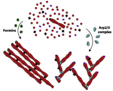
a) Branched network – Arp2/3
Arp2/3 is a complex of seven subunits. Among them, Arp2 and Arp3 are actin-related proteins and mimic an actin dimer. The complex acts as a template for further F-actin elongation and overcomes the limiting step of nucleation (Goley and Welch, 2006). It binds to the side of pre-existing filaments and branches a new F-actin at an angle of 70°. The new filament grows from its barbed end, while interactions with capping proteins limit its length (Amann and Pollard, 2001).
Nucleation by Arp2/3 leads to the formation of a branched network of short actin filaments. The WASP family and the WAVE family are the main regulators of its activity (Molinie and Gautreau, 2018).
b) Non-Branched Network – Formins
Formin family proteins share conserved FH1 and FH2 domains and their activity is controlled by the Rho family GTPases. The FH1 domain is known to interact with profilin and the FH2 domain with actin. Formins promote both nucleation and elongation of F-actin. They interact processively with the barbed end of the growing filament and protect it from capping proteins (Higashida et al.,
23 2004).
Nucleation by formins leads to long, unbranched actin filaments. Actin crosslinkers further organize this network. They bridge two actin filaments and form actin bundles with parallel, antiparallel or mixed F-actin depending on the crosslinker (Blanchoin et al., 2014).
Formin and Arp2/3 are the best described actin nucleators, which give rise to distinct network organizations with specific properties. Their functions are not exclusive, and they can cooperate to generate complex actin networks. Actin elongation also involves other proteins such as Spires or members of the Ena/Vasp family, adding another level of complexity to the network dynamics.
3.
Myosin
Other key players in the dynamics of the actin cytoskeleton are members of the myosin family. Myosins are molecular motors characterized by a globular domain that interacts with actin and has ATP hydrolysis activity. Depending on their structure, they are involved in different processes: cross-linking of F-actin with the cell membrane (class I), F-actin contraction (class II), or transport of vesicles along F-actin (class V) for the conventional myosins (Sellers, 2000).
Figure 6 Different organization of actin networks polimerized by Arp2/3 or formins.
From the same pool of G-actin (red), Arp2/3 (blue) and formins (green) polymerize actin networks with different structures. Arp2/3 leads to short, branched filaments due to the interraction of capping proteins (violets) with free filamentsbarbed end. Formins protect the growing end from the capping proteins, resulting in long filaments that are bundled by crosslinkers (cross). Adapted from Michael Bindschadler and James L. McGrath 2004.
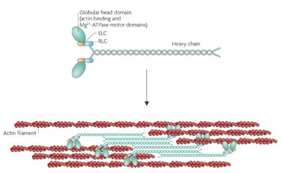
Non-muscular class II myosin (MyoII) produces contractile force by sliding antiparallel actin bundles through the energy of ATP hydrolysis. MyoII is a heterodimer complex composed of two heavy chains encompassing the globular domain, two regulatory light chains (RLC), and two essentials light chains (ELC). MyoII alone is unipolar and, therefore, ineffective in producing contractile forces. It must assemble into a bipolar mini-filament to interact with antiparallel actin bundles and cause their gliding (Vicente-Manzanares et al., 2009).
Phosphorylation of the RLC controls MyoII ATPase activity and its assembly into filaments. More than a dozen kinases have been identified to phosphorylate the RLC. The most characterized are the myosin light chain kinase (MLCK) and the Rho-associated coiled-coil containing kinase (ROCK). The signals triggering these kinases are different, MLCK is activated by calcium via calmodulin, while the GTP binding protein RhoA activates ROCK. They also differ in their specificity, MLCK seems specific to the regulatory light chain, while ROCK can phosphorylate several substrates in the cell (Vicente-Manzanares et al., 2009).
The interaction between F-actin, actin nucleators and myosin gives rise to a dynamic and tightly regulated actomyosin network. Its elongation and contraction generate pushing and pulling forces capable of deforming the cell membrane. At the cellular level, the actomyosin network is
Adapted from:
Figure 7 MyoII structure and organization in bipolar mini-filaments.
The subunits of the MyoII form a unipolar complex. Mini filaments are formed by anti-parallel interaction of the MyoII via their heavy chains. This bipolar organization allows the interaction with antiparallel actin bundles and their sliding with the movement of globular domains. Adapted from Vicente-Manzanares M et al 2009.
25
integrated into large structures that lead to movement. I will describe these structures in the next section.
B. Different types of cell protrusions
1.
Actin-rich protrusion
F-actin assembly produces picoNewton forces (Kovar and Pollard, 2004); thus, the growing of an actin network underneath the plasma membrane is sufficient to push it and create a protrusion (Pollard and Borisy, 2003).
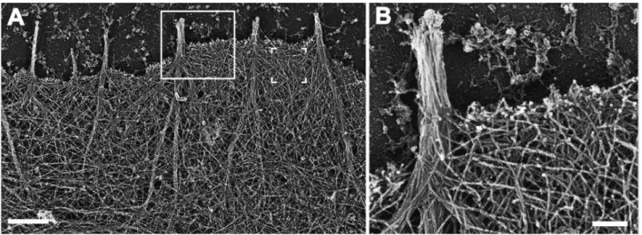
Actin-rich protrusions have a variety of shapes (Ridley, 2011). The most well-described is a thin (~200nm thick) veil assembled by cells on a 2D surface, named lamellipodia. Such actin-rich lamellar ruffles protruding in the free space have also been observed on cells migrating in 3D environments (Fritz-Laylin et al., 2017). In these structures, F-actin elongation occurs mainly against the plasma membrane. The formation of a branched network by Arp2/3 is essential for lamellipodia extension (Bailly et al., 2001), being WAVE the main Arp2/3 activator (Takenawa and Miki, 2001).
Another protrusion involved in cell movement is filopodia, described as a finger-like structure. They are formed by the elongation of unbranched actin bundles towards the plasma membrane nucleated by formins and Ena/VASP at the tip of the filopodia (Faix and Rottner, 2006). Actin crosslinkers further modulate the stability of these structures (Khurana and George, 2011). N-WASP mediated Arp2/3 activation also promotes filopodia formation (Miki et al., 1998).
Branched and unbranched actin networks are not mutually exclusive in the different protrusions. Studies inhibiting actin elongation mediated by Arp2/3 or by formins have revealed their collaboration in the formation of complex structures. For example, the formin mDia2 generates long actin filaments essential for the maintenance of lamellipodia (Fig.8) (Yang et al., 2007). Similarly, the formins FMNL2 and FMNL3 are not essential for the formation of the lamellipodia, but they enhance the density and the mechanical stability of the network and are required for efficient force generation (Kage et al., 2017a). At the cellular level, distinct actin networks polymerized by formins or Arp2/3 have also been observed in migrating cell protrusions (Ponti et al., 2004; Wilson et al., 2013). Generally, Arp2/3 and formins have specific roles in leukocytes that help to coordinate their migration and immune functions. For instance, cell motility requires only the formin-dependent network (Vargas et al., 2016; Wilson et al., 2013), while Arp2/3