Université de Montréal Étude de la toxicité causée par le gène C9orf72 dans la Sclérose Latérale Amyotrophique Par Martine Therrien Département de pathologie et biologie cellulaire Faculté de médecine Thèse présentée à la Faculté des études supérieures En vue de l’obtention du grade de Ph.D. En pathologie et biologie cellulaire -option système nerveux Janvier 2016 ©Martine Therrien, 2016
R
ÉSUMÉLa Sclérose Latérale Amyotrophique (SLA) est une maladie neurodégénérative qui affecte les neurones moteurs. 10% des cas sont des cas familiaux et l’étude de ces familles a mené à la découverte de plusieurs gènes pouvant causer la SLA, incluant SOD1,
TARDBP et FUS. L’expansion de la répétition GGGGCC dans le gène C9orf72 est, à ce jour,
la cause la plus connue de SLA. L’impact de cette expansion est encore méconnu et il reste à déterminer si la toxicité est causée par un gain de fonction, une perte de fonction ou les deux.
Plusieurs gènes impliqués dans la SLA sont conservés entre le nématode
Caenorhabditis elegans et l’humain. C. elegans est un vers transparent fréquemment
utilisé pour des études anatomiques, comportementales et génétiques. Il possède une lignée cellulaire invariable qui inclue 302 neurones. Aussi, les mécanismes de réponse au stress ainsi que les mécanismes de vieillissement sont très bien conservés entre ce nématode et l’humain. Donc, notre groupe, et plusieurs autres, ont utilisé C. elegans pour étudier plusieurs aspects de la SLA.
Pour mieux comprendre la toxicité causée par l’expansion GGGGCC de C9orf72, nous avons développé deux modèles de vers pour étudier l’impact d’une perte de fonction ainsi que d’un gain de toxicité de l’ARN. Pour voir les conséquences d’une perte de fonction, nous avons étudié l’orthologue de C9orf72 dans C. elegans, alfa-1 (ALS/FTD
moteurs causant une paralysie et une dégénérescence spécifique des neurones moteurs GABAergiques. De plus, les mutants sont sensibles au stress osmotique qui provoque une dégénérescence. D’autre part, l’expression de la séquence d’ARN contenant une répétition pathogénique GGGGCC cause aussi des problèmes moteurs et de la dégénérescence affectant les neurones moteurs. Nos résultats suggèrent donc qu’un gain de toxicité de l’ARN ainsi qu’une perte de fonction de C9orf72 sont donc toxiques pour les neurones. Puisque le mouvement du vers peut être rapidement évalué en cultivant les vers dans un milieu liquide, nous avons développé un criblage de molécules pouvant affecter le mouvement des vers mutants alfa-1 en culture liquide. Plus de 4 000 composés ont été évalués et 80 ameliore la mobilité des vers alfa-1. Onze molécules ont aussi été testées dans les vers exprimant l’expansion GGGGCC et huit diminuent aussi le phénotype moteur de ces vers.
Finalement, des huit molécules qui diminent la toxicité causée par la perte de fonction de C9orf72 et la toxicité de l’ARN, deux restaurent aussi l’expression anormale de plusieurs transcrits d’ARN observée dans des cellules dérivées de patient C9orf72. Avec ce projet, nous voulons identifier des molécules pouvant affecter tous les modes de toxicité de C9orf72 et possiblement ouvrir de nouvelles avenues thérapeutiques.
Mots clés: sclérose latérale amyotrophique; C. elegans; C9orf72; criblage de molecules; dégénérescence
A
BSTRACTAmyotrophic lateral sclerosis (ALS) is a neurodegenerative disorder affecting the motor neurons. 10% of the cases are familial and using those families, many genes were shown to be involved in ALS pathogenesis, including SOD1, TARDBP and FUS. The GGGGCC repeat found in the first intron of C9orf72 is, to this day, the most common genetic cause of ALS. Many hypotheses have been speculated to explain the toxicity of the pathogenic GGGGCC repeat, including loss and gain of function mechanisms.
Many proteins involved in amyotrophic lateral sclerosis (ALS) are evolutionarily conserved in the worm Caenorhabditis elegans. C. elegans is a transparent nematode widely used for anatomical, behavioural and genetic studies. It possesses an invariant cell lineage that includes 302 neurons in the adult nematode. Also, cellular stress responses and survival mechanisms are genetically regulated and conserved from the nematode and human. Therefore, our group, and others, have used C. elegans to model different aspects of neurodegenerative diseases including ALS.
To better understand the toxicity caused by the GGGGCC repeat expansion in
C9orf72, we have developed two C. elegans models to understand either the impact of the
loss of function of C9orf72 or the gain of toxicity of the RNA containing the GGGGCC repeat. To understand the loss of function, we have characterized the orthologue of
C9orf72 in C. elegans, alfa-1 (ALS/FTD associated gene homolog). Mutant alfa-1 worms
exhibit motor impairments leading to paralysis and neurodegenereation of the GABAergic neurons. Mutant worms are also sensitive to osmotic stress which can lead to increased neurodegeneration. On the other part, exposure of C. elegans neurons to the
RNA containing the GGGGCC repeat causes also motor problem and degeneration affecting the motor neurons. Therefore, our data suggest that both loss of function of
C9orf72 and toxic gain of function are detrimental to neurons.
Since motor dysfunctions in worms can be easily accessed in liquid culture, we have screened more than 4,000 FDA approved compounds in the alfa-1(ok3062) worms. 80 molecules were shown to improve alfa-1 impaired function and eleven of those were also tested for their effect to reduce the neurotoxicity caused by the GGGGCC repeat RNA. Eight molecules were shown to affect both types of neurotoxicity.
Finally, from these eight molecules that can improveboth types of toxicity, two were shown to restore the abnormal RNA expression observed in C9orf72 patient-derive cells. With this project, we aimed to identify molecules that can affect the loss of C9orf72 toxicity and the toxic gain of RNA function containing the GGGGCC repeat to hopefully open new therapeutic avenues for ALS patients.
Key words: Amyotrophic lateral sclerosis; C. elegans; C9orf72; drug screening; neurodegeneration
Table of content
T
ABLE OFC
ONTENTS RÉSUMÉ ... IV ABSTRACT ... VI LIST OF FIGURES ... XI LIST OF TABLES ... XIV LIST OF ABBREVIATIONS ... XV ACKNOWLEDGMENTS ... XVIIICHAPTER 1: INTRODUCTION ... 20
ALS ... 20
Disease incidence and development ... 20
Treatment ... 21
Genetics ... 22
Pathological characteristics of ALS ... 26
Similarities between ALS and other neurodegenerative disorders ... 27
C9ORF72 ... 31
Genetics ... 31
C9orf72 Expression ... 34
Function of C9orf72 ... 36
Conservation of C9orf72 across species ... 37
REPEAT DISORDERS ... 39
Myotonic Dystrophy and the toxicity of RNA foci ... 39
polyglutamine diseases and Abnormal translation ... 40
Fragile X syndrome and toxicity of a decrease of gene expression ... 42
C. ELEGANS ... 43
Current models to understand ALS ... 43
Lifespan and genome of C. elegans ... 44
C. elegans nervous system ... 45
Stress response pathways in C. elegans ... 47
C. elegans tool box ... 49
ALS C. elegans models ... 51
Other disease related C. elegans models ... 53
CHAPTER 2 ... 56
INTRODUCTION ... 56
MANUSCRIPT :DELETION OF C9ORF72 RESULTS IN MOTOR NEURON DEGENERATION AND STRESS SENSITIVITY IN C. ELEGANS ... 58
Authors and affiliations ... 58
Authors contribution ... 58 Manuscript ... 58 Abstract ... 58 Introduction ... 60 Results ... 61 Discussion ... 66
Materials and Methods ... 69
Acknowledgments ... 73
References ... 73
FIGURES ... 76
CHAPTER 3 ... 85
INTRODUCTION ... 85
MANUSCRIPT:DRUGS SCREENING IDENTIFIES MOLECULES THAT CAN ALLEVIATE THE TOXICITY CAUSED BY LOSS OF C9ORF72 AND BY TOXIC GGGGCC REPEAT EXPANSION RNA IN IN VITRO AND IN VIVO MODELS ... 87
Abstract ... 87 Introduction ... 89 Results ... 90 Discussion ... 98 Methods ... 101 REFERENCE ... 106 FIGURES ... 109 Supplementary material ... 116 DISCUSSION ... 126 INTRODUCTION ... 126
Summary of work presented ... 126
ANIMAL MODELS OF C9ORF72 ... 128
Summary of new C9orf72 models ... 128
Conclusion ... 129
perspective ... 131
MODES OF TOXICITY OF C9ORF72 ... 134
Summary of advancements of research ... 134
Conclusion ... 137
Perspectives ... 140
ALS DRUG DEVELOPMENT ... 142
Challenges for the development of ALS drugs ... 143
Conclusion ... 145
Perspectives ... 147
CONCLUSION ... 149
ANNEXE I... I MANUSCRIPT:FET PROTEINS REGULATE LIFESPAN AND NEURONAL INTEGRITY ... I Authors and affiliations: ... i
Author Contributions ... i
Abstract: ... ii
Introduction ... iii
Results ... v
Discussion ... xii
Materials and Methods ... xv
Acknowledgements: ... xix REFERENCES: ... XX FIGURE ... XXIV Supplementary Material ... xxx
L
IST OF FIGURES Figure 1.1- Genetic causes of ALS Figure 1.2- C9orf72 gene and isoforms Figure 1.3- C. elegans GABAergic motor neurons Figure 1.4- The insulin-IGF signalling pathway Figure 1.5- RNAi machinery in C. elegans Figure 2.1- alfa-1 is the orthologue of C9orf72 in C. elegansFigure 2.2- Age-dependant motility defects and neurodegeneration in alfa-1(ok3062) mutants Figure 2.3- alfa-1(ok3062) mutants are sensitive to osmotic stress Figure 2.4-Genetic interactions between alfa-1(ok3062), TDP-43 and FUS Figure 2.S1- alfa-1(ok3062) have a normal progeny and lifespan Figure 2.S2- alfa-1 RNAi caused motility impairment Figure 3.1- Expression of GGGGCC50 is detrimental to C. elegans neurons Figure 3.2- alfa-1 loss of function and exposure to GGGGCC50 RNA are both neurotoxic Figure 3.3- Molecules that alleviate alfa-1/ C9orf72 loss of expression phenotypes Figure 3.4- Molecules that alleviate GGGGCC50 RNA phenotypes
Figure 3.5- Small-molecule restoration of C9orf72 abnormal transcriptome
Figure 3.S1- GGGGCC50 RNA construct
Figure 3.S2- Tissue specific exposure to alfa-1 RNAi and GGGGCC50 RNA
Figure 3.S3- Molecules that alleviate paralysis phenotype caused by exposure to GGGGCC50 RNA
Figure 3.S4- Molecule that do not restore abnormal expression of IL6, BACE2 and
PCDHG4A Figure 4.1- C9orf72 potential modes of toxicity Figure 4.2- Outline of drug development Figure AI.1-Deletion of fust-1 causes motility impairment and loss of neuronal integrity Figure AI.2-fust-1 functions within the IIS to regulate lifespan Figure AI.3- Oxidative and osmotic stress responses require fust-1 Figure AI.4-fust-1 expression is induced by osmotic stress and IIS Figure AI.5-Maintenance of neuronal integrity by fust-1 is not regulated by the IIS Figure AI.S1-fust-1 contains the functional domains of human FUS, EWSR1 and TAF15 Figure AI.S2- FUST-1 overexpression strain Figure AI.S3-Loss of fust-1 does not affect lifespan
Figure AI.S4-Overexpression of fust-1 rescues stress sensitivity of fust-1(tm4439)
Figure AI.S5-Representative picture of fust-1p::GFP worm
L
IST OF TABLES Table 1.1-ALS genes Table 2.S1-Statistic tables for paralysis tests Table 2.S2-Statistic tables for lifespan experiments Table 3.S1- Statistic tables for figure 3.1 Table 3.S2- Statistic tables for figure 3.2 Table 3.S3- Molecules identified to increase alfa-1(ok3062) motility in liquid culture Table 3.S4-Transcripts misregulated in C9orf72 patient-derived cells Table 3.S5- C. elegans strains used in Chapter 3 Table AI.S1- Statistic tables of figure AI.1 Table AI.S2- Statistic tables of figure AI.2 Table AI.S3- Statistic table of figure AI.5 Table AI.S4- List of strainsL
IST OF ABBREVIATIONS ◊ a.a.: amino acid◊ ALS: amyotrophic lateral sclerosis ◊ BAC: bacterial artificial chromosome
◊ CRISPR/Cas9 clustered regulatory-interspaced short palindromic repeats/CRISPR associated protein 9 ◊ DENN: differentially expressed in normal and neoplastic cells ◊ DM1: myotonic dystrophy type 1 ◊ DNA: deoxyribonucleic acid ◊ EMS: ethyl methanesulfonate ◊ ER: endoplasmic reticulum ◊ fALS: familial amyotrophic lateral sclerosis ◊ FDA: food and drug administration ◊ FTD: frontotemporal dementia ◊ GFP: green fluorescent protein ◊ HSP: hereditary spastic paraplegia ◊ IIS: insulin-IGF signaling pathway ◊ iPSC: induced pluripotent stem cell ◊ IRES: internal ribosomal entry site ◊ mRNA: messenger ribonucleic acid ◊ PCR: polymerase chain reaction ◊ RAN translation: repeat associated non-ATG translation ◊ RNA: ribonucleic acid ◊ RNAi: interference ribonucleic acid
◊ sALS: sporadic amyotrophic lateral sclerosis ◊ SCA2: spinocerebellar ataxia type 2 ◊ SMA: spinal muscular atrophy ◊ TMP: 4,5’,8-trimethylpsoralen ◊ UTR: untranslated region
It matters that you don’t just give up Stephen Hawking
A
CKNOWLEDGMENTS Wow it’s almost over already!I heard somewhere that we are the average of the 5 people we hang out the most with. I don’t know where I stand among those 5 people but I can say that I was very lucky to be surrounded by very talented people during this journey. I would like to thank all members of the Rouleau’s and Parker’s laboratories. Thank you for teaching me, for sharing with me your knowledge and for helping me. Special thank to Guy Rouleau who was not afraid to welcome a CEGEP student in his lab 10 years ago. Thanks for giving me this great opportunity. Also, I would like to thank Patrick and Alex. You helped me built this project and I’m very proud of what I have accomplished with you. Thanks for supporting me and trusting me. I think you both share this enormous curiosity toward science and it is always great to work with people that love what they do. I have learnt so much from Alex’s calmness and Patrick’s crazy ideas…because they are not that crazy most of the time. I’ll miss talking science with both of you. I have spent 10 years in the Rouleau lab and so many things have happened during those years. The sad part of our field is that if we do well, people come and go very fast. My road crossed the ones of many people and I am grateful to have met you all. I have shared much laughter, memories, drinks and sweet treats with many of you. I leave this project in the good hands of James and Amélie, good luck to both of you, I know you will succeed. Throughout the years, some colleagues became friends, very good friends. Hélène, Pascale, Loubna, Sandra and Claire, thanks for always having time for me, support is important is this big lab. Thanks Kathrin, life needs this little sparkle that you have. Very special thank to Simon and Anaïs. Thanks for understanding how weird I can
be…probably because you also are. A Ph.D is not always easy and you were there in the good and in the less good moments, thanks!
On a more personal side, I would like to thank my ‘non-scientist’ friends; Marlène, Laurence, Carol, and my family; Nicolas, Carole and Guy. Thanks for supporting me even though you did not always understand why I really needed to count worms at odd hours. I truly believe that life is about balance and as much as I like what I do, I am grateful that you reminded me sometimes that it was time to take a little break from the lab. Without knowing it, you were part of this journey and you made me better at it. Merci énormément ! Martine
C
HAPTER1:
I
NTRODUCTION1ALS
DISEASE INCIDENCE AND DEVELOPMENT
Amyotrophic lateral sclerosis (ALS) is a fatal, late onset, neurodegenerative disorder. It is speculated that most cases of ALS are sporadic (sALS) (∼90%), but ∼10% are familial (fALS). Many groups have suggested that these numbers are probably an underestimation of the fALS cases 3,4. There is no definitive criterion for fALS but the general consensus is that the presence of ALS in either a first or second degree relative constitutes the familial form of the disease 5. ALS cases with no known family history are therefore referred to as sALS. However, sALS and fALS are clinically indistinguishable 4.
The disease is characterized by the loss of motor neurons in the brainstem and spinal cord leading to muscle weakness, fasciculation and wasting 6. Symptoms typically start in a specific region of the body, causing either bulbar (25%), cervical or lumbar onset (75%), which specifically characterizes the type of ALS. Ultimately most motor neurons become affected and death by respiratory failure usually occurs 3-5 years after the onset of symptoms when the respiratory muscles are denervated.
1 This chapter is inspired by literature reviews written by Martine Therrien, including 1, 2 and Therrien et al.
Incidence of ALS in western countries is 2-16/100 000 individuals but it affects people worldwide 6. Men are more at risk than women to develop sALS, but the risk ratio is the same for fALS cases 6. The age of onset varies between 47-63 years old with a slightly earlier age of onset in fALS cases; however the incidence decreases drastically after the age of 80 years 6. Diagnosis of ALS is usually a long process and a definite diagnosis is often only made when the patient’s symptoms do not fit the symptoms of any other possible conditions. ALS can be mistaken with other motor neuron or nerve diseases or lesions; neuromuscular junction disorders; or myopathies6. Clinicians base their diagnostics on the presence of upper and lower motor neuron signs in the same region of the body6 and the lack of neuroimaging, electrophysiological and pathological evidences of other diseases that could explain the symptoms 7. According to the ‘El Escorial’ and ‘Airlie House’ diagnostic criteria, which include the main elements for the classification of ALS patients, to obtain a ‘definite’ ALS diagnosis, patients must have clinical evidences of upper and lower motor neuron signs in three distinct regions of the body. At diagnosis, only 40% of patients with a family history of ALS fit the ‘definite’ diagnostic criteria while at death, 10% of all patients still remain with only a ‘possible’ diagnostic of ALS 8.
TREATMENT
Riluzole, an inhibitor of glutamate release, is the only drug presently used to alleviate the symptoms of ALS patients 6. It has a modest effect, increasing survival from 3-6 months. Therefore, symptomatic treatment is the sole alternative for patients. Multidisciplinary
teams of physiotherapists, occupational therapists, respiratory physicians and neurologists guide patients throughout the development of the symptoms and this type of therapy was shown to reduce the risk of death by 45%, within 5 years 6. Finally, weight loss and respiratory failure are the main outcomes in ALS patients, thus the use of a ventilator and percutaneous gastrostomy are used to manage symptoms and delay death. GENETICS Most cases of ALS are sALS (90%), but 10% are fALS and exhibit a Mendelian pattern of inheritance 9. The study of fALS has led to the identification of many disease causative genes 10 that explain fALS and sALS cases (Figure 1.1 and Table 1.1).
In 1993, mutations in the gene
SOD1 (Superoxide dismutase 1) were
linked to many fALS cases 11 and a few unrelated cases of sALS 12. Since then, more than 160 mutations have been linked to sALS and fALS 13. SOD1 is a ubiquitous enzyme that catalyzes the removal of superoxide into oxygen and peroxide. Mutations are thought to affect the folding of the protein causing a toxic gain of function of the mutant proteins that accumulate at the mitochondria 14,15. Even though it is not the only possible mechanism, it is speculated that mitochondrial dysfunction is central to SOD1 pathogenesis 15.
Figure 1.1: Genetic causes of ALS. In red are the fALS cases and blue the sALS explained by a genetic mutation. In grey are the sALS and fALS cases with unknown cause
The identification of TDP-43 as the main constituent of the aggregates found in post-mortem patient spinal cord and brain tissues 16 led to the identification of mutations in
TARDBP (Tar DNA binding protein), the gene encoding TDP-43 protein 17,18. Since then, more than 40 mutations in this gene were shown to be causative of fALS and sALS 13,17,18. TDP-43 is a ubiquitous protein that contains two RNA binding domains, a glycine rich domain and nuclear import and export signals 19. In the wild-type state, TDP-43 shuttles between the cytoplasm and the nucleus, but its specific function is unknown. It was identified as an important player in various aspects of RNA processing, including, transcription, translation, splicing and micro RNA processing; and involved in RNA transport and stress granules formation 19. Shortly after the identification of TARDBP, mutations in FUS (Fused in sarcoma) were found in ALS patients 20,21. FUS is also a RNA binding protein that shares many functions with TDP-43 22. It is speculated that mutations in FUS and TARDBP cause a toxic gain of function of the mutant proteins that relocalize in the cytoplasm, but much remains to be understood about their specific role and toxicity in motor neurons.
With the fast evolution of sequencing techniques, many genes were recently linked to ALS 10 (Table 1.1). To this day, over twenty genes have been speculated to be involved in ALS pathogenesis. Studies to clearly evaluate the importance of the individual genes have been undertaken 23,24 but large studies are required to unambiguously confirm their contributions to ALS pathogenesis in different subtypes of patients and in different regions of the world.
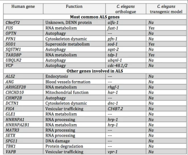
Table 1.1 ALS genes. Genes linked to ALS, their C. elegans orthologues and a summary of the transgenic C. elegans models published. The importance of the gene is based on 23,24
Sequencing and resequencing of large ALS cohorts have led to the identification of many individuals with more than one disease causative mutation (reviewed by Lattante et
al 25). These mutated genes might reflect low penetrance genes, or genes that cannot induce ALS alone. However, it cannot be excluded that those genes interact to vary disease onset, progression and symptoms.
New genetic concepts are emerging to explain some sALS cases. One of the main hypotheses explaining sALS cases was the impact of the environment in the development of the disease 26 . Groups have studied the impact of smoking, exercise and exposure to heavy metals and pesticides in different ALS cohorts (reviewed by Al-Chalabi & Hardiman 26). However, epidemiological studies like these can be challenging and few are reliable, most of the time because of the lack resources available to do the study 26. Therefore, no specific environmental factors have been found thus far to clearly explain sALS cases.
With the rapid advancement of sequencing technologies, new genetic concepts have emerged to explain sALS. Amongst these is the concept of de novo mutations which was previously shown to be a key player for many neurodevelopmental and psychiatric diseases 27,28. De novo mutations arise during the fertilization of germline and results in mutations found in the offspring that were absent in the parents, therefore causing sALS with a genetic cause. Mutations in a few known ALS genes were found in some sALS patients 29,30. FUS is the gene that is the most affected by de novo mutations so far. Many novel and previously identified mutations affecting the coding region and splicing of FUS were linked to early onset sALS 31-34, hence suggesting that de novo mutations can have a role in sALS cases.
Many pathological pathways were speculated to play a role in the pathogenesis of ALS. At the functional level, many of the genes linked to ALS share common cellular roles. RNA processing, mitochondrial dysfunction, stress response and protein degradation have emerged to be important pathological concepts. However, for most of the genes, the exact cellular function and the impact of the mutant proteins still remain to be established. It is still necessary to elucidate how these mutant genes can be specifically detrimental to motor neurons and how disease progression can vary among individuals with the same mutation.
PATHOLOGICAL CHARACTERISTICS OF ALS
Aside from the motor neurons, which cells contribute to ALS pathogenesis is still under evaluation. TDP-43 inclusions are observed in glia and neurons of the motor cortex, but also in the brainstem, the spinal cord and in white matter 35. Based on the pathological observation that protein inclusions are also present in glial cells, different groups have speculated that these non-neuronal cells may participate in the death of the motor neurons. Using SOD1 mouse models, it was shown that onset of symptoms require expression of mutant protein in the motor neurons but the mutant protein also had to be expressed in non-neuronal cells such as astrocytes and microglia to affect to disease progression 36,37. Therefore indicating that dysfunction of different cell types might be involved in ALS pathogenesis. However, thus far these types of in vivo observations were only made for
opposite results 38,39 therefore, more studies are required to examine the contribution of non-neuronal cells in ALS pathogenesis.
Protein aggregates are often observed in neurodegenerative disorders and it is also the case for ALS. TDP-43 is the main constituent of the aggregates observed in motor neurons of fALS and sALS 16. Protein aggregates containing FUS, p62, SOD1, UBQLN2 and
C9orf72 dipeptides repeat proteins are also observed in post-mortem, brain and spinal cord
tissues of different subset of ALS patients 7,40. Protein aggregates are observed in motor neurons of the motor cortex, brainstem and spinal cord as well as in glial cells in those regions 35. It is interesting to note that some aggregates are mutually exclusive suggesting independent toxic pathways in motor neurons7. However, it is still unclear if these aggregates are toxic or protective. Many proteins that were found in aggregates as well as the proteins encoded by some ALS genes were shown to participate to protein homoeostasis and could therefore affect the formation and/or clearance of these aggregates 41,42. Abnormal protein homoeostasis leading to the presence of aggregates or inclusions is a hallmark of many neurodegenerative disorders so it is difficult to know if these are specific to ALS or are a general feature of age-related neuronal death.
SIMILARITIES BETWEEN ALS AND OTHER NEURODEGENERATIVE DISORDERS
FRONTOTEMPORAL DEMENTIA (FTD)
FTD is a group of non-Alzheimer dementia characterized by atrophy of frontal and/or temporal lobes leading to behavioural changes or language decline 43. It is characterized by
pathological protein inclusions in which either TDP-43, TAU or FUS proteins are found in the affected regions of the brain 43. Most cases are sporadic but 10-20% of cases have a genetic component. Mutations in MAPT (microtubule-associated protein tau), GRN (Granulin) or C9orf72 are linked to FTD and are the most common genetic causes of FTD 43.
For many years, it was speculated that ALS solely causes motor dysfunction, however, it is now established that subsets of ALS patients also exhibit cognitive deficits. Patients’ cognitive dysfunctions range from mild cognitive deficits to dementia fitting the criteria of FTD. 50% of ALS patients have been shown to develop cognitive deficits 44,45 and 15-20% were shown to fit the criteria for FTD. Also, up to 27% of FTD patients exhibit motor dysfunction 46. Therefore, it is now accepted that ALS and FTD are along the same pathogenic continuum where patients range from pure motor neuron symptoms to pure dementia with many individuals found to have a mixture of both. Mutations in C9orf72,
TARDBP, and UBQLN2 were found to be causative of ALS and FTD 17,18,47-49. It is still unclear how in a single family, the same genetic variant can cause either ALS and FTD (for an example of C9orf72 50). Therefore, much remains to be investigated about the similarities and differences leading to ALS or FTD pathogenesis.
POLYGLUTAMINE DISORDERS
ALS proteins are also found in aggregates of other neurodegenerative disorders 51-53. An example is the presence of TDP-43 and FUS proteins in aggregates of polyglutamine disorders. Those proteins have been shown to co-localize to the polyglutamine aggregates in Huntington’s Disease in cells and tissue mouse model, and in post-mortem tissues of
patients affected by Machado-Joseph Disease 54,55. FUS and TDP-43 were also shown to interact with ATXN-2 (encoded by ATXN2(Ataxin-2) gene) 56,57 a protein that contains a polyglutamine tract that has been shown to cause Spinocerebellar ataxia type 2 (SCA2) when the polyglutamine repeat is longer than 34 units 58. Intermediate CAG repeat (i.e. repeat length around 28 units) found in ATXN2, have been shown to be a modifier of pathogenesis in TDP-43 model organisms and sALS patients 56 , highlighting the special relationship between ALS and CAG repeat disorders.
COMMON PATHWAYS IN NEURODEGENERATIVE DISEASES
Neurodegenerative disorders share many common pathological pathways. Impaired protein degradation, endoplasmic reticulum (ER) stress and neuroinflammation are some examples.
Many neurodegenerative diseases are characterized by the composition of the protein aggregates that are observed in tissues of affected individuals. Alzheimer’s disease is characterized by the presence of amyloid plaques and tau filaments 59, Parkinson’s disease is characterized by the presence of Lewy bodies, which are composed mainly of alpha-synuclein protein 60 and most polyglutamine diseases are characterized by inclusions containing the expanded polyglutamine tract61. As mentioned above (section Pathological
characteristics of ALS), impaired protein homoeostasis is a key feature of ALS pathogenesis.
Therefore, abnormal protein synthesis or degradation leading to the formation of cytoplasmic or nuclear protein aggregates is a recurrent theme in neurodegeneration. However, the role that these aggregates play in pathogenesis is still unclear.
Also, ER stress has been a recurrent theme regarding ALS pathogenesis in many models as well as in patients 62 . In addition, neuroinflammation, was observed in patient post-mortem tissues and has been shown to alleviate phenotypes observed in different model organisms 63. Similar toxic mechanisms were also implicated in Parkinson’s disease and Alzheimer’s disease 64-67. These data suggest that neurodegenerative disorders share many common toxic pathways and insight into a specific disease could be gained by comparing them.
C9
ORF72
GENETICS
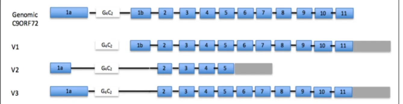
Many linkage studies using families affected by ALS and/or FTD from different areas of the world identified a region on chromosome 9p as a common genetic cause of ALS 68-70. In 2011, two independent groups simultaneously identified a causative ALS gene on chromosome 9p as being the non-coding repeat expansion GGGGCC found in the first intron of the C9orf72 gene. C9orf72 generates three alternatively spliced transcripts; two of which (V1 and V3) produce the long protein form of C9orf72 (481 a.a) and the other transcript (V2) produces a shorter form (222 a.a.) (Figure 1.2). The GGGGCC repeat is located in the promoter region of the V1 isoform and the first intron of the two others (Figure 1.2). In the first reports, the size of the repeat was suggested to be less than 10 units in healthy individuals. Its size in affected individuals appeared to be at least 30 units; while it was already clear that the number of repeats could reach hundreds and even thousands of units in patients 47,48. More recent reports have since then identified some ALS and/or FTD cases with repeat size as small as 22 units 71,72. Nonetheless several studies have now
Figure 1.2: C9orf72 gene and isoform structure. V1 refers to transcript NM_145005, V2 refers to transcript NM_018352 and V3 refers to transcript NM_1256054
reported healthy individuals with 20-30 repeats 71-73 so the minimal repeat length that can trigger ALS/FTD still remains to be precisely established.
Following these first reports, many others have followed and identified the GGGGCC expansion as being a major cause of ALS and/or FTD in individuals from European countries (Finland74, France75, United Kingdom, Ireland76, Spain77, Italy78, Holland79, Grece80, Portugal81, Belgium and Poland), the Americas (USA82, Canada50, Brezil83) and Australia 84. Most individuals are heterozygous for the expansion, carrying an expanded and a wild-type allele, but a few cases were identified to be homozygous 85,86. In one homozygous case, disease severity was stronger than what is typical of FTD cases 86 but in the other one, the disease progression and severity of ALS/FTD symptoms were similar to typical ALS/FTD cases 85. Interestingly, with the exception of the Kii peninsula of Japan 87, most studies done in Asian populations have shown that C9orf72 is not a major genetic cause of ALS in this area of the world (Korea 88,89, China 90,91, Japan 92-94, Taiwan 95).
Even though the ALS/FTD symptoms have a variable age of onset, several groups reported almost complete penetrance of the expansion by the age of 80 96,97. However, a few individuals were reported with an expanded GGGGCC repeat and did not develop neurodegenerative symptoms after this age 92,98 .
CHARACTERISATION OF REPEAT LENGHT
Since the expansion in C9orf72 is a pure G/C repeat, its quantification has been challenging. Repeat primed PCR can accurately only detect repeats smaller than 60 units and Southern
blot analysis requires large amounts of DNA so it has not been carried out for most individuals. Also, when using the same DNA samples, groups using repeat primed PCR to quantify the repeat length often misquantified it, suggesting that interpretation of repeat size should be done carefully, if not exclusively done by Southern blotting 99.
Anticipation is a concept by which the number of repeats can expand in size from one generation to the next and correlates with an increase in disease severity and an earlier age of onset. This phenomenon is observed in most coding and non-coding repeat disorders 100. Some researchers have speculated a link between age of onset or survival and repeat size in C9orf7296,101. Also, one group reported major anticipation in C9orf72 patients 102, with disease affecting children 7 years younger than the age at which their parents became affected. Nonetheless, other independent teams could not replicate these findings and the importance of anticipation in C9orf72 toxicity remains a matter of debate 47,48,74,103,104 . The small sample sizes might be the reasons for these conflicting observations.
Most studies that evaluated the repeat length have used genomic DNA prepared from peripheral blood. However, in repeat disorders it is often observed that the repeat length varies across different tissues 105. Many reports showed similar results for the GGGGCC expansion of C9orf72 and neuronal cells seem to exhibit an increase in repeat length when compared to non-neuronal cells of the same individual 104,106,107 . Therefore suggesting that the repeat length expressed in neurons is probably underestimated.
OTHER MUTATIONS
Aside from the GGGGCC repeat found in the first intron of C9orf72, different groups have tried to identify mutations outside this region in ALS and FTD patients. Different groups have confirmed that most expansion carriers share a common haplotype that includes 10 nucleotides surrounding the repeat 47,92,94,108.
Outside the expansion region, a 10 bp deletion has also been identified in the first intron in a few FTD patients 109 and some missense variations were found in the coding region of C9orf72 in sALS 110. However, the pathogenic contribution of these variants is still unclear.
C9ORF72 EXPRESSION
EXPRESSION OF C9ORF72 IN MICE
The expression of C9orf72 was studied in mice by using a Lac-Z insertion in the mouse
C9orf72 sequence 111 and by in situ hybridization 112. In adult animals, expression was found in the brain, spinal cord, spleen, kidney and testes 112, whereas no expression was observed in muscle, heart, lungs and liver 111. Expression of mouse C9orf72 during development was reported in neurons and different organs during embryonic stages 112. mRNA and protein expression of the different isoforms was shown to change during development 113 . For example, isoform 1 is the most expressed at postnatal day 1 and is found mainly in the nucleus. Isoform 2, however, is expressed mainly in the cytoplasm at postnatal day 1, but its expression increases in the nucleus, during development, to reach
its maximum at postnatal day 56 113. In primary mouse culture, cortical neurons show expression of C9orf72 in neurites and growth cones 113 . Thus, experiments in mice show that C9orf72 is expressed during development and adulthood in the nervous system and suggest that the different isoforms could have different roles during development.
EXPRESSION OF C9ORF72 IN HUMANS
In humans, expression of C9orf72 was studied with in situ hybridization probes and showed expression in neuronal cells of the spinal cord 111. Using qRT-PCR, expression of the C9orf72 isoforms 1 and 2 was shown in cervical spinal cord, cerebellum and motor cortex in post-mortem tissues 114. In the central nervous system, the highest expression of
C9orf72 occurs in the cerebellum and the lowest in the putamen 48. Expression in cultured fibroblasts is low and seems to increase when the cells are used for the differentiation of iPSC-neurons 114,115, suggesting an important tissue specific expression of C9orf72.
Few antibodies are commercially available to clearly detect C9ORF72 protein, however, one study has confirmed C9ORF72 protein expression in frontal and cerebellar cortex 116. Novel antibodies were recently generated to detect the long and short C9ORF72 isoforms and confirmed the expression of both isoforms in motor, temporal and frontal cortexes as well as cerebellum and lumbar spinal cord 117.
The exact cellular expression and localization of C9orf72 in humans still need to be elucidated, but preliminary results suggest localization at the nuclear membrane for the short isoform, and in mischaracterized cytoplasmic puncta for the long isoform 117. It is
interesting to mention that in Western blotting, the isoforms were found in different solubility fractions, where the long isoform seems to be more insoluble than the short one 117, suggesting that the cytoplasmic puncta might be insoluble and less dynamic.
In conclusion, expression analysis of C9orf72 in mice and humans has confirmed the importance of this protein in the nervous system during development and adulthood. Furthermore, even though these results remain to be confirmed, expression analysis suggests a role for C9ORF72 protein in the cytoplasm and at the nuclear membrane.
FUNCTION OF C9ORF72
Very little was known about the regular function of C9orf72 when it was first linked to ALS. Bioinformatic analysis showed that the full sequence of C9ORF72 protein shares many characteristics with DENN proteins (Differentially Expressed in Normal and Neoplastic cells) 118,119. DENN proteins are highly conserved guanine nucleotide exchange factor (GEF) proteins involved in endocytosis and intracellular trafficking 120. In motor neurons, C9ORF72 protein was shown to colocalize with some RAB proteins and regulates autophagy, partially confirming that C9ORF72 can act as a DENN protein 121. Recently, different in vivo models suggested that the GGGGCC expansion can affect RNA export from the nucleus and different modifier screens conducted in model organisms have shown that nuclear import and export proteins could modify this toxicity 122-124. These data are also supported by the fact that the short protein isoform of C9ORF72 was shown to be located at the nuclear membrane 117, suggesting a function at C9ORF72 at the nuclear
membrane. However, a recent study also suggested that the presence of cytoplasmic aggregates alone could affect nucleo-cytoplasmic shuttling independently of the function of the proteins found in the aggregates 125. Therefore, knowing that C9orf72 positive patients exhibit many protein aggregates, the toxicity caused by the expanded GGGGCC and the function of C9ORF72 protein at the nuclear membrane still remains to be confirmed.
Additionally, the pathogenic GGGGCC repeat expansion was shown to affect the formation of stress granules 121,126,127. Even though other ALS proteins, such as FUS and TDP-43, were also shown to participate in stress granule formation (reviewed by Ling et al 128), little is known about this function of C9orf72 in regards to ALS pathogenicity.
The functions of C9orf72 in normal and disease states remain to be elucidated in different cell types. However, it seems to affect RNA metabolism and endosomal trafficking, two important pathways that were previously shown to be involved in ALS pathogenesis.
CONSERVATION OF C9ORF72 ACROSS SPECIES
Model organisms are important tools to learn about the function of new proteins. Therefore, conservation of C9orf72 in different species was examined. The GGGGCC repeat found in the first intron of the gene has only been found in primates and it is not conserved in mice or lower model organisms. However, the gene is highly conserved (over 90% identity) in chimpanzees and marmosets 111. In lower organisms, most amino acids are conserved in mouse, rat, chick embryo, zebrafish with between 66%-98% identity in these
organisms 111. Most conserved residues are distributed across the protein suggesting that its function is conserved across the species 118.
R
EPEAT DISORDERSALS is not the only disease that can be caused by a nucleotide repeat expansions. Nucleotide repeats represent 30% of the human genome and vary in length and frequency 129. Many repeats were shown to cause neuro-developmental or neurodegenerative disorders when the repeat exceeds a certain threshold 130. Repeats can be found in the coding or non-coding regions, introns or UTRs, of the affected genes and cause either a toxic loss and/or gain of function mechanism. Here are a few examples of well-studied repeat disorders and how those repeats can induce cellular toxicity.
MYOTONIC DYSTROPHY AND THE TOXICITY OF RNA FOCI
Myotonic dystrophy type 1 (DM1) is a common repeat disorder and the most common muscular dystrophy. It is clinically similar to myotonic dystrophy type 2. The onset of symptoms can be observed during birth, childhood or at adulthood leading to a wide variety of symptoms including mental retardation, muscle degeneration, heart defects and cataracts 131. It is caused by a CTG repeat in the 3’ UTR of DMPK1 (Dystrophia myotonica protein kinase) gene 132-134. Healthy individuals have between 5-35 units of the CTG repeat, whereas patients have at least 50 units. Repeat can be as long as thousands of units and disease severity correlates with repeat length. Most of the research concerning the toxicity of RNA foci arises from studying DM1. The transcript containing the expanded repeat causing DM1 accumulates in the nucleus of muscle cells. Interestingly, it was shown that those RNA foci sequester muscleblind-like (MBLN), a protein that binds to CUG repeats 135, leading to its loss of function. Knock-out
mice of Mbln recapitulates features of DM1 including myotonia, heart defects and abnormal splicing 135 suggesting that toxicity observed in DM1 reflects the loss of function of Mbln in muscle cells. Therefore, RNA foci can be toxic to cells by sequestrating proteins. POLYGLUTAMINE DISEASES AND ABNORMAL TRANSLATION One of the most common toxic nucleotide repeat is the CAG expansion. CAG codon encodes for glutamine and coding and non-coding CAG pathogenic expansions were found in a dozen genes causing a wide variety of symptoms 136. While the genetic causes underlying these diseases are known, the toxic mechanisms involved are unclear and no treatment is available. Aside from the CAG repeat encoding gene, genes encoding a CTG repeat were found to be transcribed in the anti-sense direction leading to the production of toxic transcript containing CAG and CUG codons 137 therefore increasing the spectrum of disorders caused by CAG repeats. The presence of a long C/G rich repeat in a transcript can lead to the formation of an abnormal secondary structure 138. Knowing that translation is highly sensitive to RNA secondary structure, researchers have studied the impact of expanded toxic repeat on translation.
The gene ATXN3 (Ataxin-3) contains a coding CAG repeat in its 3’ end . When the repeat reaches 55 units, it causes Machado-Joseph disease, also called Spinocerebellar ataxia type 3 (SCA3), a neurodegenerative disorder causing the loss of Purkinje cells in the cerebellum 139. The CAG repeat causes the production of toxic long polyglutamine tracts. However, when the polyglutamine tract is encoded by CAA codons, which also leads to the production of polyglutamine, no toxicity is observed140-142. Therefore, it was hypothesized
that a change in reading frame, called ribosomal frameshifting, occurred along the CAG tract during translation leading to the production of polyalanine instead of polyglutamine. This phenomenon was shown in cell models and model organisms 141,142. Ribosomal frameshifting is widely characterized in viruses that use this method to increase the efficiency of their genomes by encoding more than one protein from a single RNA transcript 143.
Another method used by viruses to increase the efficiency of their genome is called internal ribosomal entry site (IRES)144. It was shown that the secondary structure of the RNA that contains a CAG repeat can attract the cellular translation machinery and initiate translation in a non-ATG manner, a process called repeat-associated non-ATG translation (RAN translation). Even though RAN translation does not act exactly like an IRES, it was speculated that the structure formed by the repeat can act in a similar manner145. Interestingly, the phenomenon was shown to happen along the ATXN8 (Ataxin-8) and HTT (Huntingtin) transcripts, causing Spinocerebellar ataxia type 8 and Huntington’s disease, respectively, when the CAG repeats that is encoded in the ATXN8 and huntingtin genes are above a certain threshold 146,147. When RAN translation occurs, expression of polyglutamine is independent of the presence of the ATG starting codon upstream of the repeat 148,149. Since no ATG is used, translation can occur in all reading frames along the sense and anti-sense transcripts, leading to the production of numerous polypeptides from a single CAG repeat containing transcript. This phenomenon was also observed along the CGG non-coding repeat of FMR1(Fragile X mental retardation 1) gene, the genetic cause of Fragile-X-associated tremor ataxia, leading to the production of polyglycine and polyalanine peptides 150.
FRAGILE X SYNDROME AND TOXICITY OF A DECREASE OF GENE EXPRESSION
The CGC repeat found in 5’ UTR of FMR1 is an example of how a repeat expansion can affect its own gene expression. In healthy individuals, the repeat length varies between 6-55 units. When the expansion is longer than 200 units, its causes a neurodevelopmental disorder called Fragile-X syndrome 151. The disease is characterised by mental retardation, as well as behavioural and social problems similar to autism spectrum disorders 136. FMRP, the protein encoded by FMR1, is an RNA binding protein that shuttles between the nucleus and the cytoplasm and directly binds to mRNA. In Fragile-X syndrome patients, a hypermethylation of the CpG islands located in the 5’ UTR has been observed causing a loss of FMR1 expression 151. Interestingly, a knock-out mouse model of FMR1 exhibits many features similar to patient symptoms including hyperactivity, anxiety behaviours and cognitive deficits 136. Therefore, these data suggest that a decreased expression of FMR1 is sufficient to induce the phenotypes observed in Fragile-X syndrome.
Interestingly, if the CGC repeat length is intermediate (i.e. between 70-200 units), expression of the FMR1 RNA is elevated and was shown to cause a neurodegenerative disease called Fragile-X tremor/ataxia syndrome. This disease is characterised by loss of Purkinje cells and atrophy of the cerebellum. Intranuclear aggregates containing proteins and the FMR1 mRNA are observed in patients post-mortem tissues and are speculated to cause toxicity by sequestrating other RNA binding proteins, but the toxicity of these RNA foci is not as clear as for DM1152.
C.
ELEGANS CURRENT MODELS TO UNDERSTAND ALS With more than 20 genes now linked to ALS, the consequences of the mutant proteins need to be evaluated. Since the identification of SOD1 in 1993, many mouse models have been established to understand the function of the mutant proteins in normal and disease states. Transgenic mouse models expressing mutant SOD1 were able to successfully model the ALS progression and the motor neuron phenotypes observed in patients (among others 153-155).However, SOD1 mutations are found in only a small fraction of ALS patients. Even though some models seem promising to understand different aspects of ALS pathogenesis, transgenic animals expressing either TDP-43 and FUS proteins cannot recapitulate the involvement of these proteins in motor neuron integrity as well as the SOD1 models. By itself, expression of wild-type TDP-43 in mouse causes neuronal loss, decreased survival and pathological characteristics of ALS 156,157. When expressing mutant TDP-43 proteins, some models exhibit phenotypes related to ALS pathology, motor phenotypes, decreased survival and astrogliosis, but surprisingly, many models exhibited only minimal neuronal loss and TDP-43 aggregates 158-163 . Expression of wild-type and mutant FUS in mouse spinal cord can recapitulate pathological characteristics of ALS but, again, in neither cases was neuronal loss observed 162,164. Also, ALS mouse models have failed over the past years to correctly predict the efficacy of drugs that were tested in clinical trials, which led many researchers to speculate that alternative approaches should be developed to evaluate potential drugs 7,165-167In recent years, some groups have started using human derived induced pluripotent stem cell (iPSC) derived neurons to study ALS pathogenesis. Since these iPSC neurons are derived from patient cells, they represent the complete genetic background of the patients. In the case of C9orf72 where the repeat is difficult to manipulate genetically, for cloning for example, using the full expansion in the gene context would be ideal. However, iPSC-derived neurons are costly, time- consuming, and only a fraction of the neurons (20-30%) exhibit C9orf72 pathological characteristics (among others168,169).
The recent identification of many new genes, and the failure of many of the mammalian models to fully represent ALS pathogenesis, have driven the use of small animal models, including yeast, zebrafish, flies or worms. Many disease-related genes are highly conserved among species, plus model organisms can be easily genetically manipulated and have rapid reproduction cycles.
The worm Caenorhabditis elegans is a multicellular, transparent nematode that is used to study many areas of biology, including aging and stress pathways 170. Also, with its fully sequenced genome, and its cell lineage fully characterized, it is a highly studied and well characterized model organism. Therefore, C. elegans was chosen by our group to model and understand ALS.
LIFESPAN AND GENOME OF C. ELEGANS
C. elegans is a transparent, non-parasitic roundworm. In the wild, the worm is found in soil
population are male worms that can fertilize the hermaphrodite. In laboratory conditions, worms are grown on nematode growth media plate and are fed with E. coli 171.
Under normal conditions, the lifecycle of C. elegans includes four larval stages (L1-L4) and adulthood. When under harsh environmental conditions during development, such as high temperature, low food supply or high population density, the L1 worm switches to a dauer larval stage instead of the L2 larval stage172 . The dauer larvae have a distinct morphology and metabolism that allow the worm to survive in difficult conditions172. When conditions become favourable, the animal leaves the dauer stage and returns to its normal life cycle172 . When kept at 20°C, it takes 3-4 days for a wild-type worm to begin laying eggs and it will produce more than 200 during its lifespan. The lifecycle of C. elegans is temperature sensitive; kept at 15°C the worms develop slower and temperatures above 25°C can be harmful. At 20°C, a wild-type worm lives on average 20 days. Different signs of aging are visible in C. elegans including loss of motility, presence of necrotic cells, presence of oxidized protein, deterioration of different tissues and decline in immune function 173. The complete genome of C. elegans was sequenced in 1998 174. It consists of more than 19,000 genes distributed across six chromosomes, whereas 40% of these are found in higher organisms 175. Many genetic biochemical pathways are highly conserved between C.
elegans and human including different stress response pathways, the insulin-IGF,
apoptosis, necrosis and the innate immune response pathways176-179 .
C. ELEGANS NERVOUS SYSTEM
The C. elegans nervous system includes 302 neurons in hermaphrodite worms. Neurons are classified by their functions, their locations or by the neurotransmitter they express.
Ongoing studies aim to completely characterize the interactome of the C. elegans nervous system. C. elegans have four types of neurons; motor neurons, sensory neurons, interneurons and polymodal neurons 180.
Two types of motor neurons coordinate the movement of the worms; the gamma-aminobutyric acid (GABA) and the cholinergic neurons. Both types of neurons are located on the dorsal side and innervate muscle cells on the dorsal and ventral side of the worm. Cholinergic neurons are involved in locomotion, egg laying, feeding and male mating (reviewed by Rand 181 ). Acetylcholine is synthesized by choline acetyl transferase (CHA-1 protein), loaded in vesicles by vesicular transporter (UNC-17protein) and secreted at the synaptic cleft where it activates the acetylcholine receptors on the post-synaptic cells. Subsequently, it is hydrolyzed to be recycled by acetylcholine esterases (ACHE protein) and re-enter the pre-synaptic cell (CHO-1 protein).
GABAergic neurons are inhibitory neurons involved in locomotion and defecation (for a review see Jorgensen182). They are activated by cholinergic neurons. GABA is synthesized by glutamic acid decarboxylase (UNC-25 protein), loaded in vesicles by a vesicular transporter (UNC-47 protein) and will activate the inhibitory GABA receptor (UNC-49 protein) on the post-synaptic cells and causing relaxation of the muscle cells.
The tight coordination between the GABAergic and cholinergic neurons allows for the movement of the worm. Hence, when one side of the worm is contracting due to cholinergic activation, the opposite side is relaxing due to GABA inhibition, therefore causing this smooth sinusoidal movement along the animal body 182(Figure 1.3).
Deletion mutant worms of any of the genes involved in GABA or acetylcholine production or secretion, or pharmacological alteration of these pathways will cause abnormal locomotion of the animals. The mutant worms are often used as control when studying worm’s locomotion and the promoter of these genes are used to expressed proteins specifically in the motor neurons both of which are often used in ALS worm research.
STRESS RESPONSE PATHWAYS IN C. ELEGANS
Stress response is a major part of C. elegans research. Animals can be easily subjected to many different types of environmental stress and many genes involved have turned out to be involved in human diseases such as cancer, infantile diseases and neurodegenerative
Figure 1.3: C. elegans GABAergic motor neurons. Upper: The GABAergic neurons include 20 ventral cord motor neurons innervating either dorsal or ventral muscles, 4 ring motor neurons (green) innervating the head and 3 interneurons (pink, yellow and blue). Bottom: tight coordination between the GABAergic and cholinergic (Ach) motor neurons allow for contraction of one side of the body and relaxation of the opposite side of the animal causing the sinusoidal movement of the animal. Figure inspired by 182
diseases 183,184. The insulin and insulin-like growth factor signaling (IIS) pathway is among the most studied stress response pathways in C. elegans. It functions by activating a cell surface receptor, in C. elegans DAF-2 (abnormal DAuer Formation 1), that acts to induce a cascade of kinases, including AGE-1 (AGEing alteration1) and AKT-1(AKT kinase family 1), and promotes cell death by inhibiting different transcription factors such as DAF-16 (abnormal DAuer formation 16) and SKN-1(SKiNhead 1) (Figure 1.4). This pathway was shown to also play a role in aging, longevity, fat metabolism, and in different neuronal phenotypes176. Mutants that result in reduced IIS are long-lived, stress resistant , exhibit increased fat content and have a reduced neuronal decline.
The role of the IIS in aging and stress response is mainly regulated by the transcription factor DAF-16. In its unphosphorylated form, DAF-16 is activated and moves to the nucleus where it induces expression of genes associated with longevity and stress resistance. When the IIS is activated, DAF-16 becomes phosphorylated and is retained in the cytoplasm where it cannot affect gene expression 176.
IIS was shown to play an important role in aging of the nervous system. Aged neurons in C. elegans display increased branching and neuronal defects, both of which are delayed when the IIS is reduced. Also, DAF-16 is involved in proteostasis and chaperone expression which were also shown to affect neuronal defects. However, not all long-lived mutants acting through DAF-16 affect aging of the nervous system 173
Figure 1.4: The insulin-IGF signaling pathway. In black are the C. elegans genes and in blue their human orthologues
C. ELEGANS TOOL BOX
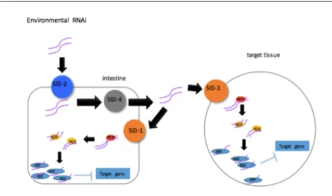
Different consortiums and groups have generated many mutant worm strains. Most of these strains are generated by mutagenesis, generating small deletions randomly in the genome. Ethyl methanesulfonate (EMS), and 4,5’,8-trimethylpsoralen (TMP) are mainly used for this and deletions or point mutations are generated185,186. After mutagenesis, the mutants are then screened for visible phenotypes, such as lethality, progeny numbers, developmental problems, and sequenced to identify the mutations generated. Using this method a large proportion of the genome has been mutagenized. The few genes for which mutations could not be generated are now being inactivated using the most recent genetic tools including the clustered regulatory-interspaced short palindromic repeats/CRISPR associated protein 9 (CRISPR/CAS9) method. Forward genetics is a valuable tool to identify genes that are involved in a specific function. However, it can be time consuming and the sequencing and identification of the proper mutation that is causing the observed phenotype are the major rate-limiting steps. Therefore, reverse genetic tools have also been developed in C. elegans. RNA interference (RNAi) is the process by which one can decrease the expression of a gene by targeting the degradation of its RNA through expressing a complement strand to the target RNA. Most organisms and cells express the machinery to cause gene silencing but it is particularly powerful in C. elegans 187. The worms express a systemic RNAi machinery that includes RNA-directed RNA polymerase that allows for the amplification of the RNA strand, also the RNA strand can be easily expressed in worms 187. For example, RNA can be expressed in the bacteria that the worms eat and will end up in their intestine. There, it will be transported to different cells across the organism. SID (systemic RNAi defective) proteins are important
for export and intake of the RNA. After being taken up by the cell, RNA is cleaved and processed using DCR (Dicer related) and RDE (RNAi defective) proteins. The RNA strand is then
amplified by the RRF (RNA-dependant RNA
polymerase family) family of proteins and binds to its target mRNA causing its degradation (Figure 1.5). Mutations affecting any of these genes make the worm partially or completely resistant to RNAi.
The RNAi process is highly efficient in almost all cell types with the exception of the nervous system. Neurons do not express the SID proteins, and therefore cannot uptake the RNA strand. However, genetic manipulations have allowed the expression of the SID proteins specifically in neurons resulting in animals sensitized to RNAi in the nervous system 188. Using similar transgenic and deletion mutant worms, many transgenic worms are available with tissue or cell specific RNAi sensitivity 188-190.
Finally, one of the first uses of green fluorescent protein (GFP) was in C. elegans 191. The worm is transparent, so expression of the GFP protein can be expressed in fusion with a known protein allowing for direct visualization of the protein at different stages of the
Figure 1.5: RNAi machinery in C. elegans. RNA strand in the intestine will be send to all cells of the animal and uptake by SID-1. Then, RNA will be process by DCER, RDF and RRF proteins and bind to its target RNA to degrade it
worm’s life cycle. Translational or transcriptional GFP reporters are also available for many genes to visualize the expression and localization of genes and proteins and the tissue and cellular levels.
The first transgenic worms were created by inserting transgenes in the genome using ultraviolet (UV) or gamma radiation192. This caused random integration of the transgene into the genome, often in multiple copies. This technique frequently resulted in copy-number variation and integration site effects rather than specifically looking at a phenotype caused by expression of a sole transgene. Now, new technologies are available and single insertion site methods 193 and CRISPR/Cas9 194 have led to the generation of new transgenic models with low expression levels of the transgene and targeting the endogenous genes in their genome contexts (promoter, regulatory regions, etc.). Hopefully, these new models will better recapitulate what is observed in non-transgenic conditions. ALS C. ELEGANS MODELS C. elegans have been previously used to model ALS (Table 1.1) 1,175. Transgenic models in which neuronal and non-neuronal expression of human SOD1, TDP-43 and FUS proteins in
C. elegans have been characterized (Table 1.1) 195-203. The mutant proteins induced an abnormal stress response and protein aggregates in many cases 195-198,200-202. Transgenic expression of mutant SOD1, TDP-43 and FUS proteins in neurons caused neuronal loss and motility 195,198,199,203.
Being the most studied ALS protein in C. elegans, TDP-43 is a good example of the variety of experiments that can be carried in worms to understand its role in neurodegeneration. Many models using various expression patterns of TDP-43 wild-type