Direction des bibliothèques
AVIS
Ce document a été numérisé par la Division de la gestion des documents et des archives de l’Université de Montréal.
L’auteur a autorisé l’Université de Montréal à reproduire et diffuser, en totalité ou en partie, par quelque moyen que ce soit et sur quelque support que ce soit, et exclusivement à des fins non lucratives d’enseignement et de recherche, des copies de ce mémoire ou de cette thèse.
L’auteur et les coauteurs le cas échéant conservent la propriété du droit d’auteur et des droits moraux qui protègent ce document. Ni la thèse ou le mémoire, ni des extraits substantiels de ce document, ne doivent être imprimés ou autrement reproduits sans l’autorisation de l’auteur.
Afin de se conformer à la Loi canadienne sur la protection des renseignements personnels, quelques formulaires secondaires, coordonnées ou signatures intégrées au texte ont pu être enlevés de ce document. Bien que cela ait pu affecter la pagination, il n’y a aucun contenu manquant.
NOTICE
This document was digitized by the Records Management & Archives Division of Université de Montréal.
The author of this thesis or dissertation has granted a nonexclusive license allowing Université de Montréal to reproduce and publish the document, in part or in whole, and in any format, solely for noncommercial educational and research purposes.
The author and co-authors if applicable retain copyright ownership and moral rights in this document. Neither the whole thesis or dissertation, nor substantial extracts from it, may be printed or otherwise reproduced without the author’s permission.
In compliance with the Canadian Privacy Act some supporting forms, contact information or signatures may have been removed from the document. While this may affect the document page count, it does not represent any loss of content from the document.
UNIVERSITÉ DE MONTRÉAL
Effect of the unfolded protein response on
MHC class 1 antigen presentation
par
Diana Paola Granados
Programme de biologie moléculaire Faculté de médecine
Mémoire présenté à la Faculté de médecine en vue de l'obtention du grade de
Maître ès Science (M.Sc.) en biologie moléculaire
April, 2008
UNIVERSITÉ DE MONTRÉAL Faculté de médecine
Ce mémoire intitulé:
Effeet of the unfolded protein response on MDC class 1 antigen presentation
Par:
Diana Paola Granados
a été évalué par un jury composé des personnes suivantes:
président-rapporteur
directeur de recherche
RÉSUMÉ
L'infection virale et la transfonnation néoplasique engendrent du stress dans le réticulum endoplasmique (RE). En conséquence, une grande proportion de cellules qui devraient être reconnues par le système immunitaire, sont des cellules stressées. Lors d'un stress du RE, les cellules déclenchent une réponse envers les protéines mal repliées (UPR). L'UPR régule les deux processus clés qui contrôlent la présentation antigénique par les molécules du complexe majeur d'histocompatibilité de classe 1 (CMH 1) : la synthèse et la dégradation des protéines. Nous avons voulu détenniner si l'UPR affecte la présentation antigénique et comment. Tout d'abord, l'impact de l'UPR sur l'expression globale du CMH 1 ainsi que sur la présentation du peptide SIINFEKL dérivant de la protéine ovalbumine a été évalué. Des cellules EL4 transfectées de façon stable avec des vecteurs codant pour des variantes de la protéine HEL-SIINFEKL ont été stressées à l'aide d'agents phannacologiques ou encore soumis à une carence en glucose. Nos résultats indiquent que l'UPR diminue l'expression du CMH 1 à la surface des cellules, mais n'a pas d'effet au niveau de l'ARN messager. Conséquemment, la présentation de SIINFEKL par les molécules H2Kb était
diminuée dans les cellules stressées, autant de façon chimique que physiologique. De plus, les cellules stressées présentaient préférentiellement des complexes CMH I-peptide provenant de la variante protéique qui se localise dans le RE par rapport à ceux dérivant de la protéine cytosolique. Par ailleurs, suite à une élution des peptides avec de l'acide, la génération de peptides provenant de la protéine localisée dans le RE était moins affectée que dans le cas de la protéine cytosoliqué. Nos résultats démontrent que le stress du RE altère la présentation de complexes CMH I-peptide et qu'il régule de façon différentielle l'expression de peptides dérivant du RE ou du cytosol. Ainsi, notre recherche montre que le stress du RE, qui est une caractéristique des cellules infectées ou transfonnées, peut affecter les signaux nécessaires pour la reconnaissance par le système immunitaire.
Mots clés: CMH de classe l, présentation antigénique, antigène/peptide, stress du RE, réponse envers les protéines mal repliées
SUMMARY
Viral infection and neoplastic transformation trigger endoplasmic reticulum (ER) stress. Thus, a large proportion of the cells that must be recognized by the immune system are stressed cells. Cells respond to ER stress by launching the unfolded prote in response (UPR). The UPR regulates the two key processes that control major histocompatibility complex class l (MHC I)-peptide presentation: protein translation and degradation. We therefore asked whether and how the UPR impinges on MHC I-peptide presentation. We evaluated the impact of the UPR on global MHC l expression and on presentation ofthe Ovalbumin-derived SIINFEKL peptide. EL4 cells stably transfected with vectors coding HEL-SHNFEKL protein variants were stressed with pharmacological agents or exposed to glucose deprivation. UPR decreased surface expression ofMHC l at the protein but not the mRNA level. Consequently, presentation ofSIINFEKL by H2-Kb
molecules was reduced in chemically or physiologically stressed cells. Notably, stressed cells preferentially presented MHC I-peptides derived from an ER-retained as opposed to a cytosol-Iocalized protein variant. Furthermore, generation of new H2Kb-SIINFEKL complexes after acid strip was less affected for ER- than for cytosol-derived SIINFEKL. Our results show that ER stress impairs MHC I-peptide presentation, and that it differentially regulates expression of ER- vs. cytosol-derived peptides. This work indicates how ER stress, a typical feature of infected and malignant cells, can impinge on cues for adaptive immune recognition.
Keywords: MHC c1ass I, antigen presentation, peptide/antigen, ER stress, unfolded protein response
TABLE OF CONTENT
RÉSUMÉ ...111
SUMMARY ... IV LIST OF ABBREVIATIONS ... IX ACKNOWLEDGEMENTS ...Xl
1. INTRODUCTION ... 12 1.1. General introduction ... 121.2. MHC I-antigen processing and presentation pathway ... 12
1.2.1. Bringing the inside out: the immunopeptidome ... 13
1.2.2. Peptide processing in the cytoplasm ... 14
1.2.3. Generation of peptide-receptive MHC 1 molecules ... 15
1.2.4. Peptide processing in the ER ... 16
1.2.5. Peptide loading and presentation ... 18
1.3. The origin of peptides for display by MHC 1 molecules ... 19
1.3.1. Exogenous proteins as source of peptides ... 19
1.3.2. Endogenous proteins as source of peptides ... 20
1.3.2.1. Peptides derived from stable proteins ... 21
1.3.2.2. Rapidly versus slowly degraded polypeptides ... 21
1.3.2.3.Driving force for peptide generation: the DRiPs hypothesis ... 22
1.3.2.4. Cryptic translation as a source of naturally processed peptides ... 25
1.3.2.5. Peptides derived from peptide splicing ... 25
1.3.2.6. Peptides derived from proteins destined to the secretory pathway ... 26
1.4. Translation and Protein Folding ... 26
1.4.1. mRNA partitioning and translation in the ER ... 27
1.4.2. Protein folding in the ER ... 29
1.4.2.1. N-glycosylation and the CNXlCRT cycle ... 29
1.4.2.2. The BiP/GRP78 pathway ... 31
1.4.2.3. Oxidative folding ... 31
1.5. Protein quality control in the secretory compartment ... 31
1.5.1. ERAD mechanism ... 31
1.5.1.1 Substrate recognition ... 33
1.5.1.2. Transport across the ER membrane ... 34
1.5.1.3. Release in the cytosol and degradation ... 34
1.5.2. Autophagy ... 35
1.6. ER stress and the Unfolded protein response ... 35
1.6.1. How cells cope with ER stress ... 35
1.6.2. The UPR acts through three main signal transduction pathways ... 36
1.6.2.2. IRE1 activates the transcriptional arm of the UPR ... 37
1.6.2.3. Activation of the transcription al arm of the UPR by ATF6 ... 38
1.6.2.4. Other effects of the UPR ... 38
1.6.3. The UPR in disease and normal physiology ... 39
2. RESEARCH PROJECT ... 41
2.1. Rationale ... 41
2.2. Research question and hypothesis ... 41
2.3. Objectives ... 41
2.3.1. General objective ... 41
2.3.2. Specifie objectives ... ~ ... 42
3. METHODOLOGY ... 43
3.1. Model for in vitro quantification of MHC I-peptide complexes ... 43
3.1.1. DNA constructs ... 43
3.1.2. LocaUzation of HEL protein variants ... 43
3.1.3. Assessment of Kb-SIINFEKL surface expression ... 43
3.2. Engineering of Kb-SIINFEKL stable transfectant cellUnes ... .44
3.2.1. Stable transfectants ... 44
3.3. Assessment of the effect of UPR on MHC I-peptide presentation ... .44
3.3.1. Stress treatment ... 45
3.3.2. UPR induction ... 45
3.3.3. MUC 1 expression ... 45
3.3.4. Measurement of existent and newly generated Kb-SIINFEKL complexes . .45 4. MANUSCRIPT ... 46
4.1. Déclaration des coauteurs de l'article ... 46
4.2. Déclaration de l'étudiant concernant l'article ... 47
4.3. ER stress affects processing ofMHC class I-associated peptides ... 48
Abstract ... 49
Background ... 50
Results ... 52
Discussion ... 57
Conclusions ... 60
Materials and Methods ... 61
Abb reviations ... 64
Authors' contributions ... 64
Acknowledgements ... 64
References ... 65
5. SUPPLEMENTARY RESLILTS ... 77
5.1.1. DNA constructs and localization of HEL protein variants ... 77
5.1.2. Presentation of SIINFEKL derived from HEL protein variants ... 78
5.1.3. Expression ofthe BiP/GRP78 protein as marker ofUPR activation ... 80
6. DISCUSSION ... 81
6.1. Activation of the UPR in different celilines ... 81
6.2. Activation of the UPR during glucose starvation ...•... 82
6.3. Why UPR activation impairs MHC 1 surface expression? ... 83
6.4. Why does UPR differentially affect expression of ER- versus cytosol-derived peptides? ... 85
6.5. What might be the impact of the UPR on immune recognition of infection and malignancy? ... 87
7. CONCLUSIONS AND FUTURE PROSPECTS ... 89
LIST OF FIGURES
IN"rRODUCTION ...
12
Figure 1. MUC I-antigen presentation pathway ... 14
Figure 2. Schematic depiction of the MHC 1 PLC. ... 16
Figure 3. The MHC 1 processing pathway ... 17
Figure 4. Possible sources of proteins presented on MHC 1 molecules ... 21
Figure 5. Intracellular polypeptide degradation pools ... 22
Figure 6. The DRiPs hypothesis ... 23
Figure 7. An alternative to the DRiPs model. ... 25
Figure 8. The SRP-ribosome cycle ... 28
Figure 9. The CNXlCRT cycle ... 30
Figure 10. Proteasomal degradation of ERAD targets ... 32
Figure Il. The UPR ... 36
MANUSCRIPT ... 46
Figure 1. UPR impairsMHC 1 surface expression but not mRNA level. ... 69
Figure 2. EL4 stable transfectants express the SIINFEKL peptide derived from HEL targeted to the ER or to the cytosol. ... 70
Figure 3. Induction of UPR decreases cell surface levels of H2Kb and H2Db in EL4 stable transfectants ... 71
Figure 4. Different peptide abundance in cells ex pressing ER- or cytosol-localized HEL du ring ER stress ... 72
Figure 5. Glucose starvation-induced ER stress impairs H2Kb and H2Db expression. pools ... 73
Figure. 6. Increased presentation of SIINFEKL derived from ER-localized relative to cytosolic proteins during glucose starvation ... 74
Figure 7. HEL expressioin during glucose starvation ... 75
Figure 8. ER stress impinges on the MHC 1 peptide repertoire ... 76
SUPPLEMENTARY RESULTS ... 77
Figure SI. Schematic representation ofthe HELconstructs and subcellular localization of the protein variants ... 77
Figure S2. Kb-SIINFEKL presentation in HeLa-Kb cells transfected with the constructs encoding HEL destined to different cellular comparments. .. ... 79
APC ATF4 ATF6 CHOP CNX CRT CTLs COS DAPI DC DMEM DRiPs DTT EDEM EGFP eIF EL4 ER ERAD ERAAP ERGIC-53 ERSE FACS GPI GRP78 HCMV HEK HEL HeLa HLA HMGR HSP IFN-y IRE 1 IRES
LIST OF ABBREVIAllONS
Antigen-presenting cellActivating transcription factor 4 Activating transcription factor 6
~2 -microglobulin
Heavy chain binding protein
CIEBP homologous protein Calnexin
Calreticulin Cytotoxic T ce Ils
Green African monkey fibroblast 4' ,6-diamidino-2-phenylindole Dendritic cell
Dulbecco's modified Eagle's medium Defective ribosomal products
Dithiotheritol
ER degradation-enhancing a-mannosidase-like protein Enhanced green fluorescent prote in
Eukaryotic initiation factor Mouse thymocytes
Endoplasmic reticulum
Endoplasmic reticulum-associated degradation
Endoplasmic reticulum aminopeptidase associated with antigen processing Endoplasmic reticulum-Golgi interrnediate compartment-53
Endoplasmic reticulum stress response element Fluorescence-activated cell sorting
Glycophosphatidylinositol 78 kDa glucose-regulated protein Human cytomegalovirus
Human embryonic kidney Hen egg lysozyme
Human cervix adenocarcinoma Human leucocyte antigen
3-hydroxy 3-methylglutaryl coenzyme A reductase Heat shock protein
Interferon gamma
Inositol-requiring enzyme-l InternaI ribosomal entry sites
mAb MFI MHC MHCI mTOR MTS NAC NK NLS ORF PCR PDI PERK PLC
RAC
RDP ROS RT-qPCR SDP SDS-PAGESRP
TAP TCR TPPII UGGT UPR UTR XBPI Monoclonal antibody Mean fluorescence intensity Major histocompatibility complex Major histocompatibility complex class I Mammalian target of rapamycinMitochondrial targeting sequence
Nascent polypeptide-associated complex Natural killer
Nuclear localization signal
Open reading frame
Polymerase chain reaction Protein disulfide isomerase PKR-related ER kinase Peptide-Ioading comlex
Ribosome-associated complex Rapidly degraded polypeptides Reactive oxygen species
Quantitative real-time reverse transcriptase polymerase chain reaction
Slowly degràded polypeptides
Sodium dodecyl sulphate-polyacrylamide gel electrophoresis Signal recognition particle
Transporter associated with antigen processing T cell antigen receptor
Tripeptidyl peptidase II
UDP-glucose glycoprotein glucosyltransferase Unfolded protein response
Untranslated region
X-box-binding prote in 1
ACKNOWLEDGEMENTS
None of this would have been possible without the generous help of many people. First,1 wi1llike to thank the founder of the "immunopeptidomeux" group, Claude Perreault, for opening the doors of his lab to me, for his wise guidance during my Masters studies and for being an excellent mentor.
Many thanks to Étienne Caron for teaching me different techniques, for reading my texts, for the valuable scientific discussions we had and for his patience in the first months when 1 was trying to understand not only the experimental techniques but also French. 1 also thank him for his contagious passion for the immunopeptidome and his encouragement. Thanks to the other "amateurs de l'immunopeptidome", Danielle de Verteuil and Marie-Pierre Hardy, for their encouragement and discussions. Special thanks to Marie-Marie-Pierre for her technical help and for reading my dissertation and offering valuable advice. 1 am also grateful to the other members of the lab for sharing with me their expertise and for their technical advice. Thanks also to past members, Vincent Rineau and Chantal Baron, who help me at the beginning ofmy project. Thanks to all ofthem for providing a pleasant working ambience.
1 would also like to thank Danièle Gagné, Christian Charbonneau and Pierre Chagnon from the core facilities for their excellent assistance in flow cytometry, microscopy and RT-qPCR, respectively.
Special thanks to Benjamin Turgeon for his love and his support in the good and difficult moments. 1 thank him for teaching me how to do western blots, for correcting my disseration and my texts in french, but specially for giving me advices on how to succeed and have fun during graduate studies.
Of course, 1 am very grateful to my family that ev en in the distance where always there, supporting me and encouraging me to make my dreams come true. Thanks to my father for teaching me how to analyze and work hard, to my mother for teaching me how to write and be organized, and to my sister for being an example ofhappiness and positivism.
Finally, 1 would like to thank the government of Canada for giving me the scholarship that allowed me to carry on graduate studies in Canada. 1 am also grateful to the Cole Foundation for their financial support during the past year.
1. INTRODUCTION
1.1. General introduction
A fundamental feature of infected and neoplastic cells is that they are stressed cells (Gleimer & Parham, 2003; Shin et al., 2003; Marciniak & Ron, 2006). Stressed cells express specific antigens that are recognized by T lymphocytes leading to eventual eIimination of tumor or infected cells (van der Bruggen & Van den Eynde, 2006; Hammer et al., 2007). The recognition of these antigens depends on the ability of cells to display their intracellular contents in the form of peptides bound to the major histocompatibility complex c1ass 1 (MHC 1) molecules on the surface (Gleimer & Parham, 2003; Shin et al., 2003; Marciniak & Ron, 2006). Generating this complex repertoire of peptides and loading them on the MHC 1 molecules for export to the cell surface, is accompli shed by the antigen processing and presentation pathway.
Peptides suitable for loading the MHC 1 molecules, as well as peptide-receptive MHC 1 molecules are generated in the endoplasmic reticulum (ER) (Hammer & Shastri, 2007). The ER stands at the crossroad of another fundamental process: maturation and folding of proteins destined to the secretory pathway. Eventually, the folding of proteins can be perturbed by viral infection and neoplastic transformation, which trigger ER stress (Marciniak & Ron, 2006). Stressed cells respond by launching the unfolded protein response (UPR), an adaptive response that allows survival to limited stress but leads to apoptosis in the presence of overwhelming stress (Rutkowski et al., 2006; Szegezdi et al., 2006). In the following section 1 will review these fundamental processes that constitute the basis of my research work.
1.2. MHC I-antigen processing and presentation pathway
The end result ofthe antigen processing pathway is the display of peptide-bound MHC 1 molecules. These complexes are expressed on the cell surface, where they can be recognized by T cell antigen receptors (TCRs) of CD8+ T cells (Gleimer & Parham, 2003; Shin et al., 2003; Marciniak & Ron, 2006). These peptides derive from the degradation ofvirtually aIl proteins inside the cell (Shastri et al., 2002) and represent proteins that are being translated at any given time (Jensen, 2007). In a stepwise manner, the antigen processing pathway generates and protects the proteolytic intermediates until they yield the final peptides that can fit the MHC 1 in the ER. This leads to MHC I-peptide complexes that are ready to be exported and presented. In the following section, we will see how this highly specialized pathway, operating with essentially conserved components, is nevertheless capable of generating
highly diverse sets of peptides that satisfy a large number of different MHC 1 molecules.
1.2.1. Bringing the inside out: the immunopeptidome
The repertoire of peptides presented by MHC 1 molecules, estimated to be composed of hundreds of thousands, is known as the MHC 1 immunopeptidome (Shastri et al., 2002; Shastri et al., 2005). Under steady-state conditions (in the absence of infection), cell surface MHC 1 molecules are associated solely with self-peptides. These peptides, colIectively referred to as the self-MHC 1 immunopeptidome (Istrail et al., 2004), play vital roles. They shape the repertoire of developing thymocytes (Huseby et al., 2003; Starr et al., 2003; Baldwin & Hogquist, 2007), transmit survival signaIs to mature CD8+ T celIs (Marrack & Kappler, 2004) and influence mating preferences and other behaviors in mice (Slev et al., 2006). Thus, the self-MHC 1 immunopeptidome links the intracelIular milieu with the surrounding environment of almost aIl celI types (Shastri et al., 2002).
The self-MHC 1 immunopeptidome also helps amplify responses against intracelIular pathogens (Anikeeva et al., 2006) and allows immunosurveillance of neoplastic celIs (Dunn et al., 2004; Zitvogel et al., 2006). It reflects the state of the celI since mutated genes, genes involved in differentiation or genes overexpressed in tumors modify and shape the self-MHC 1 immunopeptidome (van der Bruggen & Van den Eynde, 2006). Moreover, viral proteins constitute a source of peptides that also molds the MHC I-peptide repertoire (YewdelI, 2007). Altogether, these modifications render the otherwise invisible internaI proteome available for surveillance by cytotoxic CD8+ T cells, which have the ability to detect and eliminate cells expressing viral proteins or tumor antigens (Shastri et al., 2002; Jensen, 2007).
The immunopeptidome or MHC I-peptide repertoire is also involved III immunopathology since it can be targeted by autoreactive T cells that initiate autoimmune diseases and alloreactive T cells that cause graft rejection and graft-versus-host disease (Perreault et al., 1990; Liblau et al., 2002). The aforementioned roles of the immunopeptidome highlight its immunotherapeutic potential. In line with this, peptides overexpressed and/or specific to neoplastic cells can be used as targets in cancer immunotherapy (Singh-Jasuja et al., 2004; van der Bruggen & Van den Eynde, 2006; Purcell et al., 2007; Fortier et al., 2008).
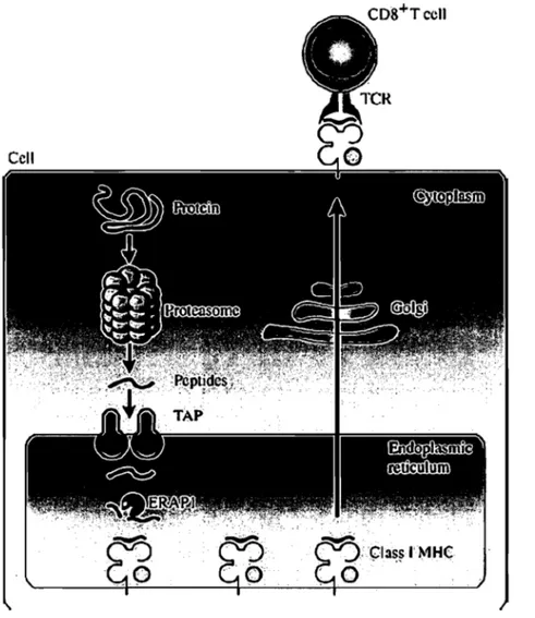
The MHC 1 immunopeptidome is the result oftwo merging cellular pathways (Figure 1). One pathway generates the peptides suitable for loading the MHC 1 molecules, whereas the second pathway produces peptide receptive MHC 1 molecules in the ER (Hammer et al., 2007).
Figure 1. MUC l-antigen presentation pathway. Cytoplasmic proteins are degraded by the proteasome and other proteases. Resultant short peptides are transferred to the lumen of the ER by TAP in an ATP-dependent manner. Long peptides are trimmed by ER aminopeptidases such as ERAP 1. Peptides bound to MHC 1 molecules are exported via the secretory pathway and presented at the cell surface for surveillance by CD8+ T cells. Adapted from (Hanada & Yang, 2005).
1.2.2. Peptide processing in the cytoplasm
Generation of peptides suitable for loading MHC 1 molecules starts in the cytoplasm. This is the major site of protein degradation, because even ER pro teins are retrieved into the cytosol for turnover (Shastri et al., 2005). Thus, this compartment is the logical site for beginning the peptide-processing pathway (the pathway by which peptide substrates for MHC 1 binding are made). Short peptides ofvariable lengths (2-25 amino acids) are generated from degradation of endogenous proteins in the cytoplasm through the action of the proteasome and other proteases (Stoltze et al., 2000a; Stoltze et al., 2000b; Jensen, 2007). Treatment of
cells with proteasome inhibitors has revealed that most but not all MHC I-peptide complexes require the prote as orne for their generation (Luckey et al., 1998). So, the majority of peptides are produced by the multicatalytic proteasome.
Besides being the site where antigenic peptides are bom, the cytoplasm constitutes the site where most peptides are rapidly destroyed (Reits et al., 2003; Jensen, 2007). This rapid degradation by cytoplasmic peptidases limits the availability of peptides and accounts for the inefficiency of the peptide presentation pathway (Shastri et al., 2005). More than 99% of peptides are degraded by cytosolic peptidases before they reach the ER (Yewdell et al., 2003). Cytosolic chaperones, su ch as Hsp70 (Heat shock prote in 70) and Hsp90 (Heat shock prote in 90), are thought to prote ct peptides from exhaustive degradation (Kunisawa & Shastri, 2003).
Proteasomes are thought to generate the final carboxyl-terminal residues of MHC-binding peptides (Cas cio et al., 2001), but additional trimming at the ami no terminus is required for most peptide epitopes (Jensen, 2007). Various cytosolic aminopeptidases are responsib1e for c1eaving the amino terminus (Rammensee, 2006). Of particu1ar interest is the cytoplasmic tripeptidyl peptidase II (TPPII), invo1ved in trimming of proteasoma1 products over 15 amine acids in length (York et al., 2006). TPPII has also been shown to participate in proteasome independent pathway for epitope generation (Guil et al., 2006). However, a delicate balance must be maintained because excess trimming by cytosolic peptidases can also destroy MHC 1 peptides.
At this point, these peptides (usually less than 12 amino acids) constitute precursors of the MHC I-peptide repertoire and are ready to be translocated into the ER. However, as we will see later, these precursors need further trimming in the ER in order to be suitable for loading MHC 1 molecules.
1.2.3. Generation of peptide-receptive MHC 1 molecules
As mentioned above, production of the MHC 1 immunopeptidpme requires a concomitant pathway that generates peptide receptive MHC 1 molecules in the ER. The MHC 1 molecules are heterodimers of a polymorphic heavy chain (a chain) and ~2-microglobulin (~2-m) (Zhang & Williams, 2006). Similar to what occurs with the peptide precursors, both polypeptides are cotranslationally translocated into the ER (Hammer et al., 2007).
Once in the ER, early folding and oxidation of the MHC 1 heavy chain and ~2-m are mediated by the chaperone ca1nexin (Jensen, 2007). These events are followed by release from
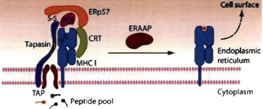
calnexin and assembly with the MHC 1 peptide-loading complex (PLC) (Jensen, 2007) (Figure 2). The PLC comprises many components, inc1uding the soluble chaperone calreticulin (CRT), the transmembrane protein tapasin, the oxidoreductase ERp57, protein disulfide isomerase (POl) and the heterodimeric transporter associated with antigen processing (TAP) (Zhang & Williams, 2006; Jensen, 2007). The luminal domain of TAP acts as a binding platform for calreticulin and ERp57, supporting the correct folding of MHC 1 in the PLC (Cresswell, 2005; Cresswell et al., 2005). This multisubunit strucmre keeps the MHC 1 molecules in a peptide-receptive state (Hammer & Shastri, 2007). Nevertheless, MHC 1 heterodimers need to be stabilized by binding to a high-affinity peptide.
ERAAP
-
Il Cel surface+
.//
.~ •• '~fl'''Htt.tl.H~~.41'IH'''H ••• I.~~141~~HH.1 TAP .... __ ~ ~ Peptide pool CytoplasmFigure 2. Schematic depiction of the MHe 1 PLC. The MHC 1 molecule folds with the assistance of the chaperones calnexin (not shown) and calreticulin (CRT). The multisubunit PLC is centered on TAP. The adaptor molecule tapasin interacts with TAP and the MHC 1 molecule and forms a covalent disulphide bond (S-S) with ERpS7 with the help of POl. ERAAP trims N-terminal extensions ofantigenic precursors, resulting in the generation of the final MHC I-peptide complex that is transported to the cell surface. Adapted from (Hammer et al., 2007).
1.2.4. Peptide processing in the ER
Besides retaining empty MHC 1 molecules, TAP is responsible for the active transport of peptide precursors into the lumen of the ER (Neefjes et al., 1993). This provides the empty MHC 1 heterodimers with easier access to the incoming peptide supply (Cresswell et al., 2005). TAP is a heterodimer composed of the TAPI and TAP2 molecules and shows substrate specificity when selecting peptides for translocation (Seliger et al., 1997; Gramme & Neefjes, 2002). lt is important to mention that the import of sorne peptides appears to be TAP-independent (Lautscham et al., 2003).
Peptide precursors are translocated in a naked fonn into the ER and can then follow one or more different fates. They can bind MHC 1 molecules, bind ER chaperones, be trimmed by ER aminopeptidases, be degraded or be retrotranslocated back into the cytosol (Elliott &
Neefjes, 2006). Together, these processes keep the concentration of peptides low in the ER su ch that only the most recent peptides are available for MHC 1 binding and do not have to compete with those that arrived earlier (Yewdell et al., 2003).
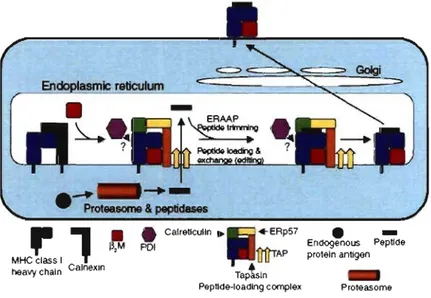
MHC 1 molecules are promiscuous, and they bind many different peptides with certain amino acid preferences at key positions (anchor residues) which constitute binding motifs (Rammensee et al., 1999). High-quality peptides that confer stability to the MHC 1 molecules share two important properties: the precise length and amino acid sequence required for a given MHC c1ass I-binding motif (Hammer et al., 2007). Peptides entering the ER via TAP bind to newly synthesized MHC 1 molecules immediately, as long as the C-tenninus is correct and the appropriate motif is present (Rammensee, 2006). At this stage, the length is not yet crucial (Rammensee, 2006). Peptides with a proper MHC 1 motif, but still too long on their N-tenninus, do bind to MHC 1 molecules. Initial peptide binding is followed by peptide exchange and editing in the ER (Elliott & Williams, 2005) (Figure 3) .
.
~...
-Proteasome & peptidase6
MHC~SI
1
heavy chain CalnexJn
• • Calretlculln • lf,M POl Taptsln Peptlde·loadlng complex
.
-Endogenous Peptlde proteln antigen ProteasomeFigure 3. The MUe 1 processing pathway. MHC I heavy chains initially assemble with ~2-m , followed by recruitment into the PLC in the ER. Endogenous peptides, generated in the cytoplasm through the action ofproteasomes and other peptidases are transported into the ER via TAP. ERAAP mediates final N-tenninal trimming before or after binding to MHC I molecules. PLC promo tes peptides loading and exchange, providing quality control for the preferential export of kinetically stable MHC I-peptide complexes. Adapted from (Jensen, 2007).
aminopeptidases. The key enzyme responsible for generation of quality peptides and final amino-terminal trimming in the MHC c1ass I pathway is the ubiquitously expressed ER amino peptidase associated with antigen processing, ERAAP in mice (ERAP1I2 in humans) (Serwold et a1., 2002). ERAAP recognizes the peptide carboxyl terminus and trims the amino terminus to generate peptides 8-10 residues in length (Chang et a1., 2005). This enzyme is induced by interferon gamma (IFN -y), a proinflammatory cytokine that enhances antigen presentation (Saric et a1., 2002; Serwold et a1., 2002). Importantly, ERAAP serves a unique function in modifying the MHC I-peptide repertoire and influencing CD8+ T cell responses; ERAAP deficiency leaves sorne peptides unaffected, whereas others are either absent or dramatically upregulated (Hammer et a1., 2006).
Peptide precursors can also bind ER chaperones (Spee et a1., 1999). PDI appears to be the most efficient peptide-binding ER chaperone, as it binds to peptides of different length and sequence (Park et a1., 2006). Importantly, binding of peptides to PDI protects them from degradation (Elliott & Neefjes, 2006).
Finally, peptides not containing a fitting motif and thus not bound to MHC 1 molecules, are trimmed and destroyed by ERAAP (Kanaseki et a1., 2006), or retrotranslocated back into the cytosol for ER-associated degradation (ERAD) (Elliott & Neefjes, 2006). In this way, they no longer compete for space in the local compartment.
1.2.5. Peptide loading and presentation
The PLC represents the precise point of intersection between the pathways of peptide processing and peptide presentation. The PLC orchestrates the final assembly of MHC 1 molecules with peptides (now 8-11 amino acids), delivered into the ER by TAP, for generation of stable MHC c1ass I-peptide complexes (Jensen, 2007). Its chief function is to provide 'quality control' by selectively retaining MHC 1 molecules loaded with suboptimal peptides for replacement by higher-affinity quality peptides (Hammer et a1., 2007).
When any ofthe PLC constituents are missing or are inhibited by viruses, intracellular MHC 1 molecules can suffer unfolding, degradation and indiscriminate peptide loading, aU of which can compromise the stability, expression and function ofMHC I-peptide complexes at the cell surface (Hammer et a1., 2007).
Finally, after successful peptide loading and customization, MHC c1ass 1 -peptide complexes are released from the PLC and are transported through the Golgi cistemae and the constitutive secretory pathway to the cell surface (Jensen, 2007). Indeed, only 1-2 out
of every 10,000 peptides generated by the proteasome bind to MHC 1 molecules (Yewdell et aL, 2003).
Different MHC alleles have different motifs and bind different set of peptides. The number of class 1 MHC molecules per cell is estimated at 50,000-100,000 (Gakamsky et al., 2000). Since as many as 2x10⁶ peptides are estimated to be generated every second, only a
small minority of peptide epitopes can be presented on a MHC 1 at any time (Princiotta et al., 2003). Most peptides are represented at fewer than ten copies per celL However, it has been estimated that only three copies of the antigenic peptide are sufficient for target celllysis by cytotoxic T ceUs (CTLs) (Purbhoo et al., 2004).
1.3. The origin of peptides for display by MHC
1 molecules
The complexity ofthe MHC I-immunopepidome reflects the equally complex milieu of intracellular proteins (Shastri et al., 2005). It is widely known that peptides displayed by MHC 1 molecules derive from degradation of proteins acquired from an exogenous source or from proteins endogenously synthesized, in processes referred to as cross-presentation or direct presentation, respectively (Shastri et al., 2005). When and how intraceUular proteins are chosen for entry into the antigen processing pathway? This is an interesting yet not completely solved question. What is now clear, however, is that the MHC I-peptide repertoire is not a random sample of the proteome: many abundant proteins do not generate MHC I-associated peptides white sorne low abundant proteins have a major contribution to the immunopeptidome (Caron et al., 2005; Milner et al., 2006). In the foUowing section, 1 will give sorne examples of generation of peptides from exogenous proteins and de scribe more in detail the mechanisms by which endogenous proteins can give rise to peptides.
1.3.1. Exogenous proteins as source of peptides
Uptake of exogenous antigens occurs routinely in antigen-presenting ce Us (APCs) such as macrophages and dendritic cells (DCs), which represent the sentinels for initiating naïve CD8+ T-cell responses (Mellman & Steinman, 2001). The transferred antigens can take many different forms, ranging from cell debris from apoptotic or necrotic ceUs to proteins or chaperone-associated peptides (Trombetta & Mellman, 2005). For instance, DCs can ingest infected ceUs or cancer ceUs and derivè antigens from these sources in a mechanism known as cross-presentation (J ensen, 2007). AIso, exogenous protein sources are clearly important in the presentation of peptides derived from intracellular pathogens such as Listeria monocytogenes
There are many pathways for cross-presentation, including dependent and TAP-independent mechanisms, ev en though the former seem to dominate. Exogenous antigens can be transferred from the endosome to the cytosol, where they are digested by proteasomes and loaded onto MHC 1 molecules in a TAP-dependent manner (Trombetta & Mellman, 2005). It remains unclear, however, what the mechanisms are by which they traverse the enosomal membrane and reach the cytoplasm. Suggested mechanisms involve transient physical rupture of the endosomal membrane or the action of a specific channel or translocator (Trombetta & Mellman, 2005). Altematively, antigens might make use of an established retro grade pathway leading from endosomes to the ER via the Golgi. Form the ER, they may reach the cytosol using the translocation channel involved in retro translocation during prote in degradation (Trombetta & Mellman, 2005). As in the MHC 1 processing pathway, there is a balance between peptide generation and destruction by proteolytic enzymes in cross-presentation. Mechanisms that reduce the activity of endosomal hydrolases in DCs have recently been shown to enhance the efficiency of cross-presentation (Jensen, 2007).
To some extent, exogenous antigens can also be presented on MHC 1 molecules in a TAP-independent manner, indicating that they do not require transport to the cytosol and can thus be loaded in the endocytic pathway (Trombetta & Mellman, 2005). This pathway may involve peptide exchange in recycling endosomes or on the cell surface, and a recent study has demonstrated that the lysosomal protease cathepsin S is important in generating peptides presented through this pathway (Jensen, 2007). Recently, it has been demonstrated that peptides can be transferred from virally-infected cells to professional APCs through gap junctions (Neijssen et al., 2005).
1.3.2. Endogenous proteins as source of peptides
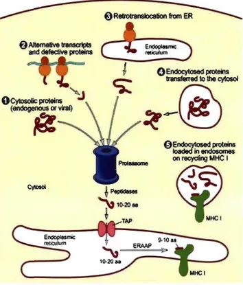
Most endogenous proteins destined for presentation on MHC 1 molecules are fed to the proteasome to initiate fragmentation. However, the source of proteasomal substrate is quite varied. Peptide ligands for MHC 1 molecules can be derived from cryptic transcription products, defective ribosomal products (DRiPs), "stable" proteins or from retrotranslocation of proteins destined to the secretory pathway from the ER to the cytosol (Figure 4).
6 AIIemative IntnscripCs
and defecIive proteins
OCytosoIic proteins (endogenous or viral)
~
{) ReIroIransloc8tion from ERc
t
=:"'
:)
1
c;::
0Endoeytœed proteins Ioaded in endosomes on recyding t.1HC 1~
Figure 4. Possible sources ofproteins presented on MHC 1 molecules. MHC 1 ligands have been shown to derive from various sources, including (1) cytosolic proteins, (2) alternative translation products and DRiPs, (3) proteins retrotranslocated to the cytosol from the ER, and (4) internalized proteins transferred to the cytosol. Adapted from (Trombetta & Mellman, 2005).
1.3.2.1. Peptides derived trom stable proteins
MHC I-peptide ligands can be obtained from "stable" proteins, as evidenced by presentation of several species of posttranslationally modified peptides: N-glycosylated peptides, de-aminated peptides (Mosse et al., 1998), phosphopeptides (Zarling et al., 2006)
or peptides with modified cysteine residues (Chen et al., 1999).
1.3.2.2. Rapidly versus slowly degraded polypeptides
As mentioned above, most of the peptides that enter the MHC 1 presentation pathway are generated by proteasome-mediated cleavage of polypeptides (Schubert et al., 2000). Note that the process of fragmenting a protein for generating an antigenic peptide is mutually exclusive from the process ofusing the same protein molecule to fold into a biologically active form. In principle, proteins can be sampled either at the beginning, the end, or anywhere in the middle oftheir lifespan. Proteins exhibit a wide range of degradation rates: from minutes
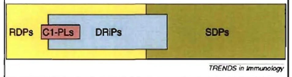
have been segregated into two general pools: (1) those degraded with an average half-life of -10 minutes, named rapidly degraded polypeptides (RDPs), and (2) those degraded with
an average half-life from hours to weeks (in average -2000 minutes), referred to as slowly
degraded polypeptides (SDPs) (Yewdell & Nicchitta, 2006) (Figure 5).
DAiPs
TRENDS in lmmunology
Figure 5. IntracelluIar polypeptide degradation pools. Polypeptides segregate into two
general pools: those degraded with an average half-life of ~ 10 min (RDP) and those degraded
with an average half-life of ~2000 min (SDP). DRiPs belong to the RDP or to the SDP pool,
although it seems likely lhat most nalurally generated DRiPs belong to the RDP pool. MHC I-peptide ligands (CI-PLs) seem to be predominantly derived from subslrates in the RDP pool. These substrates include DRiPs and c1eaved leader sequences. Adapted from (Yewdell & Nicchitta, 2006).
A significant proportion of peptides appears to result from the degradation of newly synthesized, but rapidly degraded polypeptides as opposed to slowly degraded polypeptides
(Reits et al., 2000; Schubert et al., 2000). These RDPs are presumably weil represented
by either misfolded or erroneous proteins, which have been called DRiPs (Yewdell et
al., 1996; Princiotta et al., 2003; van der Bruggen & Van den Eynde, 2006). Remarkably,
RDPs could constitute 1 % or Jess of newly synthesized proteins, yet still provide ail of the
peptides presented by MHC 1 molecules (Yewdell & Nicchitta, 2006). Strong evidence has
been published supporting the idea that translation and protein folding must be error prone (Jensen, 2007) and that newly generated polypeptides, including DRiPs, represent the main
source of antigens entering the MHC 1 loading pathway (Yewdell, 2005; Yewdell & Nicchitta,
2006). Thus, it has been suggested that, MHC 1 molecules preferentially sample what is being
translated as opposed to what has been translated (Qian et al., 2006a; Qian et al., 2006b).
Accordingly, the MHC 1 peptide repertoire has been shown to be biased toward peptides
derived from highly abundant transcripts (Fortier et al., 2008). To note, long-lived intact proteins can also contribute to the peptide pool although possibly in a lesser extent (Yewdell
& Nicchitta, 2006).
1.3.2.3.Driving force for peptide generation: the DRiPs hypothesis
over very slowly with half-lives of many hours if not days (Eisenlohr et al., 2007). Yewdell
et al. pointed out that this rate is inconsistent with in vitro assays showing that cells become
recognizable by CD8+ T cells soon after they are infected and that peptide production must commence very shortly after protein synthesis (Yewdell et al., 1996).
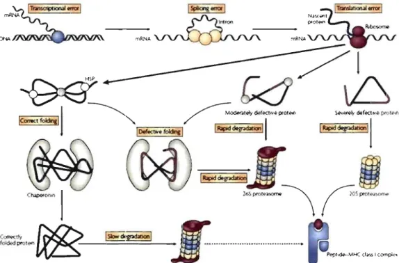
Consequently, Yewdell et al. proposed that immediate peptide supply is driven not by senescence of mature proteins but by newly synthesized proteins that are defective, termed DRiPs (Yewdell et al., 1996). These DRiPs, comprising polypeptides that fail to achieve its native structure, owing to imperfections in transcription, splicing, translation, post-translational modifications or protein folding, are ftagged by the quality-control machinery and rapidly degraded (Yewdell et al., 1996) (Figure 6).
Figure 6. The ORiPs hypothesis.Accurnulated errors during protein production (transcription,
translation and fol ding) can lead to defective proteins that are instantly recognized by the quality-control rnachinery and targeted for rapid degradation, with sorne products ultirnately becorning MHC I-bound epitopes. Misfolded proteins are subdivided into severely rnisfolded proteins that are degraded by the 20S proteasorne and rnoderately rnisfolded proteins that are degraded by the 26 proteasorne. Adapted frorn (Eisenlohr et al., 2007).
The DRiPs hypothesis was initially supported by sorne indirect evidence. First, early studies showed that mutant proteins that cause disease, as weil as misfolded proteins induced in the presence of certain compounds, are immediately degraded after their synthesis (Knowles et al., 1975; Rieder et al., 1975). Second, a substantial fraction ofnewly synthesized proteins is rapidly turned over. This fraction has been estimated to correspond to 40% and
30% per hour (Wheatley et al., 1980; Yewdell et al., 1996; Schubert et al., 2000). Third, increasing the degradation rate of an antigen results in a substantially higher level of epitope production (Townsend et al., 1988). The DRiPs hypothesis was subsequently supported by experimental results showing that peptide production ceased 30 minutes after inhibition of protein synthesis, suggesting that mature prote in turnover is too slow to make a meaningful contribution to the peptide pool (Reits et al., 2000; Yewdell, 2005).
Nevertheless, sorne aspects of the DRiPs hypothesis have been questioned (Eisenlohr et al., 2007). First, a DRiP has not yet been identified nor produced (Yewdell, 2005). Second, a window of30 minutes for peptide production, implies a very short half-life of 15 minutes or less for the substrates from which the peptides are derived (Princiotta et al., 2003) and a quickly disposaI of defective proteins. This seems not to be in line with current concepts of prote in production and quality control arguing against the DRiPs model (Eisenlohr et al., 2007). On one side, there is an ever growing list of 'natively' or 'intrinsically' unfolded proteins that bypass the quality control machinery and are not degraded (Eisenlohr et al., 2007). On the other side, many misfolded proteins can be rescued by prolonged interaction with HSPs (Markossian & Kurganov, 2004; True, 2006). Concordantly, increased degradation is not always the fate of misfolded proteins. They can enter aggregates that resolve very slowly or not at all, becoming candidates for ubiquitin-mediated autophagy and not proteasomal degradation (Bukau et al., 2006). The DRiPs model has also been called into question by recent evidence showing that newly synthesized polypeptides are mostly protected from prote as omal degradation during and immediately after translation and that preexisting proteins represent the main proteasome substrates (Vabulas & Hartl, 2005).
(
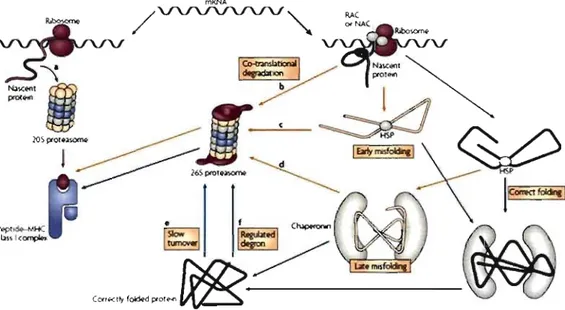
A recent alternative model, not exduding the DRiPs hypothesis, proposes that a subset of nascent polypeptides is stochastically delivered to the 20S proteasome owing to neglect by the folding machinery (Eisenlohr et al., 2007) (Figure 7). This subset would presumably correspond to 25% of all synthesized proteins (Qian et al., 2006a). For a given antigen, the basallevel of peptide presentation from immediately degraded substrate by the 20S proteasome will be supplemented by the cohort of newly synthesized proteins that is successfully intercepted by the folding machinery (Eisenlohr et al., 2007). The more defective the prote in is, the sooner and more intense the presentation of the peptide will be, which is due to more rapid rejection by the quality control machinery.
Figure 7. An alternative to the DRiPs modeI. Most mRNAs are translated by ribosomes associated with HSPs such as RAC and NAC, resulting in partial compaction of the nascent polypeptide and its delivery to downstrearn chaperones. If the prote in carries an overt degradation signal (degron), then degradation starts immediately (b). If it carries a covert degron, then it is rapidly degraded after having achieved the mature state (f). Proteins are identified as defective at different steps towards maturation (a and c). A small fraction of mRNAs is translated by unengaged ribosomes and these unfolded protein species are degraded by the 20S proteasome (a). Adapted from (Eisenlohr et al., 2007).
1.3.2.4. Cryptic translation as a source of naturally processed peptides
ln addition to conventional translation products, cells can also generate peptide ligands for MHC 1 molecules from cryptic translation products. Cryptic translation refers to polypeptides that are synthesized by unconventional translational mechanisms. These include peptides encoded by open reading frames contained within 5' and 3' untranslated regions, alternative open-reading frames, introns, or intron-exon junctions (Bullock & Eisenlohr, 1996; Mayrand et al., 1998; Shastri et al., 2002; Cardinaud et al., 2004). The list of MHC I-peptides derived from cryptic translation has been steadily growing (Ho & Green, 2006). Many examples of peptides ofviral origin or in tumor cells have been described (Ho & Green, 2006). Cryptic translation also operates in nonnal professional as weIl as non-professional APCs (Schwab et al., 2003). Although cryptic peptides are expressed at low abundance within the MHC I-peptide pool, they have been shown to induce tolerance in transgenic mice that generate cryptic peptides, and elicit CD8+ T-cell responses in nonnal mice (Schwab et al., 2003). Besides cryptic translation, translational errors, including ribosomal frameshifting, can have biological relevance in generating CD8+ T cell detenninants (Zook et al., 2006).
It has been reported that MHC 1 ligands can contain sequences that are not contiguous in the original protein but are spliced together from neighboring peptides (Hanada et al., 2004; Vigneron et al., 2004; Hanada & Yang, 2005; Warren et al., 2006). They represent spliced peptides generated by the proteasome. This suggests that the number of MHC I-peptides could be potentially larger than anticipated.
1.3.2.6. Peptides derived From proteins destined to the secretory pathway
Secretory and membrane proteins are also a known source of MHC I-peptides. Nevertheless these proteins have to exit the ER because there are no proteasomes present in the ER lumen (Wojcik & DeMartino, 2003). Proteins destined to the secretory pathway can gain access to the cytosol after being retrotranslocated from the ER, in a process that typically results in ubiquitination and proteasomal degradation, defined as ER-associated degradation (ERAD, see below) (Tsai et al., 2002). Many peptides derived from transmembrane or secretory proteins correspond to sequences derived either from transmembrane regions or signal sequences (Shastri et al., 2005).
1.4. Translation and Protein Folding
Protein synthesis is energetically the most expensive process in the cell. It places heavy demands upon the cell in terms of requirements of both amino acids and metabolic energy (Proud, 2007). Not surprisingly, translation rates are tightly regulated to match the requirements of the cell/tissue with the supply of metabolic energy and amine acids. Regulating the overall rate of protein synthesis is important not only for modulating cell and tissue metabolisni and growth, but also for controlling gene expression (Proud, 2007). Cell proliferation also depends upon maintaining an adequate rate of prote in synthesis (Scheper et al., 2007).
Protein synthesis is mediated by ribosomes. Ribosomes comprise two subunits: the 'large' subunit (60S in mammals) and the 'small' subunit (40S in mammals) (Scheper et al., 2007). The process of translation itself requires several other proteins, which are known as translation factors (Scheper et al., 2007). rnRNA translation is conventionally divided into three stages: initiation, elongation and termination. During initiation, the ribosome and the tRNA for the first amino acid residue (methionyl-tRNA) are positioned at the first codon (AUG) of the mRNA. During elongation, the polypeptide chain is assembled step-by-step as dictated by the ORF of the mRNA. When the ribosome encounters a stop codon, termination occurs, resulting in release of the completed polypeptide and the ribosomal subunits (Scheper
et al., 2007).
Accuracy is essential at aIl these stages, particularly in locating the start codon. For most mRNAs, this involves a process whereby the 40S ribosomal subunit is recruited to the mRNA's 5' cap structure. Together with the methionyl-tRNA and certain translation initiation factors, the 40S subunit then scans along the 5' untranslated regions (5'UTR or 'leader') of the mRNA to find the start codon. In sorne cases, features within the 5' UTR allow the 40S subunit to enter downstream of the 5' cap, largely obviating the need for scanning. Such internaI ribosomal entry sites (IRES) were first found in certain viral RNAs, but also occur in a subset ofhuman cellular messengers (Jackson, 2005). Nevertheless, in eukaryotes, most mRNAs are translated in a cap-dependent manner.
Both 5' and 3' UTRs can contain other elements that modulate the efficiency ofmRNA translation. Furthermore, sequences in the 3' UTR can affect translation through interaction of the poly-A-binding prote in with eukaryotic initiation factor (eIF) 4F (Mangus et al., 2003).
It is worth to note that translation also occurs in mitochondria. N onetheless, the components of the mitochondrial protein synthesis machinery are quite distinct from their cytosolic counterparts, generally being more similar to those of bacteria (Scheper et al., 2007).
After synthesis, proteins must rapidly fold to perform their biological activities. Folding takes place in three main subcellular compartments: cytosol, ER and mitochondria. Each organelle is equipped with a specific set of chaperones and folding enzymes (Anelli & Sitia, 2008). Whereas it is generally accepted that the ER functions uniquely in the biogenesis of secretory and integral membrane proteins, it has been recently shown that the ER membrane also supplies a substantial portion ofnewly synthesized proteins to the cytosol (Stephens & Nicchitta, 2008). The following section, l will concentrate on the mechanism by which proteins destined to the secretory pathway are produced, since it is the most studied and best understood.
1.4.1. rnRNA partitioning and translation in the ER
mRNA partitioning between the cytosol and the ER compartments is an ubiquitous, highly conserved property of eukaryotic cells that serves to create a dramatic compartmentalization of protein synthesis: in general, mRNAs encoding cytosolic proteins undergo translation on free ribosomes, whereas mRNAs encoding secretory and integral membrane proteins are translated on ER-bound ribosomes (Nicchitta et al., 2005) (Figure
8).
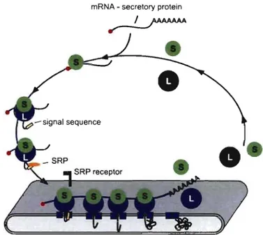
mRNA - secretory protein
__ I ___ ,/AAAAAA .".
Figure 8. The SRP-ribosome cycle. Cytosolic ribosomes engaged in the translation of mRNAs encoding secretory or membrane proteins, are targeted through the SRP pathway to the ER membrane. At the ER, the signal sequence engages the protein-conducting channel and prote in translocation ensues. Termination of the protein synthesis leads to the release of ribosomal subunits from the ER membrane to the cytosol. L: large ribosomal subunit, S: small ribosomal subunit. Adapted from (Nicchitta et al., 2005).
A considerable fraction of the proteome consists of molecules that are either secreted or inserted into membranes, to act as ligands and receptors, respectively (Anelli & Sitia, 2008). Proteins destined to the secretory pathway are directed to the ER through a predominantly hydrophobie signal sequence (Schroder & Kaufman, 2005). They are synthesized on ER-bound ribosomes, and are cotranslationally translocated into the ER lumen where they attain their native conformation, before being transported to the Golgi and downstream compartments (Anelli & Sitia, 2008).
In general, in the absence of an encoded signal sequence, rnRNAs undergo continued translation on cytosolic ribosomes (Nicchitta et al., 2005). In the case ofmRNAs containing a signal sequence, the mRNA-ribosome-nascent chain complex is relayed to the membrane by the signal recognition partic1e (SRP) early in translation (Nicchitta et al., 2005). The SRP elicits a suppression oftranslation and enables the trafficking of complexes to the ER (Meyer
& Dobberstein, 1980). Then the protein traverses the ER membrane through an aqueous channel, named the Sec61 p complex (Schroder & Kaufman, 2005). Sequential interactions of the mRNA-ribosome-nascent chain complex with the SRP receptor and with Sec61p, results in the release of the SRP, the binding of the ribosome to Sec61 p, and a coupled process of protein synthesis and protein translocation (Stephens & Nicchitta, 2008).
The signal peptide is then cleaved off by signal peptidase (Schroder & Kaufman, 2005). Indeed, the signal sequence influences the timing ofN-linked glycosylation and signal sequence cleavage (Rutkowski et al., 2003). AIso, inefficient cleavage can result in prolonged interaction of the protein with ER chaperones (Stevens & Argon, 1999).
1.4.2. Protein folding in the ER
Initial stages ofprotein folding, which consist of shie1ding and compacting hydrophobie domains, are carried out by chaperones of the HSP family (Frydman, 2001). Specialized HSPs, positioned at the exit channel of the ribosome, rapidly intercept nascent polypeptides. ln yeasts and higher eukaryotes, this function is carried out by two different complexes: the heterodimeric nascent polypeptide-associated complex (NAC) and the HSP70-associated ribosome-associated complex (RAC) (Wegrzyn & Deuerling, 2005; Wegrzyn et al., 2006). Partially compacted pro teins are then transferred to the chaperonins, which shepherd the substrate through the final stages of condensation (Eisenlohr et al., 2007).
Many principles goveming protein folding in the cytoso1 apply to the ER. However, protein folding in the ER is more complex than protein folding in the cytosol because proteins are posttranslationally modified (Schroder & Kaufman, 2005). The ER is unique in sustaining a set of covalent modifications, which include removal of the signal sequence, disulfide bond formation, N-glycosylation and glycophosphatidylinositol (GPI) addition. To note, removal of signal sequence and N-glycosylation are unique to secretory proteins (Anelli & Sitia, 2008). Different chaperones and folding assistants de termine different folding pathways, su ch as the calnexin/calreticulin (CNXlCRT) cycle or the Binding Prote in (BiP, also called GRP78) pathway. The choice of one or another is dictated by the localization of the N-glycans: the closer these are to the N-terminus, the higher the tendency to use CNXlCRT
as a chaperone system (Molinari & Helenius, 2000). Very rarely glycoproteins are found to bind simultaneously BiP/GRP78 and CNXlCRT (Anelli & Sitia, 2008). AIso, certain proteins that are produced in large amounts are assisted by substrate- or tissue-specifie chaperones (Anelli & Sitia, 2008).
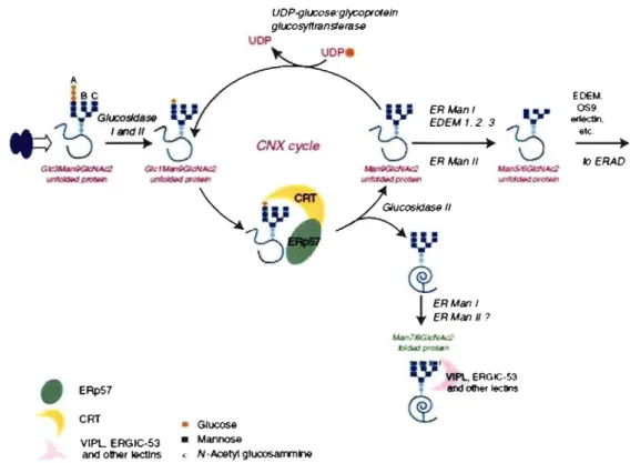
1.4.2.1. N-glycosylation and the CNX/CRT cycle
N-glycosylation involves binding ofa preformed oligosaccharide (Glc3Man9GlcNAc) to consensus Asn-X-Ser/Thr residues (Figure 9). The sugar moieties are then progressively trimmed by resident enzymes. Soon after synthesis, glucosidases 1 and II sequentially rem ove the three glucoses from the A branch of the oligosaccharide. UDP-glucose glycoprotein
glucosyltransferase (UGGT) adds back a glucose residue to N-glycans (Taylor et al., 2004). The produced monoglucosylated proteins (Glc 1 Man9GlcNAc2) can then interact with CNX or CRT, two ER chaperones with lectin activity (Williams, 2006). Besides retaining misfolded proteins and preventing their aggregation (see Protein quality control section), CNX and CRT promo te oxidative folding via interactions with ERp57. Then, by removing the terminal glucose, glucosidase II dissociates the substrate from CNX/CRT (Anelli & Sitia, 2008). If the prote in has attained its native structure, it can now proceed along the secretory pathway by bulky flow or by interaction with specific lectin transporters such as ERGIC-53 (ER-Golgi intermediate compartment-53) or VIPL (Anelli & Sitia, 2008). However, if unfolding persists, the protein enters the CNXlCRT cycle again. On the other side, mannose trimming causes exit of terminally misfolded proteins from the cycle (Anelli & Sitia, 2008). The fate of misfolded proteins will be discussed in further sections.
A Be •
Itl
D
~
~
• ERpS7 CRT • Glucose • Mannose UDP UOP-g/UCOS9:glycoprOf9/n g/ucosyltmns/9f8S9 UDPe CNXcycie VIPL EAGIC-S3an<! olller I9ctJns • N -Acetyl glucosarrrnne
@,
1 ERMan/+-
ER Man Il ?7
'
VIP\.. ERGIC-53@.,
aM olier lecUns EDEM. 0$9 ....
-.
etc, klERAOFigure 9. The CNXlCRT cycle. After transfer of the preformed core oligosaccharide onto nascent proteins, glucosidase 1 and II sequentially remove two terminal glucoses. The resulting monoglucosylated protein interacts with CNX and CRT, which in association with ERp57 prevents aggregation and facilitates folding_ Removal of the glucose by glucosidase Il liberates the protein form CNX and CRT. If the protein has attaint its native structure, it follows the secretory pathway by bulky fiow or by interaction with transporters. If unfolding persists, UGGTI places a single glucose back onto the protein, causing its entrance into the CNX/CRT cycle. Mannose trimming causes exit ofmisfolded proteins that can be targeted to ERAD. Adapted from (Anelli & Sitia, 2008)
1.4.2.2. The BiPIGRP78 pathway
The affinity of BiP/GRP78 for substrates depends on ATP. Thus, substrates can undergo cycles of BiP/GRP78 binding and release, depending on ATP hydrolysis (Anelli & Sitia, 2008). This process is regulated by hsp40-like co-chaperones containing a J domain (ERdj) (Shen et al., 2002).
1.4.2.3. Oxidative fa/ding
A hyper-oxidizing environment in the ER lumen may inhibit folding of proteins with multiple disulfides. Therefore, oxidative folding relies primarily on the PDI pathway. PDI or PDI-like proteins catalyze disulfide bond formation in the ER (Anelli & Sitia, 2008). After transferring a disulphide bond to nascent proteins, PDI is re-oxidized by members of the Ero 1 flavoprotein family (Anelli & Sitia, 2008).
1.5. Protein quality control in the secretory compartment
As we have seen, the ER provides an environment that facilitates the folding and assembly of newly synthesized secretory and transmembrane proteins and actively participates in the quality control of these proteins. The term 'ER quality control' refers to the processes of conformation-dependent molecular sorting of secretory proteins (Hurtley & Helenius, 1989). By these means, actively folding proteins are retained in the ER and shielded from degradation pathways, folded proteins are destined for export and packaged into transport vesicles, and misfolded proteins are retained and ejected into the cytosol for ubiquitin-dependent degradation (Meusser et al., 2005; Ismail & Ng, 2006).
Quality control in the ER is achieved by two independent mechanisms. The first one is the productive folding mechanism, which is involved in folding of ER-proteins and recognition ofmisfolding (Oda et al., 2006). The system must be able to distinguish between molecules that are actively folding, fully folded and misfolded (lsmail & Ng, 2006), a process that involves certain chaperones (Carvalho et al., 2006). Nevertheless, detailed information is lacking on how pro teins are initially selected for degradation (Denie et al., 2006). The second mechanism, comprising terminal steps of ER quality control, is termed ERAD, retrotranslocation or dislocation (Meusser et al., 2005). Recently, it has been suggested that autophagy could also play a role in protein quality control (Bemales et al., 2007).
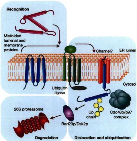
The high flux of proteins into the ER together with the complicated multidomain nature of many secreted proteins, inevitably results in sorne fraction of proteins becoming tenninally misfolded (Denic et al., 2006). The ER must specifically identify tenninally misfolded proteins in an environment dominated by structurally similar folding intennediates. Proteins transiting the ER can be soluble or membrane bound with significant portions in the lumen, the membrane and the cytosol. To monitor misfolding despite this topological diversity and the misfolded substrate selection problem, the cell has developed distinct ERAD pathways. Substrates are targeted to an appropriate ERAD pathway depending on the site of the misfolded region. For instance, membrane and soluble proteins with luminal lesions are targeted to the ERAD-L pathway, whereas membrane proteins with misfolded cytoplasmic domains use the ERAD-C pathway (lsmail & Ng, 2006). Recently the ERAD-M pathway has been proposed for membrane proteins with misfolded intramembrane domains (Carvalho et al., 2006). These pathways diverge in their components, which are still not completely characterized. In general, the ERAD mechanism occurs in four steps: substrate recognition, translocation across the ER membrane, release in the cytosol and degradation (Figure 10).
265 proteasome
Rad23p1Dsk2p
Degradation Dislocation and ubiqultlnation
Figure 10. Proteasomal degradation of ERAD targets. Aberrant proteins are recognized within the ER lumen by the quality-control machinery and escorted to a putative channel that facilitates their export to the cytoplasm. Exposed lysine residues are ubiquitinated by ubiquitin ligases. Dislocation is completed with the help of the Cdc48p/p97 and membrane-extracted substrates are conveyed to the proteasome by accessory factors such as Rad23p and Dsk2p. Adapted from (Meusser et al., 2005).
Importantly, misfolded proteins are not the only substrates for ERAD, which is also required for the regulated turnover of ER resident proteins. The regulated breakdown of a key enzyme of the mevalonate pathway, HMG CoA, is controlled by this system (Hampton, 2002). Other examples include the o-opioid receptor and the MHC 1 (Shamu et al., 1999; Petaja-Repo et al., 2001). In addition, it is now clear that elements of the ERAD system are exploited by human pathogenic viruses. For example, two membrane-anchored proteins encoded by the human cytomegalovirus (HCMV), US2 and US Il, bind to MHC 1 molecules in the ER and initiate their dislocation into the cytoplasm where they are degraded by the proteasome (Wiertz et al., 1996).
1.5.1.1 Substrate recognition
Polypeptides that do not me et the quality control standards are retained within the ER and delivered to the ERAD ligases. In mammalian cells, the 1igase CHIP targets defective proteins whose cytoplasmic domains are recognized by the cytoplasmic chaperones of the HSP70-HSP90 family (Meacham et al., 2001).
Polypeptides with native cytoplasmic domains are then examined for proper folding of the lumenal regions. The secretory pathway recognizes substructures within proteins such as hydrophobic patches, unpaired cysteines and immature glycans (Meusser et al., 2005). Proteins trapped in the CNX/CRT cycle, which normally retain and assist the folding of immature proteins in the ER, are eventually trimmed by ER mannosidase 1 (Meusser et al., 2005). This leads to their recognition by EDEM, a lectin likely to be specific for Man8GlcNAc2-oligosaccharides. Presumably, EDEM then targets misfolded glycoproteins for degradation (Molinari et al., 2003; Oda et al., 2003). This mechanism allows the selective elimination of proteins that have failed to mature properly even after extensive folding attempts. Much less is known on how terminally misfolded proteins that lack N-glycans are targeted to destruction (Anelli & Sitia, 2008).
Trimming of N-linked glycans is not the only mechansim that targets misfolded proteins for degradation. Aberrant proteins probable undergo partial unfolding and reduction before retrotranslocation (Anelli & Sitia, 2008). Both BiP/GRP78 and PDI have been implicated in this process (Molinari, 2002). BIP/GRP78 and other chaperones associate with hydrophobic surfaces of these proteins, whereas PDI and other oxidoreductases bind free thiols and control the formation of disulphide bonds between correct pairs of cysteine residues (Tsai et al., 2001; Wang & Chang, 2003; Schroder & Kaufman, 2005).