Targeted disruption of the MHC class II Aa gene in C57BL/6 mice
8
0
0
Texte intégral
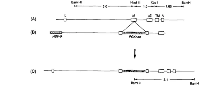
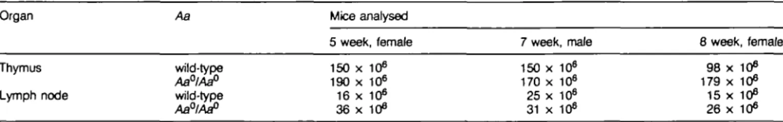
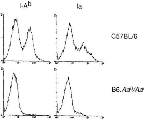
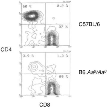
Figure
+2
Documents relatifs