CIITA-induced occupation of MHC class II
promoters is independent of the cooperative
stabilization of the promoter-bound
multi-protein complexes
Jean Villard, Annick Muhlethaler-Mottet, Se´verine Bontron, Bernard Mach
and
Walter Reith
Louis-Jeantet Laboratory of Molecular Genetics, Department of Genetics and Microbiology, University of Geneva Medical School, CMU, 9 Avenue de Champel, 1211 Geneva 4, Switzerland
Keywords: CIITA, MHC class II, multi-protein complex, stabilization
Abstract
Precise regulation of MHC class II expression plays a crucial role in the control of the immune response. The transactivator CIITA behaves as a master controller of constitutive and inducible MHC class II gene activation, but its exact mechanism of action is not known. Activation of MHC class II promoters requires binding of at least three distinct multi-protein complexes (RFX, X2BP and NF-Y). It is known that the stability of this binding results from cooperative interactions between these proteins. We show here that expression of CIITA in MHC class II–cells triggers occupation of the promoters by these complexes. This observation raised the possibility that the effect of CIITA on promoter occupation is mediated by an effect on the cooperative stabilization of the DNA-bound multi-protein complexes. We show, however, that the presence of CIITA does not affect the stability of the higher-order protein complex formed on DNA by RFX, X2BP and NF-Y. This suggests other mechanisms for CIITA-induced promoter occupancy, such as an effect on chromatin structure leading to increased accessibility of MHC class II promoters. This ability of CIITA to facilitate promoter occupation is undissociable from its transactivation potential. Finally, we conclude that this effect of CIITA is cell-type specific, since expression of CIITA is not required for normal occupation of MHC class II promoters in B lymphocytes.
Introduction
MHC class II molecules are heterodimeric cell surface proteins implicated in at least three crucial processes of the immune response (1). First, they initiate and sustain antigen-specific immune responses by presenting antigenic peptides to the receptor of CD41 Thlymphocytes. MHC class II expression
is also an absolute requirement for superantigen-induced T cell activation. Second, they shape the T cell repertoire by directing positive and negative selection events in the thymus. Finally, they also participate in activation of the antigen-presenting cells (APC) on which they are expressed. In contrast to the relatively ubiquitous cell type distribution of MHC class I molecules, constitutive MHC class II expression is largely restricted to highly specialized APC such as dendritic cells, B lymphocytes and macrophages. In addition, MHC
Correspondence to: J. Villard
Transmitting editor: H. R. MacDonald Received12 August 1998,accepted24 November 1998
class II genes can be induced in many other cell types, in particular by IFN-γ.
Both the constitutive and inducible modes of MHC class II expression are regulated at the level of transcription by a short promoter proximal enhancer containing four highly conserved cis-acting DNA sequences referred to as the S, X, X2 and Y boxes (2). Most of what is currently known about the molecular mechanisms controlling MHC class II gene transcription via thesecis-acting DNA sequences has come from studies of genetic defects in MHC class II regulation, in particular from patients having a rare genetic disease called MHC class II deficiency (2–5). Elucidation of the genetic defects underlying this disease has allowed us to clone four transcription factors (CIITA, RFX5, RFXANK and RFXAP)
462 Role of CIITA in MHC class II promoter occupation directly implicated in MHC class II gene regulation. RFX5, RFXANK and RFXAP are three subunits of RFX, a ubiquitously expressed multimeric DNA-binding complex that binds spe-cifically to the X box of MHC class II promoters (3,6–8). In contrast to RFX, CIITA is a non-DNA binding transactivator characterized by a highly regulated pattern of cell type-specific and inducible expression. It is the differential expres-sion of CIITA that controls the constitutive and inducible pattern of MHC class II gene transcription (9,10). Interestingly, the complex control of CIITA expression is achieved by the differential activation of multiple alternative promoters (11).
The RFX complex interacts cooperatively with two other transcription factors, X2BP (12,13) and NF-Y (14), such that binding of all three complexes to MHC class II promoters is strongly stabilized (13,15–18). The formation of these stable higher-order protein–DNA complexes is essential forin vivo occupation and activation of MHC class II promoters. In contrast to RFX, CIITA is believed to be recruited to the MHC class II promoter by protein–protein interactions with DNA bound factors such as RFX5 and is thought to activate transcription by contacting the basal transcription machinery via an N-terminal activation domain (19–23). On the other hand, CIITA was until now not thought to be involved in promoter occupation, because MHC class II promoters are occupied normally (24,25) in B cell lines in which the CIITA gene is severely mutated (9,26)
Here, we have analyzed the dependence of MHC class II promoter occupancy on CIITA, and examined the effect of CIITA on cooperative binding between RFX, X2BP and NF-Y in two different situations; constitutive MHC class II expression in B cells and induced expression in MHC class II– cells transfected with CIITA or stimulated with IFN-γ. In contrast to what was previously believed, the results demonstrate that CIITA indeed plays a key role in promoter occupancy, but that this is only evident in certain cell types. While CIITA is dispensable for constitutive promoter occupancy in B cells, it is clearly required for promoter occupancy induced in fibroblasts and other MHC class II–cells by IFN-γ(27,28) or
by transfection with CIITA (29 and this study). Since MHC class II promoter occupation is known to be dependent on the cooperative assembly of a strongly stabilized multi-protein complex containing RFX, X2BP and NF-Y (13,15–18), it seemed likely that this CIITA-induced promoter occupation could be due to a direct effect on the stability of this complex. Surprisingly, our results reveal that CIITA-induced promoter occupancy is not the result of an effect of CIITA on the stabilization resulting from cooperative binding between RFX, X2BP and NF-Y. Indeed, both the concentrations of these DNA binding proteins in nuclear extracts and the stability of the higher-order complexes they can form on the MHC class II promoter are independent of the presence of CIITA. These results suggest that CIITA facilitates in vivo occupation of MHC class II promoters indirectly by affecting the accessibility of the promoter DNA rather than directly by affecting the concentration or binding affinity of the multi-protein RFX– X2BP–NF-Y complex.
Methods
Cell culture and transfections
The B lymphoblastoid cell line Raji, its CIITA-deficient variant RJ2.2.5, the human melanoma cell line GL 19, GL 19
trans-fected with CIITA, the monocytic cell line C119/9 [subclone derived from U937 (30)] and C119/9 transfected with CIITA were grown in RPMI 1640. HeLa cells and HeLa transfected with CIITA were grown in DMEM. Both media were supple-mented with glutamine, 10% heat-inactivated FCS and anti-biotics. Cells were incubated at 37°C in 5% CO2. For induction of MHC class II expression, cells were incubated for 48–72 h with 500 U/ml of recombinant human IFN-γ. GL 19 melanoma cells were transfected as described using calcium phosphate precipitation followed 4 h later by a glycerol shock (31). C119/9 cells (30) (13107 cells) were
washed in cold PBS, resuspended in 0.5 ml PBS containing 10µg of linearized CIITA plasmid and transfected by electro-poration with a BioRad electropulser using a 300 V/960 µF pulse. After selection with hygromicin HLA class II1 cells were selected with Dynabeads. HeLa cells were transfected as above by electroporation with a 250 V/960µF pulse. Stably transfected cells were isolated after selection with hygromicin for 15 days and analyzed for MHC class II expression by FACS (polymorphic HLA-DR mAb 2.06) as previously described (9). Plasmids used for transfections were the empty EBO-Sfi expression vector, EBO-Sfi containing a full-length CIITA cDNA (9), EBO-sfi containing the mutated CIITA-allele 2 of patient BCH (32), EBO-sfi containing the mutated CIITA-gene of patient BLS-2 (9) and EBO-sfi containing the dominant negative NLS-D5 form of CIITA (33).
In vivofootprint analysis
In vivo footprint experiments were performed as described previously (34) using Vent DNA polymerase (New England Biolabs, Beverley, MA) for all steps of the ligation-mediated PCR reaction (LM-PCR). The sequences of the oligonucleo-tides and the incubation temperatures used for the LM-PCR reactions were as described (34).
Electrophoretic mobility shift assays (EMSA)
Nuclear extracts were prepared from 2–83108 cells as
described (15,16). The DRA-XY (–150 to –59 bp), DRA-XX2 (–144 to –70 bp) and DRA-Y (–89 to –49 bp) oligonucleotide probes (Fig. 1) were prepared as described (35). With the exception of the following modifications, EMSA experiments were performed essentially as described (15,16). Experiments were performed at room temperature for binding of NF-Y, RFX 1 NF-Y, RFX 1 X2BP and RFX 1 NF-Y 1 X2BP or on ice for binding of RFX. Binding mixtures were pre-incubated for 30 min prior to addition of 40,000 c.p.m. of the suitable 32
P-labeled oligonucleotide probes. Probes used were DRA-XX2 for binding of RFX and RFX1 X2BP, DRA-Y for binding of NF-Y, and DRA-XY for binding of RFX1 NF-Y and RFX 1 X2BP1 NF-Y. Reactions were then incubated for a further 45 min to allow binding to proceed to completion, after which they were supplemented with a 500-fold molar excess of unlabeled probe and incubated for various times (Figs 4 and 5) prior to gel electrophoresis. Binding was done in 20 µl reactions containing 8µg of nuclear extract, 5 mM MgCl2, 50
µg of BSA, 0.01% NP-40, 10 ng of a methylated pBR322 oligonucleotide (see 15,16) and the following competitor DNAs. Amounts of added poly(dI–dC)·poly(dI–dC) and sonicated denaturedEscherichia coli DNA were respectively 1 and 0.5µg for NF-Y, 0.8 and 0.4 µg for RFX, 0.6 and 0.3
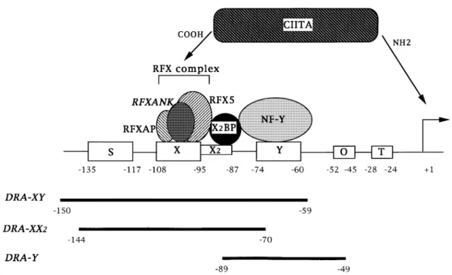
Fig. 1.Schematic representation of the HLA-DRA promoter. The S, X, X2 and Y boxes are present in all MHC class II promoters, and are crucialcis-acting regulatory sequences controlling transcription of MHC class II genes. T represents the TATA box. The octamer sequence (O) is present in the HLA-DRA promoter but not in other MHC class II genes. The DRA-XY, DRA-XX2 and DRA-Y oligonucleotides used in EMSA experiments are shown below. The RFX complex binding to the X box is composed of three subunits (RFX5, RFXANK and RFXAP) (4,6–8). NF-Y and X2BP are nuclear proteins that bind to the Y and X2 boxes respectively. CIITA is believed to be recruited to MHC class II promoters by protein–protein interactions with one or more of the DNA bound factors.
µg for RFX 1 X2BP, 1 and 0.5µg for RFX1 NF-Y, and 0.5 and 0.25µg for RFX1 NF-Y 1 X2BP. An aliquot of 50 ng of an X2 box competitor oligonucleotide was also included for binding of RFX and RFX1 NF-Y.
Results
Induction of CIITA expression in MHC class II–cells leads to the occupation of MHC class II promoters
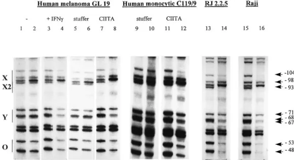
In vivo footprint experiments were performed to analyze promoter occupation. The promoter is occupied constitutively in the MHC class II1B cell line Raji (Fig. 2, lanes 15 and 16). Protections are particularly clear at the guanosine residues –104 in the X box, –98 and –93 in the X2 box, –71, –68, and –67 in the Y box, and –53 and –48 in the octamer sequence. This pattern is identical to what has been described previously (24). In RJ2.2.5, a CIITA-deficient and MHC class II–variant
derived from Raji, the promoter is also occupied normally (Fig. 2, lanes 13 and 14). The same has been observed in other CIITA-deficient B cell lines (24,25). Clearly, therefore, promoter occupation in B cells is not dependent on CIITA. It is also not dependent on transcriptional activity because the MHC class II genes are not expressed in these CIITA-deficient B cells.
The situation is very different for cell types in which expres-sion of MHC class II genes is induced by stimuli such as
IFN-γ. The promoter is bare in the uninduced human melanoma cell line GL 19 (Fig. 2, lanes 1 and 2) and the monocytic cell
line C119/9 (Fig. 2, lanes 9 and 10). Two approaches for studying the effect of CIITA on promoter occupancy in these cells were explored; induction with IFN-γand transfection with a CIITA expression vector. Induction of MHC class II gene expression in GL 19 by treatment with IFN-γis accompanied by the appearance of an occupied promoter (Fig. 2, lanes 3 and 4). As judged by the pattern of protected G residues, occupation of the X, X2 and Y boxes in the induced GL 19 cells is identical to that observed in Raji B cells. As described previously (27) occupation of the octamer sequence is less evident in the induced cells than in B cells. Similar results have been observed for other IFN-γ inducible cell lines, including HeLa (27,28).
De novo expression of CIITA is an obligatory intermediate step in the signal transduction pathway leading to the induc-tion of MHC class II genes by IFN-γ. To determine whether the induction of promoter occupation by IFN-γ is a direct consequence of CIITA expression, uninduced GL 19 cells were transfected with a CIITA expression vector. Cells trans-fected with empty expression vector were used as controls. The results demonstrate that transfection with CIITA is suffi-cient to obtain normal promoter occupancy (Fig. 2, lanes 7 and 8) and activate expression of MHC class II genes. The pattern of protected G residues observed in the CIITA-transfected cells is identical to that observed in the IFN-γ -induced cells (Fig. 2, lanes 3 and 4). The same results are observed in the monocytic cell line C119/9, where transfection with CIITA also induced an occupied promoter (Fig. 2, lanes 11 and 12). In contrast to what is observed in B cells, these
464 Role of CIITA in MHC class II promoter occupation
Fig. 2.In vivofootprint analysis of the HLA-DRA promoter in cells lacking or expressing CIITA.In vivofootprint analysis was performed with uninduced GL 19 cells (lanes 1 and 2), GL 19 cells induced with IFN-γ(lanes 3 and 4), GL 19 cells transfected with an empty expression vector (stuffer, lanes 5 and 6), GL 19 cells transfected with a CIITA expression vector (lanes 7 and 8), C119/9 monocytic cells transfected with an empty vector (lanes 9 and 10), C119/9 transfected with CIITA (lanes 11 and 12), RJ2.2.5 cells (lanes 13 and 14) and Raji cells (lanes 15 and 16). Lanes 2, 4, 6, 8, 10, 12, 14 and 16: the cells were treated with DMS, the DNA was purified and cleaved at methylated nucleotides with piperidine, and cleaved fragments spanning the DRA promoter were amplified by LM-PCR and analyzed by sequencing gel electrophoresis. Lanes 1, 3, 5, 7, 9, 11, 13 and 15: control samples representing unoccupied DNA were obtained by performing the procedure with naked DNA purified from the same cells. Positions of the X, X2, Y and Ocis-acting sequences, and the protected positions observed in IFN-γ-treated GL 19 cells (lane 4), GL 19 cells transfected with CIITA (lane 8), C119/9 cells transfected with CIITA (lane 12), RJ2.2.5 (lane 14) and Raji (lane 16) are indicated.
results demonstrate that in other cells types, occupation of MHC class II promoters requires CIITA. MHC class II promoter occupancy thus exhibits a cell type-dependent requirement for CIITA.
The ability of CIITA to induce promoter occupation is undisso-ciable from its transactivation potential
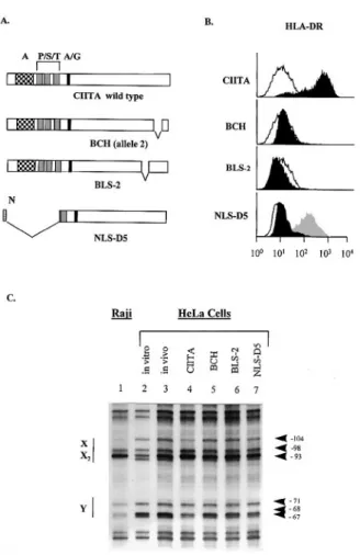
The ability of CIITA to activate transcription of MHC class II genes relies on two different functions that map to two different regions of the protein. The N-terminal region of CIITA contains a transcription activation domain that is believed to contact the basal transcription machinery (19–23). The C-terminus of CIITA, on the other hand, is believed to mediate protein– protein interactions required for recruitment to the MHC class II promoter (20–22). To determine whether promoter occupation induced by CIITA could be attributed to one or both of these functional domains we analyzed three different mutants of CIITA. In B cell lines from patients having mutations in the CIITA gene (BLS-2 and BCH) MHC class II promoters are normally occupied even though the genes are transcriptionally silent. It therefore seemed possible that the mutated forms of CIITA of these patients could retain the capacity to drive occupation of the promoter. In both patients, CIITA is disrupted by a small deletion in the C-terminal part of the gene (Fig. 3A). The NLS-D5 mutant of CIITA has been demonstrated to function as a dominant negative molecule (Fig. 3B) (33). This mutant has a deletion of the N-terminal part of the gene and has lost the activation domain of CIITA (3,20,21,33). On the other hand, NLS-D5 retains intact the C-terminal region of
CIITA that is believed to be required for its recruitment to MHC class II promoters (20,21). We therefore thought that the capacity to induce promoter occupation might be main-tained by this mutant. To determine whether the NLS-D5, BCH and BLS-2 versions retained the ability to induce promoter occupation, they were transfected into HeLa cells. These three mutated forms of CIITA are unable to activate expression of MHC class II genes (Fig. 3B). In addition NLS-D5 has a strong dominant negative effect that severely hinders induc-tion of MHC class II expression by IFN-γ(Fig. 3B). Promoter occupation could not be observed in any of the three transfec-tants, while promoter occupation could be induced normally by transfection with wild-type CIITA (Fig. 3C). These results suggest that the transactivation activity of CIITA is undissoci-able from its ability to induce the occupation of MHC class II promoters and requires the same functional domains. The same conclusion has recently been arrived at by another study using a different series of CIITA mutants (29).
Stability of the complexes bound to MHC class II promoters is independent of CIITA
Previousin vitro binding studies have demonstrated that RFX binds cooperatively with two other MHC class II promoter binding proteins, X2BP and NF-Y (13,15–18). These cooperative binding interactions strongly enhance the stability with which RFX, X2BP and NF-Y are bound to their respective X, X2 and Y box target sites. The strongly stabilized binding observed in the higher order RFX1 X2BP 1 NF-Y complex is essential for promoter occupationin vivo. Indeed, in cell
Fig. 3.In vivofootprint analysis of the HLA-DRA promoter in HeLa cells expressing mutated CIITA proteins. (A) Schematic view of CIITA and the different mutants analyzed. Mutant BCH (allele 2) has a deletion of 27 amino acids in the C-terminus (∆1079–1106). Mutant BLS-2 has a deletion of 23 amino acids in the C-terminus (∆ 940–963). The dominant-negative NLS-D5 mutant has a nuclear localization signal (NLS) at its N-terminus and has a deletion of the first 294 amino acids. A, acidic region; P/S/T, Pro/Ser/Thr-rich regions; A/G, ATP/GTP-binding motif. (B) Cell surface expression of HLA-DR in HeLa cells transfected with the mutated CIITA constructs. Black profiles: cells transfected with the wild-type or mutated forms of CIITA. Open profiles: cells transfected with empty expression vector. To demonstrated the dominant negative effect of D5, the NLS-D5 (black profile) and empty vector transfected cells (gray profile) were treated with IFN-γto induce MHC class II expression. (C) In vivofootprint analysis was performed as outlined in Fig. 2 with Raji cells (lane 1) and HeLa cells transfected with empty vector (lanes 2 and 3), wild-type CIITA (lane 4), the BCH mutant (lane 5), the BLS-2 mutant (lane 6) and the NLS-D5 mutant (lane 7). In the case of the empty vector transfected samples, DMS treatment was done directly with the cells (lane 3,in vivo) or with the purified DNA (lane 2,in vitro). Positions that are protected in Raji (lane 1) and wild-type CIITA-transfected cells (lane 4), but not in the other transfectants (lanes 3 and 5–7) are indicated at the right.
lines deficient in RFX the formation of these higher-order complexes is abolished and the entire promoter remains bare (16,17,25).
Considering this key role of cooperative binding in MHC class II promoter occupancy and the fact that CIITA expression can trigger promoter occupancy, it was logical to explore if
Fig. 4.Dissociation rate experiments performed with extracts from HeLa and HeLa transfected with CIITA. Reactions optimized for binding of RFX or NF-Y on their own, or for the simultaneous binding of RFX1 X2BP, RFX 1 NF-Y and RFX 1 X2BP 1 NF-Y (see Methods), were first incubated to allow binding to proceed to completion, and were then supplemented with an excess of unlabeled competitor DNA and continued for 0, 5, 10, 30, 60 or 120 min prior to gel electrophoresis. Nuclear extracts were from HeLa or HeLa transfected with CIITA. The experiment for RFX was done on ice in order to prolong the half-life of the RFX-DNA complex, which is,5 min at room temperature (15). All other experiments were performed at room temperature. Only the regions of the gels containing the protein–DNA complexes are shown.
Fig. 5.Dissociation rate experiments performed with extracts from Raji and RJ2.2.5. Dissociation rate experiments with nuclear extracts from Raji and RJ2.2.5 were done as described in Fig. 4.
the effect of CIITA was mediated by either an increase in the concentration of RFX, X2BP or NF-Y, or by an enhanced stability in the interactions between these proteins and their target sites. The abundance and stability of various different protein–DNA complexes were therefore analyzed by EMSA experiments. As an indication of stability, the off-rates of the protein–DNA complexes were examined. Four different cell types were included in the analysis (Figs 4–6). Two of these
466 Role of CIITA in MHC class II promoter occupation
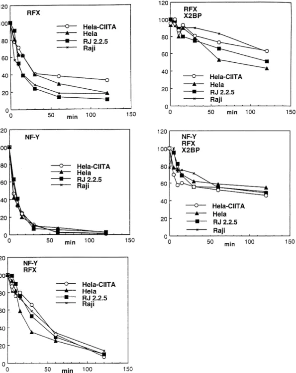
Fig. 6.Quantification of the dissociation rate experiments. The gels shown in Figs 4 and 5 were quantified by PhosphorImager analysis. The percent of the remaining protein–DNA complexes is plotted as a function of time. The amount of complex bound at time zero (addition of unlabeled competitor DNA) was taken as 100%.
lack CIITA (HeLa and RJ2.2.5) while the other two express CIITA (HeLa cells transfected with CIITA and Raji). For each cell type, off-rates were determined for complexes containing only RFX or NF-Y, and for the higher-order complexes con-taining RFX1 NF-Y, RFX 1 X2BP and RFX 1 X2BP 1 NF-Y. The off-rate for X2BP bound on its own could not be determined because its affinity for the X2 box in the absence of the other two proteins is too low to permit detection of a stable complex in EMSA experiments (15). Representative gels for HeLa and
HeLa transfected with CIITA are shown in Fig. 4, those for Raji and RJ2.2.5 are shown in Fig. 5, and quantifications of the results are shown in Fig. 6. Identical results were also obtained in experiments comparing RJ2.2.5 with CIITA-trans-fected RJ2.2.5 and uninduced HeLa with IFN-γ-treated HeLa (data not shown). Four major conclusions can be drawn from the data shown here. First, expression of CIITA does not change the levels of RFX, X2BP and NF-Y detectable in the nuclear extracts. This holds true both for B cells (Raji) and
HeLa. Second, as observed previously (13,15,16), in all cell types the interaction of RFX, X2BP and NF-Y with the promoter is strongly stabilized in the higher-order RFX1 X2BP, RFX 1 NF-Y and RFX1 X2BP 1 NF-Y complexes. Third, there are no significant differences between B cells (Raji and RJ2.2.5) and HeLa cells (transfected or not with CIITA) with respect to the stability of any of the different complexes. Fourth, no differences in off-rates are observed between cells that lack CIITA (uninduced HeLa and RJ2.2.5) and those that express it (Raji and HeLa transfected with CIITA), indicating that CIITA does not influence the stability of any of the complexes. Taken together these results demonstrate that the effect of CIITA on promoter occupancy is not mediated by an increase in the expression levels of RFX, X2BP and NF-Y, or by an enhancement in the efficiency with which these proteins can form stable higher-order complexes on the promoter.
Discussion
A combination of biochemical and genetic evidence has formally demonstrated that the DNA binding complex RFX and the non-DNA binding transactivator CIITA are both key components of the molecular machinery that controls the activity of the MHC class II promoters (3,4,36,37). Although both are essential for MHC class II expression, their respective modes of action are believed to be quite different. RFX binds cooperatively with two other MHC class II promoter binding proteins, X2BP (12,13) and NF-Y (14), to generate a very stable higher-order protein–DNA complex and this stable binding is required for promoter activation (13,15–18). Forma-tion of this higher-order complex is clearly required for occupa-tion of MHC class II promoters in vivo, because these promoters remain bare (24,25) in cells in which formation of the higher-order complex is abrogated by mutations des-troying RFX (3,6) or by mutations in the Y box of MHC class II promoters (38). In the case of CIITA, existing evidence suggests that it is recruited to the promoter by protein–protein interactions with one or more DNA bound factors such as the RFX5 subunit of RFX and that it activates transcription by contacting components of the basal transcription machinery via an acidic activation domain (19–22). In contrast to RFX, CIITA has been believed to be dispensable for promoter occupation in vivo because occupancy remains essentially normal in mutant B cell lines lacking functional CIITA (24,25). However, we now demonstrate here that CIITA does in fact also play a role in promoting promoter occupation, but that this is not evident in all cell types: while CIITA is not necessary for promoter occupation in B cells, it is required in cell types in which MHC class II expression depends on induction with stimuli such as IFN-γ(Fig. 2). This is consistent with similar results presented in another recent study using cell types distinct from those used here (29). The analysis of different mutants of CIITA suggests that the transactivation potential of CIITA and its ability to influence promoter occupation map to the same functional domains of the protein (Fig. 3). Moreover, we demonstrate that CIITA affects promoter occupation by a mechanism that is distinct from the stabiliza-tion that results from the cooperative binding between RFX, X2BP and NF-Y (Figs 4–6).
Two different MHC class II promoter occupancy phenotypes
can be observed in cells lacking CIITA (Fig. 2). Promoter occupancy is normal in B lymphoblastoid cells such as RJ2.2.5 which lack functional CIITA (Fig. 2). On the other hand, the promoter is bare in CIITA–cells such as uninduced GL 19 melanoma cells, C119/9 monocytic cells and HeLa cells (Figs 2 and 3) (24,25,27,28). This cannot be explained by a difference in the expression levels of proteins known to be required for occupation, i.e. RFX, X2BP and NF-Y (Figs 4 and 5). Surprisingly, it is also not the result of differences in the stability of the higher-order protein–DNA complexes formed by cooperative binding between these three proteins (Figs 4–6). It can also be excluded that promoter occupancy simply reflects active transcription, because neither mutant B cells lacking CIITA (RJ2.2.5) nor the uninduced melanoma cells, monocytes and HeLa cells express MHC class II genes. Consequently, it is likely that the difference in promoter occupancy observed between these two MHC class II– cell
types reflects a difference in the accessibility of the DNA within the chromatin structure at the promoter.
A difference in promoter accessibility is not observed only when comparing B cells and MHC class II–cells. Changes in
promoter accessibility also appear to occur during differenti-ation within the B cell lineage. Developmental extinction of MHC class II expression in plasmocytes is accompanied by the appearance of a bare promoter phenotype that can be reversed by transfection with CIITA (34 and data not shown). As described here for IFN-γinducible cells, the bare promoter in plasmocytes is not a consequence of the absence of RFX, X2BP or NF-Y, or of a reduced ability to form stable higher-order protein–DNA complexes (34). Compared to MHC class II– B cells, plasmocytes thus seem to acquire a tighter chromatin structure prohibiting occupation of MHC class II promoters by RFX, X2BP and NF-Y.
Transfection with CIITA is sufficient to overcome the access-ibility barrier opposing binding of RFX, X2BP and NF-Yin vivo to the MHC class II promoter in MHC class II– cells. How CIITA achieves this remains a matter of speculation. Several possibilities come to mind. One possibility is that interaction of CIITA with RFX, X2BP and/or NF-Y contributes an additional stabilizing effect that is not detectable by in vitro binding assays. It is also possible that this interaction induces a conformational change in the RFX–X2BP–NF-Y complex that is required to maintain stable binding of these proteinsin vivo. Alternatively, perhaps CIITA facilitates access of the DNA binding proteins to the DNA because it has a direct or indirect effect on the chromatin structure at the promoter: it could, for example, recruit chromatin remodeling factors such as histone acetylases (39,40) or the Swi/Snf complex (41,42). Clearly, the precise mode of action of CIITA is far from elucidated and the novel observations made here point to several potential mechanisms that need to be explored.
Acknowledgments
We thank Paolo Silacci for the C119/9 cell line and Madeleine Zufferey for excellent technical assistance. This work was supported by the Louis-Jeantet Foundation and by the Swiss National Science Foundation.
468 Role of CIITA in MHC class II promoter occupation
Abbreviations
APC antigen-presenting cell
EMSA electrophoretic mobility shift assay LM-PCR limited-mobility polymerase chain reaction NLS nuclear localization signal
References
1 Cresswell, P. 1994. Assembly, transport, and function of MHC class II molecules.Annu. Rev. Immunol.12:259.
2 Glimcher, L. H. and Kara, C. J. 1992. Sequences and factors: a guide to MHC class-II transcription.Annu. Rev. Immunol.10:13. 3 Mach, B., Steimle, V., Martinez-Soria, E. and Reith, W. 1996.
Regulation of MHC class II genes: lessons from a disease.Annu. Rev. Immunol.14:301.
4 Villard, J., Lisowska-Grospierre, B., van den Elsen, P., Fischer, A., Reith, A. and Mach, B. 1997. Primary MHC class II deficiency: genetic and molecular definition of a novel complementation group.N. Engl. J. Med.337:748.
5 Boss, J. M. 1997. Regulation of transcription of MHC class II genes.Curr. Opin. Immunol.9:107.
6 Steimle, V., Durand, B., Barras, E., Zufferey, M., Hadam, M. R., Mach, B. and Reith, W. 1995. A novel DNA binding regulatory factor is mutated in primary MHC class II deficiency (Bare Lymphocyte Syndrome).Genes Dev.9:1021.
7 Durand, B., Sperisen, P., Emery, P., Barras, E., Zufferey, M., Mach, B. and Reith, W. 1997. RFXAP, a novel subunit of the RFX DNA binding complex is mutated in MHC class II deficiency. EMBO J.16:1045.
8 Masternak, K., Barras, E., Zufferey, M., Conrad, B., Corthals, G., Aebersold, R., Sanchez, J. C., Hochstrasser, D. F., Mach, B. and Reith, W. 1998. A gene encoding a novel RFX-associated transactivator is mutated in the majority of MHC class II deficiency patients.Nat. Genet.20:273.
9 Steimle, V., Otten, L. A., Zufferey, M. and Mach, B. 1993. Complementation cloning of an MHC class II transactivator mutated in hereditary MHC class II deficiency (or bare lymphocyte syndrome).Cell75:135.
10 Steimle, V., Siegrist, C., Mottet, A., Lisowska-Grospierre, B. and Mach, B. 1994. Regulation of MHC class II expression by interferon-gamma mediated by the transactivator gene CIITA. Science265:106.
11 Muhlethaler-Mottet, A., Otten, L. A., Steimle, V. and Mach, B. 1997. Expression of MHC class II molecules in different cellular and functional compartments is controlled by differential usage of multiple promoters of the transactivator CIITA.EMBO J.16:2851. 12 Hasegawa, S. L. and Boss, J. M. 1991. Two B cell factors bind the HLA-DRA X box region and recognize different subsets of HLA class II promoters.Nucleic Acids Res. 19:6269.
13 Moreno, C. S., Emery, P., West, J. E., Durand, B., Reith, W., Mach, B. and Boss, J. M. 1995. Purified X2 binding protein (X2BP) cooperatively binds the class II MHC X box region in the presence of purified RFX, the X box factor deficient in the bare lymphocyte syndrome.J. Immunol.155:4313.
14 van Huijsduijnen, R. H., Li, X. Y., Black, D., Matthes, H., Benoist, C. and Mathis, D. 1990. Co-evolution from yeast to mouse: cDNA cloning of the two NF-Y (CP-1/CBF) subunits.EMBO J.9:3119. 15 Reith, W., Kobr, M., Emery, P., Durand, B., Siegrist, C. A. and
Mach, B. 1994. Cooperative binding between factors RFX and X2bp to the X and X2 boxes of MHC class II promoters.J. Biol. Chem.269:20020.
16 Reith, W., Siegrist, C. A., Durand, B., Barras, E. and Mach, B. 1994. Function of major histocompatibility complex class II promoters requires cooperative binding between factors RFX and NF-Y. Proc. Natl Acad. Sci. USA91:554.
17 Durand, B., Kobr, M., Reith, W. and Mach, B. 1994. Functional complementation of MHC class II regulatory mutants by the purified X box binding protein RFX.Mol. Cell Biol.14:6839. 18 Louis-Plence, P., Moreno, C. and Boss, J. 1997. Formation of
a regulatory factor X/X2 box-binding protein/nuclear factor-Y multiprotein complex on the conserved regulatory regions of HLA class II genes.J. Immunol.159:3899.
19 Mahanta, S. K., Scholl, T., Yang, F. C. and Strominger, J. L. 1997. Transactivation by CIITA, the type II bare lymphocyte syndrome-associated factor, requires participation of multiple regions of the TATA box binding protein.Proc. Natl Acad. Sci. USA94:6324. 20 Riley, J. L., Westerheide, S. D., Price, J. A., Brown, J. A. and
Boss, J. M. 1995. Activation of class II MHC genes requires both the X box and the class II transactivator (CIITA).Immunity2:533. 21 Zhou, H. and Glimcher, L. H. 1995. Human MHC class II gene transcription directed by the carboxyl terminus of CIITA, one of the defective genes in type II MHC combined immune deficiency. Immunity2:545.
22 Scholl, T., Mahanta, S. K. and Strominger, J. L. 1997. Specific complex formation between the type II bare lymphocyte syndrome-associated transactivators CIITA and RFX5.Proc. Natl Acad. Sci. USA94:6330.
23 Fontes, J. D., Jiang, B. and Peterlin, B. M. 1997. The class II trans-activator CIITA interacts with the TBP-associated factor TAF II 32.Nucleic Acids Res.25:2522.
24 Kara, C. J. and Glimcher, L. H. 1991.In vivofootprinting of MHC class II genes: bare promoters in the bare lymphocyte syndrome. Science252:709.
25 Kara, C. J. and Glimcher, L. H. 1993. Threein vivo promoter phenotypes in MHC class II deficient combined immunodeficiency.Immunogenetics37:227.
26 Brown, J. A., He, X. F., Westerheide, S. D. and Boss, J. M. 1995. Characterization of the expressed CIITA allele in the class II MHC transcriptional mutant RJ2.2.5.Immunogenetics43:88.
27 Wright, K. L. and Ting, J. P. 1992.In vivofootprint analysis of the HLA-DRA gene promoter: cell-specific interaction at the octamer site and up-regulation of X box binding by interferon gamma. Proc. Natl Acad. Sci. USA89:7601.
28 Kara, C. J. and Glimcher, L. H. 1993. Developmental and cytokine-mediated regulation of MHC class II gene promoter occupancy in vivo.J. Immunol.150:4934.
29 Wright, K., Chin, K., Linhoff, M., Skinner, C., Brown, J., Boss, J., Stark, G. and Ting, J. 1998. CIITA stimulation of transcription factor binding to major histocompatibility complex class II and associated promotersin vivo.Proc. Natl Acad. Sci. USA95:6267. 30 Willheim, M., Silacci, P., Gessl, A., Spittler, A., Sze´pfalusi, Z., Samorapoompichit, P., Agis, H., Mayr, W., Scheiner, O., Fo¨rster, O., Mach, B. and Boltz-Nitulescu, G. 1995. Tumor necrosis factor-α induction of major histocompatibility complex class II antigen expression is inhibited by interferon-gamma in a monocytic cell line.Eur. J. Immunol.25:3202.
31 Siegrist, C. A., Martinez-Soria, E., Kern, I. and Mach, B. 1995. A novel antigen-processing-defective phenotype in major histocompatibility complex class II-positive CIITA transfectants is corrected by interferon-gamma.J. Exp. Med.182:1793. 32 Bontron, S., Steimle, V., Ucla, C. and Mach, B. 1997. Two novel
mutations in the MHC class II transactivator CIITA in a second patient from MHC class II deficiency complementation group A. Hum. Genet.99:541.
33 Bontron, S., Ucla, C., Mach, B. and Steimle, V. 1997. Efficient repression of endogenous major histocompatibility complex class II expression through dominant negative CIITA mutants isolated by a functional selection strategy.Mol. Cell. Biol.17:4249. 34 Silacci, P., Mottet, A., Steimle, V., Reith, W. and Mach, B. 1994.
Developmental extinction of major histocompatibility complex class II gene expression in plasmocytes is mediated by silencing of the transactivator gene CIITA.J. Exp. Med.180:1329. 35 Herrero Sanchez, C., Reith, W., Silacci, P. and Mach, B. 1992.
The DNA binding defect observed in MHC class II regulatory mutants concerns only one member of a family of complexes binding to the X box of class II promoters.Mol. Cell. Biol.12:4076. 36 Chang, C. H., Guerder, S., Hong, S. C., van Ewijk, W. and Flavell, R. A. 1996. Mice lacking the MHC class II transactivator (CIITA) show tissue-specific impairment of MHC class II expression. Immunity4:167.
37 Clausen, B., Waldburger, J., Schwenk, F., Barras, E., Mach, B., Rajewski, K., Fo¨rster, I. and Reith, W. 1998. Residual MHC class II expression on mature dendritic cells and activated B cells in RFX5-deficient mice.Immunity8:143.
Criscitiello, M., Cogswell, P., Clarke, J. B. and Ting, J. P. 1994. CCAAT box binding protein NF-Y facilitatesin vivorecruitment of upstream DNA binding transcription factors.EMBO J.13:4042. 39 Mizzen, C., Yang, X., Kokubo, T., Brownell, J., Bannister, A.,
Owen-Hughes, T., Workman, J., Wang, L., Berger, S., Kouzarides, T., Nakatani, Y. and Allis, C. 1997. The TAF(II) 250 subunit of TFIID has histone acetyltransferase activity.Cell87:1261.
40 Hartzog, G. and Winston, F. 1997. Nucleosomes and transcription: recent lessons from genetics.Curr. Opin. Genet. Dev.7:192. 41 Peterson, C. 1996. Multiple switches to turn on chromatin.Curr.
Opin. Genet. Dev.6:171.
42 Pugh, B. 1996. Mechanisms of transcription complex assembly. Curr. Opin. Cell. Biol.8:303.