Université de Montréal
Systems biology of the human
MHC class I immunopeptidome
par
Diana Paola Granados
Programme de Biologie Moléculaire Faculté de Médecine
Thèse présentée à la Faculté de Médecine en vue de l’obtention du grade de
Docteur en biologie moléculaire option biologie de systèmes
Octobre, 2013
Université de Montréal Faculté de Médecine
Cette thèse intitulée :
Systems biology of the human
MHC class I immunopeptidome
présentée par : Diana Paola Granados
A été évaluée par un jury composé des personnes suivantes: François Major, Président-rapporteur
Claude Perreault, Directeur de recherche John David Rioux, Membre du jury
Silvia Vidal, Examinateur externe Michel Desjardins, Représentant du Doyen
RÉSUMÉ
Le système de différenciation entre le « soi » et le « non-soi » des vertébrés permet la détection et le rejet de pathogènes et de cellules allogéniques. Il requiert la surveillance de petits peptides présentés à la surface cellulaire par les molécules du complexe majeur d’histocompatibilité de classe I (CMH I). Les molécules du CMH I sont des hétérodimères composés par une chaîne lourde encodée par des gènes du CMH et une chaîne légère encodée par le gène β2 -microglobuline. L’ensemble des peptides est appelé l’immunopeptidome du CMH I. Nous avons utilisé des approches en biologie de systèmes pour définir la composition et l’origine cellulaire de l’immunopeptidome du CMH I pré-senté par des cellules B lymphoblastoïdes dérivés de deux pairs de fratries avec un CMH I identique. Nous avons découvert que l’immunopeptidome du CMH I est spécifique à l’individu et au type cellulaire, qu’il dérive préférentiel-lement de transcrits abondants, est enrichi en transcrits possédant d’éléments de reconnaissance par les petits ARNs, mais qu’il ne montre aucun biais ni vers les régions génétiques invariables ni vers les régions polymorphiques. Nous avons également développé une nouvelle méthode qui combine la spectromé-trie de masse, le séquençage de nouvelle génération et la bioinformatique pour l’identification à grand échelle de peptides du CMH I, dont ceux résultants de polymorphismes nucléotidiques simples non-synonymes (PNS-ns), appelés antigènes mineurs d’histocompatibilité (AMHs), qui sont les cibles de réponses allo-immunitaires. La comparaison de l’origine génomique de l’immunopepti-dome de sœurs avec un CMH I identique a révélé que 0,5% des PNS-ns étaient représentés dans l’immunopeptidome et que 0,3% des peptides du CMH I se-raient immunogéniques envers une des deux sœurs. En résumé, nous avons découvert des nouveaux facteurs qui modèlent l’immunopeptidome du CMH I et nous présentons une nouvelle stratégie pour l’indentification de ces pep-tides, laquelle pourrait accélérer énormément le développement d’immuno-thérapies ciblant les AMHs.
MOTS CLÉS : complexe majeur d’histocompatibilité, antigène de leucocytes humains, immunopeptidome, antigène mineur d’histocompatibilité, spectro-métrie de masse, lignées de cellules B lymphoblastoïdes, séquençage de nou-velle génération
ABSTRACT
The self/nonself discrimination system of vertebrates allows detection and rejection of pathogens and allogeneic cells. It requires the surveillance of short peptides presented by major histocompatibility class I (MHC I) molecules on the cell surface. MHC I molecules are heterodimers that consist of a heavy chain produced by MHC genes and a light chain encoded by the β2-microglobulin
gene. The peptides presented by MHC I molecules are collectively referred to as the MHC I immunopeptidome. We employed systems biology approaches to define the composition and cellular origin of the self MHC I immunopep-tidome presented by B lymphoblastoid cells derived from two pairs of MHC-identical siblings. We found that the MHC I immunopeptidome is subject- and cell-specific, derives preferentially from abundant transcripts, is enriched in transcripts bearing microRNA response elements and shows no bias toward invariant vs. polymorphic genomic sequences. We also developed a novel per-sonalized approach combining mass-spectrometry, next-generation sequenc-ing and bioinformatics for high-throughput identification of MHC I peptides including those caused by nonsynonymous single nucleotide polymorphisms (ns-SNPs), termed minor histocompatibility antigens (MiHAs), which are the targets of allo-immune responses. Comparison of the genomic landscape of the immunopeptidome of MHC-identical siblings revealed that 0.5% of ns-SNPs were represented in the immunopeptidome and that 0.3% of the MHC I-pep-tide repertoire would be immunogenic for one of the siblings. We discovered new factors that shape the self MHC I immunopeptidome and present a novel strategy for the identification of MHC I-associated peptides that could greatly accelerate the development of MiHA-targeted immunotherapy.
KEYWORDS : major histocompatibility complex, human leukocyte antigen, immunopeptidome, minor histocompatibility antigens, mass-spectrometry, B lymphoblastoid cell lines, next-generation sequencing
TABLE OF CONTENTS
RÉSUMÉ ������������������������������������������������������������������������������������������������������������������� ii ABSTRACT �������������������������������������������������������������������������������������������������������������� iii LIST OF FIGURES ������������������������������������������������������������������������������������������������������ ix LIST OF TABLES ������������������������������������������������������������������������������������������������������ xii LIST OF ABBREVIATIONS ���������������������������������������������������������������������������������������� xiii ACKNOWLEDGEMENTS �����������������������������������������������������������������������������������������xviii OVERVIEW �������������������������������������������������������������������������������������������������������������� xx CHAPTER 1
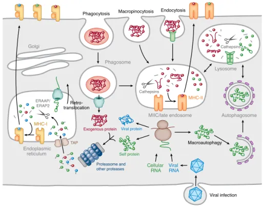
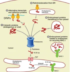
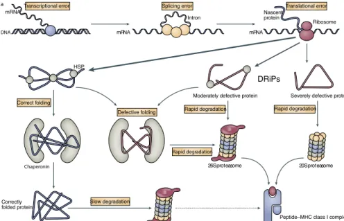
1. Introduction...2 1�1 The innate and adaptive immune system ������������������������������������������� 2 1�2 T lymphocytes ����������������������������������������������������������������������������������� 4 1�3 The human major histocompatibility complex (MHC) ������������������������� 5 1�4 The classical MHC class I and II molecules ���������������������������������������� 6 1�4�1 The MHC II molecules and associated peptides ���������������������������� 7 1�4�2 The MHC I molecules and associated peptides ����������������������������� 7 1�5 Antigen processing and presentation ����������������������������������������������� 10 1�5�1 MHC II antigen processing and presentation ������������������������������ 10 1�5�2 MHC I antigen processing and presentation ������������������������������� 12 1�5�2�1 Peptide processing in the cytoplasm ������������������������������������� 13 1�5�2�2 Generation of peptide-receptive MHC I molecules ����������������� 15 1�5�2�3 Peptide processing in the ER ������������������������������������������������ 16 1�5�2�4 Peptide loading and presentation ����������������������������������������� 17 1�5�2�5 The end of MHC I life ������������������������������������������������������������ 18 1�5�3 Alternative MHC I antigen presentation pathways ����������������������� 19 1�5�3�1 Cross-presentation �������������������������������������������������������������� 21 1�6 The origin of MHC I-associated peptides ������������������������������������������ 23 1�6�1 Endogenous proteins as source of peptides ������������������������������� 24 1�6�1�1 Rapidly versus slowly degraded polypeptides ���������������������� 24 1�6�1�2 The DRiPs hypothesis ����������������������������������������������������������� 25 1�6�1�3 Stable proteins as source of peptides ����������������������������������� 28 1�6�1�4 Cryptic translation as a source of naturally processed peptides �������������������������������������������������������������������������������������������������������� 28 1�6�1�5 Peptides derived from peptide splicing ��������������������������������� 29
1�6�1�6 Peptides derived from proteins destined to the secretory path-way �������������������������������������������������������������������������������������������������� 30 1�7 The MHC I immunopeptidome: exposing the inside of the cell to the immune system ������������������������������������������������������������������������������������� 30
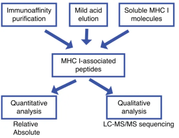
1�7�1 Biological roles of the MHC I immunopeptidome ������������������������ 31 1�7�2 Characterization of the immunopeptidome by mass spectrometry ����������������������������������������������������������������������������������������������������������� 33 1�7�3 Immunoinformatics and prediction of the immunopeptidome ������� 36 1�8 Objectives ��������������������������������������������������������������������������������������� 37 1�8�1 Research questions and hypothesis �������������������������������������������� 37 1�8�2 General objective ����������������������������������������������������������������������� 38 1�8�3 Specific objectives ���������������������������������������������������������������������� 38 1�9 Cellular model ��������������������������������������������������������������������������������� 39 1�8 References ��������������������������������������������������������������������������������������� 40 CHAPTER 2
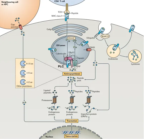
2 Origin and plasticity of MHC I-associated self peptides ... 60 2�1 Abstract ������������������������������������������������������������������������������������������ 61 2�2 Authors’ contributions ��������������������������������������������������������������������� 62 2�3 Background ������������������������������������������������������������������������������������� 63 2�4 The nature and role of the immune self recognized by CD8 T cells �� 66 2�5� A synopsis of MHC I processing – making the most out of misbegot-ten polypeptides ������������������������������������������������������������������������������������ 67 2�6 Different types of proteasomes generate different MIP repertoires ��� 68 2�7 The SMII is complex and is not a representative excerpt from the pro-teome ���������������������������������������������������������������������������������������������������� 70 2�8 The SMII conceals a tissue-specific signature ����������������������������������� 71 2�9� Neoplastic transformation has a broad impact on the SMII ������������� 72 2�10 Viral infection causes presentation of cryptic self MIPs ������������������ 73 2�11 The SMII conveys to the cell surface an integrative view of cellular regulation ���������������������������������������������������������������������������������������������� 74 2�12 The immunogenicity of neo-MIPs ��������������������������������������������������� 75 2�13 The complexity and plasticity of the SMII – A challenge for self toler-ance ������������������������������������������������������������������������������������������������������ 76 2�14 The MHC class II immunopeptidome ���������������������������������������������� 78
2�13 Perspective –toward a more comprehensive definition of the immune self �������������������������������������������������������������������������������������������������������� 79 2�15 Take-home messages �������������������������������������������������������������������� 80 2�16 Acknowledgements ����������������������������������������������������������������������� 81 2�17 References ������������������������������������������������������������������������������������� 82 CHAPTER 3
3. MHC I-associated peptides preferentially derive from transcripts bearing miRNA response elements ... 95
3�1 Authors’ contributions ��������������������������������������������������������������������� 96 3�2 Abstract ������������������������������������������������������������������������������������������ 97 3�3 Introduction ������������������������������������������������������������������������������������ 98 3�4 Material and Methods ���������������������������������������������������������������������� 99 3�5 Results ������������������������������������������������������������������������������������������ 103 3�6 Discussion ������������������������������������������������������������������������������������� 118 3�7 Authorship ����������������������������������������������������������������������������������� 120 3�8 Acknowledgements ����������������������������������������������������������������������� 121 3�9 Disclosure of Conflict of Interests �������������������������������������������������� 121 3�10 References ����������������������������������������������������������������������������������� 122 3�11 Supplemental methods ���������������������������������������������������������������� 126 3�12 Supplemental Figures ������������������������������������������������������������������ 129 3�13 Supplemental Tables ������������������������������������������������������������������� 132 CHAPTER 4
4 Minor histocompatibility antigens ...134 4�1 Origin of MiHAs ����������������������������������������������������������������������������� 134 4�2 MiHAs and allo-recognition ������������������������������������������������������������ 137 4�3 MiHA-based immunotherapy ���������������������������������������������������������� 138 4�4 The arduous identification of MiHA ����������������������������������������������� 141 4�6 References ������������������������������������������������������������������������������������� 150 CHAPTER 5
5 Impact of Genomic Polymorphisms on the Repertoire ...159 of Human MHC Class I-Associated Peptides ...159 5�1 Authors’ contributions ������������������������������������������������������������������� 160
5�2 Graphical abstract ����������������������������������������������������������������������� 161 5�3 Abstract �������������������������������������������������������������������������������������� 162 5�4 Introduction �������������������������������������������������������������������������������� 163 5�5 Results ���������������������������������������������������������������������������������������� 165 5�6 Discussion ������������������������������������������������������������������������������������� 180 5�7 Methods ���������������������������������������������������������������������������������������� 183 5�8 References ������������������������������������������������������������������������������������� 192 5�9 Acknowledgments ������������������������������������������������������������������������� 196 5�10 Author contributions ������������������������������������������������������������������ 196 5�11 Additional information ����������������������������������������������������������������� 196 5�13 Competing Financial Interests ������������������������������������������������������ 197 5�14 Supplementary Information ��������������������������������������������������������� 198 5�15 Supplementary Data ������������������������������������������������������������������� 205 CHAPTER 6
6 Discussion...207 6�1 The influence of specific HLA allelic products on the MHC I immuno-peptidome ������������������������������������������������������������������������������������������ 207 6�2 The MHC I immunopeptidome is cell-specific and subject–specific 208 6�3 Influence of the transcriptome on the MHC I immunopeptidome ���� 210 6�4 The immunopeptidome preferentially derives from transcripts bearing microRNA response elements (MREs) ��������������������������������������������������� 211 6�5 Which factors determine the number of MHC I peptides generated by a transcript? ���������������������������������������������������������������������������������������� 213 6�6 A novel personalized approach for the identification of MHC I-associ-ated peptides including MiHAs ����������������������������������������������������������� 214 6�7 Proposed improvements to our personalized approach: the database ����������������������������������������������������������������������������������������������������������� 216 6�8 Proposed improvements to our personalized approach: peptide elu-tion and MS analyses ��������������������������������������������������������������������������� 217 6�9 Impact of genomic polymorphisms on the MHC I immunopeptidome ����������������������������������������������������������������������������������������������������������� 219 6�10 MHC I peptides encoded by conserved and polymorphic genomic regions ������������������������������������������������������������������������������������������������ 221 6�11 Identification of potential MiHAs in the general population ���������� 222
6�12 How many human MiHA exist? ����������������������������������������������������� 223 Perspectives ...225 Study of the MHC I immunopeptidome from a systems biology perspective ����������������������������������������������������������������������������������������������������������� 225 Model to unravel the factors that control which MHC I peptides are dis-played and in which quantity ��������������������������������������������������������������� 226 Conclusion ...228 References ...230 APPENDIXES
APPENDIX 1 ...xxiii ER stress affects processing of MHC class I-associated peptides ��������� xxiii Abstract �������������������������������������������������������������������������������������������xxiv Background ������������������������������������������������������������������������������������� xxv Results ���������������������������������������������������������������������������������������������xxvi Discussion ����������������������������������������������������������������������������������������� xli Conclusions �������������������������������������������������������������������������������������� xlv Methods ��������������������������������������������������������������������������������������������xlvi Abbreviations����������������������������������������������������������������������������������������� l Authors’ contributions ��������������������������������������������������������������������������� l Acknowledgements ������������������������������������������������������������������������������� l References ������������������������������������������������������������������������������������������� lii APPENDIX 2 ...lviii
Deletion of Immunoproteasome subunits imprints on the transcrip-tome and has a broad impact on peptides presented by major histo-compatibility complex I molecules ���������������������������������������������������lviii
LIST OF FIGURES
CHAPTER 1Figure 1� Location and organization of the human MHC complex on chro-mosome 6 �������������������������������������������������������������������������������������������������� 6 Figure 2� Three-dimensional structures of peptide-bound MHC I and MHC II molecules �������������������������������������������������������������������������������������������������� 8 Figure 3� Examples of peptide-binding motifs of some HLA molecules�������� 9 Figure 4� Simplified overview of the MHC II antigen processing and presen-tation pathway ����������������������������������������������������������������������������������������� 11 Figure 5� General overview of the MHC I antigen processing and presenta-tion pathway� ������������������������������������������������������������������������������������������� 12 Figure 6� The MHC I antigen processing and presentation pathway ����������� 14 Figure 7� Basic and some alternative antigen processing and presentation pathways �������������������������������������������������������������������������������������������������� 20 Figure 8� Endogenous and exogenous sources of MHC I-associated peptides ���������������������������������������������������������������������������������������������������������������� 24 Figure 9� The DRiPs hypothesis ���������������������������������������������������������������� 26 Figure 10� Simplified flowchart of analysis of MHC I-associated peptides by MS ����������������������������������������������������������������������������������������������������������� 34 CHAPTER 2
Figure 1� Plasticity of the SMII ������������������������������������������������������������������� 77 CHAPTER 3
Figure 1� General features of MIPs eluted from B-LCLs from 2 HLA-disparate sibships ������������������������������������������������������������������������������������������������� 104 Figure 2� MIP source proteins are very different in the two sibships, but are implicated in similar biological pathways and are functionally interconnect-ed ���������������������������������������������������������������������������������������������������������� 106 Figure 3� MIPs derive preferentially from abundant transcripts ��������������� 109 Figure 4� The MIP repertoire is functionally connected to the transcriptome �������������������������������������������������������������������������������������������������������������� 111 Figure 5� The MIP-coding transcriptome is enriched in transcripts containing miRNA-binding sites ������������������������������������������������������������������������������� 113 Figure 6� MIPs from HLA-disparate sibships derive from different sets of
transcripts regulated by similar miRNomes �������������������������������������������� 116 Figure S1� Comparison of the predicted MHC I binding affinity (IC50) of MIPs ������������������������������������������������������������������������������������������������������� 129 Figure S2� Random changes of mRNA expression thresholds set to define expression categories do not affect the differences in expression between transcripts encoding MIPs and all the transcripts expressed in B-LCLs ���� 129 Figure S3� The MIP-transcriptome connectivity correlates with the level of expression of the transcripts in the transcriptome ��������������������������������� 130 Figure S4� MIPs coded by miRNA targets are overrepresented in the mouse and human immunopetidomes ��������������������������������������������������������������� 131 CHAPTER 4
Figure 1� Mechanisms of generation of MiHA disparities ������������������������� 135 CHAPTER 5
Figure 1� High-throughput genoproteomic strategy used for the identifica-tion of polymorphic MIPs on B-LCLs from 2 HLA-identical siblings ���������� 166 Figure 2� Integrative view of the genomic landscape of the MIP repertoire of HLA-identical siblings ����������������������������������������������������������������������������� 168 Figure 3� HLA-identical siblings present similar but not identical MIP reper-toires ����������������������������������������������������������������������������������������������������� 171 Figure 4� Overview of MiHAs identified following analysis of genomic and peptidomic data from our two subjects �������������������������������������������������� 172 Figure 5� Unshared MIPs encoded by polymorphic loci are immunogenic 175 Figure 6� Frequency of ns-SNPs in the MIP coding exome ������������������������ 179 Supplementary Figure S1� Global false discovery rate (FDR) and predicted binding affinity allow discrimination between MIPs and contaminant (non-MIP) peptides ����������������������������������������������������������������������������������������� 200 Supplementary Figure S2� The global false discovery rate (FDR) allows en-richment of MIPs and affects the proportion of small (8-9mers) and long peptides (10-11mers) identified ������������������������������������������������������������� 201 Supplementary Figure S3� Comparison of MIPs identified using UniProt vs� personalized databases built with next generation sequencing data ������� 202 Supplementary Figure S4� Quantification of surface HLA-ABC before and after peptide elution ������������������������������������������������������������������������������� 203
Supplementary Figure S6� Differential expression of non-polymorphic MIPs does not correlate with differences in MIP-coding genes or exons between subjects ������������������������������������������������������������������������������������������������� 205 CHAPTER 6
APPENDIXES
Figure 1� EL4 stable transfectants express the SIINFEKL peptide derived from HEL targeted to the ER or to the cytosol ����������������������������������������xxvii Figure 2� Induction of ER stress in EL4 cells ������������������������������������������� xxix Figure 3� ER stress impairs MHC I surface expression ���������������������������� xxxi Figure 4� ER stress impairs cell surface MHC I expression through posttran-scriptional mechanism(s) ��������������������������������������������������������������������� xxxiii Figure 5� Differential effects of ER stress on surface expression of various glycosylated proteins ����������������������������������������������������������������������������xxxv Figure 6� ER stress inhibits protein synthesis through phosphorylation of eIF2α in EL4 stable cell lines �����������������������������������������������������������������xxxvi Figure 7� Increased presentation of SIINFEKL peptide derived from ER-local-ized relative to cytosolic HEL protein during ER stress ������������������������xxxviii Figure 8� Stability of cytosolic HEL and ER-retained HEL during ER stress �� xli
LIST OF TABLES
CHAPTER 3Table 1� SNP frequency (SNPs/bp) in MIPs and in the human exome ������� 107 Table 2� Representative set of enriched miRNA binding sites present in tran-scripts that are source of MIPs in B-LCLs from both sibships ������������������� 114 CHAPTER 4
Table 1� Features of known MiHAs associated to HLA I and HLA II molecules �������������������������������������������������������������������������������������������������������������� 146 CHAPTER 5
Table 1� MiHAs resulting from ns-SNVs in the MIP-coding region and detect-ed in (A) one of the two subjects or in (B) both subjects ������������������������� 173
LIST OF ABBREVIATIONS
AACE Angiotensin-converting enzyme AE Average expression
Allo-HSCT Allogeneic hematopoietic stem cell transplantation APC Antigen-presenting cell
ARF Alternative open-reading frame
B
β2-m β2-microglobulin BCR B cell receptor
BiP Binding immunoglobulin protein B-LCL B lymphoblastoid cell line
bp Base pairs
C
C Constant
CDS Coding sequence
CEPH Centre d’Etude Polymorphism Humain CFSE Carboxyfluorescein succinimidyl ester CID Collision induced dissociation
CLIP Class II-associated Ii peptide CNV Copy number variants
CP Constitutive proteasome TECs Thymic epithelial cells CTLs Cytotoxic T cells
D
D Divesity DC Dendritic cell
DLI Donor lymphocyte infusion DRiPs Defective ribosomal products
E
EBV Epstein-Barr virus ER Endoplasmic reticulum
ERAAP Endoplasmic reticulum aminopeptidase associated with an-tigen processing
ERAP1 Endoplasmic reticulum aminopeptidase associated with an-tigen processing 1
ERAP2 Endoplasmic reticulum aminopeptidase associated with an-tigen processing 2
F
FACS Fluorescence-activated cell sorting FDR False discovery rate
FPKM Fragments per kilobase of exon model per million mapped reads G GvHD Graft-versus-host disease GvL Graft-versus-leukemia GvT Graft-versus-tumor H
HCD High collision induced dissociation HSV Herpex simplex virus
HLA Human leucocyte antigen
HPLC High-performance liquid chromatography HSP Heat shock protein
I
IDE Insulin-degrading enzyme IEDB Immune epitope database IFN-γ Interferon gamma
INDEL Insertion/deletion IP Immunoproteasomes
IARP Insulin-regulated aminopeptidase IRES Internal ribosomal entry sites
ITAM Immunoreceptor tyrosine-based activating motifs
J, K
J Joining
KIR Killer cell immunoglobulin-like receptor
M
MFI Mean fluorescence intensity MHC Major histocompatibility complex
MHC I Major histocompatibility complex class I MHC I I Major histocompatibility complex class II MHC I II Major histocompatibility complex class III MIIC MHC class II compartment
MiHA Minor histocompatibility antigen MIPs MHC I-associated peptides
MiRNA MicroRNA
MRE microRNA response element MS Mass spectrometry
MSigDB Molecular Signature Database mTEC Medullary thymic epithelial cells
N, O
NK Natural killer
ns-SNP Non-synonymous single nucleotide polymorphism ns-SNV Non-synonymous single nucleotide variation ORF Open reading frame
P
PBMC Peripheral blood mononuclear cell PCR Polymerase chain reaction
PCT Protein coding transcriptome PDI Protein disulfide isomerase PLC Peptide-loading complex
R
RDP Rapidly degraded polypeptides
RT-qPCR Quantitative real-time reverse transcriptase polymerase chain reaction
S
SCX Strong cation exchange SOM Self-organizing maps
s-SNP Synonymous single nucleotide polymorphism SDP Slowly degraded polypeptides
siRNA Small interfering RNA
SNP Single nucleotide polymorphism
T
TAP Transporter associated with antigen processing TAPBPR tapasin-related protein
TCR T cell receptor
TOP Thimet oligopeptidase
TP Thymoproteasome
TPPII Tripeptidyl peptidase II
U
UGT1 UDP-glucose-glycoprotein glucosyltransferase 1 UTR Untranslated region
V, W
V Variable
ACKNOWLEDGEMENTS
This work would have not been possible without the generous advice and sup-port of many people. First, I wish to express my deepest thank to “le plus grand amateur de l’immunopeptidome”, Claude Perreault, for giving me the chance to be part of his team, for his wise guidance during my Masters and Ph.D. studies, his contagious passion for research and for being an excellent mentor.
I would like to thank all past and present members of the laboratory for shar-ing with me their expertise, for their support and for providshar-ing a pleasant working ambience. I am especially grateful to my first “lab mentor” Étienne, for teaching me different techniques and for all the valuable scientific discus-sions. Thanks to all the people whom I had the fortune to work with: Danielle, Marie-Pierre, Caro, Tara, Wafaa, Olivier, Antoine, Vydia and Céline. I specially thank Caro for her constant support and technical help, and Tariq and Dev for all the fruitful discussions and experiences we gained in building our multidis-ciplinary team. I learned a lot from all of you! I am specially grateful to Benja-min, Marie-Pierre and Dev for their comments on some sections of this thesis. I would also like to thank the members of the jury and of my thesis commit-tees for their valuable time and their contribution to my education and to the advancement of our work: Sébastien Lemieux, John Rioux, Silvia Vidal and François Major. I am also grateful to Pierre Thibault and Sébastien for thought-ful comments and discussions, as well as many people from IRIC’s core facili-ties for their excellent assistance and/or significant contribution to my work: Raphaëlle Lambert, Pierre Chagnon, Simon Drouin, Jean-Philippe Laverdure, Geneviève Boucher, Patrick Gendron, Danièle Gagné, Gaël Dulude and Chris-tian Charbonneau.
I would like to express my deepest gratitude to Benjamin for his encourage-ment and his support in the good and difficult moencourage-ments. I thank him for cor-recting my texts in French, listening to my presentation practices, but specially for giving me so many valuable advices during my graduate studies. I thank him for being “Mum and Dad” so many times. Without you, I wouldn’t have
been able to finish this long road. I thank “mis pequenos tesoritos”, Maïté and Salomé, for shining my life and providing me with a work-family balance. I am also very grateful to my parents, my sister and my family-in-law who even in the distance where always supporting me and encouraging me to make my dreams come true.
Finally, I would like to thank our blood donors as well as the Programmes de
biologie moléculaire de l’Université de Montréal, IRIC and the following
finan-cial agencies for their contribution to my education: Foreign Affairs Canada, the Cole Foundation and the Canadian Institutes for Health Research (CIHR).
OVERVIEW
The ability to discriminate between self and non-self is a fundamental re-quirement for life. Multicellular organisms use self/non-self discrimination primarily in immune defense. The adaptive immune system of jawed verte-brates has taken advantage of the protein fragments (peptides) generated by the ubiquitin-proteasome degradation machinery to use them as flags in self/ non-self discrimination. These protein fragments, collectively known as the MHC I immunopeptidome, need to be processed and presented on MHC class I molecules on the surface of the cell. Under steady state conditions, the MHC I immunopeptidome is composed solely of self peptides. The immune sys-tem keeps track of intracellular protein content by sampling the universe of self peptides produced in search of: i) altered (transformed) peptides or neo-self peptides that may reveal dysfunction (e.g. cancer, stress, inflammation) or ii) non-self peptides resulting from alteration of steady state conditions (e.g. infection, pregnancy, transplantation). While the adaptive immune system is vital for combating pathogens and neoplastic transformation, it represents a significant barrier for foreign (allogeneic) transplantation in a clinical context. Moreover, alteration of the self/nonself discrimination system may lead to autoimmune diseases.
The MHC I immunopeptidome is the end result of the antigen processing and presentation pathway, which behaves as a complex system involving input, processing and output of data. Complex systems can be studied from a ho-listic perspective. Systems biology is an inter-disciplinary field that combines high-content multiplexed measurements with computational methods to bet-ter understand and model biological function at various scales. Accordingly, recent large-scale (-omic) studies have yielded unprecedented insights into the genesis, molecular composition and plasticity of the MHC I immunopeptidome. The aim of the present multidisciplinary work was to unravel the biogenesis and composition of the MHC I immunopeptidome of human B lymphoblastoid cell lines by applying data-driven systems biology approaches.
The results from this work will be presented in six chapters and two appen-dixes. The first chapter includes the general introduction and objectives. The
second chapter corresponds to a published review article about the origin and plasticity of MHC-I associated self peptides. The third chapter includes a pub-lished article in which we report the relationship between the MHC I immuno-peptidome, the transcriptome and the microRNAome. The forth chapter cor-responds to a review article to be submitted about identification methods and molecular mechanisms responsible for the generation of MHC I peptides that cause immune reactions in allogeneic transplantation (i.e. minor histocompat-ibility antigens). The fifth chapter includes a submitted manuscript showing the impact of single nucleotide polymorphisms on the MHC I immunopepti-dome. The results from this work are collectively discussed in the sixth chap-ter. The first appendix is a published work that I started during my Masters studies and completed during my Ph.D. studies. It shows the effect of ER stress on the processing of MHC class I-associated peptides. The second appendix is the summary of my contribution to a published article showing the impact of immunproteasome deletion on the repertoire of MHC I-associated peptides. Our studies reveal how various factors in different functional genomic levels such as genomic polymorphisms, transcript abundance and the presence of microRNA response elements, influence the human self MHC I immunopepti-dome. Moreover, this work significantly expand the number of sequenced and characterized human MHC I peptides and constitute a precious resource of exome, transcriptome and miRNAome sequencing and analyses of human B lymphoblasts. Lastly, we provide a novel approach relying on next-generation sequencing, bioinformatics and mass spectrometry for high-throughput dis-covery of minor histocompatibility antigens. Therefore, our work provides ma-jor insights on the biogenesis of the MHC I immunopeptidome and contributes to the advancement of cancer immunotherapy.
1. Introduction
1.1 The innate and adaptive immune systemIn vertebrates, the effective recognition and elimination or containment of in-fectious microorganisms is achieved by the synergic and complementary ac-tion of the innate and adaptive immune systems [1]. The immune system is not only equipped with potent effector mechanisms to clear pathogen, toxins and allergens, but it also has the ability to distinguish self from non-self and avoid damaging self-tissues that can lead to auto-immune diseases [2]. This immunosurveillance mechanism is known as self tolerance and it is mani-fested both in the innate and the adaptive immune responses [2].
The innate immune (or non-specific) system allows initial host defense against microbial pathogens and comprises innate mechanisms that are encoded in their mature functional forms by germline genes [2]. It includes i) physical bar-riers such as respiratory, gastrointestinal and genitourinary epithelia, ii) small molecules including complement proteins and defensins that are constitutive-ly present in fluids or are released from activated cells, iii) soluble proteins such as cytokines, chemokines and enzymes, and iv) membrane-bound recep-tors and cytoplasmic proteins of phagocytic immune cells that bind molecular patterns of microbial origin [3]. The innate immune system is non-specific as the distinction of microbial pathogens from host cells is made through recog-nition of conserved molecules shared by many microbes [2]. These molecules are recognized by a limited repertoire of receptors, such as the Toll family, expressed on phagocytic immune cells [4]. Because the recognition molecules are expressed broadly on a large number of cells, the cells are rapidly activated (within hours of contact) and this constitutes the initial host response [2]. Of note, the efficacy of the innate response is not increased by previous exposure to the same pathogen [3].
By contrast, the adaptive (or specific) immune system is composed of small numbers of cells with specificity for any individual pathogen. These special-ized cells are T and B lymphocytes that express antigen-specific receptors on their cell surface [2]. The T cell receptors (TCR) and the B-cell receptors (BCR)
are acquired during the lifetime of the organism as a result of somatic rear-rangement of gene segments and allow the formation of millions of different antigen receptors, each with a unique specificity for a different antigen [2]. In this way, the adaptive immune system provides flexibility to respond to nu-merous and highly variable targets. This response is less rapid (within days of contact) than the innate response since the responding cells must proliferate after encountering the antigen to attain sufficient numbers to mount an effec-tive response [2,3]. Hence, the adapeffec-tive response generally arrives temporarily after the innate response in host defense [2]. Following recognition of specific antigens of a given microorganism, the lymphocytes and the antibodies they produce persist as immunological memory and are rapidly protective on re-exposure to the same pathogen, albeit in an antigen-dependent manner [1]. The phenomenon of immunological memory is exploited in vaccination, in which antigens are inoculated to stimulate an individual’s adaptive immunity to a pathogen [1].
Both B and T lymphocytes provide defense against extracellular pathogens via recognition of antigens, although by different mechanisms. B lymphocytes (or B cells) mature in the bone marrow and trigger what is typically known as the humoral immune response (or antibody-mediated system). This response is characterized by recognition of intact antigens in the extracellular milieu through secreted immunoglobulins (antibodies) produced by B cells and sur-face immunoglobulins that compose the BCRs on the sursur-face of B cells [3]. By contrast, T lymphocytes (or T cells) develop in the thymus from common lymphoid progenitors and trigger the cellular immune response (or cell-me-diated system). In this response T cells are activated by antigen-presenting cells (APCs) (e.g. dendritic cells (DCs), macrophages, B cells) and consequently eliminate infected cells and activate other cells of the immune system [3,5]. Activation of T lymphocytes is initiated upon recognition of peptide fragments of antigen (i.e. epitopes) presented by MHC molecules encoded by the major histocompatibility complex (MHC) on the surface of APCs [3]. Hence, T cells recognize a molecular complex composed of a self-component (the MHC) and a non-self structure (the epitope) [2].
1.2 T lymphocytes
T lymphocytes are classified into several T cell subsets that are distinguished based on the composition of their TCR (αβ or γδ), their antigenic specificity (determined by expression of the CD4+ or CD8+ coreceptor molecules) and their state (e.g. naive, effector, regulatory) [6]. The composition of the TCR de-termines its specificity for a given target [7]. While T cells that express the αβ TCR react to peptides presented by MHC molecules, those that express the γδ TCR are not MHC-restricted and play a role in the surveillance of tissue stress [6]. αβ T cells (referred as T cells hereinafter) constitute the majority of the T cell population in lymphoid organs [5]. The αβ TCR is an heterodimer made from two separate chains that arise from somatic gene rearrangement of vari-able (V), diversity (D), joining (J) and constant (C) gene fragments during T cell development [7]. In this process, nucleotides are inserted and deleted at V(D) J junctions in each chain, resulting in an extensive repertoire of TCRs, whose reactivity against self MHC-peptide complexes is screened thereafter during thymic selection [7].
In the thymus, thymic selection of T cells is achieved through positive and negative selection. During positive selection, immature double-positive (CD4+CD8+) thymocytes that are capable of forming a minimal interaction between the αβ TCR and self MHC-peptide complexes on cortical thymic epi-thelial cells, are rescued [8,9]. Positive selection is assumed to enrich the rep-ertoire of self-MHC-restricted T cells capable of react against potential for-eign antigens [10]. Hence, double-positive T cells bearing a TCR that do not bind MHC-self-peptide complexes die by neglect. During negative selection (or clonal deletion), thymocytes are exposed to a variety of tissue-specific peptides ectopically expressed and presented by medullary epithelial cells and medullary APCs [6,8]. In this process, T thymocytes that bind with high avid-ity to self MHC-peptide complexes are eliminated [8]. The removal of these self-reactive T cells is essential for preventing autoimmunity and illustrates one mechanism of central tolerance [6]. Double-positive thymocytes passing both positive and negative selection develop into mature CD4+ or CD8+ T cells expressing large amounts of TCRs [5]. This pre-immune repertoire of naive T lymphocytes potentially reactive to foreign but not self antigens, exit the
thymus and migrate to secondary lymphoid organs, where they can encounter non-self peptides presented by MHC molecules on APCs and thereafter prolif-erate and differentiate into effector T lymphocytes [11]. Activation of CD8+ (or cytotoxic) T cells (CTLs) and CD4+ (or helper) T cells is triggered by rec-ognition of peptides bound on MHC class I and class II molecules, respectively, encoded by the major histocompatibility complex (MHC) genes (see next sec-tion). Upon activation, CD4+ T cells produce cytokines that activate other cells including macrophages and B cells and thereby regulate cellular and humoral immune responses, while CD8+ T cells differentiate into effector T cells that directly contact infected or transformed cells and destroy them via release of perforin and granzymes [5].
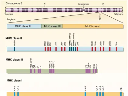
1.3 The human major histocompatibility complex (MHC)
The major histocompatibility complex (MHC) is a large multigenic region found in most vertebrates containing at least 128 genes in humans, of which more than 20% encode proteins of the immune system [4,12]. The human MHC is the most gene-dense and polymorphic region and is part of the so-called extended MHC region, covering 7.6 Mb of the short arm of the chromosome 6 [4]. The MHC region contains, among others, highly polymorphic genes encod-ing the aforementioned MHC molecules that present protein-derived peptides (antigens) to T cells [1]. These type of antigen-presenting molecules are known as classical MHC molecules [3]. There are also structurally related molecules that are monomorphic or oligomorphic, known as non-classical MHC mol-ecules [3,13]. Non-classical MHC molmol-ecules are involved in immune and non-immune processes and some can function in the presentation of peptide anti-gens [13]. Besides, the MHC region contains genes involved in the processing of antigens [4].
For historical reasons, the human MHC is also referred to as the human leuko-cyte antigen (HLA) complex, since the first MHC gene products were discov-ered on the surface of leukocytes [4]. Three distinct regions have been identi-fied within the human MHC: the MHC class I (MHC I), MHC class II (MHC II) and MHC class III (MHC III) (Figure 1). The MHC I and II regions encode both classical MHC and non-classical MHC molecules. The MHC I region contains
among other genes, the classical MHC I genes (also known as MHC Ia genes)
HLA-A, HLA-B and HLA-C and the more ancient non-classical MHC I genes
(also known as MHC Ib genes) HLA-E, HLA-F and HLA-G [4,13]. The MHC II re-gion covers the classical MHC II genes HLA-DP, HLA-DQ and HLA-DR, as well as non-classical MHC II genes HLA-DM and HLA-DO [4]. Some non-classical MHC I molecules play a role in activating specialized classes of T cells, whereas non-classical MHC II molecules regulate peptide loading into classical MHC class II molecules. The MHC III region is located between the class I and class II regions and encloses miscellaneous non-HLA genes (e.g. MICA, MICB) encod-ing proteins with or without immune function [12].
1.4 The classical MHC class I and II molecules
MHC I and MHC II molecules are cell-surface glycoproteins with a similar three-dimensional structure and similar function in presenting peptide fragments or antigens to the immune system [15]. Nevertheless, these molecules differ in their tissue distribution and in the type of antigenic peptides they display that reflects different antigen processing pathways [16]. The 3 classical human MHC I genes (HLA-A, HLA-B and HLA-C) are co-dominantly expressed on the cell surface of all nucleated cells and play a major role in adaptive immunity.
Figure 1. Location and organization of the human MHC complex on chromosome 6. Adapted with permission
from [14]. Copyright Massachusetts Medical Society
Telomere Telomere Chromosome 6 Regions q Centromere p 6p 21 .3 1 DRA DRB3 DRB2 DRB1 DQ A1 DQB1 DOB DMB DMA DO A DP A1 DPB1 DPB2 P4 50 , C 21B C4B BF C2 HSPA 1B HS PA 1A HS PA 1L C4A LTB TNF- a-LT A HLA-E HLA -C HLA-B M ICA M ICB
HLA-A HLA-G HLA-F HFE TAPBP PSMB9 (LMP2) TAP1 PSMB8 (LMP1) TAP2
MHC class II
MHC class I MHC class III
On the contrary, surface expression of MHC II molecules is restricted mainly to APCs such as macrophages, B cells and DCs, although it can be induced by interferon-γ (IFN-γ) and other stimuli in non-APCs like mesenchymal stromal cells, fibroblasts, endothelial cells and activated human T cells [5,16].
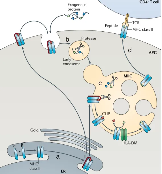
1.4.1 The MHC II molecules and associated peptides
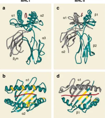
MHC II molecules are transmembrane glycoproteins with short cytoplasmic domains, composed of one α and one β chain. The membrane-proximal re-gion consists of one conserved domain that is part of the α subunit and an-other conserved domain of the β subunit (Figure 2c). MHC II molecules exhibit enormous amino acid sequence variation in the region that interacts with the peptide named the peptide-binding groove [17] (Figure 2d). Consequently, dif-ferent alleles bind difdif-ferent sets of peptides. The groove is formed by the jux-taposition of the N-terminal regions of the α and β chains. The peptide-binding groove is open at both ends and hence, long peptides, generally between 15 and 24 amino acids long, can bind and overhang the binding groove resulting in more or less restrictive binding motifs [17] (Figure 2d).
1.4.2 The MHC I molecules and associated peptides
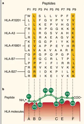
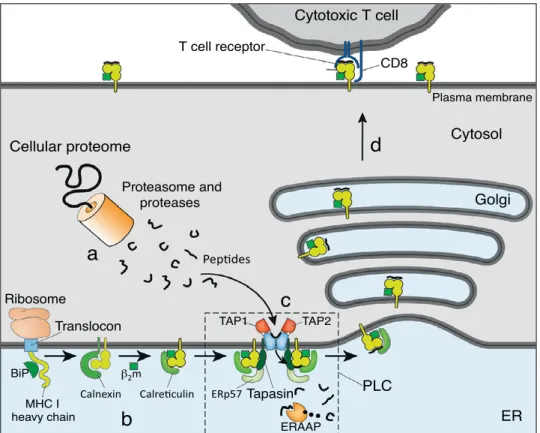
The molecular target of the TCR of CD8+ T cells is the complex formed by the MHC I and the associated peptide [2]. MHC I molecules are cell-surface het-erodimers composed of a polymorphic transmembrane 44-kd heavy (α) chain and a 12-kd invariant light chain, known as β2-microglobulin (β2m) [2,14,16] (Figure 2a). The heavy chain consists of 3 extracellular domains (α1, α2 and α3), a transmembrane domain and a short intracellular domain that anchors the protein in the cell membrane [18]. The α1 and α2 domains form a platform with a groove, in which antigenic peptides can bind noncovalently through the N and C termini [17] (Figure 2a). The main anchor residues of the peptide to the peptide-binding groove are frequently the second and the last C-terminal residue [15] (Figure 3b). In some exceptional cases, the main anchors are lo-cated in positions P3 (as in HLA-A1-associated peptides) and P5 (as in HLA-B8-associated peptides). The peptide-binding groove is blocked at both ends by bulky aromatic amino acids that typically limit the length of the bound peptide
to 8-10 residues [15] (Figure 2b). Less frequently, MHC I molecules can bind longer peptides [19-21], in which case the central part of the peptide protrudes and the binding is presumably less stable [15,22].
A prominent characteristic of the human MHC I molecules is their high degree of polymorphism [1,23]. The IMGT/HLA database of the international ImMuno-GeneTics project (http://www.ebi.ac.uk/ipd/imgt/hla) registers thousands of MHC I alleles in their last release (2013-07-25): 2,365 HLA-A, 3,005 HLA-B and 1,848 HLA-C alleles. Besides, HLA-B is the most polymorphic gene in the hu-man genome [24]. Most polymorphisms in exons 2 and 3 of the HLA-A, HLA-B and HLA-C genes lead to amino acid substitutions in the floor and sides of the peptide-binding groove of the corresponding MHC I protein [17,25]. Thereby, different MHC I allelic products display distinct amino acid preferences at key
c a d b I I C H M I C H M α2 α1 α3 β2m α1 α2 α1 β1 α1 α2 β1 β2
Figure 2. Three-dimensional structures of peptide-bound MHC I and MHC II molecules
(a) and (b) HLA-A2 molecule complexed with influenza derived peptide. (c) and (d) HLA-DR1 molecule complexed with influ-enza derived peptide. Highly polymorphic residues proximal to the peptide-binding groove are highlighted in yellow; the peptide is shown in red. Adapted with permission from [17].