© The Author(s) 2020. Published by Oxford University Press on behalf of the European Society of Human Reproduction and Embryology.
This is an Open Access article distributed under the terms of the Creative Commons Attribution NonCommercial-NoDerivs licence (http://creativecommons.org/licenses/by-nc-nd/4.0/), which permits non-commercial reproduction and distribution of the work, in any medium, provided the original work is not altered or transformed in any way, and that the work properly cited. For commercial re-use, please contact journals.permissions@oup.com
ESHRE PAGES
Recommendations for the surgical
treatment of endometriosis. Part 2:
deep endometriosis
†‡¶
Working group of ESGE, ESHRE, and WES, Joerg Keckstein
1,*,
Christian M. Becker
2, Michel Canis
3, Anis Feki
4, Grigoris F. Grimbizis
5,
Lone Hummelshoj
6, Michelle Nisolle
7, Horace Roman
8,9,
Ertan Saridogan
10, Vasilios Tanos
11, Carla Tomassetti
12,
Uwe A. Ulrich
13, Nathalie Vermeulen
14, and
Rudy Leon De Wilde
151Endometriosis Centre Dres. Keckstein, Richard-Wagner Strasse 18, 9500 Villach, Austria2Nuffield Department of Obstetrics and Gynaecology, University of Oxford, John Radcliffe Hospital Womens Centre, OX3 9DU Oxford, UK 3Department of Gynaecological Surgery, University Clermont Auvergne CHU, Estaing 1 Place Lucie Aubrac, 63000 Clermont-Ferrand, France 4Department of Obstetrics and Gynecology, HFR Fribourg Hopital cantonal, 1708 Fribourg, Switzerland 51st Department of Obstetrics and Gynecology, Medical School Aristotle University of Thessaloniki, Tsimiski 51 Street, 54623 Thessaloniki, Greece 6World Endometriosis Society, London N1 3JS, UK7Hôpital de la Citadelle, Department of Obstetrics & Gynecology, 4000 Liège, Belgium8Endometriosis Centre, Clinic Tivoli-Ducos, Bordeaux, France 9Department of Obstetrics and Gynaecology, Aarhus University Hospital, Aarhus, Denmark10Reproductive Medicine Unit, Elizabeth Garrett Anderson Wing Institute for Women’s Health, University College Hospital, NW1 2BU London, UK11Department of Obstetrics and Gynecology, Aretaeio Hospital, 2024 Nicosia, Cyprus 12Department of Obstetrics and Gynaecology, Leuven University Fertility Centre, University Hospital Leuven, 3000 Leuven, Belgium 13Department of Obstetrics and Gynaecology, Martin Luther Hospital, 14193 Berlin, Germany14ESHRE, Central office - Meerstraat 60, BE 1852 Grimbergen, Belgium15University Hospital for Gynecology, Carl von Ossietzky Universitat Oldenburg, 26129 Oldenburg, Germany
*Correspondence address. Endometriosis Centre Dres. Keckstein Villach, Richard-Wagner Strasse 18, 9500 Villach, Austria. E-mail: joerg@keckstein.at; guidelines@eshre.eu https://orcid.org/0000-0002-3943-3300
Submitted on November 27, 2019; resubmitted on November 27, 2019; editorial decision on January 13, 2020
STUDY QUESTION:How should surgery for endometriosis be performed?
SUMMARY ANSWER:This document provides recommendations covering technical aspects of different methods of surgery for deep endometriosis in women of reproductive age.
WHAT IS KNOWN ALREADY:Endometriosis is highly prevalent and often associated with severe symptoms. Yet compared to equally prevalent conditions, it is poorly understood and a challenge to manage. Previously published guidelines have provided recommendations for (surgical) treatment of deep endometriosis, based on the best available evidence, but without technical information and details on how to best perform such treatment in order to be effective and safe.
STUDY DESIGN, SIZE, DURATION:A working group of the European Society for Gynaecological Endoscopy (ESGE), ESHRE and the World Endometriosis Society (WES) collaborated on writing recommendations on the practical aspects of surgery for treatment of deep endometriosis.
PARTICIPANTS/MATERIALS, SETTING, METHODS:This document focused on surgery for deep endometriosis and is complemen-tary to a previous document in this series focusing on endometrioma surgery.
MAIN RESULTS AND THE ROLE OF CHANCE:The document presents general recommendations for surgery for deep endometriosis, starting from preoperative assessments and first steps of surgery. Different approaches for surgical treatment are discussed and are respective of location and extent of disease; uterosacral ligaments and rectovaginal septum with or without involvement of the rectum, urinary tract or extrapelvic endometriosis. In addition, recommendations are provided on the treatment of frozen pelvis and on hysterectomy as a treatment for deep endometriosis.
LIMITATIONS, REASONS FOR CAUTION:Owing to the limited evidence available, recommendations are mostly based on clinical expertise. Where available, references of relevant studies were added.
WIDER IMPLICATIONS OF THE FINDINGS:These recommendations complement previous guidelines on management of endometrio-sis and the recommendations for surgical treatment of ovarian endometrioma.
STUDY FUNDING/COMPETING INTEREST(S):The meetings of the working group were funded by ESGE, ESHRE and WES. Dr Roman reports personal fees from ETHICON, PLASMASURGICAL, OLYMPUS and NORDIC PHARMA, outside the submitted work; Dr Becker reports grants from Bayer AG, Volition Rx, MDNA Life Sciences and Roche Diagnostics Inc. and other relationships or activities from AbbVie Inc., and Myriad Inc, during the conduct of the study; Dr Tomassetti reports non-financial support from ESHRE, during the conduct of the study; and non-financial support and other were from Lumenis, Gedeon-Richter, Ferring Pharmaceuticals and Merck SA, outside the submitted work. The other authors had nothing to disclose.
TRIAL REGISTRATION NUMBER:na
Key words: endometriosis / laparoscopy / surgery / deep endometriosis / extrapelvic / frozen pelvis / hysterectomy / good practice recommendations
†ESHRE Pages content is not externally peer reviewed. The manuscript has been approved by the Executive Committee of ESHRE. ‡This paper has been approved by the Executive Committees of the ESGE and WES.
¶This article has been co-published with permission in HROpen and FACTS, VIEWS & VISION in Obgyn.
WHAT DOES THIS MEAN FOR PATIENTS?
This paper was produced by a European working group looking at the different types of surgery for endometriosis, a common condition where tissue, which is similar to the lining of the womb, is found elsewhere in the body. The working group looked specifically at how best to treat a type of the disease, named deep endometriosis. Drug therapies may be used to treat endometriosis, but when endometriomas are found and need treatment, surgery is often used. There are risks associated with surgery, and especially repeated surgery, including adhesions.
The working group looked at the main types of surgery which are used to treat deep endometriosis, focussing on the different locations in the pelvis where the lesions can be found. The paper discusses in detail how different types of surgery should be performed, taking potential risks into consideration, and stresses that careful planning and involving different surgeons specialising in bowel or bladder is essential to ensure the best outcomes.
Introduction
Endometriosis is highly prevalent, yet compared to equally prevalent conditions it is poorly understood and a challenge to manage. It has been estimated that more than 176 million women worldwide suffer from endometriosis and its associated symptoms including infertility, cyclical and non-cyclical abdominal pain, dysmenorrhea, dyspareunia,
dysuria and dyschezia (Adamson et al., 2010). It is generally accepted
that no correlation exists between the severity of such pain symptoms and the extent of disease as characterised by the most commonly used revised American Fertility Society/American Society for Reproductive
Medicine (rASRM) staging system (Vercellini et al., 2007); however,
with the use of other classification systems, such as ENZIAN, a
correla-tion with pain symptoms may exist (Montanari et al., 2019). It has been
shown that women with surgically verified endometriosis reported the highest pain symptoms compared to women with other gynaecologic
pathology (Schliep et al., 2015). However, the correlation between
location and depth of endometriotic lesions and pain location is poor
(Hsu et al., 2011). Therefore, it could be relevant to include histology
when classifying the disease (Bouquet de Joliniere et al., 2019).
Endometriosis may be categorized into three entities: peritoneal endometriosis, ovarian endometriotic cysts (endometrioma) and deep endometriosis (DE) (previously known as deep infiltrating
endometrio-sis or DIE) (Nisolle and Donnez, 1997). In addition to medical therapy
with hormones and analgesics, surgery has been shown to significantly
improve endometriosis-associated symptoms (Duffy et al., 2014;Byrne
et al., 2018). However, like medical intervention, surgery is not always
successful and is also associated with clinically relevant risks (Chapron
et al., 1998; Becker et al., 2017). Treatment failure can be partially attributed to the heterogeneity of endometriosis and, in the case of surgical intervention, is directly correlated with factors such as surgical
. . . . . . . . . . . . . . . . . . . . . . . . . . . . . . . . . . . . . . . . . . . . . . . . . . . . . . . . . . . . . . . . . .
experience, the complexity of each case and anatomical locations of the disease.
A working group comprising members of the European Society for Gynaecological Endoscopy (ESGE), ESHRE and the World Endometriosis Society (WES) has set out to produce a series of recommendations on the practical aspects of endometriosis surgery. The first part of this series on endometrioma surgery was published
in 2017 (Working group of ESGE-ESHRE and WES et al., 2017).
This second part focuses on the surgical management of DE. After general considerations, such as definition and anatomical specifications, this publication concentrates on recommendations related to pre-operative management and surgical technique respective of location and extent of disease. Choices of different treatment options for DE and the selection of patients that would benefit from surgery are beyond the scope of this document. Conservative treatment, including pain management, has to be considered thoroughly.
Materials and Methods
Previously published guidelines have provided recommendations on the management of endometriosis, based on the best available
evi-dence (Johnson et al., 2013;Dunselman et al., 2014;Ulrich et al., 2014).
However, these guidelines were not intended to provide recommen-dations on the technical details of surgical procedures.
Due to the scarcity of evidence on these technical details, the current recommendations are primarily based on expert opinion on best clinical practice. Studies and trials of these approaches have been cited in the text, when available.
In addition to the recommendations, the working group has set up a web platform with videos on the different options available for
surgery of DE. The web platform is accessible through the following
link (https://www.eshre.eu/surendo) or via the ESGE, ESHRE or WES
websites.
(Results) Good Practice
Recommendations
Deep Endometriosis
Definition of DE
Rokitansky was probably the first to describe DE in the uterus (
Roki-tansky C., 1860; Hudelist et al., 2009; Tsui et al., 2015). Thomas
Cullen later described DE, although he described it as adenomyosis
of the round ligament (Cullen, 1896). In modern times, using laser
excision, Martin et al. described in a landmark study that the lesions in approximately one-third of the women were penetrating more than
4 mm under the peritoneal surface (Martin et al., 1989). Cornillie
et al. then investigated the activity of endometriosis tissue at different
sub-peritoneal depths and suggested that DE was invasive (Cornillie
et al., 1990); however, clear evidence of infiltration is still missing. Histologically, the investigators found active glandular and stromal tissue as defined by the presence of mitoses and glycogen accumulation 5 mm below the peritoneal surface. Fibromuscular hyperplasia, cystic transformation of the glands and perivascular mononuclear inflam-matory cells were noted. Other definitions of DE have been used related to the location of the disease including the involvement of bowel, bladder, ureter, vagina, parametrium (cardinal ligament) and
diaphragm (Keckstein, 2017). Another definition by Bazot described
DE as a fibrous/muscular infiltration of organs and anatomical struc-tures containing endometrial tissue below the peritoneum, regardless
of the depth of infiltration (Bazot and Darai, 2017).
For the purpose of this publication, and the surgical treatment of the disease, the working group is defining DE as the involvement of
endometrial-like tissue with a depth of more than 5 mm (Koninckx
et al., 2012).
Morphological considerations
Infiltration of the abdominal and pelvic parietal peritoneum by endometriotic tissue can lead to involvement of retroperitoneal structures depending on the location and depth of the endometriotic nodule. DE is often associated with fibrotic changes. Thus, retraction of surrounding structures is common, and this is to be considered during pre-operative and intra-operative planning and assessment of the optimal surgical approach.
Surgeons must have a significant knowledge of pelvic anatomy in order to have an approach to a grossly distorted surgical field. Thus, pelvic anatomical landmarks represent essential points of reference to start procedures such as mobilization of the pelvic viscera, wide peri-toneal resections or the identification of further anatomical structures to be preserved, such as bowel, ureter, vessels and parasympathetic and orthosympathetic pelvic neural fibres in nerve-sparing procedures
(Ceccaroni et al., 2018). The preparation or dissection of specific
anatomical spaces (Latzko, Okabayashi, Yabuki), which have been described by various authors, helps to identify these landmarks in order
to facilitate a complete and safe excision of the deep lesions (Yabuki
et al., 2005;Ceccaroni et al., 2018;Hudelist et al., 2018;Puntambekar
and Manchanda, 2018). . . . . . . . . . . . . . . . . . . . . . . . . . . . . . . . . . . . . . . . . . . . . . . . . . . . . . . . . . . . . . . . . . . . . . . . . . . . . . . . . . . . . . . . . . . . . . . . . . . . . . . . . . . . . . . . . . . . . . . . . . . . .
The abdominal (upper third) part of the retroperitoneally located ureter follows the ventral side of the psoas muscle. Entering the pelvis, the ureter crosses dorsally to the ovarian artery and vein and then ventrally to either the common (left side) or external (right side) iliac artery. It then courses ventrally directly below the peritoneum of the pelvic sidewall and runs antero-laterally from the uterosacral ligament. Before entering the urinary bladder, the ureter crosses the uterine artery caudally. Ureteral blood supply originates from the renal artery (upper third), ovarian artery and abdominal aorta (middle third) as well as the internal iliac arteries and smaller vessels (lower third) and is usually located at the lateral side of the ureter. Plenty of anastomoses of these blood vessels exist within the ureteral adventitia where also the ureteral nerve plexus can be found. Anatomic variations are common; for example, the prevalence of a duplex ureter has been described in 0.5–6% of cases and has to be taken into consideration when removing or ablating endometriotic lesions or nodules in the
pelvis (Phillips et al., 1987). In addition, the normal anatomical position
of the ureter can be altered significantly in the presence of DE, such as medial displacement due to surrounding fibrosis and/or infiltration. Ureteral stricture or complete obstruction may lead to hydro-ureter and hydronephrosis (this is further discussed in the section on urinary tract endometriosis).
The bilateral inferior hypogastric plexus supplies the pelvic organs, such as the rectum, bladder and cervix, with sympathetic and parasym-pathetic nerve fibres, and receives visceral afferent fibres. The
sympa-thetic nerve fibres come from the superior hypogastric plexus (L3–L4)
via the hypogastric nerve, which follows the common and the internal iliac artery: they are found in the pararectal space medial and dorsal to the ureter. The parasympathetic fibres originate from the pelvic and
sacral splanchnic nerves (S2–S4).
The intraperitoneally located sigmoid and the retroperitoneal rectum are often involved in DE. Endometriotic nodules in the recto-vaginal septum result in adherence of the recto-sigmoid to the lower dorsal side of the uterus, cervix and vagina. The large bowel wall consists of multiple layers, all of which can be invaded
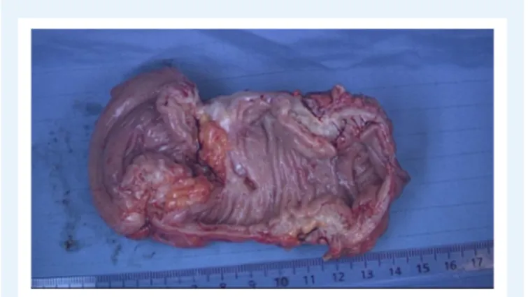
by endometriotic tissue (Fig. 1). The sigmoid colon receives its blood
supply from multiple branches of the inferior mesenteric artery. There exist anastomoses between the sigmoid arteries and the superior rectal (superior haemorrhoidal) artery, itself a branch of the inferior mesenteric artery, supplying the rectum and creating further anastomoses with the middle rectal (middle haemorrhoidal) artery, a branch of the inferior vesical artery which originates from the internal iliac artery. Distally, the rectum receives some blood supply from the inferior rectal (inferior haemorrhoidal) artery, which mainly supplies the anal canal originating from the internal pudendal artery, another branch of the internal iliac artery. Again, anastomoses exist between the middle and inferior rectal arteries.
Like any subperitoneal (deep) endometriosis, nodules in the vesico-uterine pouch (ventral cul-de-sac) can involve or invade surrounding structures, such as the underlying urinary bladder, eventually displacing the round ligaments medially. The bladder wall consists of mucosa, a submucosal layer (lamina propria) containing blood vessels and nerve fibres, a muscularis propria (detrusor muscle) consisting of an inner and outer longitudinal and a middle circular muscle layer, and either a serosal layer or adventitia. The ureters, after passing about 1 to 2 cm laterally to the uterine cervix and coursing ventral to the lateral border of the vagina, enter the bladder postero-laterally in an oblique angle.
Figure 1 The layers of the recto-sigmoid colon, as graphical representation (upper figure), in eosin-stained healthy recto-sigmoid colon (lower left) and eosin-stained endometriosis-affected recto-sigmoid colon (lower right).1 serosa (sigmoid) or Adventitia (rectum);
2 subserosa; 3 tunica muscularis (outer longitudinal layer, inner circular layer) with plexus myentericus in between; 4 submucosa with plexus submucosus, blood, and lymphatic vessels; 5 mucosa.
The urinary bladder receives its blood supply from the superior, middle and inferior vesical arteries (branches of the internal iliac artery) as well
as the vaginal (branch of uterine artery) artery (see alsoFig. 2).
Classification
The most commonly used classification system of endometriosis, the rASRM classification, does not provide sufficient information for DE; correlation with symptoms is poor, and it does not predict surgical
diffi-culty level or outcome (Johnson et al., 2017). Several systems classifying
and documenting the extent of the DE have been developed,
includ-ing the ENZIAN classification (Keckstein et al., 2003b;Tuttlies et al.,
2005;Stiftung Endometriose Forschung (Foundation for Endometriosis
Research), 2011) (Fig. 3), the Visual Numeric Endometriosis Surgical
Score (VNESS) system (Abdalla AL and S., 2015) and those proposed
by Chapron et al. and Adamyan (Adamyan, 1993; Chapron et al.,
2003). The ENZIAN classification showed a significant correlation
between the extent of the disease, difficulty and length of surgery and
symptoms (Haas et al., 2013a;Haas et al., 2013b;Haas et al., 2013c;
Morgan-Ortiz et al., 2019;Montanari et al., 2019). The different
scor-. . . . . . . . . . . . . . . . . . . . . . . . . . . . . . . . . . . . . . . .
ing systems with their advantages and disadvantages are summarised in
Table I.
In addition to classifying endometriosis, the documentation of sur-gical findings, such as in Endometriosis Fertility Index (EFI), has been
found to have a prognostic value in infertile women (Adamson and
Pasta, 2010;Adamson, 2013).
The working group emphasises the importance of classification of DE, but also the limitations of the existing systems, specifically with regard to the scoring of the severity of disease. The ideal classification system for DE should define accurately the anatomical location, size of the lesions and level of involvement of the adjacent organs. It should also be reproducible and should help the surgeon in the planning and execution of surgery.
The working group recommends documenting the following information:
• the location of DE lesions;
• uterosacral ligaments, including whether ureters are infiltrated;
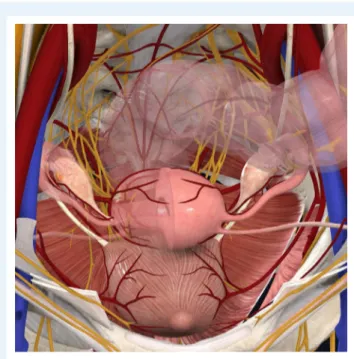
Figure 2 Anatomical landmarks in DE surgery (vessels, nerves, ureter, bladder, bowel) which may be involved and have to be respected carefully. DE: deep endometriosis. (Image
courtesy of Complete Anatomy (reprinted with permissions).)
• bowel, including involvement of muscularis layer;
• bladder, including involvement of muscularis and ureteral ostia;
• other sides in the pelvis;
• extrapelvic locations;
• involvement of the ovaries;
• the sizes of the lesions;
• the number of lesions;
• the degree of involvement of adjacent organs and structures.
The current paper is structured according to the different locations of DE, with further subdivision on extent (where relevant).
Pre-operative assessment and preparation
for surgery
Pre-operative assessment of patients with suspected DE aims to estab-lish a diagnosis, evaluate extent of disease and determine the optimal surgical approach. This includes a thorough medical history, clinical examination and imaging. When assessing the medical history and symptoms, the focus should be directed on symptoms that could indicate the presence of DE lesions in specific organs/locations. These include, but are not limited to, cyclical haematuria and cyclical
rec-tal bleeding (Chattot et al., 2019). It is very important to record
co-morbidities and take them into consideration when deciding on surgery.
It is helpful to use a validated symptom questionnaire for data collection, for audit and comparison. Women’s symptoms, such as pain, should be assessed, for instance using a visual analogue scale and
. . . . . . . . . . . . . . . . . . . . . . . . . . . . . . . . . . . . . . . . . . . . . . . . . . . . . . . . . . . . . . . . . . . . . . . . . . . . . . . . . . . . . . . . . . . . . . . . . . . . . . . . . . . . . . . . . . . . . . . . . . . .
general quality of life questionnaires (e.g. the World Endometriosis Research Foundation (WERF) set up the Endometriosis Phenome and Biobanking Harmonisation Project (EPHect) Endometriosis Patient
Questionnaire) (Vitonis et al., 2014;Bourdel et al., 2015;Vanhie et al.,
2016). Other questionnaires can be used to assess specific aspects of
endometriosis, including sexual function, urinary function, bowel func-tion, recovery and depression and anxiety. These symptoms should be assessed, taking into account uterine bleeding pattern and associated dysmenorrhea, as painful symptoms associated with irregular periods can be managed by starting and/or adapting a medical treatment.
Fertility plans and indications for surgery should be discussed before
the surgical procedure, for instance using the EFI (Adamson et al.,
2010). Choices of different treatment options (surgery, IVF) for DE and
the selection of patients that would benefit from surgery are beyond the scope of this document. However, IVF could be proposed as the first step in patients who have been operated previously, who have a low ovarian reserve and/or when there is male factor infertility
(Dunselman et al., 2014). Oocyte freezing may also need to be
dis-cussed as an option in cases of coexistent ovarian endometrioma
(Working group of ESGE-ESHRE and WES et al., 2017).
A surgical history is essential. Previous operation reports should be read in detail and any pictures/videos reviewed carefully. The surgeon should know if the retroperitoneal space was opened and, if it was, its side should be noted. It is also essential to know whether ureterolysis and/or bowel dissection was performed. Extensive ureteral dissection always impairs ureteral vessels, so that even a very cautious and meticulous repeat ureterolysis may induce ureteral ischemia. Similarly, previous bowel dissection, and/or previous rectal shavings, implies that the bowel wall may be compromised by previous procedures, so the risk of bowel injury and/or postoperative fistula may be increased. As the risks of intra- and/or postoperative urinary or intestinal com-plications are considered much higher in women who have under-gone previous extensive procedures, this should be taken in account during counselling the woman, decision-making and organisation of the surgical team. History of previous ovarian cystectomy should be taken into account when planning and performing surgery on ovarian endometrioma in particular in women who may wish to conceive in the
future (Working group of ESGE-ESHRE and WES et al., 2017).
Clinical examination
Clinical examination in women with suspected DE includes not only a physical examination of the pelvis but also the inspection and pal-pation of the abdomen. The examination may need to be extended beyond the pelvis, depending on the symptoms of the woman. Loca-tion and extent of disease can sometimes be determined by clinical
examination (Ripps and Martin, 1992; Koninckx et al., 1996; Bazot
et al., 2009). There should be special emphasis on the visualization of DE in the vagina by inspection of the dorsal fornix with a bivalved speculum.
Vaginal examination can facilitate the detection of infiltration or nodules of the vagina, uterosacral ligaments or pouch of Douglas. It could also contribute to the assessment of the extent of disease to the pelvic sidewall, which is important to evaluate the risk of trauma to the hypogastric plexus and/or the ureter. Rectovaginal digital examination may allow the detection of infiltration or mass involving the
rectosig-moid or adnexal masses (Ripps and Martin, 1992;Koninckx et al., 1996;
Ta b le I Classification syst e ms fo r d e e p e ndometriosis. Classification syst e m Scorin g of compar tment s Scorin g of se v e rity P o sitiv e aspe ct s N egativ e a spe c ts ... ... ... ... ENZIAN Classification ( Ke c k st e in et al ., 2003b ) ( T u ttlies et a l., 2005 )( Stiftung Endometriose Fo rsc hung (F oundation for Endometriosis R e sear ch ), 2011 ) R e tr operitoneal structur e s ar e di vided into the fo llo wing thr e e compar tments: -A : recto vaginal sep tum and vagina -B : Sac ro uterine ligament to p elvic w all -C : rectum and sigmoid colon R e tr operitoneal distant locations: -FA: adenomy o sis -FB: in vo lv ement o f the bladder -FU: intrinsic in volv e ment of the u re ter -FI: bo w e ldisease cr anial to the re ctosigmoid junction -FO: o ther locations , suc h as abdominal wall endometriosis Se ve rit y is rated in the same wa y for all co mpar tments: -G rade 1: in vasion < 1c m -G rade 2: in vasion 1–3 cm -G rade 3: in vasion > 3c m R e lati ve ly good morphological desc rip tion p ro vided Cor relation b etw een symp toms and in volv e d co mpar tments Additional aspects can be calc u lated (e.g . the antic ipated operating time, risk of co mplication d uring and after surger y) Inter n ational ac cep tanc e o f the classification is still lo w—mostl y used in Eur o pe Cha p ro n ( Chapr o n et a l., 2003 ) Ve n tr al D IE -A 1: Bladder Dorsal DIE -P 1: Uter osac ral ligament -P 2 : V ag in a -P 3: Intestine (solel y -with o r w ithout vaginal infiltration-or multiple intestinal location) No sc oring L ink e d to an o perati ve pr oc edur e R e por ted in tw o p ublications b y the authors , n o fur ther inf o rm ation No t w idel y used Adam y a n ( Adam yan, 1993 ) Adamyan Stage I: endometrio tic lesions ar e co nfined to the re cto vaginal ce llular tissue in the ar ea of the vaginal vault. Adamyan Stage II: endometrio tic tissue in vades the cer vix and penetrates the vaginal wall, causing fibr o sis and small cyst for mation. Adamyan Stage Ill: lesions spr ead into the sac ro-uterine ligaments and the re ctal ser o sa. Adam yan Stage IV : the rectal wall, re cto-sigmoid zo ne, and re ctouterine peritoneum ar e co mpletel y in vo lv ed, and the rectouterine p ouc h is to tall y o bliterated No sc oring N o t published in p eer re vie w ed jour nal No t w idel y used Visual Numeric Endometriosis S urgical Scor e (VNESS) (Abdalla AL and S ., 2015 ) Eight locations: Left adnexa—Left p elvic side wall -L eft uter osac ral ar e a/v e ntral compar tment (UVF)—dorsal co mpar tment (POD)/right uter osac ral ar e a—right p elvic side wall—right adnexa Fo r e ac h location, a sc o re betw een 0 and 4 re pr esenting the se verit y o f endometriosis 0: No visible e ndometriosis 1: Superfic ial e ndometriosis 2: DIE w ith n o attac h ment to visc era 3: DIE loosel y adher ent to visc e ra 4: DIE d ensel y adher e nt to visc era O R in vading m usc u laris O R k issing o varies Intuiti ve and easy to re member . Visuall y h elpf ul No t p ublished in p eer re vie w ed jour nal No t w idel y used DIE; d eep infiltrating endometriosis , UVF: Left uter osac ral ar e a/v e ntral compar tment POD: Dorsal co mpar tment
Figure 3 Revised ENZIAN-classification for DE. The system classifies the clinical findings of endometriosis according to their localisation
(com-partment) and size (<1 cm, 1–3 cm, >3 cm). The ENZIAN classification focusses on the three dimensions (compartments) in the pelvis: A = craniocaudal axis or compartment (rectovaginal space, vagina), B = laterodorsal axis (uterosacral and cardinal ligaments), C = dorsal axis (rectosigmoid). Other localisations as uterus, bladder, ureter, other bowel involvement and extragenital localisations are respected as well and described with suffix F). The ENZIAN Classification is under revision (2019) again which is under publication.
recommended to assess the lateral and dorsal extension of the disease allowing detection of the patients who are at risk of hypogastric vessels injury and/or hypogastric plexus damage. It also allows the surgeon to evaluate the mobility of the nodule of the dorsal cul-de-sac and thus to predict how difficult the surgery may be.
Imaging and other investigations
The ESHRE Guideline on the Management of Endometriosis recom-mends assessing the ureter, bladder and bowel involvement by
addi-. . . . . . . . . . . . . . . . . . .
tional imaging, if there is a suspicion based on history or physical
exam-ination of DE, in preparation for further management (Dunselman
et al., 2014). Imaging for suspected involvement of bladder, bowel
and ureters may start with ultrasonography (US) (Guerriero et al.,
2016). Other imaging techniques, such as MRI (Fig. 4) including neuro
MRI, and computed tomography (CT) (only in selected cases as it is associated with unacceptably high radiation exposure) (Exacoustos et al.,
2014;Guerriero et al., 2018) can also be used. Assessment of MRIs
Figure 4 MRI picture of the pelvis. DE in the rectum with small,
dense adhesions between the posterior wall of the cervix and anterior wall of the rectum (right circle) and adenomyosis (left circle).
and sigmoidoscopy may give additional information about stenosis of the bowel. These investigations aim to determine the location, size and number of DE lesions (nodules or plaques) as well as the level of infiltration (depth of invasion, length of infiltration, stenosis) into the organ/structure involved. Furthermore, the identification of lesions on/in the pelvic wall (i.e. sacral root) and other extragenital localisation (abdominal wall, inguinal canal, diaphragm, lung, etc.) with specific imaging techniques is relevant, as it has an important impact on the surgical treatment and its planning. Kidney sonography is mandatory in every patient with DE potentially involving the ureters to prevent overlooking silent hydronephrosis.
Bowel. Where appropriate, involvement of bowel muscularis and the distance between the inferior border of the lowest bowel lesion and the anal verge should be evaluated as these would be expected to have an impact on the type of surgery that will be performed. Limitations of the accuracy of these investigations should be kept in mind.
Colonoscopy identifies stenosis or intraluminal lesions, which are rare, but unfortunately it does not give sufficient information about the presence, localisation and size of endometriosis in the bowel wall
(Fig. 5).
US is the first-line imaging modality for the assessment of pelvic endometriosis. It has been demonstrated that most of the deep lesions in the lower colon can be identified with a high sensitivity and specificity
(Hudelist et al., 2009;Hudelist et al., 2011;Exacoustos et al., 2017).
It may have limitations with respect to field of view and operator
dependence (Fig. 6).
Transrectal ultrasonography (TRUS) may be used for rectosigmoid involvement but could not be adequately assessed for other anatomical sites because of scant heterogeneous data.
MRI is usually performed as an additional examination in complex cases or prior to surgery and is highly accurate in the evaluation of endometriosis. . . . . . . . . . . . . . . . . . . . . . . . . . . . . . . . . . . . . . . . . . . . . . . . . . . . . . . . . . . . . . . . . . . . . . . . . . . . . . . . . . . . . . . . . . . . . . . . . . . . . . . . . . . . . . . . . . . . . . . . . . . . .
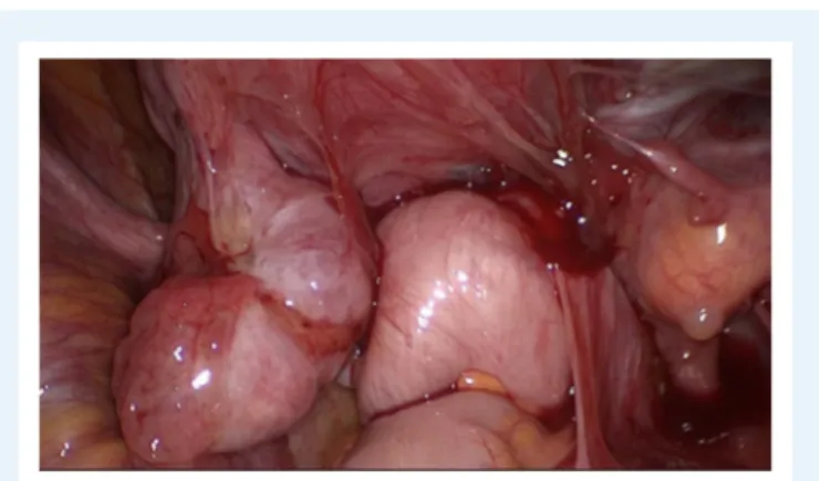
Figure 5 Deep endometriosis of the rectosigmoid. The
extent of the white nodules (multifocal) which infiltrate the muscular layer is not visible by colonoscopy and also difficult to identify com-pletely by laparoscopy.
Figure 6 Transvaginal ultrasound of the rectosigmoid with signs of infiltration of the muscular layer. Length > 3 cm Enzian
C3. Hypodense ultrasound pattern (halfmoon-shaped) represents the hyperplasia of the lamina muscularis with inclusion of epithelium, stroma and fibrosis.
Diagnostic accuracies were higher for transvaginal ultrasonography (TVUS or TVS) with bowel preparation (TVUS-BP) and rectal water contrast (RWC-TVS) and for 3.0 T MRI than for conventional methods,
although the paucity of studies precluded statistical evaluation (
Nisen-blat et al., 2016). TVUS for DE is highly dependent on the experience
of the operator and the quality of the US equipment. The additional use of vaginal and rectal contrast US gel can further enhance the image
(Grammatico et al., 2017).
Multi-detector computed tomography enema (MDCT-e) is another technique which might have a high diagnostic performance for
rectosig-moid and other bowel endometriosis (Ferrero et al., 2011).
Virtual colonoscopy may add new information to that provided by
MRI (Mehedintu et al., 2018). Virtual colonoscopy is a single
non-invasive short procedure. It provides information about lesions in the
whole length of the colon (esp. sigmoid, ileum, caecum) (van der Wat
et al., 2013). However, a dry bowel preparation is necessary, and the risk of irradiation has to be considered, as with the MDCT-e scan.
If rectal bleeding (haematochezia) is reported by the patient, colonoscopy is indicated for differential diagnosis of primary bowel disease.
Bladder. TVS is sufficient to diagnose bladder endometriosis in the majority of cases. Typically, lesions are located at the dorsal wall or the fundus of the bladder. They can extend into the vesico-cervical space and the ventral wall of the uterus. A partially filled bladder can optimize sonographic assessment, while a full bladder can make it
more challenging (Guerriero et al., 2018). MRI is often not necessary to
diagnose bladder endometriosis in addition to TVS, but it can be helpful to identify the relation between the nodule and the ureteral ostia and if
additional complex rectovaginal endometriosis is suspected (Carfagna
et al., 2018;Aas-Eng et al., 2019).
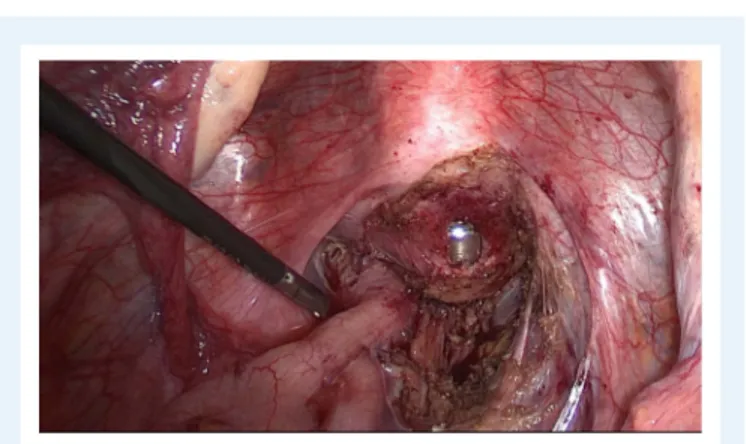
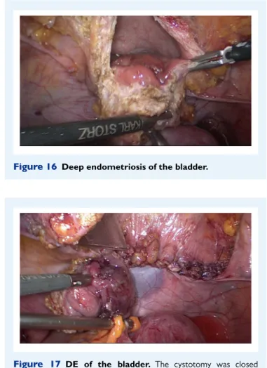
Pre- or intra-operative cystoscopy is recommended for bladder endometriosis as it allows visualization of the blueish, submucosally protruding nodules. It is important to localize the lesion precisely
in relation to the ureterovesical junction (UVJ) (Collinet et al., 2006;
Fadhlaoui et al., 2015). Whilst the mucosa (urothelium) itself is rarely
infiltrated, the nodule usually protrudes into the bladder cavity and may reach considerable size. Involvement of the outer layer of the detrusor muscle cannot be excluded by cystoscopy. A biopsy is only required for differential diagnosis when other diseases are suspected, such as urothelial carcinoma and/or interstitial cystitis. If surgery of bladder endometriosis is planned, the placement of ureteral stents can be advantageous.
Urodynamic evaluation to assess bladder function can be useful in case of bladder problems. It may have a place in distinguishing pre-existing bladder dysfunction from that developed postoperatively, as surgery for DE may induce de novo dysfunction, potentially caused by
surgical nerve damage (de Lapasse et al., 2008). The use of a specific,
validated questionnaires, such as International Prostate Symptom Score
(I-PSS) (Barry et al., 1992) and Bristol Female Lower Urinary Tract
Scale (BFLUTS) (Jackson et al., 1996), may improve the preoperative
work-up.
Ureter and kidney. Assessment of the kidney is also necessary to rule
out asymptomatic hydronephrosis (Lusuardi et al., 2012).
Endometrio-sis of the ureter is very likely if hydronephroEndometrio-sis in women with endometriosis is present, so abdominal ultrasound is the gold standard in this situation. Hydronephrosis requires a functional work-up (side separate clearance) in order to assess the renal function. Imaging, including intravenous urography (IVU), high-resolution TVUS, mercap-toacetyltriglycine (MAG3) renal scan (or radioisotope renography), MRI and/or contrast CT, is usually performed according to local protocols. Studies have shown the value of TV ultrasound scanning of ureters in patients with DE; in about 50% of the cases with ureteral involvement, obstruction can already be visualised by TVUS (cases
lack-ing hydronephrosis—early stage obstruction) (Carfagna et al., 2018).
In case of hydronephrosis, the function of the kidney has to be checked before surgery.
Nerves. The involvement of nerves in DE is of great importance to
the patient as well as to the surgeon (Possover et al., 2011;Chiantera
et al., 2018).
Both the disease and radical removal of endometriosis may lead to the destruction of the nerve fibres with corresponding symptoms, which can be very debilitating for the patient.
. . . . . . . . . . . . . . . . . . . . . . . . . . . . . . . . . . . . . . . . . . . . . . . . . . . . . . . . . . . . . . . . . . . . . . . . . . . . . . . . . . . . . . . . . . . . . . . . . . . . . . . . . . . . . . . . . . . . . . . . . . . . .
Endometriosis close to the sympathetic and parasympathetic nerve fibres (hypogastric plexus and splanchnic nerves) can lead to a dys-function of pelvic organs (e.g. dysdys-function of the bladder as well as
disturbance of vaginal lubrication and intestinal dysfunction) (Possover,
2014).
Involvement of somatic nerves, such as the sacral plexus and the sciatic nerve, leads to corresponding neurological symptoms or deficits.
In recent years, neuropelveology (Possover et al., 2015), a new field,
has become established in endoscopy and gained great importance. Laparoscopy provides an optimal surgical approach to the pelvic somatic nerves and allows micro-neurosurgery as a therapeutic approach. Both the exact pre-operative diagnosis in cases of suspected nerve involvement and the specialized surgical techniques to protect the nerves or even eliminate endometriosis close to the nerve are reserved for specialists who have undergone appropriate training in
diagnosis and surgical treatment (Rabischong et al., 2018).
The current document draws particular attention to the importance of these procedures. However, these interventions require special training and detailed instructions on them would be the subject of another publication.
Extrapelvic lesions. The diagnostic approach for extra-pelvic endometri-otic lesions includes physical examination (palpation), MRI and US. MRI can help to visualise the extent and to plan the surgery. A complete diagnostic evaluation will minimise the risk of incomplete resection.
Abdominal wall endometriosis including scars (secondary to Cae-sarean sections or hysterectomy using open route), the umbilicus and the inguinal region are thoroughly explored using either low depth US or MRI examinations. The size and depth of nodules, and the involvement of muscles or aponeurosis, should be checked before the surgery in order to maximise the chances of a complete excision. Large defects after excision have to be closed with the help of a mesh.
MRI (especially high definition) may reveal endometriosis of the diaphragm, usually when the lesions are larger than 5 mm, or when they presented recent bleeding (T1 frontal, axial and sagittal views). How-ever, small lesions may be overlooked during the pre-operative assess-ment and may be only intra-operatively revealed. When laparoscopic examination of the diaphragm is carried out using a trans-umbilical endoscope, the surgeon should be aware that only the ventral part of the diaphragm can be explored. However, lesions located behind the liver in the hepatophrenic cul-de-sac are routinely associated with visible satellite lesions spread on the ventral part of the diaphragm
(Ceccaroni et al., 2012).
Informed consent
Informed consent should be relevant to the patient and must cover the extent of the surgery that may be performed and its potential complications. Pros and cons of alternatives to the proposed treatment
must be presented (Bolton, 2015). Short- and long-term side effects
of surgery should be explained. The use of current patient information leaflets or evidence-based online resources, with references/links to best practice guidelines, should be considered to provide a sufficient source of information that the woman can review in her own time. This may also be helpful as evidence of appropriate pre-operative counselling.
Multidisciplinary surgical team
The surgical team should be organised according to the needs of the planned or anticipated procedure(s). A bowel surgeon, a urologist, a thoracic surgeon and even a plastic surgeon may need to be involved. The gynaecologist should lead the team as his/her understanding of endometriosis and the woman’s symptoms and needs is crucial in planning the surgery. The gynaecologist advocates for the patient and sets up a unique care plan, together with the team and patient, to improve the woman’s pain, fertility and quality of life. Such a care plan should consider input from other disciplines related to the tech-nical aspect of procedures. A multidisciplinary team meeting before the surgery may be helpful. The team should be informed well in advance in order to plan the procedure and to organise their time adequately.
If an ileostomy or a colostomy is planned, this should be discussed extensively with the woman and the site of the stoma may be decided and eventually drawn on the skin before the procedure.
Pre-operative strategies for a safe and complete excision of the
lesion
Bowel preparation. Different types of bowel lumen cleaning can be helpful in cases of lesions in the dorsal compartment for the following reasons:
• an empty bowel gives more space in the pelvis during the dissection;
• the use of rectal probes or manipulators in a clean bowel will cause
less contamination with faeces on the perineum, especially when the vagina has to be opened;
• in the case of opening the bowel, it will minimise faecal soiling in the
abdomen.
According to the literature data on colorectal surgery, mechanical
bowel preparation and enemas are widely used (Guenaga et al., 2011;
Oliveira et al., 2016), though specific trials on the usefulness of bowel
preparation in endometriosis surgery (where the vaginal fornix dorsal is often opened) are not available. Whereas none of these techniques have a proven benefit on the reduction of postoperative complications
(Guenaga et al., 2011), in cases where a low anastomosis is expected
mechanical bowel preparation is better than an enema (Platell et al.,
2006). The use of intraluminal antibiotic decontamination of the bowel
to reduce clinically relevant anastomotic leakage can be considered
(McDermott FD et al., 2016).
Ureteral stenting. Pre-operative placement of ureteral stents is suggested when: surgery of large bladder endometriosis is planned; ureteral endometriosis is suspected pre-operatively; hydronephrosis is present; or there is a history of previous ureteral surgery.
Uterine manipulator and rectal probe. The use of a uterine manipulator achieves maximum mobility of the uterus, thereby improving visualiza-tion and facilitating dissecvisualiza-tion. A rectal probe could also be helpful in moving the bowel, although it could be hindered by stenosis due to a deep nodule or severe bowel adhesions. Tactile feedback between the dissecting instruments and the rectal and vaginal probes, handled by an assistant, helps to identify the correct planes of cleavage. Bowel and adnexal suspension can improve visualization and access to the pouch
of Douglas (Einarsson and Wattiez, 2016) (Figs 7and8).
. . . . . . . . . . . . . . . . . . . . . . . . . . . . . . . . . . . . . . . . . . . . . . . . . . . . . . . . . . . . . . . . . . . . . . . . . . . . . . . . . . . . . . . . . . . . . . . . . . . . . . . . . . . . . . . . . . . . . . . . . . . .
Figure 7 Deep endometriosis in the vagina, rectovaginal septum and the anterior wall of the rectum (Enzian A, C compartment). Two different ways to excise the nodule. A
manipu-lator and a sponge or rectal probe is in situ for a better presentation of the nodule during the excision procedure. (Reprinted with permissions fromKeckstein and Hucke, 2000).
Figure 8 Frozen pelvis with invisible deep endometriosis (bilateral ovary, left cardinal ligament, ureter left, anterior wall of the rectum).
Strategy of the surgical intervention
Each surgeon needs a strategy for the operation, which is influenced by many factors including the size, activity and localization of endometrio-sis as well as the age and expectations of the patient, and the results of previous interventions.
Advanced endometriosis in a young patient with a desire to have children may be operated differently than in a patient over 40 years of age with pain as the main symptom.
The surgeon is often confronted with the conflict between complete removal of endometriosis and the need for preservation of organs affected by the disease.
Another challenge is tackling multi-organ involvement, which requires a complex intervention possibly in a multidisciplinary setting.
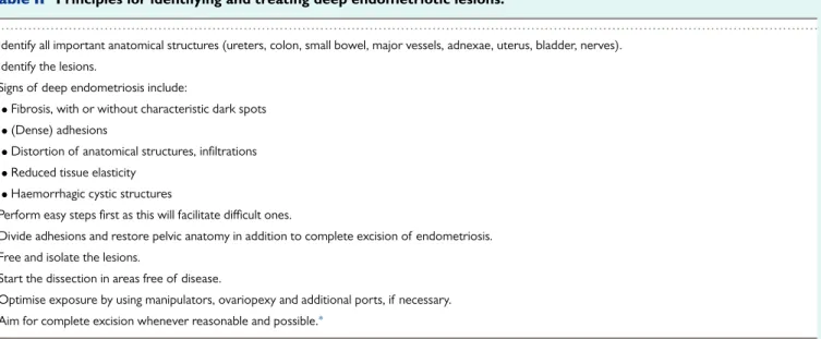
Table II Principles for identifying and treating deep endometriotic lesions.
...
• Identify all important anatomical structures (ureters, colon, small bowel, major vessels, adnexae, uterus, bladder, nerves). • Identify the lesions.
• Signs of deep endometriosis include:
• Fibrosis, with or without characteristic dark spots • (Dense) adhesions
• Distortion of anatomical structures, infiltrations • Reduced tissue elasticity
• Haemorrhagic cystic structures
• Perform easy steps first as this will facilitate difficult ones.
• Divide adhesions and restore pelvic anatomy in addition to complete excision of endometriosis. • Free and isolate the lesions.
• Start the dissection in areas free of disease.
• Optimise exposure by using manipulators, ovariopexy and additional ports, if necessary. • Aim for complete excision whenever reasonable and possible.∗
∗When deciding that a part of the disease may be left behind, the surgeon should remember that if an extensive dissection has been performed to access this part of the disease, reoperation will be extremely difficult and sometimes almost impossible. If excision is considered to be too risky, it is likely to be even more difficult and dangerous if a reoperation is needed due to recurrent pain or other severe symptoms (such as stenosis of the bowel or ureter). Ideally, surgeons would be prepared to manage all aspects of the disease.
An important limitation is the risk of postoperative complications. For this reason, occasionally a limited radicality or surgery in several steps may be chosen, for example simultaneous segmental resection of intestinal endometriosis and ureteral re-implantation in hydronephrosis may be avoided.
Decisions for the strategy before and during surgery depend in particular on the situs and surgeon’s experience, and they have to be made on a case-by-case basis.
Open versus endoscopic surgery (or robotic assisted)
Endoscopic access has become standard for the treatment of endometriosis, including DE. It is obvious that the endoscopic procedures are advantageous due to better views and access to lesions in the depth of the pelvis, as well as lower postoperative morbidity. Minimal trauma to the abdominal wall and the healthy peritoneum, lack of dehydration and the use of microsurgical techniques improve the outcome, especially in patients with infertility. Furthermore, there are anatomical sites or endometriosis findings that can exclusively be reached/treated only by an endoscopic procedure (e.g. neuropelveology).
Endoscopic operations require special instruments and equipment as well as a high level of training and experience of the surgeon. The access route should be chosen according to the clinical findings and the existing options.
However, a laparotomy (sometimes with midline incision) may occa-sionally be more effective than several inadequate laparoscopies. The advantage of a laparotomy for treatment of severe endometriosis to identify and completely eliminate it lies in the ability of having a better tactile feedback. In this situation, microsurgical operation techniques should still be used.
Robot-assisted surgery has gained importance in the treatment of endometriosis over the last 10 years. Special features of the instru-ments may facilitate difficult steps of the procedures and some of the benefits of laparotomy are thus incorporated into endoscopic surgery.
. . . . . . . . . . . . . . . . . . . . . . . . . . . . . . . . . . . . . . . . . . . . . . . . . . . . . . . . . . . . . . . . . . . . . . . . . .
Several studies have shown that the results of robot-assisted surgery in DE are equivalent to those of conventional laparoscopy, but not superior.
In the current document, particular attention is given to conventional endoscopic procedures.
Initial steps of DE surgery—patient
positioning in view of anticipated long
duration of surgery
Prevent pressure sores and compartment syndrome by using:
• anti-embolism stockings for thromboprophylaxis, and additional
prophylaxis with postoperative low molecular weight heparin is usually recommended after this type of pelvic surgery (follow local guidelines);
• body warmer to maintain the core temperature;
• boots, lithotomy with soft stirrups, legs flat, intermittent pneumatic
compression devices.
The woman is placed in the modified dorsolithotomy position and her legs are placed in surgical stirrups carefully avoiding trauma to the leg nerves. As the surgery is often long, particularly in women with previous multiple surgeries and/or in obese or moderately overweight patients, mobilization and/or massage of the legs can be performed every 2–4 h or between different surgical phases. Application of intermittent pneumatic compression devices or sequential compres-sion devices has been proposed to limit the risks of lower limb
compartment syndrome (Tomassetti et al., 2009;Gould et al., 2012).
Arms should be positioned carefully to avoid shoulder restraints or pressure, especially in steep Trendelenburg position during surgery.
• Examination under anaesthesia is generally recommended for DE.
Figure 9 DE involving the uterosacral ligament, vagina and the rectum. The extent of the disease is not visible during diagnostic
laparoscopy. Spaces have to be opened in order to get access to the entire lesion.
Figure 10 Dissection of the posterior compartment. Right
uterosacral ligament has already been resected, the vagina is partially open with the manipulator in place.
beneficial particularly when rectovaginal nodules are not obviously visible.
• Antibiotics can be used according to local guidelines.
• Systematic laparoscopic inspection and documentation is
recom-mended. After insertion of the laparoscope, the upper abdomen including diaphragm and appendix/caecum should be inspected, preferably prior to placing the patient in (adequate) Trendelenburg.
• The placement of secondary trocars for the various instruments
should be individualized according to the anatomical situation and surgical needs.
The basic principles to identify and treat deep endometriotic lesions
are stated inTable II.
DE of the uterosacral ligaments and
rectovaginal septum with or without
involvement of the rectum
The extent of the surgical procedure is determined by the size of the lesions, their location, their number (single or multifocal), and the
degree of infiltration (Figs 9and10).
. . . . . . . . . . . . . . . . . . . . . . . . . . . . . . . . . . . . . . . . . . . . . . . . . . . . . . . . . . . . . . . . . . . . . . . . . . . . . . . . . . . . . . . . . . . . . . . . . . . . . . . . . . . . . . . . . . . . . . . . . . . .
Definitions of bowel infiltration and surgical procedures
The pre-operative clinical diagnostic workup including clinical exami-nation (vaginal and rectal), TVUS, and an optional MRI is necessary in order to identify the bowel wall infiltration pre-operatively. A negative colonoscopy does not exclude the intramural presence of DE.
In case of muscularis infiltration by the nodule, the distance of the inferior border of the most distal bowel lesion to the anal verge should be evaluated, as this may impact on the type of surgery performed.
If the lesions are only located in or on the serosa without infiltration of the muscularis layer, it can be treated with superficial resection
(serosal shaving) (Koninckx et al., 2012;Vanhie et al., 2016).
If deep (infiltrating) lesions involve the muscularis layer, sometimes the submucosa and even the mucosa, partial or full thickness removal
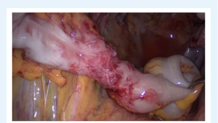
by shaving, discoid or segmental bowel resection is necessary (
Kon-inckx et al., 2012;Donnez and Roman, 2017) (Figs 5and7).
First steps of surgery
Uterine, vaginal and rectal set up. A uterine manipulator is used to improve exposure of the cul-de-sac, particularly in the presence of an enlarged uterus due to fibroids and/or severe adenomyosis, which makes the procedure more difficult. In some cases, a sponge can be placed in the dorsal fornix of the vagina. A rectal probe should also be available to mobilize the rectum in order to determine its position and attachment to the vaginal wall and to evaluate the elasticity of the tissue and degree of stenosis, taking care to avoid inadvertent rectal laceration. During the surgery, these three manipulators can be mobilized individually in order to clearly identify the limit between the
vagina, the rectal wall, and other pelvic structures (Figs 7, 9and10).
Preparing the operating field. Prior to starting the operation, a vagi-nal exam using a bivalved speculum and digital (vagivagi-nal and rectal) examination for the evaluation of the dorsal fornix (mucosa protru-sion/retraction or invasion) and the extension to the pelvic sidewalls is recommended.
The following steps may facilitate the surgical procedure: ovariolysis and ovariopexy, sigmoid mobilization, ureterolysis, and the identifica-tion of ligaments and rectosigmoid colon.
Ovariolysis The mobilization of fixed ovaries on the pelvic sidewall improves the view of the operative field, particularly the lower struc-tures in the cul-de-sac, which simplifies the identification of the ureters. If present, endometrioma should be drained and managed according
to previous recommendations (Working group of ESGE-ESHRE and
WES et al., 2017). This may be done immediately after the drainage of
the cyst or after having removed all other deep lesions in the pelvis. Endometriosis in the ovarian fossae should also be removed.
Temporary ovariopexy Suspension of the ovaries with sutures (curved or straight needle through the abdominal wall) or special devices can maximize access to the pelvic structures and especially the pararectal spaces and the pelvic sidewalls.
Mobilisation of the sigmoid: (starting from the ‘white line of Toldt’) Mobilize the sigmoid colon off its attachments to the abdominal wall and pelvic sidewall to expose the left adnexa and underlying structures such as the left pararectal space and ovarian fossa.
Ureterolysis It is advisable to identify the position of the ureter on the pelvic rim or upper pelvic sidewall at the beginning of the procedure. The ureter should be followed down to the cardinal ligament and the