ORIGINAL ARTICLE
SIRT6 regulates the cigarette smoke-induced
signalling in rheumatoid arthritis synovial fibroblasts
Anna Engler&Fabienne Niederer&Kerstin Klein&Renate E. Gay&Diego Kyburz&Giovanni G. Camici&Steffen Gay&Caroline Ospelt
Received: 10 December 2013 / Revised: 14 February 2014 / Accepted: 21 February 2014 / Published online: 19 March 2014 # Springer-Verlag Berlin Heidelberg 2014
Abstract
Cigarette smoking is a recognized environmental risk factor for the development and progression of rheumatoid arthritis (RA). RA synovial fibroblasts (RASF) actively contribute to inflammation and joint destruction in this chronic inflamma-tory autoimmune disease. In the current study, we investigated the influence of cigarette smoke on the inflammatory and matrix-destructive properties of RASF. Furthermore, the func-tional role of Sirtuin 6 (SIRT6) in the regulation of the signal-ling induced by cigarette smoke or by tumor necrosis factor alpha (TNFα) was elucidated. We demonstrated that stimula-tion with cigarette smoke extract (CSE) enhances the pro-inflammatory and matrix-destructive potential of RASF by inducing the production of pro-inflammatory cytokine inter-leukin 8 (IL8) and the matrix-destructive enzyme matrix me-talloproteinase 1 (MMP1), but not of IL6 and MMP3. More-over, we could show that the expression of MMP1 is specif-ically regulated by SIRT6. Treatment of RASF with CSE or TNFα increased the levels of SIRT6. The expression of SIRT6 was also enhanced in vivo in synovial tissues of RA smokers
and in joints of mice exposed to cigarette smoke. Silencing of SIRT6 specifically increased basal as well as CSE- and TNFα-induced production of MMP1, demonstrating that SIRT6 plays an important role in restricting MMP1 expres-sion. In conclusion, the upregulation of SIRT6 in RASF under CSE or TNFα stimulation functions as a counterregulatory mechanism attenuating the production of the matrix-destructive enzyme MMP1. This is the first study revealing the protective function of SIRT6 in the cigarette smoke-induced signalling.
Key messages
& Cigarette smoke induces pro-inflammatory and matrix-destructive responses in RASF.
& Cigarette smoke enhances the expression of SIRT6 in vitro and in vivo.
& TNFα increases the levels of SIRT6.
& SIRT6 diminishes MMP1 production under cigarette smoke extract and TNFα stimulation.
Keywords Rheumatoid Arthritis . Smoking . Sirtuins . Matrix metalloproteinases . Cytokines
Introduction
Rheumatoid arthritis (RA) is an inflammatory disorder affect-ing multiple joints and leadaffect-ing to progressive destruction of cartilage and bone. Along with inflammatory cells, RA syno-vial fibroblasts (RASF) actively contribute to inflammation and joint destruction [1]. RASF exhibit an activated and aggressive phenotype, characterized by increased resistance to apoptosis, enhanced production of inflammatory cytokines, matrix-degrading enzymes, and pro-angiogenic factors [2].
Electronic supplementary material The online version of this article (doi: 10.1007/s00109-014-1139-0 ) contains supplementary material, which is available to authorized users.
A. Engler
:
F. Niederer:
K. Klein:
R. E. Gay:
D. Kyburz:
S. Gay:
C. Ospelt (*)Center of Experimental Rheumatology, University Hospital Zurich, Gloriastrasse 25, 8091 Zurich, Switzerland
e-mail: caroline.ospelt@usz.ch
A. Engler
:
F. Niederer:
R. E. Gay:
D. Kyburz:
S. GayZurich Center of Integrative Human Physiology (ZIHP), University of Zurich, Winterthurerstrasse 190, 8057 Zurich, Switzerland G. G. Camici
Institute of Physiology, University of Zurich, Winterthurerstrasse 190, 8057 Zurich, Switzerland
Multiple mechanisms including genetic predisposition, epige-netic changes, interaction with matrix components, and in-flammatory mediators contribute to the activation of RASF [3]. The impact of environmental factors on the activated phenotype of RASF is unknown. Smoking is a recognized environmental risk factor not only for the development of RA, but also for the severity of established RA. Specifically, smoking was associated with increased disease activity and progression [4–6]. RA patients with a history of smoking were reported to show poor response to anti-tumor necrosis factor (TNF) therapy and RA heavy smokers, to have the poorest drug survival [7,8]. The role of cigarette smoke in the devel-opment of collagen-induced arthritis (CIA) in mice is less clear. Lindblad and colleagues have shown that cigarette smoke exposure delays the onset and progression of CIA in mice [9]. However, in two other studies the treatment of mice with cigarette smoke condensate extract augmented the devel-opment of CIA [10,11].
Since synovial fluid is a passive ultrafiltrate from the blood, substances in the blood stream, e.g., drugs and their metabolites, can be found or even accumulate in the synovial fluid and possibly alter joint physiology [12, 13]. Indeed, gene expression in synovia from smoking RA patients was significantly altered compared to nonsmoking RA patients [14]. Furthermore, compo-nents of cigarette smoke were shown to increase inva-sion of RASF in Matrigel in vitro [15]. However, the specific regulators and the signalling pathways that are affected by cigarette smoke in RA are poorly investi-gated. In particular, it is not clear how cigarette smoke regulates the gene expression that influences the activity of RASF.
Sirtuins (SIRTs) regulate a broad range of cellular process-es including aging, metabolism, cell survival, gene transcrip-tion, stress responses, and inflammation [16]. In mammals, seven members of the sirtuin family (SIRT1–SIRT7) were identified that regulate the activity of histones, transcription factors, and other target proteins by posttranslational modifi-cations [17, 18]. We have previously reported that SIRT1 overexpression in RA synovium contributes to pro-inflammatory cytokine production and apoptosis resistance [19]. Another member of the sirtuin family, SIRT6, has gained much attention in recent years because of its role in DNA repair and genomic stability as well as in suppression of tumor formation and NF-κB signalling [20]. In a recent publication by Lee et al., the overexpression of SIRT6 was shown to inhibit the expression of NF-κB target genes and to decrease the severity of CIA in mice, demonstrating an important protective function of SIRT6 [21].
In the current study, we investigated the influence of the environmental risk factor of cigarette smoke on the gene expression that regulates the inflammatory and matrix-destructive properties of RASF. We demonstrated that an
increase in SIRT6 levels is important for restriction of ciga-rette smoke- and TNFα-induced production of the matrix-destructive enzyme MMP1.
Methods
Patient samples/cell culture
Synovial tissues were obtained from RA patients undergoing joint replacement surgery at the Schulthess Clinic Zurich after written informed consent according to the principles of the 1964 Declaration of Helsinki. For all experiments with human and mouse tissues, ethical approval was obtained at the Swiss Ethical commission. All RA patients fulfilled the American College of Rheumatology criteria for the classification of RA [22]. For fibroblast cultures, tissue specimen were digested with collagenase and cultivated in Dulbecco’s modified Ea-gle’s medium (DMEM) supplemented with 10 % heat inactivated fetal calf serum, 50 IU/ml penicillin-streptomycin, 2 mM l-glutamine, 10 mM HEPES, and 0.2 % fungicide. For experiments, RASF from passages 4 to 8 were used. The characteristics of patients used for detection of SIRT6 in synovial tissues are shown in Table1.
Animals
Male C57BL/6 mice (n = 6) were exposed to cigarette smoke from research cigarettes (University of Kentucky 1R4F) twice a day for a total of 6 h, 5 days per week in a whole-body smoking chamber (Teague TE-10). Cigarettes were smoked at a rate of a 2 s, 35 ml puff each minute and smoke particulate concentration was kept at 25.2 mg/m3. Control mice (n = 8) were exposed to filtered ambient air. After 3 weeks, mice were eutha-nized, and mRNA from dissected joints was isolated. Stimulation assays
Cigarette smoke extract (CSE) was prepared using a modification of the method described by Vassallo et al. [23]. One-hundred-percent CSE was prepared by bubbling the smoke of one cigarette into 10 ml of cell culture medium at a rate of one cigarette per 7 min. The generated CSE was sterile-filtered and diluted to 5 % CSE with cell culture medium. The pH of 5 % CSE and of the control cell culture medium was equal and remained unaltered during the cultivation. CSE prepara-tions were freshly prepared for each experiment and monitored by measuring the optical density at 320 and 350 nm. The cigarettes used contained 10 mg tar, 0.8 mg nicotine, and 10 mg carbon monoxide (Marlboro
Red). Confluent cells were stimulated with 5 % CSE or 10 ng/ml TNFα for 24 h.
Transfection experiments
RASF were transfected using the Basic Nucleofector Kit for Primary Mammalian Fibroblasts (Amaxa/Lonza). For SIRT6 silencing, 10 μM SIRT6-validated small interfer-ing RNA (siRNA) or control siRNA (both Qiagen) were used. Transfected RASF were cultured for 48 h and subsequently stimulated with CSE or TNFα.
RNA isolation and quantitative real time PCR
Total RNA was isolated using the RNeasy Miniprep Kit (Qiagen) including DNase treatment. RNA was dis-solved in RNAse-free water, and the optical density was determined at 260 nm in a spectrophotometer. RNA was reverse transcribed using random hexamers a n d m u l t i s c r i b e r e v e r s e t r a n s c r i p t a s e ( A p p l i e d Biosystems). Samples without addition of reverse tran-scriptase were used as negative controls (non-RT). Rel-ative quantification of mRNA levels was performed by SYBRGreen Real time PCR using the ABI Prism 7700 Sequence Detection System (Applied Biosystems) for 40 cycles. As endogenous controls, hypoxanthine phosphoribosyltransferase 1 (HPRT1) was used for hu-man samples and 18 s rRNA assay for murine samples (Life Tech-Applied Biosystems). The differences of the comparative threshold cycles (Ct) of sample and endog-enous control were calculated (dCt). Relative expression levels were calculated following the formula ddCt = dCt (sample stimulated)–dCt (sample unstimulated), relative expression was calculated using the expression 2−ddCt. For primer sequences see supplementary table S1.
Enzyme linked immunosorbent assay (ELISA)
Proteins in cell supernatants were detected with OptEIA® Kits (BD Pharmingen) for IL6, IL8, and TNFα, and with DuoSet ELISA Development Kits (R & D Systems) for MMP1 and MMP3. Data were analyzed using Revelation v4.22 software (Dynex Technologies).
Immunohistochemistry
Formalin-fixed, paraffin-embedded synovial tissue sections were deparaffinised, pretreated with EDTA (pH 9), blocked for endogenous peroxidase activity (3%H2O2) and for
non-specific binding (5 % goat serum/1 % bovine serum albumin), and incubated with rabbit IgG (Jackson ImmunoResearch) or rabbit anti-SIRT6 antibodies (D8D12, Cell Signaling, #12486). SIRT6-positive cells were visualized with biotinyl-ated goat anti-rabbit antibodies (Jackson ImmunoResearch) and DAB (3.3′-Diaminobenzidin, Vector Laboratories). For double stainings, the slides were stained with anti-SIRT6 as described above. Following, the slides were pretreated with 10 mM citrate buffer (pH 6) or 1 mg/ml trypsin, blocked with 5 % goat serum/1 % bovine serum albumin, and incubated with mouse anti-prolyl 4-hydroxylase beta (Acris Antibodies) or with mouse anti-CD68 (Dako). Slides were incubated with biotinylated goat anti-mouse antibodies (Jackson ImmunoResearch). Prolyl 4-hydroxylase beta-positive cells were visualized with HistoGreen (Linaris) and CD-68-positive cells with Vector Blue Alkaline Phosphatase Sub-strate Kit (Vector Laboratories). Slides stained with single anti-SIRT6 or IgG control were counterstained with hematox-ylin. The images were obtained on a Zeiss Axio Imager Z1 microscope through a Zeiss Plan-Neofluar 10×/0.30 or 40×/ 0.75 numerical objective aperture and acquired by a Zeiss AxioCam HRc camera and the AxioVision 4.6.3.SP1 imaging
Table 1 Patient characteristics used for detection of SIRT6 in synovial tissues
Age, disease duration, and CRP were not significantly different between RA smokers and RA nonsmokers
ND not determined, NSAIDs non-steroidal anti-inflammatory drugs, DMARDs disease-modifying anti-rheumatic drugs, RF rheumatoid factor, CRP C-reactive protein
Characteristics RA smoker (n=11) RA non smoker (n=11) Age; years, mean (range) 60 (53–73) 64 (40–78)
Sex; female/male 7/4 10/1
Number of cigarettes per day; mean (range) 13 (2–20) – Disease duration; years, mean (range) 17 (4–28) (2 patients ND) 25 (4–50)
CRP; mg/l, mean (range) 9 (0.1–19.3) (4 patients ND) 6 (0.1–20.2) (6 patients ND) RF positive 9/10 (1 patient ND) 5/5 (6 patients ND) Medications DMARDs 5 8 NSAIDs 4 2 steroids 4 1 biologics 1 4 none 1 2
software (Carl Zeiss). The intensity of SIRT6 staining was evaluated by two blinded observers, using a gradual scoring scale from 0 (no staining) to 4 (strong staining).
Immunoblotting
Whole cell lysates were prepared for immunoblotting as previously described [19]. Nuclear fractions were enriched by differential centrifugation at 4 °C. RASF were washed in PBS and harvested by cell scraping. Following centri-fugation, the cell pellet was incubated in homogenization medium (150 mM MgCl2, 10 mM KCl, 10 mM TrisHCl (pH 6.7)) for 5 min. The cell suspension was homoge-nized using a glass-glass homogenisator and centrifuged at 1,000 g for 10 min. The pellet was lysed in RIPA buffer. The following antibodies were used: anti-SIRT4 (Abcam), anti-SIRT6 (Cell Signaling, #2590), anti-Vimentin (Abcam), or anti-histone H3 (Abcam). Horseradish perox-idase (HRP)-labeled species-specific secondary antibodies (Jackson ImmunoResearch) and enhanced chemilumines-cence (GE Healthcare) were used for visualization.
Statistical analysis
Statistical analysis was performed using GraphPad Prism 5.0 software. Values are presented as mean±standard error of mean (SEM). After testing the values for Gaussian distribu-tion, data were analyzed using one sample t test, Mann– Whitney, or Wilcoxon matched-pairs signed rank test as indi-cated. Two-way analysis of variance (ANOVA) was used to determine synergistic or additive effects. p values <0.05 were considered as significant.
Results
Cigarette smoke alters the expression of sirtuins in vitro and in vivo
To investigate the impact of cigarette smoke on the expression of sirtuins, RASF from nonsmokers were stimulated with 5 % CSE for 24 h. Among all sirtuins examined (SIRT1–7), the transcriptional level of SIRT4 was 4.3-fold (p=0.01) and
Fig. 1 Stimulation of RASF with cigarette smoke extract (CSE) increases the expression of sirtuins. a Confluent monolayer cultures of synovial fibroblasts from patients with RA (n=6) were treated with 5 % CSE for 24 h. Expression of SIRT1–7 mRNA was measured by real time PCR using specific primers and normalized to the housekeeping gene HPRT1. Changes in the transcriptional levels of SIRTs are shown as fold induction compared to untreated cells (dotted line). (b–c) Protein expression of SIRT4 and SIRT6 was increased in synovial fibroblasts from patients with RA (=RASF, n=4) stimulated with CSE. Molecular weights are indicated in kiloDalton (kDa). Bars show the mean±SEM. n= number of patients investigated. *p<0.02, by one sample t test
SIRT6 mRNA was 2.7-fold (p = 0.02) enhanced in CSE-stimulated RASF (Fig.1a). Elevated expression of SIRT4 and SIRT6 was confirmed at the protein level by immunoblot (Figs.1b, c).
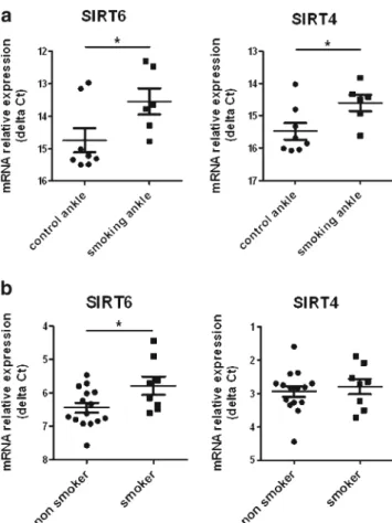
Increased mRNA expression of SIRT4 and SIRT6 was detected in vivo in joints of mice exposed to cigarette smoke for 3 weeks. The basal mRNA levels of SIRT4 were 1.8-fold higher (ΔCT smoking 14.6±0.6; ΔCT control 15.5±0.7; p= 0.04) and of SIRT6, 2.3-fold higher (ΔCT smoking 13.6±0.9; ΔCT control 14.8±1.0; p=0.04) in joints of mice exposed to cigarette smoke as compared to control mice (Fig. 2a). In human synovial tissue samples from RA patients, smokers had 1.6-fold higher mRNA expression of SIRT6 (ΔCT smokers 5.8±0.7; ΔCT nonsmokers 6.5±0.6; p=0.04) as compared to nonsmokers (Fig. 2b). The levels of SIRT4 mRNA were not significantly different in synovial tissues of RA patients who smoked.
We further investigated the expression of SIRT6 in synovial tissue sections from RA patients by immuno-histochemistry. Scoring of SIRT6 staining showed in-creased expression of nuclear SIRT6 in synovial tissues of RA smokers when compared to RA nonsmokers (Figs. 3a, b). To detect the specific cells in the synovium that express SIRT6, we performed double stainings with SIRT6 and prolyl 4-hydroxylase beta as a fibroblast marker or CD68 as a macrophage marker. Strong expression of SIRT6 was detected in prolyl 4-hydroxylase beta-positive SF and in CD68-positive mac-rophages (Fig. 3c). Interestingly, the levels of SIRT6 positively correlated with the disease duration in RA smokers, but not in RA nonsmokers (Fig. 3d).
CSE increases the expression of IL8 and MMP1 in RASF The activated and aggressive phenotype of RASF is c h a r a c t e r i z e d b y i n c r e a s e d p r o d u c t i o n o f p r o -inflammatory ILs and matrix-destructive MMPs [1, 2]. In particular, TNFα strongly induces the secretion of ILs and MMPs in RASF. To investigate whether inflam-matory and matrix-destructive properties of RASF can be modulated by CSE alone and together with TNFα, RASF were stimulated with CSE, TNFα, or with TNFα and CSE in combination. RASF released 2.1-fold more MMP1 and 1.5-fold more IL8 protein (both p < 0.03) in cell culture supernatants after stimulation with CSE (Fig. 4a). Secretion of IL6 and MMP3 was not altered. Co-treatment with CSE and TNFα led to a further increase in protein release of IL8 and MMP1 compared to stimulation of RASF with TNFα alone (Fig. 4a), but did not affect TNFα-mediated secretion of IL6 or MMP3.
Real time PCR analysis confirmed 6.5-fold enhanced levels of MMP1 transcript and 3.5-fold increased expres-sion of IL8 mRNA after CSE treatment (both p < 0.02) (Fig. 4b), indicating that CSE induces changes at the transcriptional level. Stimulation of RASF with CSE and TNFα in combination further increased mRNA levels of MMP1 and IL8 compared to the stimulation with TNFα alone (Fig. 4b). Two-way analysis of variance (ANOVA) revealed that IL8 production was synergistically induced by CSE and TNFα whereas an additive effect of CSE and TNFα was detected on MMP1 expression. Moreover, mRNA levels of TNFα and IL-1β were measured; how-ever, they were under the detection limit before as well as after stimulation with CSE.
In summary, these results indicate that CSE increases the transcription and secretion of IL8 and MMP1, and further augments the strong effect of TNFα on the expression of IL8 and MMP1 thereby promoting the pro-inflammatory and matrix-destructive potential of RASF.
Fig. 2 Exposure to cigarette smoke enhances the expression of sirtuins in vivo. a Increased mRNA levels of SIRT6 and SIRT4 in the ankles of mice exposed to cigarette smoke (smoking ankle, n=6) as compared to control treated mice (control ankle, n=8). Changes in the transcriptional levels of SIRT4 and SIRT6 are shown as the difference in threshold cycle (ΔCt), relative to 18S rRNA. b Elevated expression of SIRT6 transcript in human synovial tissue samples from RA patients who smoked (smoker) as compared to RA nonsmokers (nonsmoker). Relative expression of mRNA is presented as the difference in threshold cycle (ΔCt), relative to HPRT1. Bars show the mean±SEM. *p<0.04, by Mann–Whitney test
The levels of SIRT6 are increased upon stimulation with TNFα
Since the expression of SIRT6 was altered in vitro in RASF as well as in vivo in the joints of mice and most importantly in the synovium of RA smokers, we focused on the functional role of SIRT6 in the regulation of pro-inflammatory and matrix-destructive activities of RASF. SIRT6 was shown to regulate the expression of TNFα-induced genes [21, 24]. However, the impact of TNFα on the expression of SIRT6 has not been investigated so far. To elucidate whether TNFα can modify the expression of SIRT6, RASF were stimulated with TNFα. Similar to CSE, treatment with TNFα increased
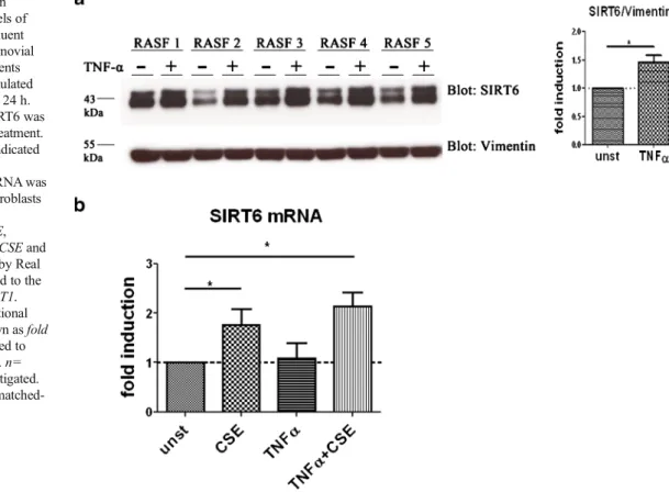
the protein levels of SIRT6 (Fig.5a). However, while CSE significantly enhanced the levels of SIRT6 mRNA, stimula-tion with TNFα did not affect the transcripstimula-tional levels of SIRT6 pointing to posttranscriptional regulation of SIRT6 protein in TNFα-treated RASF (Fig.5b).
SIRT6 attenuates CSE- and TNFα-induced MMP1 expression
To investigate whether increased SIRT6 levels influence the CSE- and TNFα-induced production of IL8 and MMP1, upregulation of SIRT6 was inhibited with siRNA during stimulation and the secretion of ILs and MMPs was measured.
Fig. 3 Expression and localization of SIRT6 in RA synovial tissues. a Representative sections of RA synovial tissues stained for SIRT6 or IgG control (inset) showing increased expression of SIRT6 (brown) in RA smokers (left). Nuclei were counterstained with hematoxylin (blue). SIRT6 staining appears in brown. Original magnification: ×100 (top) and×400 (bottom). b Staining intensities in synovial tissues of RA smokers (n=10) and RA nonsmokers (n=11) were scored. n=number
of patients investigated. Bars show the mean±SEM. *p<0.04, by Mann– Whitney test. c Representative sections of RA synovial tissue double stained for detection of SIRT6 (orange) and prolyl 4-hydroxylase beta (blue, left) as a fibroblast marker or CD68 (blue, right) as a macrophage marker. Original magnification: ×400. Levels of SIRT6 positively corre-lated with the disease duration in RA smokers (d), but not in RA nonsmokers (e)
To compare the effects of CSE and TNFα, SIRT6-silenced cells were stimulated with CSE or TNFα alone or with TNFα and CSE in combination. Basal expression as well as induction of SIRT6 after stimulation with CSE or TNFα was successfully blocked by transfection of RASF with SIRT6-specific siRNA (Fig.6a). Silencing of SIRT6 significantly increased the basal protein secretion of MMP1, but did not affect the secreted levels of MMP3, IL6, or IL8 (Fig.6b). Additionally, under stimulation with CSE or TNFα, the release of MMP1 was significantly enhanced in SIRT6-silenced cells as compared to control-transfected cells. Furthermore, under combined stimulation with TNFα and CSE, the silencing of SIRT6 led to a higher increase in MMP1 release (Fig. 6b).
To investigate whether SIRT6 can regulate the gene expression of MMP1, we measured MMP1 transcripts in SIRT6-silenced RASF. Similar to the protein levels, silencing of SIRT6 significantly increased mRNA ex-pression of MMP1 under basal conditions as well as under stimulation with CSE or with TNFα, or under stimulation with TNFα and CSE together (Fig. 6c). The expression of IL8 mRNA was not significantly altered by silencing of SIRT6.
These data show that SIRT6 reduction leads to a specific increase in the mRNA and protein expression of MMP1 under CSE as well as under TNFα stimula-tion, but does not affect the production of IL8, IL6, or MMP3. Thus, the increase in SIRT6 levels detected in
Fig. 4 Stimulation of RASF with CSE induces the expression of IL8 and MMP1. Synovial fibroblasts from patients with RA (n=7) were stimu-lated with 5 % CSE, 10 ng/ml TNFα or with CSE and TNFα in combi-nation for 24 h. a Protein release of MMP1, MMP3, IL8 and IL6 was measured in cell culture supernatants of stimulated and unstimulated (unst) cells by ELISA. b Increased expression of MMP1 and IL8 was
confirmed at the mRNA level in RASF stimulated with CSE using Real time PCR analysis and is shown as fold induction compared to untreated cells (unst). As endogenous control, the expression of HPRT1 mRNA was used for normalization. Values are mean±SEM. *p<0.05; **p<0.02; by Wilcoxon matched-pairs signed rank test
C S E a n d T N Fα-stimulated RASF might be a counterregulatory mechanism to restrict the production of MMP1 under cellular stress.
Discussion
In the current study, we have investigated the molecular pathways that are altered by the environmental factor cigarette smoke in RASF. We found that stimulation with CSE en-hances the pro-inflammatory and matrix-destructive proper-ties of RASF by inducing the expression of IL8 and MMP1. Moreover, we have shown for the first time that cigarette smoke increases the expression of SIRT6 in vivo in the joints of mice and in the synovial tissues of RA patients and in vitro in RASF. Most interestingly, we found that SIRT6 specifically attenuates the transcription and secretion of the matrix-destructive enzyme MMP1 in RASF. Thus, an increase in SIRT6 levels after stimulation with CSE or TNFα functions as a counterregulatory mechanism to restrict MMP1 produc-tion in response to CSE- or TNFα-induced MMP1 expression.
Our finding of elevated levels of SIRT6 in joints of smoke-exposed mice and in synovial tissues of smokers validates our in vitro model and supports the hypothesis that cigarette smoke constituents reach the synovial fluid and cause changes
in gene expression. Currently, no study is published that analyzed the presence of cigarette smoke ingredients in the synovial fluid. In addition to our study, indirect evidence is provided by a study showing activation of aryl hydrocarbon receptors in the synovium of smokers [14]. This receptor binds polycyclic aromatic hydrocarbons, which are formed in cigarette smoke. Another study demonstrated the presence of tetrahydrocannabinol in the synovial fluid of hashish smokers, proving that bulky molecules can be filtered in the synovial fluid [25].
SIRT6 was previously shown to be induced by oxidative stress [26], and its increased expression in smokers points to the activation of stress response pathways by smoking. This is also in line with our observations that citrullination and heat shock proteins are elevated in the joints of smoke exposed mice, both of which are known to be increased under cellular stress [27]. Interestingly, cigarette smoke and TNFα had
different effects on transcription of SIRT6. Both pro-inflammatory stimuli increased protein levels of SIRT6. How-ever, only cigarette smoke increased the transcription of SIRT6. Different regulation of SIRT6 on transcriptional and posttranslational level was recently reported by Ronnebaum and colleagues. In this study, the posttranslational regulator of SIRT6 protein was identified and it was shown that the over-expression of ubiquitin ligase CHIP increased SIRT6 protein expression without affecting SIRT6 transcription [28].
Fig. 5 TNFα stimulation increases the protein levels of SIRT6 in RASF. a Confluent monolayer cultures of synovial fibroblasts from RA patients (=RASF, n=5) were stimulated with 10 ng/ml TNFα for 24 h. Protein expression of SIRT6 was increased upon TNFα treatment. Molecular weights are indicated in kiloDalton (kDa). (b) Expression of SIRT6 mRNA was measured in synovial fibroblasts from RA patients (n=6) stimulated with 5 % CSE, 10 ng/ml TNFα or with CSE and TNFα together for 24 h by Real time PCR and normalized to the housekeeping gene HPRT1. Changes in the transcriptional levels of SIRT6 are shown as fold induction±SEM compared to unstimulated cells (unst). n= number of patients investigated. *p<0.03, by Wilcoxon matched-pairs signed rank test
The role of SIRT6 in the regulation of inflammatory medi-ators has been investigated in various cell types with contro-versial results. SIRT6 was shown to exhibit pro-inflammatory properties in T cell leukemia and pancreatic cancer cell lines [29,30]. Consistently, SIRT6 promotes the secretion of TNFα
in mouse embryonic fibroblasts [18]. However, SIRT6 can also attenuate NF-κB signalling and suppress the expression of NF-κB-dependent pro-inflammatory genes [24]. In line with this, knockdown of SIRT6 led to increased expression
of pro-inflammatory molecules and pro-angiogenic growth factors in human umbilical vein endothelial cells and primary amnion cells [31,32]. Moreover, deletion of SIRT6 in mice induced chronic liver inflammation and increased levels of pro-inflammatory cytokines in bone marrow-derived macro-phages [33]. Recently, overexpression of SIRT6 was demon-strated to reduce the inflammatory response and tissue de-struction in CIA in mice via reduction of TNFα, IL1β, RANKL, IL6, and IL17 [21]. This study also showed that
Fig. 6 SIRT6 regulates the CSE-and TNFα-induced MMP1 release. For silencing of SIRT6, synovial fibroblasts from patients with RA (n=9) were transfected with control (ctr) small interfering RNA (siRNA) or with siRNA targeting SIRT6 (S6) for 48 h and stimulated for additional 24 h with 5 % CSE, 10 ng/ml TNFα or TNFα and CSE together. a A representative immunoblot confirming successful downregulation of SIRT6 is shown. Molecular weights are indicated in kiloDalton (kDa). Vimentin was used as loading control. Reduction of the SIRT6 transcript was confirmed by Real time PCR and is presented as relative expression compared to control transfected cells (dotted line). As endogenous control, the expression of HPRT1 mRNA was used for normalization. b Protein secretion of MMP1, MMP3, IL8, and IL6 was detected in cell culture supernatants of stimulated and unstimulated (unst) RASF by ELISA. c Increased protein expression of MMP1 in SIRT6 silenced RASF was confirmed at the mRNA level using Real time PCR analysis and is shown as fold induction compared to untreated cells (n=7). As endogenous control the expression of HPRT1 mRNA was used for
normalization. Values are mean± SEM. n=number of patients investigated. *p<0.05; **p<0.01 by Wilcoxon matched-pairs signed rank test
overexpression of SIRT6 in RASF followed by treatment with TNFα abolished the expression of MMP1 via inhibition of acetylation on H3K9 at the MMP1 gene promoter. Addition-ally, the expression of MMP3 was reduced upon overexpres-sion of SIRT6. In our study, we also observed a protective effect of SIRT6 in RASF stimulated with CSE or TNFα, regarding the secretion of MMP1. The expression of MMP3, however, was not affected by SIRT6 knockdown. Different effects of SIRT6 overexpression and knockdown on MMP3 levels indicate that SIRT6 can regulate the expression of MMP3, but the loss of SIRT6 can be compensated by other mechanisms that control MMP3 production. Since MMPs play a key role in the destructive process in RA and are currently not targeted by any therapeutic approach, activators of SIRT6 activity might be an efficient tool to reduce joint destruction.
Due to its different enzymatic activities, SIRT6 can have various functions in the regulation of pro-inflammatory medi-ators. Whether the different functions of SIRT6 are dependent on the stimulation or the cell type is not known. Therefore, it might be that the functions of SIRT6 in response to inflamma-tion or smoking can vary between cell types as it was shown for the activity of SIRT6 in the regulation of cell survival. Overex-pression of SIRT6 selectively induced apoptosis in multiple cancerous human cell lines, but the survival of primary, nontransformed human cells was not affected [34], demonstrat-ing that this function of SIRT6 is cell type specific. Similarly, the silencing of SIRT6 reduced the release of IL8 in a pancreatic cancer cell line, but in our experiments the silencing of SIRT6 did not affect the expression of IL8 in primary RASF [30].
In summary, our study identified SIRT6 as a regulator of cigarette smoke- and TNFα-induced signalling. Stimulation with CSE enhances the pro-inflammatory and matrix-destructive properties of RASF and can further augment the effects of TNFα. At the same time, compensatory mecha-nisms are initiated that involve the modulation of SIRT6 expression. RASF counteract the effects of the pro-inflammatory stimuli CSE and TNFα by upregulation of SIRT6 thus attenuating the release of the matrix-destructive enzyme MMP1. Therefore, the enhancement of SIRT6 activ-ity could help to reduce the matrix-destructive potential of RASF that is promoted by environmental and inflammatory stimuli and persists despite successful suppression of inflam-mation using different therapeutic approaches.
Acknowledgments We thank Maria Comazzi and Peter Künzler for excellent technical assistance.
Conflict of interest None.
Funding This work was supported by the Zurich Center of Integrative Human Physiology (ZIHP, University of Zurich, Switzerland), the IAR Epalinges, the FP7 Masterswitch, EuroTEAM, IMI BT Cure, and the Swiss National Fund grant no. 32–120702 (to DK).
References
1. Ospelt C, Gay S (2008) The role of resident synovial cells in destruc-tive arthritis. Best Pract Res Clin Rheumatol 22:239–252
2. Neumann E, Lefevre S, Zimmermann B, Gay S, Muller-Ladner U (2010) Rheumatoid arthritis progression mediated by activated syno-vial fibroblasts. Trends Mol Med 16:458–468
3. Klein K, Ospelt C, Gay S (2012) Epigenetic contributions in the development of rheumatoid arthritis. Arthritis Res Ther 14:227 4. Masdottir B, Jonsson T, Manfredsdottir V, Vikingsson A, Brekkan A,
Valdimarsson H (2000) Smoking, rheumatoid factor isotypes and severity of rheumatoid arthritis. Rheumatology (Oxford) 39:1202– 1205
5. Papadopoulos NG, Alamanos Y, Voulgari PV, Epagelis EK, Tsifetaki N, Drosos AA (2005) Does cigarette smoking influence disease expression, activity and severity in early rheumatoid arthritis pa-tients? Clin Exp Rheumatol 23:861–866
6. Manfredsdottir VF, Vikingsdottir T, Jonsson T, Geirsson AJ, Kjartansson O, Heimisdottir M, Sigurdardottir SL, Valdimarsson H, Vikingsson A (2006) The effects of tobacco smoking and rheumatoid factor seropositivity on disease activity and joint damage in early rheumatoid arthritis. Rheumatology (Oxford) 45:734–740
7. Mattey DL, Brownfield A, Dawes PT (2009) Relationship between pack-year history of smoking and response to tumor necrosis factor antagonists in patients with rheumatoid arthritis. J Rheumatol 36: 1180–1187
8. Soderlin MK, Petersson IF, Geborek P (2012) The effect of smoking on response and drug survival in rheumatoid arthritis patients treated with their first anti-TNF drug. Scand J Rheumatol 41:1–9
9. Lindblad SS, Mydel P, Jonsson IM, Senior RM, Tarkowski A, Bokarewa M (2009) Smoking and nicotine exposure delay develop-ment of collagen-induced arthritis in mice. Arthritis Res Ther 11:R88 10. Chujo S, Okamoto S, Sunahara R, Adachi M, Yamada K, Hayashi H, Takii T, Hayakawa K, Onozaki K (2010) Cigarette smoke condensate extracts augment collagen-induced arthritis in mice. Int immunopharmacol 10:1194–1199
11. Okamoto S, Adachi M, Chujo S, Yamada K, Akita K, Itoh S, Takii T, Hayakawa K, Onozaki K (2011) Etiological role of cigarette smoking in rheumatoid arthritis: nasal exposure to cigarette smoke condensate extracts augments the development of collagen-induced arthritis in mice. Biochemi Biophys Res Commun 404:1088–1092
12. Fowler PD, Dawes PT, John VA, Shotton PA (1986) Plasma and synovial fluid concentrations of diclofenac sodium and its hydroxyl-ated metabolites during once-daily administration of a 100 mg slow-release formulation. Eur J Clin Pharmacol 31:469–472
13. Bianchi M, Ferrario P, Balzarini P, Broggini M (2006) Plasma and synovial fluid concentrations of nimesulide and its main metabolite after a single or repeated oral administration in patients with knee osteoarthritis. J Int Med Res 34:348–354
14. Kazantseva M, Highton J, Stamp LK, Hessian PA (2012) Dendritic cells provide a potential link between smoking and inflammation in rheumatoid arthritis. Arthritis Res Ther 14:R208
15. Lee J, Jeong H, Park EJ, Hwang JW, Bae EK, Ahn JK, Ahn KS, Koh EM, Cha HS (2013) A role for benzo[a]pyrene and slug in invasive properties of fibroblast-like synoviocytes in rheumatoid arthritis: a potential molecular link between smoking and radiographic progres-sion. Jt Bone Spine. doi:10.1016/j.jbspin.2013.02.009
16. Finkel T, Deng CX, Mostoslavsky R (2009) Recent progress in the biology and physiology of sirtuins. Nature 460:587–591
17. Houtkooper RH, Pirinen E, Auwerx J (2012) Sirtuins as regulators of metabolism and healthspan. Nat Rev Mol Cell Biol 13:225–238 18. Jiang H, Khan S, Wang Y, Charron G, He B, Sebastian C, Du J, Kim
R, Ge E, Mostoslavsky R et al (2013) SIRT6 regulates TNF-alpha secretion through hydrolysis of long-chain fatty acyl lysine. Nature 496:110–113
19. Niederer F, Ospelt C, Brentano F, Hottiger MO, Gay RE, Gay S, Detmar M, Kyburz D (2011) SIRT1 overexpression in the rheuma-toid arthritis synovium contributes to proinflammatory cytokine pro-duction and apoptosis resistance. Ann Rheum Dis 70:1866–1873 20. Beauharnois JM, Bolivar BE, Welch JT (2013) Sirtuin 6: a review of
biological effects and potential therapeutic properties. Mol Biosyst. doi:10.1039/c3mb00001j
21. Lee HS, Ka SO, Lee SM, Lee SI, Park JW, Park BH (2013) Overexpression of SIRT6 suppresses inflammatory responses and bone destruction in collagen-induced arthritic mice. Arthritis Rheum. doi:10.1002/art.37963
22. Arnett FC, Edworthy SM, Bloch DA, McShane DJ, Fries JF, Cooper NS, Healey LA, Kaplan SR, Liang MH, Luthra HS et al (1988) The American Rheumatism Association 1987 revised criteria for the classification of rheumatoid arthritis. Arthritis Rheum 31:315–324 23. Vassallo R, Tamada K, Lau JS, Kroening PR, Chen L (2005)
Cigarette smoke extract suppresses human dendritic cell function leading to preferential induction of Th-2 priming. J Immunol 175: 2684–2691
24. Kawahara TL, Michishita E, Adler AS, Damian M, Berber E, Lin M, McCord RA, Ongaigui KC, Boxer LD, Chang HY et al (2009) SIRT6 Links histone H3 lysine 9 deacetylation to NF-kappaB-dependent gene expression and organismal life span. Cell 136:62–74 25. Balabanova S, Kramer M, Brandle G (1993) Tetrahydrocannabinol
concentrations in synovial fluid of hashish smokers. Z Rheumatol 52: 38–40
26. Mao Z, Hine C, Tian X, Van Meter M, Au M, Vaidya A, Seluanov A, Gorbunova V (2011) SIRT6 promotes DNA repair under stress by activating PARP1. Science 332:1443–1446
27. Camici G, Brentano F, Künzler P, Kolling C, Gay RE, Michel BA, Gay S, Ospelt C (2010) Smoking changes gene expression and citrullination in joints of mice and men. Ann Rheum Dis 69(Suppl3):79
28. Ronnebaum SM, Wu Y, McDonough H, Patterson C (2013) The ubiquitin ligase CHIP prevents SirT6 degradation through noncanon-ical ubiquitination. Mol Cell Biol. doi:10.1128/MCB.00480-13
29. Bauer I, Grozio A, Lasiglie D, Basile G, Sturla L, Magnone M, Sociali G, Soncini D, Caffa I, Poggi A et al (2012) The NAD+ −dependent histone deacetylase SIRT6 promotes cytokine produc-tion and migraproduc-tion in pancreatic cancer cells by regulating Ca2+ responses. J Biol Chem 287:40924–40937
30. Montecucco F, Bauer I, Braunersreuther V, Bruzzone S, Akhmedov A, Luscher TF, Speer T, Poggi A, Mannino E, Pelli G et al (2012) Inhibition of nicotinamide phosphoribosyltransferase reduces neutrophil-mediated injury in myocardial infarction. Antioxid Redox Signal. doi:10.1089/ars.2011.4487
31. Lappas M (2012) Anti-inflammatory properties of sirtuin 6 in human umbilical vein endothelial cells. Mediat Inflamm 2012:597514 32. Lim R, Barker G, Lappas M (2013) SIRT6 is decreased with preterm
labor and regulates key terminal effector pathways of human labor in fetal membranes. Biol Reprod 88:17
33. Xiao C, Wang RH, Lahusen TJ, Park O, Bertola A, Maruyama T, Reynolds D, Chen Q, Xu X, Young HA et al (2012) Progression of chronic liver inflammation and fibrosis driven by activation of c-JUN signaling in Sirt6 mutant mice. J Biol Chem 287:41903–41913 34. Van Meter M, Mao Z, Gorbunova V, Seluanov A (2011) SIRT6
overexpression induces massive apoptosis in cancer cells but not in normal cells. Cell Cycle 10:3153–3158