Reduction of the transcription level of the mitochondrial
genome in human glioblastoma
Vladimir Dmitrenko
a, Katherina Shostak
a, Oxana Boyko
a, Olexiy Khomenko
b,
Vladimir Rozumenko
b, Tatiana Malisheva
b, Mikhail Shamayev
b,
Yuriy Zozulya
b, Vadym Kavsan
a,*
a
Department of Molecular Biology, Institute of Molecular Biology and Genetics, 150 Zabolotnogo str., 03143 Kiev, Ukraine
b
A.P. Romodanov Institute of Neurosurgery, 32 Manuilskogo str., 04050 Kiev, Ukraine Received 26 April 2004; received in revised form 28 June 2004; accepted 1 July 2004
Abstract
Screening of human fetal brain cDNA library by glioblastoma (GB) and normal human brain total cDNA probes revealed 80 differentially hybridized clones. Hybridization of the DNA from selected clones and the same cDNA probes confirmed this difference for 38 clones, of which eight clones contained Alu-repeat inserts with increased levels in GB. Thirty clones contained cDNAs corresponding to mitochondrial genes for ATP synthase subunit 6 (ATP6), cytochrome c oxidase subunit II (COXII), cytochrome c oxidase subunit III (COXIII), NADH dehydrogenase subunit 1 (ND1), NADH dehydrogenase subunit 4 (ND4), and mitochondrial 12S rRNA. The levels of all these mitochondrial transcripts were decreased in glioblastomas as compared to tumor-adjacent histologically normal brain. Earlier we found the same for cytochrome c oxidase subunit I (COXI) Serial Analysis of Gene Expression (SAGE) showed lower content of the tags for all mitochondrial genes in GB SAGE libraries and together with our experimental data could serve as evidence of general inactivation of the mitochondrial genome in glioblastoma—the most malignant and abundant form of human brain tumor.
q2004 Published by Elsevier Ireland Ltd.
Keywords: Astrocytic gliomas; Glioblastoma; Differential expression; Mitochondrial genes; Differential hybridization; Serial analysis of gene expression
1. Introduction
Malignant gliomas are very aggressive, highly invasive and neurologically destructive tumors of the central nervous system. In the most aggressive manifestation of glioma, glioblastoma (GB), median
survival ranges from 9 to 12 months despite technical progress in neurosurgery, chemical, and radiation therapy. Although a comprehensive view of the genetic lesions encountered in malignant astrocytic gliomas has been compiled, present knowledge reflects only a fraction of the biological mechanisms assumed to initiate and promote astrocytoma formation. Changes in gene expression are important determinants of normal cellular physiology and, if disturbed, directly contribute to abnormal cellular physiology, including
0304-3835/$ - see front matter q 2004 Published by Elsevier Ireland Ltd. doi:10.1016/j.canlet.2004.07.001
www.elsevier.com/locate/canlet
* Corresponding author. Tel.: 44-266-3498; fax: C380-44-266-0759.
cancer. Characterization of genes either activated or repressed in GB will contribute to our understanding of the mechanisms underlying tumor initiation and progression. Knowledge of such mechanisms is likely to improve diagnostic and prognostic evaluation of astrocytic gliomas, as well as therapeutic approaches towards these tumors.
In this study, differential hybridization of human fetal brain cDNA library with total cDNA probes of GB and human normal brain was used for the identification of differentially expressed genes. Hybridization anal-ysis revealed decreased expression in GB for seven mitochondrial (mt) genes. Mitochondria are involved either directly or indirectly in many aspects of metabolism in cancer cells and notable differences in the bioenergetics and oxidative metabolism between normal and transformed cells have been discovered [1–4]. Differences between the mitochondria of cancer cells and those of normal cells result in the respiratory deficiencies common to rapidly growing cancer cells. The mutations were identified in different mt genes. The very high frequency of mtDNA mutations and the increase of mtDNA contents in pancreatic cancer cells led the authors to speculate that mtDNA may serve as a diagnostic tool [5]. It is not clear if the increase of mtDNA mass reflects an increased replication of mtDNA in response to defective mtDNA function due to extensive mutations. There is also abundant evidence for the alteration of the mitochondrial genome transcription in different cancer cells: in some tumors and tumor cell lines, transcription of mitochondrial genes has been shown to occur at a lower rate than in normal tissue, while in different other tumors mitochondrial genes express mostly at a higher level (for example, see reviews of Refs.[6–9]).
Our results extend previous data concerning the changes of mitochondrial genes expression in tumors and show general inactivation of the mitochondrial genome in glioblastoma, the last stage of astrocytic glioma progression.
2. Materials and methods
2.1. Tumor and normal tissue samples
Samples of astrocytic gliomas, WHO grade II and grade III, GBs and other types of brain tumors were
obtained from the A P. Romodanov Institute of Neuro-surgery (Kiev). Tumors were classified on the basis of review of haematoxylin and eosin stained sections of surgical specimens according to World Health Organi-zation (WHO) criteria [10]. Surgical specimens of histologically normal brain tissue adjacent to tumors were used as a source of normal adult human brain RNA. Altogether, 41 gliomas and 3 non-glial tumors were studied. Tumors included 14 GBs (astrocytoma WHO grade IV), 15 anaplastic astrocytomas (astro-cytoma WHO grade III), 9 astro(astro-cytomas (astro(astro-cytoma WHO grade II), two oligoastrocytoma (WHO grade II), one anaplastic oligoastrocytoma (WHO grade III), one epidermoid, one angiosarcoma, and one lymphoma. 2.2. Differential hybridization of cDNA library
Human fetal brain cDNA library (library No 415, 25 week old) was received from the Resource Center/ Primary DataBase (RZPD) of the German Human Genome Project (Max Planck Institute of Molecular Genetics, Berlin) The library consists of two filters with double spotted 27648 cDNA clones on each filter. Total cDNA probes for differential hybridization were synthesized from total RNA isolated from surgical specimens of primary GB and normal adult human brain. The conditions of oligo(dT)-primed cDNA synthesis were as described previously [11], but the concentration of dCTP was reduced to 25 mM and 150–200 mCi of [a-32P]dCTP to obtain cDNA probes with high specific activity[12]. cDNA filter arrays were sequentially hybridized with tumor and normal brain cDNA probes and the coordinates of cDNA clones with altered hybridization signals between tumor and control cDNAs were determined on the gridded arrays. The clones were requested from RZPD (http://www. rzpd.de) and repeated differential hybridization of these clones was done.
2.3. RNA isolation and Northern blot analysis Total RNA was isolated from frozen tissues according to Chomczynsky and Sacchi [13] RNA (10 mg per lane) was electrophoretically separated in a 1.5% agarose gel containing 2.2 M formaldehyde and then transferred to a Hybond-N nylon membrane (Amersham Pharmacia Biotech, Austria).32P-labelled probes of mitochondrial cDNAs were produced with
RediPrime II kit (Amersham Pharmacia Biotech). The membranes were hybridized with 32P-labelled mito-chondrial cDNA probes in 50% formamide, 5!SSC, 5!Denhardt’s solution, 0.1% SDS and 100 mg/ml salmon sperm DNA at 42 8C overnight. Extensive washing was performed: twice with 2!SSC, 0.1% SDS for 15 min at room temperature; once with 2!SSC, 0.1% SDS for 30 min at 65 8C; and finally with 0.2!SSC, 0.1% SDS for 30 min at 65 8C. Subsequently, the membranes were exposed to radiographic film with an intensifying screen at K70 8C. The membranes were stripped and rehybridized with a32P-labelled human b-actin cDNA probe as a control of RNA gel loading. Densitometric analysis of hybridization signals was performed by theSCION IMAGE1.62c program. 2.4. Serial analysis of gene expression (SAGE)
Virtual Northern analysis was carried out for all mt genes by accessing SAGEmap (NCBI web site http://www ncbi.nlm.nih.gov/SAGE). Five GB SAGE libraries (GSM696: SAGE_Duke_GBM_ H1110;
GSM765: SAGE_pooled_GBM; GSM14767:
SAGE_Brain_glioblastoma_B_H833; GSM 14768: SAGE_Brain_glioblastoma_B_R336; GSM14769: SAGE_Brain_glioblastoma_B_R70) and two normal
human brain SAGE libraries (GSM676:
SAGE_BB542_whitematter and GSM763: SAGE_ normal_pool(6th)) were selected for the displaying of the differences of mt genes expression.
3. Results
3.1. Identification of genes with altered expression in GBs by differential hybridization
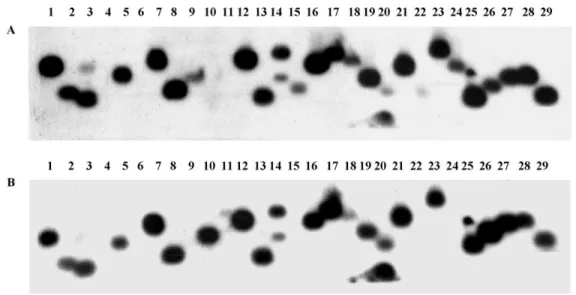
Hybridization of an arrayed human fetal brain cDNA library with total cDNA probes of GB and normal human adult brain revealed about one hundred cDNA clones with differences in the intensity of hybridization. Plasmid DNA from 80 cDNA clones received from RZPD was digested by HindIII/EcoRI restriction endonucleases, and after electrophoresis was blotted and hybridized with the same cDNA probes. Repeated differential hybridization of selected cDNA clones confirmed the results of the primary screening for 38 clones. Differential hybridization of one such plasmid DNA blot is shown inFig. 1where is possible to see that some cDNA inserts (lanes 10, 26) hybridized more intensive with a GB cDNA probe while the majority (lanes 1,5,9,15,16,19,24,25,28) hybridized more intensive with the normal adult human brain cDNA probe.
Nucleotide sequence analysis of cDNA inserts from 38 clones revealed 8 clones containing Alu-repeats and 30 clones with cDNAs corresponding to mitochondrial genes ATP6, COXI, COXII, COXIII, ND1, ND4, and 12S rRNA (Table 1).
As it was expected in accordance with the results of primary screening, Alu-containing sequences
hybridized more intensively with glioblastoma total cDNA.
3.2. Northern analysis of mt genes expression in astrocytic gliomas
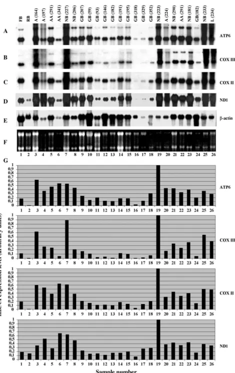
Regardless of the type or combination of data for genes with altered expression in glioblastoma versus normal brain, there is still a need to confirm and expand the expression information Northern blot hybridization seems to be the best approach for this purpose. Indeed, Northern analysis of astrocytic gliomas of different malignancy grades showed the significantly lower level of ATP6, COXIII, COXII and ND1 mRNAs in GB (sample 182) as compared to normal brain (sample 181) of the same patient
(Fig. 2). These tissue samples were used for primary screening of the human fetal brain cDNA library. Mean values of ATP6, COXIII, COXII or ND1 mRNA content in GBs was about two fold lower than in normal brain. It varied greatly in different GB samples analyzed that likely reflected the molecular heterogeneity of these tumors.
The comparison of the pairs of astrocytic tumors and corresponding adjacent tissues of normal brain showed the difference in the mt mRNAs level for anaplastic astrocytoma (Fig. 2A–D, lanes 21 and 22) and even for astrocytoma grade II (Fig. 2A–D, lanes 19 and 20).
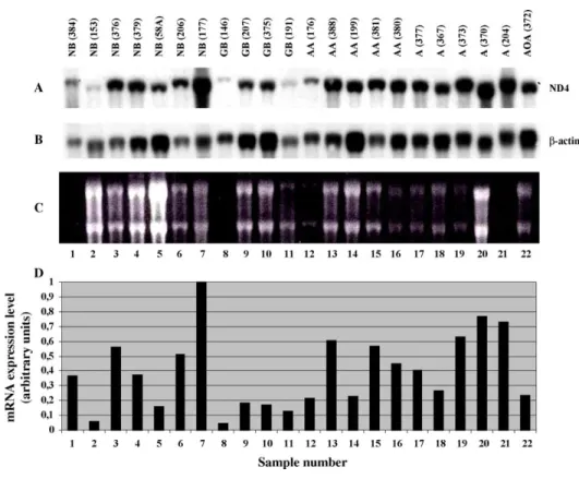
Northern hybridization with COXI (Fig. 3) and with ND4-probes (Fig. 4) showed similar patterns of the expression in normal brain and astrocytic tumors.
Table 1
Nucleotide sequences from human fetal brain (25 weeks old) cDNA library differentially expressed between glioblastoma and human normal brain
Clone Expression changes
in glioblastoma
Gene name
DKFZp415A2296 decreased mt NADH dehydrogenase subunit 1 DKFZp415D11116 decreased mt NADH dehydrogenase subunit 1 DKFZp415J1098 decreased mt NADH dehydrogenase subunit 1 DKFZp415L2318 decreased mt NADH dehydrogenase subunit 1 DKFZp415N0897 decreased mt NADH dehydrogenase subunit 1 DKFZp415N1579 decreased mt NADH dehydrogenase subunit 1 DKFZp415?1316 decreased mt NADH dehydrogenase subunit 4 DKFZp415G18136 decreased mt NADH dehydrogenase subunit 4 DKFZp415M02136 decreased mt NADH dehydrogenase subunit 4 DKFZp415P2074 decreased mt cytochrome oxidase subunit I DKFZp415G06136 decreased mt cytochrome oxidase subunit II DKFZp415E117 decreased mt cytochrome oxidase subunit III DKFZp415E1874 decreased mt cytochrome oxidase subunit III DKFZp415E1386 decreased mt cytochrome oxidase subunit III DKFZp415I1555 decreased mt cytochrome oxidase subunit III DKFZp415J19138 decreased mt cytochrome oxidase subunit III DKFZp415K05112 decreased mt cytochrome oxidase subunit III DKFZp415K1124 decreased mt cytochrome oxidase subunit III DKFZp415L07128 decreased mt cytochrome oxidase subunit III DKFZp415 decreased mt cytochrome oxidase subunit III N2116DKFZp415P2330 decreased mt cytochrome oxidase subunit III DKFZp415H0983 decreased mt ATP synthase subunit 6 DKFZp415J0588 decreased mt ATP synthase subunit 6 DKFZp415H20103 decreased mt ATP synthase subunit 6 DKFZp415I0891 decreased mt ATP synthase subunit 6 DKFZp415K0676 decreased mt ATP synthase subunit 6 DKFZp415L24135 decreased mt ATP synthase subunit 6 DKFZp415N0519 decreased mt ATP synthase subunit 6 DKFZp415N0413 decreased mt ATP synthase subunit 6
Fig. 2. Expression of ATP6, COX III, COXII and ND1 genes in normal human tissues and in brain tumors. Northern blot hybridization of
32P-labeled ATP6 (A), COXIII (B), COXII (C) and ND1 (D) cDNA probes with tumor RNA panel. Tissue types and tumor subtypes are
indicated above each lane of the blot: FB, human fetal brain; RB, rat brain; NB, human normal brain; GB, glioblastoma; AA, anaplastic astrocytoma; A, astrocytoma; L, lymphoma. (E) Northern blot hybridization of the same blot with b-actin cDNA control probe. (F) Ethidium bromide stained agarose gel. (G) Bar graphs showing relative expression of mt genes after correction for gel loading based on b-actin gene expression.
It is possible to see the decreased levels of these mRNAs in GB as compared to normal brain samples.
3.3. Analysis of the expression of mt genome in GB and normal brain by SAGE
Tag counts of all mt genes were examined in astrocytic gliomas and normal brain by accessing SAGEmap In the description to SAGE (http://www. ncbi.nlm.nih.gov/SAGE/) are some caviates that mt sequences were in large part masked or removed from Unigene but not from mapping of tags to mitochon-drial sequences. A single NlaIII tag was revealed for each of the mt genes by utilizing the ‘Retrieve by sequence’ option, and expression of all mt genes in astrocytic gliomas was analyzed by ‘Virtual North-ern’. Thus, we obtained clear evidence that transcrip-tion levels of all mitochondrial genes are lower in
glioblastoma than in the human normal brain (Table 2).
4. Discussion
Mitochondrial defects have long been suspected to play an important role in the development and progression of cancer. Several distinct differences between the mitochondria of cancer cells and normal cells have already been observed at the genetic, molecular and biochemical levels and these studies have indicated that mitochondrial dysfunction is involved in carcinogenesis. In particular, activation of the genes for COXI, COXII, and 16S rRNA was shown in rat fibroblast cells immortalized or trans-formed by viral and cellular oncogenes [14]; ND5, ND6, cytochrome b, COXII, and 16S rRNA in chemically induced rat hepatomas [15]; ND1, 12S
Fig. 3. Expression of COXI gene in normal human tissues and in brain tumors. (A) Northern blot hybridization of32P-labeled COXI cDNA probe with tumor RNA panel. Tissue types and tumor subtypes are indicated above each lane of the blot: He, human heart; K, human kidney; NB, human normal brain; GB, glioblastoma; AA, anaplastic astrocytoma; A, astrocytoma; OA, oligoastrocytoma; Ans, angiosarcoma; Ep, epidermoid; FB, human fetal brain; RB, rat brain. (B) Northern blot hybridization of the same blot with b-actin cDNA control probe. (C) Ethidium bromide stained agarose gel. (D) Bar graphs showing relative expression of COXI gene after correction for gel loading based on b-actin gene expression.
rRNA, and 16S rRNA in polyps of familial polyposis coli patients [16]. Elevated levels of ATP6, ND5, cytochrome b, COXI, and COXII mRNAs were revealed in papillary thyroid carcinomas as compared to normal thyroid tissue [17]. Sharp et al. [18] detected activation of the COXII gene by analysis of 17 breast cancer tumors, but failed to detect the alterations in ATP6, ND2 and ND4 gene expression. Other mitochondrial enzyme levels (NADH cyto-chrome c reductase, succinate cytocyto-chrome c reductase, and cytochrome c oxidase) have been shown to change also during exposure to carcinogens with cytochrome c oxidase levels remaining elevated even after discontinuation of exposure[19]; increased levels of RNA transcripts of the nuclear-encoded subunit COXIV and mitochondrially encoded COX subunits I and II have been observed in Zajdela hepatoma as compared with normal liver [20]. A study utilizing tissues from 15 colorectal cancer
patients found that ND2 mRNA levels were elevated in the malignant tissue as compared to normal tissue from the same individuals [21]. Analysis of the mitochondrial genome in colorectal cancer cell lines also revealed abnormalities; elevated expression of 16S rRNA, ND4, ND4L, cytochrome b, COXII, ATP6, and ATP8 was detected in the HT-29 cell line[22]. ND1, ND4, and COXII genes were shown to be overexpressed in B-cell non-Hodgkin’s lympho-mas in SIV-infected monkeys, 16S rRNA, COXII, ATP6, cytochrome b, ND2, and ND4—in human lymphomas[23].
In contrast to the investigations cited earlier, Zhou et al. [24] found that mitochondrial cytochrome c oxidase (COX) activity was significantly diminished in p53(K/K) cells. Their study suggests that p53 is involved in regulation of COXII at the protein level but not at the mRNA level. Decrease in COX activity was found also when comparing the specific activity
Fig. 4. Expression of ND4 gene in normal human tissues and in brain tumors. (A) Northern blot hybridization of32P-labeled ND4 cDNA probe with tumor RNA panel. Tissue types and tumor subtypes are indicated above each lane of the blot: NB, human normal brain; GB, glioblastoma; AA, anaplastic astrocytoma; A, astrocytoma; AOA, anaplastic oligoastrocytoma. (B) Northern blot hybridization of the same blot with b-actin cDNA control probe. (C) Ethidium bromide stained agarose gel. (D) Bar graphs showing relative expression of ND4 gene after correction for gel loading based on b-actin gene expression.
of the enzyme in biopsies of human colonic adenocarcinoma versus normal colon mucosa [25], in cultured rat HC252 hepatoma cells versus non-neoplastic liver [26], and in cultured carcinoma cell lines MCF-7 (breast), T47D (breast) and DU-145 (prostate) versus normal epithelial cells [27]. Faure-Vigny et al. [28] detected the lower expression of several mitochondrial genes encoding the proteins involved in energy metabolism of cancer cells in renal tumors of different types: a clear cell carcinoma, an oncocytoma, and urothelial tumors. The mean level of expression of COX subunit III gene was found to be lower in carcinoma versus normal mucosa samples [29]. A separate study reported the loss of mtDNA and mRNA coding for subunit the ND3 gene in renal cell carcinoma[30].
Our results obtained by differential hybridization of arrayed human fetal brain cDNA library with GB and normal adult brain cDNA probes with subsequent sequence analysis of differentially hybridized cDNAs, Northern blot hybridization, and SAGE, revealed decreased expression of all mitochondrial genes in glioblastoma as compared to human normal adult brain. These results suppose possible coordinated changes of mitochondrial genes expression in GB. The reason of the decrease of mt mRNA levels is not known yet; it may be a consequence of the decrease of
mt genome transcription activity, the increase of large mutations in mitochondrial DNA, or/and the decrease of quantity of mitochondria in glioblastomas. Dis-crepancies in the results concerning increase or decrease of the expression of mt genes in different tumors may be due to the different nature of various cancer types under investigation, and the reduction of all mt mRNA levels in GB may reflect the peculiarities of glioblastoma, the last grade of astrocytic glioma progression.
Acknowledgements
This work was supported in part by NATO Collaborative Linkage Grant No. 977284, grant No. F7/282-2001 (Fundamental Investigation Fond of Ukrainian Ministry of Education and Science), and by National Academy of Sciences of Ukraine in frames of the program ‘Novel Problems of Medicine and Biology and Human Environment’.
References
[1] S. Weinhouse, Oxidative metabolism of neoplastic tissues, Adv. Cancer Res. 3 (1955) 269–325.
[2] P.L. Pedersen, Tumor mitochondria and the bioenergetics of cancer cells, Prog. Exp. Tumor Res. 22 (1978) 190–274. Table 2
Results of ‘Virtual Northern’ of mt genes
Tag Tags per million Gene name
NB A (94876)* NB B (63208)* GB C (70087)* GB D (61886)* GB E (100600)* GB F (102322)* GB G (99099)* GTAAGTGTAC 664 727 28 145 238 820 151 12S rRNA CTAAGACTTC 16864 10489 1983 3651 2773 3156 3239 16S rRNA CACCTAATTG 9053 11739 2282 4314 2554 7153 15600 ATP6/ ATP8 CACTACTCAC 1443 1439 570 646 0 1915 2875 Cytochrome b ATTTGAGAAG 4163 6992 2297 2165 556 2804 1261 COXI CCCATCGTCC 11267 14001 4879 10616 4701 3107 17810 COXII TGATTTCACT 4279 4176 1383 1276 735 2413 5328 COXIII ACCCTTGGCC 6840 8748 870 1842 79 1847 2260 ND1 ACTAACACCC 6861 5505 784 1939 864 2609 2230 ND2 AGCCCTACAA 8621 8574 1169 3571 2713 5404 6518 ND3 TTCATACACC 5354 4002 3167 3183 7514 2756 14964 ND4/ ND4L AGACCCACAA 653 648 99 226 188 263 988 ND5 GGGGTCAGGG 242 221 57 145 79 107 191 ND6
SAGE libraries (total tag counts in library indicated in parentheses): A*, GSM676: SAGE_ BB542_ white matter; B*, GSM763: SAGE_ normal pool (6th); C*, GSM696: SAGE_Duke_GBM_H1110; D*, GSM765: SAGE_ pooled_GBM; E*, GSM14767: SAGE_Brain_glioblasto-ma_B_H833; F*, GSM 14768: SAGE_Brain_glioblastoma_B_R336; G*, GSM14769: SAGE_Brain_glioblastoma_B_R70.
[3] E. Carafoli, Mitochondria and disease, Mol. Aspects Med. 3 (1980) 295–429.
[4] S. Ohta, A multi-functional organelle mitochondrion is involved in cell death, proliferation and disease, Curr, Med. Chem. 10 (2003) 2485–2494.
[5] J.B. Jones, J.J. Song, P.M. Hempen, G. Parmigiani, R.H. Hruban, S.E. Kern, Detection of mitochondrial DNA mutations in pancreatic cancer offers a ‘mass’-ive advantage over detection of nuclear DNA mutations, Cancer Res. 61 (2001) 1299–1304.
[6] J.S. Penta, F.M. Johnson, J.T. Wachsman, W.C. Copeland, Mitochondrial DNA in human malignancy, Mutat. Res. 488 (2001) 119–133.
[7] J.S. Modica-Napolitano, K. Singh, Mitochondria as targets for detection and treatment of cancer, Expert Rev. Mol. Med. 2002; 1–19.
[8] J.S. Carew, P. Huang, Mitochondrial defects in cancer, Mol. Cancer 1 (2002) 1–9.
[9] W.C. Copeland, J.T. Wachsman, F.M. Johnson, J.S. Penta, Mitochondrial DNA alterations in cancer, Cancer Invest. 20 (2002) 557–569.
[10] P. Kleihues, D.N. Louis, B.W. Scheithauer, L.B. Rorke, G. Reifenberger, P.C. Burger, W.K. Cavenee, The WHO classification of tumors of the nervous system, J. Neuropathol. Exp. Neurol. 61 (2002) 215–225.
[11] T. Sambrook, E.F. Fritsch, T. Maniatis, Molecular cloning: a laboratory manual, second ed., Cold Spring Harbor Laboratory Press, Cold Spring Harbor, NY, 1989.
[12] K.O. Shostak, V.V. Dmitrenko, O.M. Garifulin, V.D. Rozumenko, O.V. Homenko, Y.P. Zozulya, G. Zehetner, V.M. Kavsan, Downregulation of putative tumor suppressor gene TSC-22 in human brain tumors, J. Surg. Oncol. 82 (2003) 57–64.
[13] P. Chomczynski, N. Sacchi, Single step method of RNA isolation by acid guanidinium thiocyanate–phenol–chloroform extraction, Anal. Biochem. 162 (1987) 156–159.
[14] N. Glaichennhaus, P. Leopold, F. Cuzin, Increased levels of mitochondrial gene expression in rat fibroblast cells immorta-lized or transformed by viral and cellular oncogenes, Eur. Mol. Biol. Org. J. 25 (1986) 1261–1265.
[15] M. Corral, B. Paris, G. Baffet, L. Tichonicky, C. Guguen-Guillouzo, J. Kruh, N. Defer, Increased level of the mitochondrial ND5 transcript in chemically induced rat hepatomas, Exp. Cell Res. 184 (1989) 158–166.
[16] A. Yamamoto, S. Horai, Y. Yuasa, Increased level of mitochondrial gene expression in polyps of familial polyposis coli patients, Biochem. Biophys. Res. Commun. 2159 (1989) 1100–1106.
[17] D.R. Haugen, O. Fluge, L.J. Reigstad, J.E. Varhaug, J.R. Lillehaug, Increased expression of genes encoding mitochondrial proteins in papillary thyroid carcinomas, Thyroid 13 (2003) 613–620.
[18] M.G.F. Sharp, S.M. Adams, R.A. Walker, Differential expression of the mitochondrial gene cytochrome oxidase II in benign and malignant breast cancer, J. Pathol. 168 (1992) 163–168.
[19] C.N. Huang, J.L. Tsai, M.T. Chen, W.J. Wu, K.W. Kuo, C.H. Huang, Changes in the activities of mitochondrial enzymes in the progress of tumorigenesis of bladder cancer, Biochem. Mol. Biol. Int. 46 (1998) 375–383.
[20] K. Luciakova, S. Kuzela, Increased steady-state levels of several mitochondrial and nuclear gene transcripts in rat hepatoma with a low content of mitochondria, Eur. J. Biochem. 205 (1992) 1187–1193.
[21] K.A. Chester, L. Robson, R.H. Begent, H. Pringle, L. Primrose, I.C. Talbot, A.J. Macpherson, S.L. Owen, G. Boxer, A.D. Malcolm, In situ and slot hybridization analysis of RNA in colorectal tumours and normal colon shows distinct distributions of mitochondrial sequences, J. Pathol. 162 (1990) 309–315.
[22] X. Lu, T. Walker, J.P. MacManus, V.L. Seligy, Differentiation of HT-29 human colonic adenocarcinoma cells correlates with increased expression of mitochondrial RNA: effects of trehalose on cell growth and maturation, Cancer Res. 52 (1992) 3718–3725.
[23] V.Z. Tarantul, G. Hunsmann, Mitochondrial polypeptides of the oxidative phosphorylation pathway as potential new targets for anti-cancer therapy, Med. Hypotheses 56 (2001) 386–387.
[24] S. Zhou, S. Kachhap, K.K. Singh, Mitochondrial impairment in p53-deficient human cancer cells, Mutagenesis 18 (2003) 287–292.
[25] A.S. Sun, K. Sepkowitz, S.A. Geller, A study of some mitochondrial and peroxisomal enzymes in human colonic adenocarcinoma, Lab. Invest. 44 (1981) 13–17.
[26] A.S. Sun, A.I. Cederbaum, Oxidoreductase activities in normal rat liver, tumor-bearing rat liver, and hepatoma HC-252, Cancer Res. 40 (1980) 4677–4681.
[27] J.S. Modica-Napolitano, S.E. Touma, Functional differences in mitochondrial enzymes from normal epithelial and carcinoma cells, in: Mitochondrial Dysfunction in Pathogen-esis, Keystone Symposia, Silverthorne, 2000 p. 64. [28] H. Faure-Vigny, A. Heddi, S. Giraud, D. Chautard, G. Stepien,
Expression of oxidative phosphorylation genes in renal tumors and tumoral cell lines, Mol. Carcinog. 16 (1996) 165–172. [29] B.G. Heerdt, H.K. Halsey, M. Lipkin, L.H. Augenlicht,
Expression of mitochondrial cytochrome c oxidase in human colonic cell differentiation, transformation, and risk for colonic cancer, Cancer Res. 50 (1990) 1596–1600.
[30] P. Selvanayagam, S. Rajaraman, Detection of mitochondrial genome depletion by a novel cDNA in renal cell carcinoma, Lab. Invest. 74 (1996) 592–599.