HAL Id: hal-01521993
https://hal.univ-lorraine.fr/hal-01521993
Submitted on 12 May 2017
HAL is a multi-disciplinary open access
archive for the deposit and dissemination of
sci-entific research documents, whether they are
pub-lished or not. The documents may come from
teaching and research institutions in France or
abroad, or from public or private research centers.
L’archive ouverte pluridisciplinaire HAL, est
destinée au dépôt et à la diffusion de documents
scientifiques de niveau recherche, publiés ou non,
émanant des établissements d’enseignement et de
recherche français ou étrangers, des laboratoires
publics ou privés.
Grégory Hoff, Claire Bertrand, Lingli Zhang, Emilie Piotrowski, Ludovic
Chipot, Cyril Bontemps, Fabrice Confalonieri, Stephen Mcgovern, François
Lecointe, Annabelle Thibessard, et al.
To cite this version:
Grégory Hoff, Claire Bertrand, Lingli Zhang, Emilie Piotrowski, Ludovic Chipot, et al.. Multiple
and Variable NHEJ-Like Genes Are Involved in Resistance to DNA Damage in Streptomyces
ambo-faciens. Frontiers in Microbiology, Frontiers Media, 2016, 7, pp.1901. �10.3389/fmicb.2016.01901�.
�hal-01521993�
doi: 10.3389/fmicb.2016.01901
Edited by: Frank T. Robb, University of Maryland, Baltimore, USA Reviewed by: Igor Kovalchuk, University of Lethbridge, Canada Andrzej Stasiak, University of Lausanne, Switzerland *Correspondence: Pierre Leblond pierre.leblond@univ-lorraine.fr
Specialty section: This article was submitted to Evolutionary and Genomic Microbiology, a section of the journal Frontiers in Microbiology Received: 07 October 2016 Accepted: 14 November 2016 Published: 28 November 2016 Citation: Hoff G, Bertrand C, Zhang L, Piotrowski E, Chipot L, Bontemps C, Confalonieri F, McGovern S, Lecointe F, Thibessard A and Leblond P (2016) Multiple and Variable NHEJ-Like Genes Are Involved in Resistance to DNA Damage in Streptomyces ambofaciens. Front. Microbiol. 7:1901. doi: 10.3389/fmicb.2016.01901
Multiple and Variable NHEJ-Like
Genes Are Involved in Resistance to
DNA Damage in Streptomyces
ambofaciens
Grégory Hoff1,2, Claire Bertrand1,2, Lingli Zhang1,2, Emilie Piotrowski1,2, Ludovic Chipot1,2,
Cyril Bontemps1,2, Fabrice Confalonieri3, Stephen McGovern4, François Lecointe4,
Annabelle Thibessard1,2and Pierre Leblond1,2*
1UMR 1128, Dynamique des Génomes et Adaptation Microbienne, Université de Lorraine, Vandœuvre-lès-Nancy, France, 2UMR 1128, Institut National de la Recherche Agronomique, Dynamique des Génomes et Adaptation Microbienne,
Vandœuvre-lès-Nancy, France,3Institute for Integrative Biology of the Cell (I2BC), CEA, Centre National de la Recherche
Scientifique, Université Paris-Sud, Orsay, France,4Institut Micalis, INRA, AgroParisTech, Université Paris-Saclay,
Jouy-en-Josas, France
Non-homologous end-joining (NHEJ) is a double strand break (DSB) repair pathway which does not require any homologous template and can ligate two DNA ends together. The basic bacterial NHEJ machinery involves two partners: the Ku protein, a DNA end binding protein for DSB recognition and the multifunctional LigD protein composed a ligase, a nuclease and a polymerase domain, for end processing and ligation of the broken ends. In silico analyses performed in the 38 sequenced genomes of Streptomyces species revealed the existence of a large panel of NHEJ-like genes. Indeed, ku genes or ligD domain homologues are scattered throughout the genome in multiple copies and can be distinguished in two categories: the “core” NHEJ gene set constituted of conserved loci and the “variable” NHEJ gene set constituted of NHEJ-like genes present in only a part of the species. In Streptomyces ambofaciens ATCC23877, not only the deletion of “core” genes but also that of “variable” genes led to an increased sensitivity to DNA damage induced by electron beam irradiation. Multiple mutants of ku, ligase or polymerase encoding genes showed an aggravated phenotype compared to single mutants. Biochemical assays revealed the ability of Ku-like proteins to protect and to stimulate ligation of DNA ends. RT-qPCR and GFP fusion experiments suggested that ku-like genes show a growth phase dependent expression profile consistent with their involvement in DNA repair during spores formation and/or germination.
Keywords: Streptomyces, non-homologous end joining, DNA repair, double strand breaks (DSBs), ligases, Ku protein, DNA damage
INTRODUCTION
Double strand breaks (DSB) are the most detrimental genomic lesions and their repair constitutes a major challenge for cell viability. In eukaryotes and prokaryotes, two main repair machineries have evolved to repair these DSBs, namely homologous recombination (HR) and non-homologous end joining (NHEJ) (Alonso et al., 2013;Deriano and Roth, 2013;Glickman, 2014). In contrast
to HR which needs an intact template to ensure repair of the break, NHEJ can bind DNA ends together without any template after a facultative DNA ends remodeling step. In multicellular eukaryotes, the NHEJ mechanism represents the predominant DSB repair pathway and requires the intervention of the end tethering heterodimer Ku70/80 as well as diverse end processing proteins (for review: Deriano and Roth, 2013). In prokaryotes, NHEJ is not ubiquitous and the minimal NHEJ machinery first described inMycobacterium consists of two actors: a Ku homolog and an ATP dependent ligase LigD (Weller et al., 2002; Della et al., 2004;Gong et al., 2005). The latter one is a multifunctional protein that carries three distinct enzymatic domains conferring nuclease, polymerase and ligase activities (Pitcher et al., 2005). Unlike DSB repair through HR, DSB repair carried out by NHEJ is mutagenic. Nucleotide addition and/or deletion are scars frequently left after DNA ends ligation (Stephanou et al., 2007;
Aniukwu et al., 2008; Gupta et al., 2011). The initial model involving two unique actors is now renewed by the discovery of alternative actors. When the ligase domain of LigD (LigDom) is defective, a back-up ligation activity is provided by LigC, another ATP dependent ligase (Della et al., 2004;Bhattarai et al., 2014). In the same way,Mycobacterium smegmatis possesses two additional
polymerases which share the same in vitro activity than the
polymerase domain of LigD (PolDom) (Zhu et al., 2012). Streptomyces are filamentous bacteria mainly found in soils, sediments and seawater as well as symbionts with plants,
fungi and animals (Seipke et al., 2012). Soil is a complex
and changing environment where Streptomyces have to adapt
to the heterogeneousness of the niche (moisture, nutrients, temperature. . .) and to cope with biotic competition. Their rich and varied secondary metabolism such as signaling molecules or chemical weapons is assumed to mediate their relationships
with other organisms (Davies and Davies, 2010; Abrudan
et al., 2015). Under limited nutritive conditions, their hyphae differentiate into long chains of exospores providing an efficient way for resistance and dissemination until suitable conditions for another growth cycle are met (Chater, 1993). While hyphae are large multi-genomic compartments, spores are known to
include a single genome (Hopwood, 2006). In contrast to
the vast majority of bacteria, Streptomyces possess a linear
chromosome whose size is ranging from 5.96 to 11.94 Mb1with
a central replication origin and terminally inverted repeats that are covalently bound to specific telomere proteins (Lin et al., 1993). TheStreptomyces genome presents a high plasticity and their chromosome is highly compartmented, with a conserved central region including mostly housekeeping genes whereas the dispensable genes are predominantly located in the chromosomal arms (Bentley et al., 2002;Ikeda et al., 2003;Kim et al., 2015). The loss of synteny at the ends of the chromosome results from horizontally acquired DNA insertion or deletion events (Choulet et al., 2006; Thibessard and Leblond, 2014). This terminal variability reveals the strong recombination activity taking place at the ends of the genome which is reminiscent of the DNA rearrangements reported several decades ago (Leblond et al., 1990; Volff and Altenbuchner, 1998). Hence,
1http://www.ncbi.nlm.nih.gov/assembly/?term=Streptomyces
Streptomyces are also subject to a high level of genetic instability which is directly linked to major genome rearrangements. Characterization of some of these rearrangements revealed large deletions, chromosome circularization, (Fischer et al., 1997;
Inoue et al., 2003), arm replacement (Fischer et al., 1998;
Uchida et al., 2003), chromosome end-to-end fusion (Wenner et al., 2003) or large DNA amplifications (Yanai et al., 2006). Molecular analyses showed that chromosome circularization and end-to-end fusion events especially rely on illegitimate recombination whereas chromosome arm replacement or DNA amplifications is dependent on HR. This terminal recombination pattern may result from higher recombination frequencies triggered by the preferential formation of DSBs in these regions. On the other hand, genome rearrangements could also be more tolerated if formed in these dispensable regions.
Nevertheless, little is known about DSB repair inStreptomyces. Like the other bacteria,Streptomyces possess a RecA homologue involved in HR (Aigle et al., 1997;Muth et al., 1997;Vierling et al., 2001;Huang and Chen, 2006). However, recent studies that we performed suggest thatStreptomyces has an atypical HR pathway. In fact, in contrast to bacterial models like Escherichia coli, Bacillus subtilis or Mycobacterium, deletion of genes encoding a potential helicase-nuclease complex supposed to be involved in the initial end resection step during HR is not viable in S. ambofaciens (Zhang et al., 2014). On the other hand, deletion
of ruvABC and recG, two loci encoding factors that have a
redundant role in migration and resolution of Holliday junction during late steps of HR, has only a mild effect on recombination efficiency (Hoff et al., 2016). Early bioinformatics studies showed that the two model species,S. coelicolor and S. avermitilis, both
possess aku ortholog suggesting also the existence of a NHEJ
pathway inStreptomyces (Rocha et al., 2005). Here, we performed in silico analyses in the fully sequenced Streptomyces genomes leading to the identification of a large panel of conserved and variable genes suspected to be involved in a NHEJ pathway. Construction of single and multiple NHEJ-like genes mutant strains inS. ambofaciens allowed us to study the impact of these different genes in DNA damage repair. All the results we obtained suggest the existence of a complex and atypical bacterial NHEJ pathway inStreptomyces.
MATERIALS AND METHODS
Phylogenetic Analyses
A total of 38 sequencedStreptomyces genomes, including that
of S. ambofaciens ATCC23877 was scanned to identify genes
putatively encoding NHEJ Ku-like or LigD-like proteins based on BLAST similarity search against the non-redundant database. A preliminary BLASTP search (e-value cut-off 10−30
; identity
threshold 30%) usingM. tuberculosis Rv0937c and Rv0938 (Ku
and LigD respectively) protein sequences as a query allow to identify and numerate the candidates. Secondly, BLASTP were
performed usingS. ambofaciens ATCC23877 NHEJ-like genes
and reciprocal best hits allowed to organize them into groups of orthologs.
For the phylogeny of Ku-like proteins, the tree was built using a maximum-likelihood method based on a GTR + G + I model. Phylogenetic trees were built and edited with Mega6 (Tamura et al., 2013). Support of the tree branches was estimated with 100 bootstrap replicates and all positions with<80% site coverage were eliminated. A total of 295 and 309 positions were respectively used for the Ku-like and the LigC/LigD protein phylogenetic trees.
Bacterial Strains and Plasmids
All strains used in this study are listed in Table 1. The
DH5α E. coli strain is used as conservation host for BACs
and plasmids. For biochemical analyses, plasmid constructions
were done in E.coli MC1061 (Casadaban and Cohen, 1980).
ER2566 (NEB) was used for kuB gene expression and Rosetta
(DE3) PLysS (Stratagene) for kuA and kuC gene expression.
E.coli strains were grown in Luria-Bertani (LB) medium at 37◦
C except for the BW25113 pKD20 thermosensitive
strain used for PCR targeting, which was grown at 30◦
C.
S. ambofaciens strains were grown at 30◦
C on Soya flour mannitol (SFM) plates except for phenotypical analyses and for liquid cultures which were performed on solid Hickey
Tresner (HT) medium. AllStreptomyces mutants derive from our
reference strain S. ambofaciens ATCC23877 (Pinnert-Sindico,
1954). Mutant strains were constructed by PCR targeting as
described by Gust et al. (2003). To summarize, a recombinant
BAC containing the locus of interest was transformed in the highly recombinogenicE. coli BW25113/pKD20 strain in order to replace the target gene by anoriT- aac(3)IV resistance disruption
cassette (Raynal et al., 2006). The ET12567 non-methylating
strain containing the mobilizing pUZ8002 plasmid was used as donor for intergeneric conjugation of the modified BAC betweenE. coli and S. ambofaciens Double-crossover (CO) events
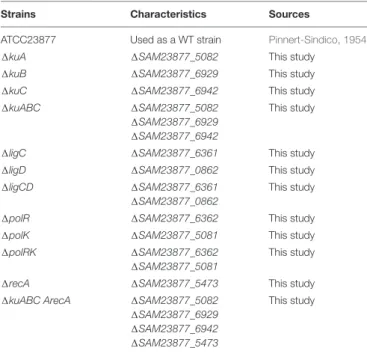
TABLE 1 | Streptomyces ambofaciens strains used in this study.
Strains Characteristics Sources
ATCC23877 Used as a WT strain Pinnert-Sindico, 1954 1kuA 1SAM23877_5082 This study
1kuB 1SAM23877_6929 This study 1kuC 1SAM23877_6942 This study 1kuABC 1SAM23877_5082
1SAM23877_6929 1SAM23877_6942
This study
1ligC 1SAM23877_6361 This study 1ligD 1SAM23877_0862 This study 1ligCD 1SAM23877_6361
1SAM23877_0862
This study
1polR 1SAM23877_6362 This study 1polK 1SAM23877_5081 This study 1polRK 1SAM23877_6362
1SAM23877_5081
This study
1recA 1SAM23877_5473 This study 1kuABC ArecA 1SAM23877_5082
1SAM23877_6929 1SAM23877_6942 1SAM23877_5473
This study
leading to the replacement of the target gene by the cassette were selected by the sensitivity to kanamycin (loss of the BAC vector part) along with the resistance to apramycin (insertion of the resistance cassette). The disruption cassette flanked by attL and attR sites was excised in the newly obtained mutant strains thanks to the introduction of pOSV508 a plasmid allowing the expression ofint and xis genes from pSAM2 (Raynal et al., 2006). Strains expressing translational EGFP fusions were also
constructed by in vivo recombination. The egfp gene with the
transcription terminator from phageλ was cloned from plasmid
pIJ8660 (Sun et al., 1999) into an oriT aac(3)IV disruption
cassette (Raynal et al., 2006). This cassette was then used as the
source to make kuA-egfp, kuB-egfp and kuC-egfp fusions. PCR
was performed using egfp-oriT-aac(3)IV as the template with
specific oligonucleotides which anneal to the linker sequence at the 50
end ofegfp and downstream of the 30
end ofaac(3)IV and introduce 40 bases of homology to the 30
ends ofkuA, kuB, or
kuC, as well as removing the stop codons. PCR products were
introduced intoE. coli BW25113/pKD20 containing the cognate
BACs with eitherkuA, kuB, or kuC, and the following steps of the procedure are described above. In these recombinant strains, the fusion genes (kuA-egfp, kuB-egfp or kuC-egfp, respectively) are the only copies ofkuA, kuB, or kuC.
Electron Beam Irradiation (EBI)
An amount of 109 spores of different Streptomyces strains was diluted in sterile water in 96 well microplates and exposed to 0, 0.1, 0.3, 0.5, 0.7, and 1 kGy of radiation doses. Processing was carried out in a Van de Graaf R
electron accelerator (VIVIRAD, Handschuheim, France) in the technological resource center Aérial (Illkirch-Graffenstaden, France). Energy delivered by the electron accelerator reach 2.2 MeV with an intensity of 10µA. Dose rate equals approximately 60 kGy/h. After irradiation, several spore dilutions were plated with the easySpiral R
apparatus (Interscience, Saint-Nom-la-Bretèche, France) on SFM Petri dishes and the number of colonies was counted after 3 days incubation at 30◦
C. For each genetic context, at least two independent mutant strains were exposed in two independent experiences.
Fluorescent Microscopy
Strains for microscopic observations were inoculated in the acute-angle junction of standard-sized microscope coverslips inserted with an angle of 45◦
in SFM agar, and grown for 48 h. The coverslips were then carefully pulled from the medium and directly mounted in a drop of sterile water on a microscope slide. Fluorescent microscopy observations were carried out using a Axiovert 200M MOT device (Carl Zeiss Micro imaging) equipped with a mercury vapor lamp and a FITC filter set (470/40 excitation, 540/50 emission). Both contrast phase and fluorescent observations were made with an X100 objective.
DNA Preparation
To verify the mutant strains, Streptomyces genomic DNA was
extracted by low binding sulfate salt method as described by
Kieser et al. (2000). Liquid HT medium was used for growth of mycelium before DNA extraction. For biochemical analyses, the
gene coding for KuA was amplified from the BAC 16ZB5.KuB
and KuC genes were amplified from the BAC 27ZF1. Primers
used for amplification were designed to flank the open reading
frames with the coding sequence of a His6-tag followed by a
NdeI site on their 50
side, and by aXhoI site on their 30
side. The resulting ORFs were cloned under the T7 promoter of the pJ411 vector (DNA2.0) giving the plasmids pSMG258, 259, 260 allowing expression of the proteins 6His-KuA, 6His-KuB, 6His-KuC, respectively (named hereafter KuA, KuB, and KuC). Sequences of the primers used in this study are available upon request.
RNA Extraction and RT-qPCR Analyses
For RNA extraction from cultures on solid medium, streaks were harvested after growth (from 24 to 60 h depending of the experiment) at 30◦
C on HT solid medium covered with cellophanes membranes. For RNA extraction after a genotoxic stress, pre-germinated spores of Streptomyces were grown until 0.2 OD600 was reached and exposed to mitomycin C (MMC,
1.5 µg.mL−1, i.e., CMI/4) for 30 min. RNA isolation was
performed using the Aurum Total RNA mini-kit (Bio-Rad) according to the manufacturer’s instructions. To optimize cell lysis, an additional sonication step (3 × 30 s) using a Bioruptor apparatus was added. The RNA quality was assessed after migration on 1% agarose gel. In order to eliminate genomic DNA contamination, the RNA samples were treated before reverse transcription with DnaseI (Ambion) in the presence of
10 U of RNase inhibitor per µg of RNA. DnaseI was then
inactivated at 75◦
C during 10 min in the presence of EDTA.
Reverse transcription was performed on 2 µg RNA with an
iScript Advanced cDNA synthesis kit (Bio-Rad) according to the manufacturer’s instructions. The absence of contamination with genomic DNA was validated by a 30 cycle-PCR on a control aliquot removed before reverse transcription using hrdB-F and hrdB-R primers. Quantitative PCR was undertaken in
a CFX96 (Bio-Rad) apparatus in a 10 µL reaction mixture
containing respectively 5 µL of SYBR supermix (Bio-Rad),
0.2 µM of each primer and 4 µL of a 10-fold dilution of
cDNA. The reaction conditions were as follow: a 3 min initial denaturation step at 95◦
C and 40 cycles of 10 s at 95◦
C and 30 s at 60◦
C. In order to check the absence of secondary
products, melting curves were realized from 65 to 95◦
C with an increase rate of 0.5◦
C/5 s. The transcription level of interest genes was analyzed using the mathematical model proposed by Pfaffl (2001). The hrdB gene, encoding the principal sigma
factor in Streptomyces, was used as the reference gene for
the normalization of the expression level as it was the case in former studies (Galet et al., 2015; Martínez-Burgo et al., 2015).
Proteins Purification
Purification of KuA
Escherichia coli Rosetta PlysS cells transformed with pSMG258
were grown at 30◦
C in 2 l of LB medium supplemented with
25 mg/mL thymine, 30 µg/mL kanamycin and 25 µg/mL
chloramphenicol to OD600 = 0.7. Production of protein was induced with 0.5 mM IPTG for 15 h at 20◦
C. Cells were harvested
by centrifugation and the pellet was resuspended in 40 mL lysis buffer A (50 mM Tris-HCl pH 8, 0.25 M NaCl). Cells were broken by a freeze/thaw step in liquid N2/37◦C and then centrifuged
1 h at 100 000 g at 4◦
C. The pellet was resuspended with
40 mL of a 20 mM Tris pH 7.5, 2.5 mM MgCl2, 0.5 mM CaCl2,
1 mg/mL DNaseI buffer. After 1 h at 20◦
C, the mixture was
centrifuged 20 min at 100 000 g at 4◦
C. All subsequent steps were carried at 4◦
C. The pellet was washed with 40 ml buffer
P1 (50 mM Tris-HCl, pH 8, 1 M NaCl), and then solubilized
with 40 mL of buffer P2 (50 mM Tris pH 8, 8 M urea). After
1 h the mixture was centrifuged 20 min at 100 000g and 3 mL
of Ni2+affinity column (Ni-NTA agarose, Qiagen) was added to
the supernatant which was transferred onto an Econo-Column (Biorad). The flow through was discarded and the resin was
washed with series of 25 mL of buffer P3 (50 mM Tris pH 8,
1% sucrose, 0.5 M NaCl) with urea concentration decreasing from 8 M to 0 M by steps of 0.5 M. The Ni-NTA column was
washed with buffer P3, then with buffer W (50 mM Tris pH
8.0, 0.5 M NaCl) supplemented with 20 mM imidazole. KuA protein was eluted in buffer W supplemented with 250 mM imidazole and then dialysed against buffer D (50 mM Tris-HCl pH 8, 0.4 M NaCl, 50% glycerol, 1 mM DTT) prior to storage at −20◦
C.
Purification of KuC
The expression ofKuC gene was induced in E. coli Rosetta PLysS cells transformed with pSMG260 as described for KuA. Cells were harvested by centrifugation and the pellet was resuspended in 40 mL lysis buffer B (50 mM Tris-HCl pH 8, 0.25 M NaCl, 0.2 mg/mL lysozyme) supplemented with Complete Antiprotease
(Roche) and incubated 1 h at 20◦
C. The mixture was then centrifuged and the pellet was treated with DnaseI, as described for KuA purification. Purification procedure of KuC at this step is the same as the one described for KuA.
Purification of KuB
ER2566 cells transformed with pSMG259 were grown at 30◦
C in 2 l of LB medium supplemented with 25 mg/mL thymine
and 30 µg/mL kanamycin and kuB gene expression was
induced as for kuA and kuC genes. Cells were harvested by
centrifugation, the pellet was resuspended in 40 mL of lysis buffer B and incubated 1 h at 20◦
C. All subsequent steps were carried at 4◦
C. Cell lysis mixture was supplemented with 0.5 M NaCl, 0.1% Triton and 0.1% Non-idet P40 and incubated 1 h. Two mL of polyethyleneimine (PEI) 10%, pH 8 was added to precipitate nucleic acids; the mixture was stirred
for 1 h and then centrifuged 1 h at 100 000 g. The soluble
phase was next precipitated by raising the concentration of ammonium sulfate (AS) firstly to 50% of saturation, KuB was mainly found in the supernatant at this step, and secondly to 80% of saturation to precipitate KuB. The corresponding
pellet was resuspended in 25 mL of buffer P1 and 3 mL of
Ni-NTA agarose, pre-equilibrated in buffer P1, were added.
The Ni-NTA agarose was washed with buffer P1, then with
buffer W supplemented with 20 mM imidazole and KuB was eluted in buffer W supplemented with 250 mM imidazole.
The eluted fraction was dialyzed against buffer D and stored at −20◦
C.
Final purification step analyzed by SDS PAGE showed that purified KuA, KuB and KuC were obtained at 90% purity and were a mix of full length and truncated forms (Supplementary Figure S1). Their concentration was determined
by UV absorption at λ = 280 nm on a NanoDrop 1000
Spectrophotometer (Thermo Scientific).
Purification of LigDBsub
LigD fromBacillus subtilis fused in C-terminal to a His6tag was
purified as previously describedMcGovern et al. (2016).
T5 Protection Assay
Fifty nanograms (∼75 fmol) of a 1001 bp linear DNA substrate with 50
-phosphorylated blunt-ends were preincubated with raising concentrations of KuA, KuB, or KuC in 19µL of buffer T5
(50 mM Tris-HCl pH 8, 1 mM DTT, 5 mM MgCl2, 40 mM NaCl
and 5% glycerol) for 30 min at 37◦
C. OneµL of T5 exonuclease
(0.5 U/µL, Biolabs) was added to the reaction mixture and
reactions were incubated for 30 min at 37◦
C. Reactions were
stopped by adding 4 µL of stop buffer (5 mg/mL proteinase
K, 2% SDS and 0.1 M EDTA) and incubated at 37◦
C for 30 min. Four microliter of DNA loading buffer (33% glycerol and 0.25% xylene cyanol) were added, and the reactions were loaded on a 0.7% agarose gel in TBE Buffer. After electrophoresis, DNA was stained by the SYBR Gold reagent (Invitrogen) and the gel was photographed using a Chemidoc apparatus (Bio-Rad).
Ligation Assay
In a final reaction volume of 20µL in the T4 DNA ligase ligation
buffer (NEB, 50 mM Tris-HCl pH 7.5, 10 mM MgCl2, 10 mM
DTT, 1 mM ATP), 50 nanograms (∼75 fmol) of the 1001 bp linear
DNA substrate were preincubated with 0.25 or 1 µM of KuA,
KuB, or KuC for 30 min at 30◦
C. Then, raising concentrations
of LigDBsub were added to reactions. Ligation reactions were
stopped after 2 h at 37◦
C by adding 4 µL of stop buffer and
analyzed as described above.
RESULTS
A “Core” and a “Variable” NHEJ Gene
Set in Streptomyces
Genes homologous to the bacterial NHEJ factors ku and ligD
were sought in the genome of our model strain Streptomyces
ambofaciens ATCC23877 (Thibessard et al., 2015). To do so,
theM. tuberculosis Rv0937c and Rv0938 (KuMtuband LigDMtub
respectively) protein sequences were used as query in a BLASTP
search. The first observation was that the LigDMtub protein
had no full length homologue inS. ambofaciens, but that each
candidate protein matched only with one single domain at a time.
Therefore, the 759 amino acid sequence of LigDMtub was split
into 3 slightly overlapping parts (1–300, 271–459, and 451–759) corresponding to the polymerase (PolDom), the nuclease (NucDom) and the ligase (LigDom) domains, respectively. In
this way, 3 Ku-like encoding genes (sharing 36, 39, and 40% of protein identity with KuMtub), 3 LigDom-like encoding genes (sharing 30, 32, and 41% of protein identity with the LigDom
of LigDMtub) and 3 PolDom-like encoding genes (sharing 29,
34, and 41% of protein identity with the PolDom of LigDMtub)
were identified inS. ambofaciens ATCC23877 genome (Figure 1).
No NucDom-like encoding gene was detected. The 3 ku-like
genes (SAM23877_5082, SAM23877_6929 and SAM23877_6942)
were named kuA, kuB, and kuC respectively. Two of the 3
LigDom-like genes (SAM23877_6361 and SAM23877_0862) were
named ligC and ligD, respectively. Indeed, SAM23877_6361
appeared to share more amino-acid identity to M. tuberculosis
LigC (LigCMtub), i.e., 60% than to the LigDom of LigDMtub
i.e., 30% and was therefore considered as LigCMtub ortholog.
Similarly,SAM23877_0862 appeared to be the closest relative of
the LigDom of LigDMtub. The third ATP-dependent DNA ligase
encoding gene detected (SAM23877_1283) would encode a LigB protein known to exhibit a strong nick sealing activityin vitro and whose defect would not alter mycobacterial NHEJ (Gong et al., 2004). Therefore, this third ligase was not included in our further investigations. The 3 PolDom-like genes (SAM23877_5081, SAM23877_6202, and SAM23877_6362) were named polK, polO andpolR, respectively. It was noticeable that 2 of the 3 PolDom like genes (polK and polR) colocalized with kuA and ligC.
KuA, KuB, and KuC proteins are composed of 365, 383, and 302 amino-acids, respectively. All these proteins harbor the conserved Ku core domain at their N-terminal side, the conserved minimal C-ter domain (red in Supplementary Figure S2), that has been shown to be required for the interaction with LigD inB. subtilis (McGovern et al., 2016), and a basic extended C-terminal domain, which is of 114, 128, and 47 aa, respectively. The pIs of these extended domains (11.64, 10.97, and 10.65) are consistent with those described for the majority of bacterial Ku proteins. This suggests that their roles in DNA binding as well as in the modulation of the ability to thread inward
the DNA molecule are conserved in Streptomyces (Kushwaha
and Grove, 2013; McGovern et al., 2016). Interestingly, a SAP domain, previously predicted in theS. coelicolor KuB homologue
(SCO0601) by Aravind and Koonin (2001) is also detected
in S. ambofaciens KuB (at position 337–370, Supplementary
Figure S2). This SAP domain is assumed to be also involved in nucleic acid binding (Aravind et al., 2000) and could modify the DNA binding abilities of KuB compare to KuA and KuC. Further experiments are required to highlight the roles and the differences of the extended C-terminal domains of these three Ku proteins.
Once this list of genes potentially involved in aStreptomyces NHEJ pathway established, the question of their conservation
in the Streptomyces genus was addressed by seeking their
homologues in 37 other Streptomyces complete genome
sequences available at http://www.ncbi.nlm.nih.gov/genome.
Therefore, S. ambofaciens KuA, KuB, KuC, LigC, LigD, PolK,
PolO and PolR were used as query in BLASTP searches. A wide variety of situations were encountered and a few of them are illustrated in Figure 1. The homologues identified
in 38 Streptomyces genomes are listed by their locus tag in
FIGURE 1 | Organization of the NHEJ-like genes in several Streptomyces genomes. The two conserved loci kuA-polK and ligC-polR are framed in dotted lines.
summarized in Figure 2. The number and position of NHEJ-like genes varied from one species to another. However, two loci are widely shared amongStreptomyces: ligC-polR which is present in all the investigated genomes andkuA-polK which is present in all the tested strains butStreptomyces vietnamensis.
Focusing on the main function expected from a functional NHEJ system, we analyzed further the Ku-like and LigDom-like encoding genes and extended our interest to the PolDom-like genes colocalizing with them.
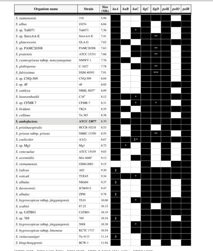
All but S. vietnamensis possess at least one kuA gene. In
contrast, kuB was detected in only 8 out of the 38 analyzed
genomes. Regarding kuC, the copy number varies from 0 to
2. Indeed, in the case of S. coelicolor, two identical copies
of kuC are included in the terminal inverted repeats of the
SCP1 linear plasmid. A few Streptomyces strains carry several
copies ofkuA genes leading us to make a distinction according
to their vicinity of polK: the neighbors of polK were called
kuA primary genes while standalone kuA genes were called
secondarykuA genes. All the ku-like genes (43 kuA, 8 kuB and
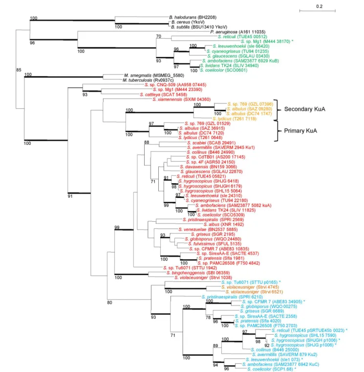
17kuC) were aligned and submitted to a Maximum Likelihood
phylogenetic analysis. The tree topology (Figure 3), together
with the fact that the primary kuA gene is almost ubiquitous,
suggested that KuA was probably present in the Streptomyces
common ancestor and evolved along with Streptomyces. The
secondary kuA gene carried by Streptomyces sp. 769, S. albulus
ZPM, S. albulus NK660, and S. lydicus shares an average
nucleotidic identity ranging from 67 to 72% with their respective primary copy. For these species, the primary and secondary KuA phylogenies are congruent, suggesting that their common
ancestor gained a second kuA copy either by gene duplication
or by gene transfer. Regarding the kuC gene, it was present
in 17 out of 38 analyzed genomes, 7 of which were plasmid-borne. It was closely related to KuA and formed a sister group of KuA within a large KuA-KuC monophyletic group that
excluded KuB. In factkuB genes formed a strong monophyletic
group and appeared much more scattered in Streptomyces
genera.
Regarding the distribution of LigC and LigD ligases, LigC encoding genes were found in all genomes and always in
the close vicinity of polR as in M. tuberculosis (Figure 1).
In contrast, LigD was detected in 29 out of the 38 cases (Figure 2). In 6 of them the encoding gene colocalized with
FIGURE 2 | Distribution of NHEJ-like genes in 38 complete genomes of Streptomyces. The presence of a homolog is mentioned by a black box and the absence of a homolog by an empty box. The number inside the boxes indicates when several homologs were identified;∗
plasmidic;∗ ∗
closed to kuC;∗ ∗ ∗
partial (cover of 35%).
FIGURE 3 | Phylogeny of Ku-like proteins identified in 38 Streptomyces strains. Streptomyces Ku-like proteins were identified in 38 Streptomyces strains and compared with the closest homologues of M. tuberculosis, M. smegmatis, B. subtilis, B cereus, B halodurans, and P. aeruginosa. The tree was built using a maximum-likelihood method with a 295 aa alignment. Only bootstrap values higher than 70% are reported. Heavy lines indicate branches supported by bootstrap values>90% (100 replicates). The scale represents mutations per amino-acid. Primary KuA are in red, secondary KuA are in orange, KuB are in green and KuC in blue. The asterisks symbolized the proteins encoded by plasmid borne-genes. The locus-tag of each sequence is mentioned in brackets. The suspected event of gene duplication is depicted by the two braces. B, Bacillus; M, Mycobacterium; P, Pseudomonas; S, Streptomyces. NRRL 8057 = DSM 46488; C34 = DSM 42122 = NRRL B-24963; MA-4680 = NBRC 14893.
kuC, and in S. cattleya, it colocalized with kuA-polK (see Figure 1). Phylogenetic analysis by Maximum Likelihood method (Supplementary Figure S3) showed that the LigC proteins
branched together withM. tuberculosis LigC with a topology that seemed quite congruent with the species relations, suggesting a vertical inheritance of this conserved locus. LigD proteins also
branched together and together with LigDom ofM. tuberculosis LigD.
Altogether, two NHEJ-like gene categories were delineated:
(i) those that are conserved among the Streptomyces and were
considered as “core” putative NHEJ factors (i.e., kuA and the
colocalizedpolK, ligC and the colocalized polR) and (ii) those that
are sporadically represented among the Streptomyces and were
considered as “variable” putative NHEJ factors (i.e.,kuB, kuC and ligD).
NHEJ-Like Genes Are Not Essential in
S. ambofaciens
After in silico identification of the “core” and “variable” NHEJ putative factors inStreptomyces, we constructed deleted mutant strains by replacing the chromosomal locus with an excisionable antibiotic resistance cassette (Raynal et al., 2006) by the use of PCR targeting (Gust et al., 2003). The whole selection procedure was performed several times simultaneously in order to select at least two independent mutant lineages for each genetic context. Table 1 summarizes the different genotypes constructed for this study. The mutations were checked by PCR amplification of the mutated locus. The maintenance of the chromosomal linearity of the mutants was checked by PCR amplification of the telomeres and of some loci scattered along the chromosome (not shown).
All the mutations, simple or multiple, were obtained with classical frequencies indicating that none of the targeted genes (“core” or “variable”) was essential. In addition, there was no significant difference between the wild-type reference strain and any of mutants in colonial morphology and sporulation under normal growth conditions (not shown).
Implication of NHEJ-Like Genes in DNA
Damage Response
Electronic beam irradiation (EBI) was shown to induce loss of viability of Bacillus spores by damaging spore coat, by altering membrane permeability or by DNA fragmentation (Fiester et al., 2012). High level energy beam such as those used in this work (2.2 MeV) is likely to trigger DSB as the most significant lesions in the spore embedded DNA. In order to test the involvement of the NHEJ-like factors to DNA damage response, spores of the wild-type S. ambofaciens reference strain as well as spores of at least two independent mutants for each genetic context were irradiated by EBI from 0.1 to 1 kGy and their survival was assessed after growth on SFM medium. The results are represented in Figure 4.
Both “Core” and “Variable” ku genes Are Involved in DNA Damage Response
Depending on the dose, deletion of kuA, which belongs to the
“core” NHEJ-like gene set, conferred a 2–4 fold increase of the sensitivity compared to the wild-type (Figure 4A). Since kuA is present in the large majority of the sequenced genome,
it appears as the major Ku actor of the putative Streptomyces
NHEJ. Surprisingly, mutations of the variable ku-like genes,
kuB and kuC, also conferred an increased sensitivity but at
a lower level, with a maximal twofold cell viability decrease reached after 0.5 and 0.7 kGy exposition for both contexts (Figure 4A). In addition, the sensitivity was aggravated when
kuB and kuC were both deleted in a 1kuA background to
generate a triple1kuABC mutant, until reaching a sevenfold cell viability diminution in comparison to the WT for the medium radiation doses (Figure 4A). This result suggests that the three Ku products participate to the response to DNA damaging agents in spores, each of them assuming their share of the DNA repair independently from the two others. Additionally, the triple 1kuABC mutation assumed to abolish any putative NHEJ repair strongly increased the sensitivity of a HR deficient context (i.e., 1recA) (Figure 4D). This last result shows that RecA and Ku-like proteins are both important contributors to the S. ambofaciens spore resistance to EBI.
Two ATP Dependent Ligases Involved in DNA Damage Response
In Mycobacterium, LigD is the main NHEJ ligase whereas
LigC only provides a back-up activity (Della et al., 2004). In
contrast to Mycobacterium where a single ligC mutation does
not impact survival rate to DNA damage, the defect of both the conservedligC and the variable ligD of S. ambofaciens conferred a significant sensitivity to EBI. Surprisingly, the deletion ofligD conferred an even higher sensitivity than the mutation of ligC
(Figure 4B). Furthermore, the 1ligCD double mutant showed
an aggravated phenotype in the same proportions as a1kuABC
triple mutant (Figure 4B). These data clearly show that the two ligase activities participate to DNA repair in response to EBI.
PolDom Genes Are Involved in DNA Damage Response
Single deletion ofpolK or polR, the two conserved homologues
of Mycobacterium PolDom colocalized respectively with kuA
andligC led to a weak sensitivity to electronic beam exposure. For both single mutants, increase of sensitivity in comparison to the wild-type does not exceed 1.5 fold for each dose
(Figure 4C). On the other hand, doublepolRK mutant strains
are approximately twice more sensitive than the single mutants (Figure 4C). Both loci are therefore involved in DNA damage response.
The ku Genes Are Differentially
Expressed during Development
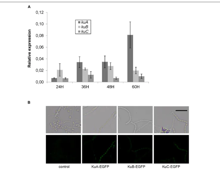
In order to investigate the spatial and temporal expression of NHEJ-like genes during development, a time-course of RT-PCR experiments was performed and translational fusions with EGFP were observed by fluorescent microscopy. First of all, the results showed that NHEJ-like genes are weakly expressed throughout the development. For instance, the expression ofligC, ligD, polK
and polR was too low to be monitored by qPCR whereas the
expression of ku genes was 10 to 50 fold lower than hrdB
expression (Figure 5A). Secondly, kuA expression was clearly
growth-phase dependent with a 10 fold increase between 24 and 60 h after germination. It is important to indicate that sporulation began to be observable after 48 h of development in these growth
FIGURE 4 | Electronic beam irradiation of S. ambofaciens strains deficient in NHEJ-like genes. Spores of mutants of NHEJ-like genes, namely single and multiple ku (A), ligase (B) and polymerase (C) mutants, were exposed to 0.1, 0.3, 0.5, 0.7, and 1 kGy of electronic beam irradiation. (D) In the same way, a1ku-like 1recA mutant also affected in HR was exposed to the same irradiation doses. In this latter case, a logarithmic scale was used for survival rate representation. Survival rates were calculated as the number of clones for a given dose relative to the number of clones unexposed to radiation. For each graph, wild-type strain is represented as a control. Each curve corresponds to data obtained from an individual mutant strain. Error bars indicate standard error of a minimum of three replicates.
conditions. There was no difference throughout the time-course
forkuB and kuC expression. Both of them were less expressed
than kuA at the late stages of development. The WT strain
was also grown to OD600 of 0.2 in HT liquid medium and
then exposed to MMC (1.5 µg·ml−1) for 30 min. RNAs were
extracted and reverse-transcribed to assess by PCR the regulation of NHEJ-like genes during a genotoxic stress. No induction or repression of NHEJ-like genes was noticeable, while the positive induction control (recA) was up-regulated up to 20 fold (not shown).
To observe the cellular localization of Ku proteins in
physiological conditions, egfp translational fusions were
constructed at kuA, kuB, and kuC chromosomal loci. Fusion
strains were inoculated on SFM agar plates and grown for 48 h before observations by fluorescent microscopy along with
a negative control strain without any egfp gene. A positive
control strain constitutively expressing egfp gene exhibited a strong green fluorescence signal in all cells throughout the developmental stages of the bacteria, proving the reporter
gene to be functional inS. ambofaciens (not shown). For KuA
fusion, the green fluorescence signal was significant in spores and segmenting aerial hyphae but not in vegetative mycelium (Figure 5B), showing a specific accumulation of KuA in spores. This result corroborates the phase dependent expression
pattern of kuA observed in the previous qPCR analysis. For
KuB and KuC fusions, although the signals were weaker than for KuA fusion, they also revealed the accumulation of KuB and KuC in spore chains. Since no significant modulation
of gene expression was detected for kuB and kuC along the
growth cycle (Figure 5A), this protein accumulation pattern suggests a translational (and/or a post-translational) regulation level.
FIGURE 5 | Growth-dependent and cell-type specific expression of NHEJ-like genes. (A) Expression level of S. ambofaciens ATCC23877 ku genes relative to hrdB after 24, 36, 48, and 60 h growth on SFM plates at 30◦
C. Error bars represent standard error. Expression of kuA after 60 h growth is significantly higher than that at 24 h. Furthermore, kuA expression at 60 h growth is significantly different than that of kuB and kuC (t-test with Bonferroni correction,α = 0.05). (B) Strains expressing EGFP fusion with KuA, KuB, or KuC were inoculated in the acute-angle junction of standard-sized microscope coverslips inserted at 45◦in SFM agar,
and grown for 48 h before observations by fluorescent microscopy. The control strain does not contain any GFP gene. Representative examples of spores or aerial mycelium are shown as a contrast phase image (upper panels) and as EGFP fluorescent channel (lower panels). The weak fluorescent foci observed mainly in mycelia were readily detected in the control strain and are not likely to correspond to GFP signal. The scale is represented by a 5µm size bar.
The Ku Proteins Protect Linear dsDNA
from the T5 Exonuclease and Stimulate
the Ligase Activity of LigD
BsubTo assess which proteins are required in a putativeS. ambofaciens NHEJ, we tried to purify all the proteins putatively implicated in this pathway. Among them, we succeeded to express and purified S. ambofaciens KuA, KuB and KuC proteins (39.1, 42.4, and 33.6 KDa respectively). To evaluate the oligomeric state of the three Ku proteins, we performed gel filtration experiments. KuA and KuB were eluted in fractions corresponding to a molecular weight of 160 and 100 kDa, respectively, strongly suggesting that KuA and KuB are multimeric (Supplementary Figure S1 and Supplementary Data Sheet S1). To date, characterized bacterial Ku are dimers. However, the elongated shape of
the B. subtilis Ku (KuBsub) dimer (McGovern et al., 2016)
led to the elution of this protein with apparent molecular weight corresponding to a tetramer. Then KuA and KuB could also be dimers displaying elongated shape, as for
KuBsub. KuC was clearly purified in different oligomeric
states ranging from 70 to more than 600 kDa. The main elution peak, corresponding to a protein of apparent molecular weight around 160 kDa, was similar to the one observed for KuA.
KuA, KuB, and KuC were able to protect a linear dsDNA molecule from the T5 exonuclease (Figure 6A), a property shared by the KuBsub protein (McGovern et al., 2016) and the
Pseudomonas aeruginosa Ku against different exonucleases (Zhu and Shuman, 2010). This result suggests that these proteins are able to bind DNA ends. Another role of Ku in the bacterial
FIGURE 6 | KuA, KuB, and KuC protect linear dsDNA from the T5 exonuclease and stimulate the ligase activity of LigDBsub. (A) A 1001 bp linear dsDNA
with 50-phosphorylated ends (3.8 nM) was incubated 30 min at 37◦C with increasing amounts of KuA, KuB, or KuC as indicated. T5 exonuclease (0.5 U) was then
added in each reaction mixtures for 30 min at 37◦
C and digested products were analyzed as described in the Section “Material and Methods.” Control experiments showing the 1001 ds DNA substrate and the nearly complete degradation of the DNA molecule by the T5 exonuclease without other proteins added are shown on the left. (B) A 1001 bp linear dsDNA with 50-phosphorylated ends (3.8 nM) was incubated with 0.25 or 1µM of KuA, KuB, or KuC as indicated. Increasing amounts
of LigDBsubwere then added (allowing final concentrations of LigDBsubindicated above the gels) and ligation products were analyzed as described in Section
“Material and Methods.” Control experiments showing the weak activity of LigDBsubalone at different concentration are shown on the left (No Ku). For comparison,
KuBsubwas assessed for its LigDBsubstimulation activity in the same conditions (right part).
NHEJ is to recruit the LigD at DNA ends and to stimulate its ligase activity. Remarkably, KuA, KuB, and KuC were able to stimulate the ligation activity of theB. subtilis LigD protein (Figure 6B), even if they were less efficient than KuBsub. However,
we observed a different concentration-dependent stimulation
of the ligase by the S. ambofaciens Ku proteins. Raising the
concentration of KuA and KuC from 0.25 to 1 µM in the
ligation assay seemed to slightly increase ligation efficiency. At
the opposite, the stimulation activity observed with 0.25 µM
of KuB appeared to be reduced when its concentration was
raised to 1 µM. A similar inhibitory effect was observed with
DISCUSSION
Streptomyces NHEJ-Like Gene
Organization Is Atypical
The presence of a NHEJ pathway in prokaryotes appears to be limited to some phyla representing about 20% of the species (estimated on a sample of 2645 complete bacterial genomes) (McGovern et al., 2016). The minimal set of NHEJ gene encodes a two partner system including the homodimeric Ku protein and a ligase polypeptide composed of the ligase enzymatic domain (LigDom) to a nuclease (NucDom) and a polymerase (PolDom) domain. The latter enzymatic activities process DNA ends before ligation. The NHEJ gene repertoire ranges from the simplest in P. aeruginosa with a single Ku and a ligase (ligase D) to the
more complex with those described inAgrobacterium (Zhu and
Shuman, 2007) or Sinorhizobium (Kobayashi et al., 2008) and Streptomyces as described in this work. Hence, while B. subtilis
andMycobacterium possess a single Ku, a single LigD and one
or two LigC, A. tumefaciens possesses three Ku, two LigD and
three LigC (Zhu and Shuman, 2007). Sinorhizobium meliloti
and other rhizobia possess up to 4 Ku, 5 LigD and one LigC (Kobayashi et al., 2008); the ligase, nuclease and polymerase domains are present in different organizations, sometimes (i) fused together in a single polypeptide or (ii) encoded by distinct genes. This situation is similar in archaea where the putative ligase, nuclease and polymerase domains are encoded by distinct genes, however, kept into an operonic organization assumed to ensure the co-regulation of the activities (Bartlett et al., 2013). However, among actinobacteria, Streptomyces appears as an exception, since other main phyla (Nocardia, Frankia, Rhodococcus, and Corynebacterium) seem to possess a multidomain LigD homologue (not shown).
In Streptomyces, several levels of complexity are also
noticeable. First, the activities putatively involved in NHEJ are encoded by standalone genes that are scattered along the genome. Consequently the repertoire is rather expanded: the number of genes ranging from none up to 4 forku, from one to two LigDom encoding genes (ligC, ligD), from 2 to 3 PolDom encoding genes, from none to one NucDom encoding gene. The total number
of NHEJ-like genes ranges from 4 in S. bingchenggensis and
S. venezuelae to 8 in S. ambofaciens and S. reticuli: 3 ku (kuA, kuB, kuC), one ligD and one ligC, and 3 polymerase genes (polK, polR, polO). These genes can fall into two groups. On one hand, the “core” set includeskuA, ligC, polK and polR which are present in almost allStreptomyces species and organized in two loci of rather conserved organization (i.e., kuA-polK, ligC-polR). The only exception appears to be inS. vietnamiensis which lacks the couplekuA-polK and the other ku genes. On the other hand, the rest of the genes constitute the “variable” gene set. An interesting feature to highlight is a gene duplication event involving kuA orkuC. The duplicated copies are either located in the variable region of the genome or present within the terminal inverted repeats of the linear replicons. This phenomenon is interesting since duplicated copies can evolve through gene divergence to raise new function. This mechanism is rather rare in bacteria but
gene duplication seems frequent in Streptomyces, notably with
large terminal duplication probably resulting from DSB repair by a mechanism related to break-induced replication.
Involvement of the “Core” and “Variable”
NHEJ-Like Gene Sets in DNA Repair
The large number of NHEJ-like genes in Streptomyces as well
as their variability questions their implication in DNA repair mechanisms. It is tempting to imply the “core” NHEJ gene set in a classical NHEJ pathway with a Ku (i.e., KuA) and an ATP-dependent DNA ligase (i.e., LigC). In addition to being the
conserved ku gene, kuA is the more expressed of the 3 ku-like
genes in the late growth phases, its defect conferred the most marked sensitivity to EBI and finally, KuA is the most efficient of the 3 Ku-like proteins in stimulating LigDBsubactivityin vitro.
It might be surprising that the NHEJ ligase could be LigC rather than LigD which is the canonical NHEJ ligase in other bacterial species. However, it should be reminded that in Streptomyces, LigD carries the only ligase domain, as does LigC. Both ligases are devoid of the PolDom domain known to be involved in its recruitment by Ku. Therefore LigD and
LigC may ensure similar functional roles inStreptomyces. One
could hypothesize that a gene rearrangement split the ligase domain from the two others (i.e., PolDom and NucDom) in a Streptomyces common ancestor. The two ligase genes would have then provided an equivalent contribution to the bacterium, finally resulting in the replacement of LigD by LigC in the “core” NHEJ set by a founder effect.
InS. ambofaciens, in which both ligC and ligD cohabits, single mutants displayed a sensitive phenotype to EBI and the double mutant displayed an aggravated phenotype. In this context, at least two hypotheses can be drawn. First, both LigC and LigD could participate to the same repair pathway; in that case, the sensitivity of single mutants and the aggravated phenotype of double mutants could be explained by a dose effect that is the need for a certain amount of ligases to cope for the induced DNA damage. In that case, both ligase genes would contribute to achieve the required level of ligase. Alternatively, they could participate to different repair pathways required to take over varied DNA damage (see below).
If the “core” NHEJ gene set indeed establishes a functional NHEJ pathway, the presence of two PolDoms in the “core” NHEJ gene set constitutes another intriguing feature, especially considering that they do not seem to have redundant functions. Hence, the sensitivity of bothpolK and polR simple mutants was
aggravated in the double mutant. As shown in M. tuberculosis
NHEJ, the role of the PolDom domain of LigD would be less to provide a polymerase activity than to recruit LigD to Ku (Pitcher et al., 2005). Then PolK and/or PolR may play a role of linker/adapter to recruit a ligase; the more likely being that PolK and PolR recruit LigC, however, on different types of DNA extremities (e.g., blunt, 30
or 50
overhanging ends). In the case of the presence of LigD (as inS. ambofaciens for example), the two PolK and PolR may recruit both LigC and LigD.
It is particularly noticeable that the “variable” genes (i.e., ligD
but also kuB, kuC) were involved in the response to EBI. The
types of lesions can result from direct transmission of energy to the DNA structure and from secondary oxidative stress (e.g., oxidized bases). In consequence, damage induced by EBI requires NHEJ but may also require other DNA repair pathways. Thus, these “variable” genes would (i) support the NHEJ encoded by the “core” gene set in an opportunistic way, (ii) constitute an alternative NHEJ pathways or (iii) constitute a repair pathway dedicated to other DNA damage single strand gaps or abasic sites). The first hypothesis would imply a dose effect of the Ku as well as the ligase proteins to explain the aggravated
phenotype of the multiple mutants (i.e.,1kuABC and 1ligCD).
In this first hypothesis, considering that prokaryote Ku act as dimers (McGovern et al., 2016), we can speculate that these three Ku proteins could act as homodimers as well as heterodimers. A data in favour of the second hypothesis (an alternative NHEJ
pathway) is the colocalization of the kuC and ligD genes in 6
Streptomyces species clustering with S. griseus (Supplementary Table S1; Figure 3). However, no clear trend toward the co-occurrence of these two “variable” genes can be noticed in the 38 analysed genomes. In B. subtilis, the recent involvement of LigDBsub and KuBsub in base excision repair along with their
major role in NHEJ supports the third hypothesis (de Ory et al., 2014, 2016).
In order to solve the question of the involvement of these genes in a NHEJ pathway, a forward-looking approach could be to specifically induce DSB formation at targeted chromosomal loci and assessed the sensitivity of the NHEJ-candidate genes to identify those truly involved in DSB repairper se.
A Late Growth Phase Role for NHEJ in
Streptomyces
The presence of a NHEJ pathway in bacteria is usually assigned to DSB repair in low rate growth phases characterizing bacterial species involved in pathogenic processes or exposed to harsh environmental living conditions. When bacteria replicate such as in laboratory conditions, sister chromosomes providing an intact template are preferentially used to ensure faithful DSB repair by HR. However, when replication is slowing down (i.e., stationary phases) or stopping (e.g., in resistance forms such as spores), DSB repair strongly relies on NHEJ. This is consistent with the fact that NHEJ genes are expressed preferentially when DSB repair cannot be ensured by a faithful process. Hence, in B. subtilis
as well as in Streptomyces, the NHEJ genes were shown to be
expressed in stationary phase and to accumulate in spores. The forespore-specific sigma factorσGinB. subtilis directly regulates ykoU (coding for LigD) and ykoV (coding for Ku), inducing their expression during the forespore development (Wang et al., 2006). In addition, B. subtilis Ku protein specifically localizes to the nucleoid of germinating spores while it disappears at later stages of germination. In Streptomyces, gene transcription analyses showed in this work an expression of theku in stationary
phase with an increase of kuA expression during sporulation.
Moreover, GFP fusions showed a predominant localization of the Ku proteins in spores. Furthermore, mutations inku and ligase genes confer sensitivity to genotoxic agents toB. subtilis as well asStreptomyces spores. Sensitivity to heat and desiccation which
are biotic parameters known to trigger DSB is also associated to
the NHEJ mutations inB. subtilis and Mycobacterium smegmatis
(Moeller et al., 2007;Pitcher et al., 2007;Stephanou et al., 2007). The presence of multipleku genes (and of other NHEJ-like genes) inSinorhizobium meliloti is assumed to correspond to the need for DNA repair at different steps of the cell cycle. Although no induction of the NHEJ pathways was observed under genotoxic conditions (i.e., IR exposure), the expression patterns of the sku genes suggest that each Ku protein has a distinct role under different conditions (free bacterial form, bacteroids). In contrast, NHEJ would not be required during the establishment of symbiosis since a strain lacking all foursku genes was shown to establish functional nodules (Kobayashi et al., 2008). As in S. meliloti, it was shown that each of the S. ambofaciens ku genes is functional since their deletion conferred a phenotype of sensitivity toward genotoxic agents. Mutant strains lackingkuA, kuB, and kuC genes showed an aggravated phenotype suggesting that each participated to the response to DNA damage. We can therefore suppose that like in S. meliloti, intervention of the Streptomyces ku genes is done under specific conditions.
Recently in Pseudomonas putida, the NHEJ was shown to
participate to another late growth phase phenomenon (Paris
et al., 2015). Hence, bacteria frequently encountered nutrient starvation during late-growth phase. Populations starving display a phenomenon called stationary-phase mutagenesis (also known as adaptive mutations) from which emerge mutants able to overcome the shortage and initiate new vegetative cycles. The underlying mechanisms notably involve unfaithful DNA polymerases during HR events (Shee et al., 2012). Although the involvement of NHEJ in late mutagenesis remains unknown, deficiency of each of the NHEJ activities triggered an altered pattern of mutations (Paris et al., 2015). The authors speculate on ability to bypass DNA lesions of the LigD polymerase domain.
Origin of the “Core” and “Variable” NHEJ
Gene Sets in Streptomyces
While the origin of the NHEJ “core” gene set is likely to
result from vertical inheritance from a common Streptomyces
ancestor, the origin of the “variable” set is questionable. The sporadic distribution of the “variable” genes may reflect either the existence of NHEJ in a bacterial common ancestor and its subsequent sporadic loss during genus/species separation. Reciprocally, it may reflect the acquisition of NHEJ genes through horizontal gene transfer in some phyla. Both hypotheses can also be combined and result in the observed complexity. The most explicit argument about their origin is their localization in the variable regions of the chromosome or in plasmids. While plasmids can be mobilized and even self-transmitted, the terminal regions of theStreptomyces chromosome is known to be targeted by recombination phenomena leading to insertions of acquired DNA and further losses (Choulet et al., 2006).
Impact of NHEJ on Genome Evolution
Whatever the evolutionary past of the NHEJ gene sets, the presence of a conserved “core” gene set enriched by additional functional “variable” genes may confer some advantage. In all
bacteria studied so far, NHEJ is not essential (Ku/LigD) for growth, at least under laboratory conditions. Mutants of NHEJ
in Mycobacterium, Pseudomonas, or Bacillus are not affected
although they show sensitivity to environmental stress (Weller et al., 2002; Jacobs et al., 2003; Gong et al., 2004, 2005). We
came to the same conclusion inS. ambofaciens although it was
particularly interesting to note that a part of the “variable” genes (i.e., kuB, kuC and ligD) was involved in response to genotoxic agents. Hence the aggravated phenotype observed in double
or multiple mutant strains (i.e., kuABC, ligCD) revealed the
cooperative actions of the multiple alleles. The “variable” genes in Streptomyces may increase the flexibility and/or efficiency of the DSB repair mechanism and influence evolution of the genome. Lessons about the role of NHEJ in genome evolution can be drawn from organisms lacking NHEJ. Hence, the only eukaryote
lacking NHEJ is the yeast Lachancea kluyveri (Gordon et al.,
2011). Compared to other species of the Saccharomycetaceae
family, this species showed a significantly low number of genomic rearrangements and the lack of telomere-to-telomere fusions. The authors suggest that the loss of the NHEJ and an alternative end joining pathway [named microhomology-mediated end joining (MMEJ) or alternative end joining (A-EJ)] is responsible for the low recombination rate assessed in this
phylum. In this way, we can hypothesize that in Streptomyces
the presence/absence of NHEJ genes in addition to the “core” NHEJ (the “variable” genes participating to a unique NHEJ or other alternative pathways) may modulate the frequency of DNA rearrangements and DNA acquisition through horizontal gene
transfer and confer different evolution rates to Streptomyces
species. These genes being themselves subject to transfer, the recombinant phenotype (i.e., hyperrecombinant) could be variable within a population and favor genetic diversification and adaptation to the environment. This situation could be reminiscent of mutator strains identified in E. coli (for review:
Giraud et al., 2001) but applied to the capacity to recombine DNA including incoming genetic information.
Future Perspectives
A key question is the relative contribution of HR and NHEJ to the evolution and the remarkable compartmentalization of the linearStreptomyces chromosome. Is there any regulation between the two pathways during the development of the bacteria? In
S. avermitilis, the deletion of the ku homologues (ku1 and ku2
corresponding to kuA and kuC respectively) was associated to
an increased efficiency of HR (Zhang et al., 2012). This may reveal the existence of a balance resulting from the competition between HR and NHEJ repair mechanisms, and that both repair pathways co-exist at least during replicating phases. Is there any spatial regulation of the DSB repair mechanisms? DSB could be healed by different mechanisms depending of their chromosomal location leading to either faithful repair or possible integration of foreign information.
AUTHOR CONTRIBUTIONS
Investigation: GH, CB, LZ, EP, LC, and SM; Computer analysis: AT and CyB; Methodology: CyB, FC, and FL; Conceptualization: CB, AT, FL, and PL; Writing original draft: GH, CB, AT, and PL; Writing-review and editing: FL, FC, CyB, CB, AT, and PL; Resources: FC, FL, and PL; Funding acquisition: CB, AT, and PL; Supervision: AT and PL.
FUNDING
This work was supported by the French National Research Agency program Streptoflux (ANR Blanc 0096_01), by ANR through the Laboratory of Excellence ARBRE (ANR-12-LABXARBRE-01) and by the Région Lorraine.
ACKNOWLEDGMENTS
We would like to thank Sébastien Riegler and Florian Kuntz from the Technological Resource center Aérial (Illkirch-Graffenstaden, France) for their technical support and their warm welcome.
SUPPLEMENTARY MATERIAL
The Supplementary Material for this article can be found online at: http://journal.frontiersin.org/article/10.3389/fmicb. 2016.01901/full#supplementary-material
REFERENCES
Abrudan, M. I., Smakman, F., Grimbergen, A. J., Westhoff, S., Miller, E. L., van Wezel, G. P., et al. (2015). Socially mediated induction and suppression of antibiosis during bacterial coexistence. Proc. Natl. Acad. Sci. U.S.A. 112, 11054–11059. doi: 10.1073/pnas.150407 6112
Aigle, B., Holl, A., Angulo, J. F., Leblond, P., and Decaris, B. (1997). Characterization of twoStreptomyces ambofaciens recA mutants: identification of the RecA protein by immunoblotting.FEMS Microbiol. Lett. 149, 181–187. doi: 10.1111/j.1574-6968.1997.tb10326.x
Alonso, J. C., Cardenas, P. P., Sanchez, H., Hejna, J., Suzuki, Y., and Takeyasu, K. (2013). Early steps of double-strand break repair in Bacillus subtilis. DNA Repair(Amst.) 12, 162–176. doi: 10.1016/j.dnarep.2012. 12.005
Aniukwu, J., Glickman, M. S., and Shuman, S. (2008). The pathways and outcomes of mycobacterial NHEJ depend on the structure of the broken DNA ends.Genes Dev. 22, 512–527. doi: 10.1101/gad.1631908
Aravind, L., and Koonin, E. V. (2001). Prokaryotic homologs of the eukaryotic DNA-end-binding protein Ku, novel domains in the Ku protein and prediction of a prokaryotic double-strand break repair system.Genome Res. 11, 1365–1374. doi: 10.1101/gr.181001
Aravind, L., Makarova, K. S., and Koonin, E. V. (2000). Holliday junction resolvases and related nucleases: identification of new families, phyletic distribution and evolutionary trajectories.Nucleic Acids Res. 28, 3417–3422. doi: 10.1093/nar/ 28.18.3417
Bartlett, E. J., Brissett, N. C., and Doherty, A. J. (2013). Ribonucleolytic resection is required for repair of strand displaced nonhomologous end-joining intermediates.Proc. Natl. Acad. Sci. U.S.A. 110, E1984–E1991. doi: 10.1073/ pnas.1302616110