HAL Id: hal-01263258
https://hal-univ-rennes1.archives-ouvertes.fr/hal-01263258
Submitted on 28 Jan 2016
HAL is a multi-disciplinary open access
archive for the deposit and dissemination of
sci-entific research documents, whether they are
pub-lished or not. The documents may come from
teaching and research institutions in France or
abroad, or from public or private research centers.
L’archive ouverte pluridisciplinaire HAL, est
destinée au dépôt et à la diffusion de documents
scientifiques de niveau recherche, publiés ou non,
émanant des établissements d’enseignement et de
recherche français ou étrangers, des laboratoires
publics ou privés.
Distributed under a Creative Commons Attribution - NonCommercial| 4.0 International
License
resynchronization therapy: an analysis of the
CeRtiTuDe cohort study
Eloi Marijon, Christophe Leclercq, Kumar Narayanan, Serge Boveda, Didier
Klug, Jonathan Lacaze-Gadonneix, Pascal Defaye, Sophie Jacob, Olivier Piot,
Jean-Claude Deharo, et al.
To cite this version:
Eloi Marijon, Christophe Leclercq, Kumar Narayanan, Serge Boveda, Didier Klug, et al..
Causes-of-death analysis of patients with cardiac resynchronization therapy: an analysis of the CeRtiTuDe
cohort study. European Heart Journal, Oxford University Press (OUP): Policy B, 2015, 36 (41),
pp.2767–2776. �10.1093/eurheartj/ehv455�. �hal-01263258�
. . . .
. . . .
ESC Clinical Registry
Causes-of-death analysis of patients with cardiac
resynchronization therapy: an analysis of the
CeRtiTuDe cohort study
Eloi Marijon
1,2,3, Christophe Leclercq
4, Kumar Narayanan
3, Serge Boveda
5,
Didier Klug
6, Jonathan Lacaze-Gadonneix
1,2, Pascal Defaye
7, Sophie Jacob
8,
Olivier Piot
9, Jean-Claude Deharo
10, Marie-Cecile Perier
3, Genevieve Mulak
11,
Jean-Sylvain Hermida
12, Paul Milliez
13, Daniel Gras
14, Olivier Cesari
15,
Franc¸oise Hidden-Lucet
16, Frederic Anselme
17, Philippe Chevalier
18, Philippe Maury
19,
Nicolas Sadoul
20, Pierre Bordachar
21, Serge Cazeau
22, Michel Chauvin
23,
Jean-Philippe Empana
3, Xavier Jouven
1,2,3, Jean-Claude Daubert
4, and
Jean-Yves Le Heuzey
1,2,3*
, for the CeRtiTuDe Investigators
1
Cardiology Department, European Georges Pompidou Hospital, Paris, France;2
Paris Descartes University, Paris, France;3
Paris Cardiovascular Research Centre, Paris, France;
4
Pontchaillou University Hospital and INSERM 1099, CIC-IT 804 Rennes, France;5
Clinique Pasteur, Toulouse, France;6
Lille University Hospital and University of Lille, Lille, France;
7
Arrhythmia Department, University Hospital, Grenoble, France;8
Epidemiology Unit, IRSN, Paris, France;9
Centre Cardiologique du Nord, Saint Denis, France;10
Cardiology Division, Hoˆpital La Timone, Marseille, France;11
French Society of Cardiology, Paris, France;12
Amiens University Hospital, Amiens, France;13
Caen University Hospital, Caen, France;
14
Nouvelles Cliniques Nantaises, Nantes, France;15
Clinique Saint Gatien, Tours, France;16
Cardiology Department, La Pitie´-Salpeˆtrie`re Hospital, AP-HP, Paris, France;17
Cardiology Division, Rouen University Hospital, Rouen, France;18
East Lyon School of Medicine, Louis Pradel Hospital, Bron, France;19
Cardiology Division, Rangueil University Hospital, Toulouse, France;20
Cardiology Division, Nancy University Hospital, Nancy, France;21
Haut-Le´veˆque Hospital, Bordeaux, France;22
Saint Joseph Hospital, Paris, France; and23
Strasbourg University Hospital, Strasbourg, France
Received 29 June 2015; revised 8 August 2015; accepted 17 August 2015; online publish-ahead-of-print 1 September 2015
See page 2777 for the editorial comment on this article (doi:10.1093/eurheartj/ehv474)
Aims The choice of resynchronization therapy between with (CRT-D) and without (CRT-P) a defibrillator remains a
con-tentious issue. Cause-of-death analysis among CRT-P, compared with CRT-D, patients could help evaluate the extent to which CRT-P patients would have additionally benefited from a defibrillator in a daily clinical practice.
Methods and results
A total of 1705 consecutive patients implanted with a CRT (CRT-P: 535 and CRT-D: 1170) between 2008 and 2010 were enrolled in CeRtiTuDe, a multicentric prospective follow-up cohort study, with specific adjudication for causes of death at 2 years. Patients with CRT-P compared with CRT-D were older (P , 0.0001), less often male (P , 0.0001), more symptomatic (P ¼ 0.0005), with less coronary artery disease (P ¼ 0.003), wider QRS (P ¼ 0.002), more atrial fibrillation (P , 0.0001), and more co-morbidities (P ¼ 0.04). At 2-year follow-up, the annual overall mortality rate was 83.80 [95% confidence interval (CI) 73.41 – 94.19] per 1000 person-years. The crude mortality rate among CRT-P patients was double compared with CRT-D (relative risk 2.01, 95% CI 1.56 – 2.58). In a Cox proportional ha-zards regression analysis, CRT-P remained associated with increased mortality (hazard ratio 1.54, 95% CI 1.07 – 2.21, P ¼ 0.0209), although other potential confounders may persist. By cause-of-death analysis, 95% of the excess mortality among CRT-P subjects was related to an increase in non-sudden death.
Conclusion When compared with CRT-D patients, excess mortality in CRT-P recipients was mainly due to non-sudden death. Our findings suggest that CRT-P patients, as currently selected in routine clinical practice, would not potentially benefit with the addition of a defibrillator.
-Keywords Heart failure † Sudden death † Cardioverter defibrillator † Competing risk † Cardiac resynchronization
*Corresponding author. Tel:+33 1 56 09 37 01, Fax: +33 1 56 09 30 47, Email:jean-yves.le-heuzey@egp.aphp.fr
&The Author 2015. Published by Oxford University Press on behalf of the European Society of Cardiology.
This is an Open Access article distributed under the terms of the Creative Commons Attribution Non-Commercial License (http://creativecommons.org/licenses/by-nc/4.0/), which permits non-commercial re-use, distribution, and reproduction in any medium, provided the original work is properly cited. For commercial re-use, please contact
journals.permissions@oup.com
European Heart Journal (2015) 36, 2767–2776 doi:10.1093/eurheartj/ehv455
by guest on January 28, 2016
http://eurheartj.oxfordjournals.org/
Introduction
Patients with congestive heart failure (HF) are at high risk of dying from its progression as well from sudden cardiac death related to
ventricular tachyarrhythmia.1Over the last decade, cardiac
resyn-chronization therapy (CRT) and implantable cardioverter defibril-lators (ICDs) have markedly improved the prognosis of HF patients, with prolongation of survival over and above that
con-ferred by medical therapy alone.2–5It has been well established
in trials that in patients with severe left ventricular (LV) systolic dysfunction, New York Heart Association (NYHA) class III/IV symptoms, and wide QRS, CRT improves symptoms/quality of
life and also reduces mortality.3,6Additionally, more recent trials
have shown beneficial reverse LV remodelling even in patients
with milder symptoms.5,7–11This has resulted in a class I
recom-mendation for CRT in appropriately selected candidates in
guide-lines framed on both sides of the Atlantic.12,13
Since most patients who are candidates for CRT will have a LV
ejection fraction (EF) of≤35%, this ‘automatically’ makes them
can-didates for an ICD as well by the current guidelines, which makes the assumption that there is universally significant excess mortality due to sudden cardiac death (SCD) among CRT-P patients who can
therefore definitively benefit from the added defibrillator.13
How-ever, concrete evidence for such a premise in a contemporary CRT-P population is lacking and in any population of this kind; competing risks for mortality need to be carefully considered. A few studies have attempted to directly compare outcomes
be-tween CRT-P vs. CRT-D subjects.14,15Furthermore, such outcome
comparisons based on observational studies have methodological limitations and may be biased. Current guidelines do not make firm recommendations in terms of the choice between CRT-P vs. CRT-D, leaving room for physician discretion. This has resulted in wide variation in the rates of implantation worldwide. For instance, the proportion of CRT implantations, which are CRT-D, reaches
.90% in most practices in the USA,16whereas it is relatively lesser
across Europe.17The use of CRT-D or CRT-P in clinical practice is
an important question with significant implications in terms of
costs,18as well as device-related complications.19,20
In this context, a better understanding of the relative contribu-tion of SCD as opposed to other competing causes of mortality in the CRT population can be very informative. A cause-of-death analysis among CRT-P vs. CRT-D patients, may represent a novel approach to this problem. Using a large, multicentre study with prospective follow-up, we evaluated the characteristics of CRT-P vs. CRT-D patients in a real-world scenario and analysed to what extent CRT-P subjects, as currently chosen in clinical practice, would have potentially additionally benefited from the presence of a back-up defibrillator.
Methods
Setting and design of the study
CeRtiTuDe, a 2-year, prospective, multicentre registry launched in Janu-ary 2008 and held under the direction of the Working Group on Pacing and Arrhythmias of the French Society of Cardiology, was funded and coordinated by the French Society of Cardiology. Its primary objective was to define the baseline characteristics and clinical outcomes of
French patients who undergo implantation of CRT systems. An analysis of the precise causes of death was planned at 2 years after device implantation.
The 41 medical centres participating in the study (Appendix) enrolled consecutive patients who, between 1 January 2008 and 31 December 2010, had undergone CRT device implantations. The criteria for CRT implantation were as per the 2007 guidelines of the European Society of Cardiology and European Heart Rhythm Association, updated in 2010. However, all CRT recipients were enrolled, in order for the registry to reflect ‘real-world’ medical practice. Each patient was then enrolled in a specific follow-up programme with clinical, ECG, echocar-diographic, and device interrogation data collected every 6 months over the following 2 years (up to 1 January 2013).
The study was conducted in accordance with Good Clinical Practice, French Law, and the French data protection law. The protocol was re-viewed by the Committee for the Protection of Human Subjects in Bio-medical Research (CCTIRS #08-522) and the data file was reported to, and authorized by, the Commission Nationale Informatique et des Lib-erte´s (French Data Protection Committee, CNIL #909048).
Baseline characteristics at implant
Individual patient data were collected, using an electronic case report form created by the Scientific Committee to record, at each participat-ing medical centre, the demographic and baseline clinical characteristics, and the implantation procedures and techniques. These data were regu-larly transferred (every 3 months) via an internet-based system to a cen-tral database created at the data management centre of the French Society of Cardiology in collaboration with the Paris Cardiovascular Re-search Center, European Georges Pompidou Hospital, Paris (INSERM Unit 970).
All variables recorded before device implantation were defined and classified using standard clinical terminology, including gender, age (stratified as ,60, 60 – 74, and≥75 years), and underlying heart disease (ischaemic vs. non-ischaemic). Renal clearance was estimated using the Cockroft and Gault’s formula, and defined as severe renal insufficiency if ,30 mL/min/1.73 m2, and QRS duration was classified as≤120, 121–
149, and≥150 ms. Left ventricular ejection fraction was measured on transthoracic echocardiograms, using Simpson’s method, and recorded as a continuous variable and also stratified as≤20, 21–35, and .35%. A history of atrial fibrillation (AF) was based on medical records, and classified as paroxysmal or permanent. In addition to AF and renal fail-ure, other co-morbidities were systematically recorded, including can-cer, chronic obstructive pulmonary disease, liver disease, diabetes mellitus, and cerebral vascular disease.
Device implant, hospital discharge,
and follow-up
The type of CRT (CRT-P or CRT-D) implanted was recorded without the manufacturer’s information. The complications recorded included infections, changes in capture threshold, lead dislodgement, haema-tomas, HF, fever, arrhythmias, pneumothorax, phrenic nerve stimula-tion, and death. Finally, drug regimens prescribed at the time of hospital discharge including beta-adrenergic blockers, anti-arrhythmics, digoxin, calcium antagonists, angiotensin-converting enzyme inhibitors or angiotensin II receptor blockers, mineralocorticoid receptor antago-nists, diuretics, and anticoagulants were recorded. Device programming was left to the discretion of the investigators at each centre, with the guiding principle being achievement of maximal biventricular pacing.
All patients were followed at 6-month intervals for 2 years by the im-planting centre till the close of study on 1 January 2013. At each follow-up, the patients underwent clinical examination, ECG, transthoracic
by guest on January 28, 2016
http://eurheartj.oxfordjournals.org/
echocardiogram, and device interrogation. In addition, information on any intercurrent events (such as hospitalization) was also recorded in the file. The above data were systematically gathered at each follow-up visit from the date of device implantation to study closure or death or heart transplantation.
Vital status, specific causes of death,
and adjudication process
The investigators at each enrolling centre recorded major clinical events, using a standardized form, and a Clinical Events Committee verified their accuracy by contacting the attending physicians or the pa-tients as required, on a yearly basis, focusing on the vital status and on the specific modes and causes of death and on major clinical events or interventions during follow-up, including changes in drug regimens, as well as interim hospitalizations. Sources to ascertain the vital status also included registries of the patients’ birthplaces, the French National Institute of Health and Medical Research (INSERM Ce´piDc Unit— Le Kremlin-Biceˆtre, France), and the French National Institute of Statis-tics and Economical Studies.
The cause-of-death were classified as sudden if the patient (i) died suddenly and unexpectedly within 1 h of symptoms in the absence of progressive cardiac deterioration, (ii) died unexpectedly in sleep, or (iii) died unexpectedly within 24 h after last being seen alive and in the usual state of health. Other cardiovascular deaths included myocar-dial infarction, HF, acute aortic syndrome, stroke, and pulmonary em-bolism. Fatal arrhythmias associated with end-stage HF were classified as non-sudden cardiovascular deaths. Deaths attributable to causes, such as cancer, infectious disease, or renal or respiratory failure, were classified as non-cardiovascular. When inadequate or no data were available, the cause of death was classified as unknown or unidentifiable. We used multiple sources to assess and finally adjudicate the cause of death, which included medical data obtained by the regional investiga-tors, pathology report, Emergency Medical Services report, as well as data from the French Center on Medical Causes of Death (INSERM Ce´piDc unit), which is able to provide the causes of death occurring in France.
Statistical analysis
This report was prepared in compliance with the STROBE checklist for observational studies.21Continuous variables are presented as mean + standard deviation and categorical variables are presented as numbers and percentages. Comparisons between groups (patients with CRT-P vs. patients with CRT-D) were made, using thex2
or Fisher’s exact tests for discrete variables and with unpaired t-tests, Wilcoxon signed-rank tests, or one-way analysis of variance for continu-ous variables. Factors associated with the implantation of CRT-P were identified, using a multiple variable, stepwise, logistic regression analysis. Kaplan – Meier curves were constructed to estimate the 2-year sur-vival, and CRT-P and CRT-D groups were compared using the log-rank test. For the cause-specific mortality, we used a competing risk analysis and estimated the cumulative incidence function. We then used Gray tests to assess the difference between the CRT-P and CRT-D groups.22 A Cox proportional hazards regression analysis was used to identify variables independently associated with overall mortality. The propor-tional hazard assumptions were tested. The crude associations between mortality and different variables (listed in Table1) were first quantified by univariate Cox regression. All covariates that reached a significance level of P , 15% were then included in an initial multivariate regres-sion model. A stepwise selection was applied to obtain a final model that included covariates with P , 5%. Given the observational design of the study and minimization of indication bias for device
implantation, propensity score analyses were conducted. We esti-mated the propensity score of receiving a CRT-P therapy by fitting a logistic regression model using age, sex, AF, LVEF, aetiology of HF, NYHA, and beta-adrenergic blockers as covariates. We then matched patients who received CRT-D therapy with those who received CRT-P in an 1 : 1 ratio using a greedy matching algorithm with a max-imum allowable difference of 0.05 (see Supplementary material online, Table S1 and Figure S1). Patients who could not be matched using these criteria were removed from the analysis. Then, the association be-tween device type and mortality was repeated after propensity score matching (462 patients). All data were analysed at INSERM, Unit 970, Cardiovascular Epidemiology and Sudden Death, Paris, using SAS ver-sion 9.4 (SAS Institute, Inc., Cary, NC, USA).
Results
Baseline characteristics and device
implantation
Overall, a total of 1705 consecutive patients were enrolled in the study and received CRT devices. The mean age of the overall popu-lation was 68.8 + 11.1 years, 33% were .75 years of age, and 77% were men. Nearly 20% had been hospitalized for decompensated HF within the previous 6 months. The heart disease was ischaemic in 47% of patients and related to non-ischaemic dilated cardiomyop-athy in 53%. Overall, 29% of patients presented with a LVEF of ≤20% at the time of implantation.
A CRT-D was implanted in 1170 patients (69%). Overall, 13% of the CRT-D group was implanted in the secondary prevention, fol-lowing symptomatic ventricular tachycardia or sudden cardiac ar-rest. Patients with CRT-P compared with CRT-D were older (75.9 vs. 65.6 years, P , 0.0001), less often male (69.5 vs. 80.8%, P , 0.0001), more symptomatic (proportion of NYHA class III/IV, 87.9 vs. 80.8%, P ¼ 0.0005), with less coronary artery disease (40.7 vs. 49.3%, P ¼ 0.003), wider QRS (160.8 vs. 154.9 ms, P ¼ 0.002), more AF (38.7 vs. 22.1%, P , 0.0001), and more
co-morbidities (≥2 comorbidities, 16.9 vs. 12.9%, P ¼ 0.04; Table1).
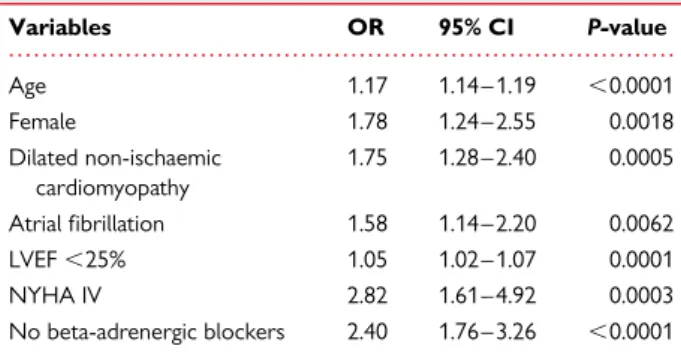
Independent variables associated with CRT-P (vs. CRT-D)
implant-ation are depicted in Table2.
Fatal periprocedural complications occurred in one patient, and death occurred before hospital discharge in five others (0.3%), due to severe cardiogenic shock. Overall, significant perioperative complications occurred in 133 subjects (7.8%) without significant difference between CRT-D and CRT-P (8.9 vs. 6.7%, P ¼ 0.20). Pulse generator pocket haematoma (2.5%), lead dislodgment (1.6%), and phrenic nerve stimulation (1.6%) were the most fre-quent complications, and the need for new intervention during the same hospital stay was observed in 40 patients (2.3%).
Follow-up, overall mortality, and specific
causes of death
The 1705 consecutive patients enrolled in the study were followed for a mean of 665.6 + 173.8 days (1.0 – 730.5 days). At 2-year follow-up (completed in 94.5% of subjects), 267 patients died, giving an overall annual mortality rate of 83.8% (95% CI 73.4 – 94.2) per 1000 person-years, with a higher rate among CRT-P, compared with CRT-D, patients [130.8 vs. 65.1 per 1000 year, respectively,
relative risk (RR) 2.01, 95% CI 1.56 – 2.58, P , 0.0001; Figure1A].
Causes-of-death analysis of patients with CRT
2769
by guest on January 28, 2016
http://eurheartj.oxfordjournals.org/
The incidence of SCD was not statistically higher in the CRT-P group compared with CRT-D (RR 1.57, 95% CI 0.71 – 3.46, P ¼
0.42) (Figure1B). The rate of hospitalization for HF was not different
between the CRT-D vs. CRT-P groups (19.6 vs. 22.0%, P ¼ 0.28). After considering potential confounding factors in a Cox pro-portional hazards regression analysis, CRT-P remained associated with increased mortality [hazard ratio (HR) 1.54, 95% CI 1.07 – 2.21, P ¼ 0.0209], as well as the presence of co-morbidities (HR 1.98, 95% CI 1.34 – 2.92, P ¼ 0.0006) and functional NYHA class IV (HR 1.85, 95% CI 1.10 – 3.11, P ¼ 0.0207). Using the propensity-matched cohort, CRT-P was associated with increased mortality (RR 2.0, 95% CI 1.22 – 3.28, P ¼ 0.01). Cardiac resynchronization therapy without defibrillator was not associated with a higher
incidence of SCD (RR 1.21, 95% CI 0.45 – 3.29, P ¼ 0.70). Forest plots showing hazard ratios of CRT-P vs. CRT-D for mortality by
dif-ferent subgroups were represented in Figure2.
However, when considering the specific cause-of-death analysis
(Table3), the increase in mortality among CRT-P patients was not
related to that in SCD, though SCD incidence was higher in the CRT-P group: 11.8 per 1000 among CRT-P vs. 7.5 per 1000 among CRT-D recipients (P ¼ 0.26). The main reasons for the almost twice-higher risk of death in the CRT-P group were an increase in non-SCD cardiovascular mortality, mainly comprising progressive HF (RR 2.27, 95% CI 1.62 – 3.18) as well as other cardiovascular mortality (RR 4.40, 95% CI 1.29 – 15.03). Overall, 95% of the excess mortality among CRT-P recipients was not related to SCD. . . . .
. . . .
. . . .
. . . .
. . . . Table 1 Characteristics of the entire registry sample and of the CRT-P vs. CRT-D recipients
Total (N 5 1705) CRT-D (N 5 1170) CRT-P (N 5 535) P-value Age (years) 68.8 + 11.1 65.6 + 10.4 75.9 + 9.0 ,0.0001 Men 1317 (77.2) 945 (80.8) 372 (69.5) ,0.0001 Heart disease Ischaemic 724 (47.0) 556 (49.3) 168 (40.7) 0.0026 Non-ischaemic 816 (53.0) 571 (50.7) 245 (59.3) QRS duration Mean (ms) 157.7 + 27.1 155.0 + 26.2 160.8 + 29.0 0.0018 Left ventricular ejection fraction
Median, % 25.5 (10.0) 25.5 (10.0) 25.5 (10.0) 0.084
≤20% 484 (29.3) 333 (29.2) 151 (29.6) ,0.0001
21 – 35% 1078 (65.3) 764 (67.1) 314 (61.5)
.35% 88 (5.3) 42 (3.7) 46 (9.0)
New York Heart Association functional class
I 16 (1.0) 14 (1.3) 2 (0.4) ,0.0001 II 250 (16.0) 194 (18.0) 56 (11.7) III 1188 (76.2) 824 (76.2) 364 (76.0) IV 106 (6.8) 49 (4.5) 57 (11.9) History of: Atrial fibrillation 445 (27.3) 248 (22.1) 197 (38.7) ,0.0001 Renal insufficiency 211 (14.4) 138 (13.0) 73 (18.2) 0.0128 COPD 264 (18.1) 198 (18.7) 66 (16.4) 0.3120 Cancer 122 (8.4) 88 (8.3) 34 (8.5) 0.9272 Miscellaneous disorders 266 (18.2) 174 (16.4) 92 (22.9) 0.0043 Drug therapy at the time of implantation
Diuretics 1045 (66.2) 752 (69.2) 293 (59.6) 0.0002 ACE inhibitors/ARB 1057 (66.9) 792 (72.9) 265 (53.9) ,0.0001
MRA 404 (25.6) 331 (30.5) 73 (14.8) ,0.0001
Beta-adrenergic blockers 945 (59.9) 732 (67.3) 213 (43.3) ,0.0001 Oral anticoagulant agent 658 (41.7) 438 (40.3) 220 (44.7) 0.0989 Antiplatelet agents 693 (43.9) 512 (47.1) 181 (36.8) 0.0001
Values are means + SD, median (IQR), or numbers (%) of observations.
ACE, angiotensin-converting enzyme; ARB, angiotensin receptor blocker; COPD, chronic obstructive pulmonary disease; MRA, mineralocorticoid receptor antagonist; CRT-D: cardiac resynchronization therapy with defibrillator; CRT-P: cardiac resynchronization therapy without defibrillator.
by guest on January 28, 2016
http://eurheartj.oxfordjournals.org/
Discussion
To the best of our knowledge, our study provides the first cause-of-death analysis comparing CRT-P with CRT-D patients in a real-world population. We demonstrate that CRT-P patients, as chosen in routine clinical practice, were older, more likely to be fe-male, with less ischaemic heart disease, more advanced HF, and greater co-morbidity burden compared with CRT-D patients. These characteristics of the CRT-P patients are in agreement with
previous reports.14,19 At 2 years, the overall mortality in the
CRT-P group was greater than that in the CRT-D group. However, importantly, this difference in mortality was mostly accounted for by an increase in non-SCD. Since SCD did not significantly contribute to the excess mortality in the CRT-P group, it suggests that the pres-ence of a back-up defibrillator would probably not have been bene-ficial in terms of improving survival for these patients. The rates of HF hospitalization were greater in the CRT-P group, which is in line with the greater HF mortality in this group. This is likely related to a sicker population with more co-morbidity, older age, and potentially more severe HF and serves to highlight that progressive HF rather than SCD may be the main driver of morbidity as well as mortality in the CRT-P population. These results from a large, prospective co-hort with robust cause-of-death adjudication need careful consider-ation in the context of the current controversy in the selection of CRT-P vs. CRT-D. Our study is not intended as a direct comparison of outcomes between CRT-D and CRT-P, and subgroup analyses should be interpreted with caution. While direct comparisons in ob-servational studies may reveal differences in death rates, knowledge of what makes up this difference takes our understanding an import-ant step further. Cause-of-death analysis, while being technically challenging to perform in a large population, represents an innova-tive, alternate approach to this problem. It also helps bring to the forefront the issue of competing risks for mortality in any population of this nature.
With the exception of patients with AF, where the evidence-base
is admittedly weaker,20CRT represents an important therapeutic
option for a growing segment of the HF population. Though the guidelines presently do not make definitive recommendations for
CRT-D vs. CRT-P, in practice, many physicians may feel compelled to use CRT-D, as a defibrillator is considered ‘necessary’ in the pres-ence of low LVEF. The effect of this choice may be even greater in light of the fact that many centres are exploring broader indications
for CRT in patients with milder symptoms and narrow QRS.23This
has important economic implications in that the incremental cost of CRT-D over CRT-P is significantly greater when compared with the cost over optimal medical therapy and this difference is even steeper
in the older age group.24The addition of a defibrillator lead can also
contribute to additional adverse events and need for repeat
proce-dures.25Thus, there is a fairly urgent need for more data such as
from the present study to tease out the putative benefits of an
added defibrillator over CRT-P,25and to better define optimal
criteria to select CRT-P or CRT-D. Since the CRT-P group had a greater proportion of non-ischaemic cardiomyopathy where the benefit from primary preventive ICD is lower, this could influence
results as well.6In the absence of proven superiority by trials and
the small survival benefit, the 2013 European Society of Cardiology Task Force was of the opinion that no strict recommendations can be made, and has preferred to merely offer guidance regarding the
selection of patients for CRT-D or CRT-P,13based on overall
clin-ical condition, device-related complications, and cost; factors fa-vouring CRT-P being advanced HF, co-morbidities, including frailty and cachexia. In contrast, factors favouring CRT-D implantation are life expectancy .1 year, stable HF, moderate functional status, is-chaemic heart disease, and lack of comorbidities being in favour of CRT-D implantation, and the French practice appears to be in agreement with this.
The only randomized trial to have CRT-P as well as CRT-D arms—the COMPANION trial did not show a significant benefit
of CRT-D over CRT-P for the primary endpoint.6However, the
study was not powered to compare these two treatments. Non-randomized studies, which have compared outcomes for these two modalities, have yielded conflicting results. Using registry-based
data, Morani et al.14showed that among patients with an European
Society of Cardiology Class IA indication for CRT, CRT-D was asso-ciated with better survival than CRT-P. Similar findings were re-ported from a US-based registry, which concluded that ‘CRT-D should be recommended to most congestive HF patients with
indi-cations for biventricular pacing’.27However, recent experience with
reasonable numbers of patients emphasized the higher risk of mor-tality among CRT-P patients compared with CRT-D, indicating that long-term benefit of an additional defibrillator may be restricted to a
selected subgroup.15,19Furthermore, logistic regression models
which are relied on in comparative studies to draw conclusions, may not adequately overcome the limitations in comparing
hetero-geneous groups.28A Bayesian network meta-analysis in 2007
con-cluded that evidence from randomized trials is insufficient to
prove the superiority of CRT-D over CRT-P.29
Whether CRT-P by itself reduces risk of arrhythmia is still a mat-ter of some debate. Long-mat-term data from CARE-HF show reduction
in SCD rates by CRT.30A mechanistic link is supported by the fact
that SCD is reduced in subjects with systolic HF and ventricular
dys-synchrony.31Recent analysis from the MADIT-CRT trial showed
that risk of ventricular arrhythmias was significantly reduced in CRT patients with normalization of LVEF. Importantly, risk of in-appropriate ICD therapy was unchanged, suggesting that these . . . .
Table 2 Independent variables associated with CRT-P (vs. CRT-D) implantation Variables OR 95% CI P-value Age 1.17 1.14 – 1.19 ,0.0001 Female 1.78 1.24 – 2.55 0.0018 Dilated non-ischaemic cardiomyopathy 1.75 1.28 – 2.40 0.0005 Atrial fibrillation 1.58 1.14 – 2.20 0.0062 LVEF ,25% 1.05 1.02 – 1.07 0.0001 NYHA IV 2.82 1.61 – 4.92 0.0003 No beta-adrenergic blockers 2.40 1.76 – 3.26 ,0.0001
LVEF, left ventricular ejection fraction; NYHA, New York Heart Association; CRT-D: cardiac resynchronization therapy with defibrillator; CRT-P: cardiac resynchronization therapy without defibrillator.
Causes-of-death analysis of patients with CRT
2771
by guest on January 28, 2016
http://eurheartj.oxfordjournals.org/
patients may be better served by a downgrade to CRT-P at device
change.32Similarly, another study showed that based on LVEF
im-provement, up to one-third of CRT-D patients no longer had an on-going indication for ICD at the time of battery change and the rate of
device therapy in this group was very low.33By inducing favourable
remodelling of the LV, CRT may reduce the substrate for ventricular arrhythmias. Some patients experience rapid reverse remodelling (‘super responders’), with major improvement in EF so that they are no longer ICD candidates. Data suggest that such patients
have excellent long-term prognosis.34,35In anticipation of rapid
im-provement of LVEF, it would seem logical to provide temporary protection against SCD such as using a life vest rather than implant-ing a defibrillator. However, although predictors of super response
have been proposed,36,37it is still difficult to identify such super
re-sponders with a high degree of confidence; thus, more work may be needed in this regard. The rationale for an anti-arrhythmic effect of CRT is also tempered to some extent by concerns over the
pro-arrhythmic effects of LV pacing.38,39Prospective follow-up of
patients with CRT-P has shown that the incidence was overall rela-tively low, and that sudden cardiac death events were likely to be preceded by recorded sustained ventricular arrhythmias, emphasiz-ing the importance of regular CRT-P device memory interrogation, as well as the potential benefit of remote monitoring in these
pa-tients, for possible urgent upgrading to CRT-D.40The possibility
of accurate and continuous surveillance to detect life-threatening arrhythmias, with upgrading of CRT-P patients to CRT-D only after Figure 1 Overall mortality incidence over time according to CRT-P and CRT-D groups. (A) Overall mortality and (B) sudden cardiac death. CRT-D: cardiac resynchronization therapy with defibrillator; CRT-P: cardiac resynchronization therapy without defibrillator.
by guest on January 28, 2016
http://eurheartj.oxfordjournals.org/
such objective documentation during follow-up, might represent a safe and cost-effective alternative to the practice of universal CRT-D implantation in all CRT candidates.
Our results should not be interpreted as a general lack of benefit from CRT-D vs. CRT-P or vice versa. Rather, we demonstrate that given currently selected CRT-P patients in the French population, addition of a defibrillator may not significantly add to survival. At least in a subset of the ‘CRT eligible’ HF population, competing risks of non-sudden death may diminish the incremental value of adding a defibrillator to CRT; therefore, all patients eligible for CRT cannot be ‘automatically’ considered as requiring a CRT-D. Thus, in a broader context, the requirement for CRT-D in similar populations needs careful consideration of the putative risks and benefits.
Relative strengths of the present study include the fact that it is prospective, multicentric with dedicated cause-of-death adjudi-cation. However, we acknowledge some limitations. First, the study was non-randomized and therefore, selection bias may have influ-enced results. The clinical decision concerning device type may af-fect subsequent management as well lead to variations in clinical
care. On the other hand, randomized trials, while being a rigorous design, have rigid selection criteria, which often do not reflect real-world scenarios. Though the results need to be interpreted with caution in view of potential confounding, it reflects actual clinical practice. Secondly, follow-up was censured at 2 years, which can in-fluence results as device utilization is a function of time; however, there were adequate events during follow-up to draw reasonable conclusions. Thirdly, information on QRS morphology was unavail-able and the extent of LBBB in the two groups may have influenced CRT outcomes. Finally, although our study suggests that mortality in this real-world CRT-P population may not be improved by upgrade to CRT-D, it does not address the question of whether, in the popu-lation implanted with a CRT-D device, CRT-P would perform just as well. Thus, this study was not intended to answer the question of whether CRT-P is comparable with CRT-D overall, but rather pro-vides a real-world assessment of cause of death in a contemporary CRT-P vs. CRT-D population, which we believe can more meaning-fully inform clinical practice. It should be borne in mind that these outcomes mainly pertain to a HF population with broad QRS.
Conclusion
In this prospective, multicentre cohort study, CRT-P patients were older, with more advanced HF, and co-morbidities when compared with CRT-D recipients. At 2-year follow-up, CRT-P patients had 2-fold higher mortality than CRT-D. By cause-of-death analysis, the excess mortality among CRT-P subjects was almost entirely related to non-SCD. Our results indicate that CRT-P patients, as currently selected in routine clinical practice, would potentially not benefit from addition of a defibrillator, emphasizing that there is still considerable room for CRT-P in the present day HF treatment.
Authors’ contributions
E.M., S.J., M.-C.P., J.-P.E., and X.J.: performed statistical analysis; J.-Y.L.H.: handled funding and supervision; E.M., J.L.-G., M.-C.P., and G.M.: acquired the data; E.M., C.L., M.C., G.M., J.-C.D., and J.-Y.L.H.: conceived and designed the research; C.L., K.N., S.B., D.K., P.D., and O.P.: drafted the manuscript; J.-C.D., J.-S.H., P.M., D.G., O.C., F.H.-L., F.A., P.C., and P.M.: made critical revision of the manuscript for key intellectual content.
Figure 2 Forest plots showing unadjusted hazard ratios of CRT-P vs. CRT-D for mortality by different subgroups. CRT-D: cardiac resynchronization therapy with defibrillator; CRT-P: car-diac resynchronization therapy without defibrillator.
. . . . Table 3 Incidence of specific causes of death among CRT-P and CRT-D recipients
Incidences (per 1000 patient-years) CRT-P (N 5 535) CRT-D (N 5 1170) Unadjusted risk ratio (95% CI) Total mortality 130.8 65.1 2.01 (1.56 – 2.58) Cardiovascular Heart failure 75.4 33.3 2.27 (1.62 – 3.18) Sudden death 11.8 7.5 1.57 (0.71 – 3.46) Others 8.3 1.9 4.40 (1.29 – 15.03) Device-related 1.2 2.8 0.42 (0.05 – 3.48) Non-cardiovascular 31.8 19.7 1.62 (1.00 – 2.62)
CRT-D: cardiac resynchronization therapy with defibrillator; CRT-P: cardiac resynchronization therapy without defibrillator.
Causes-of-death analysis of patients with CRT
2773
by guest on January 28, 2016
http://eurheartj.oxfordjournals.org/
Supplementary material
Supplementary material is available at European Heart Journal online.
Acknowledgement
The authors thank Guillaume Galidie, MD, Frankie Beganton, MS, Florence Bourrely, MS, Nicolas Estrugo, MS, and Florian Prevost, MS, for collecting data.
Funding
CeRtiTuDe was funded by grants from the French Institute of Health and Medical Research (INSERM) and from the French Society of Cardi-ology. A specific research grant support was funded specifically for the CeRtiTuDe cohort study from Biotronik, Boston Scientific, Medtronic, Sorin and St. Jude Medical. Funding to pay the Open Access publication charges for this article was provided by the French Society of Cardiology.
Conflict of interest: S.B. is a consultant for Medtronic, Inc., Boston Scientific, and Sorin Group. P.D. is a consultant for Medtronic, Boston Scientific. and Sorin Group. D.G. is a consultant for Medtronic, Boston Scientific, Saint Jude Medical, and Biotronik. C.L. received lectures and honorarium from Medtronic, Inc., Boston Scientific, St. Jude Medical, Biotronik, and Sorin Group. P.M. received lectures and honorarium from Boston Scientific, Biotronik, and Sorin Group. O.P. received lectures and honorarium from Medtronic, Inc., St. Jude Medical, Biotronik, and Sorin Group. F.H.-L. is a consultant for Medtronic, Inc., and Biotronik. S.C. is a consultant for Medtronic and Sorin Group.
Appendix
The following investigators and institutions participated in the con-ception of the registry, and in the organization, collection, storage, and analysis of the data.
Principal Investigator: Jean-Yves Le Heuzey, MD Collaborating Investigators
Ambroise Pare Clinic: Bruno Cauchemez, Alain Khemache, and Olivier Thomas.
Amiens University Hospital: Jean-Sylvain Hermida, Mathieu Kuba-la, Armelle Mathiron, and Sarah Traulle´.
Angers University Hospital: Jean-Marc Dupuis, Anthony Foucault, and Aude Tassin.
Antoine Beclere University Hospital: Vincent Algalarrondo, Sylvie Dinanian, Christophe Juin, and Claude Sebag.
Besanc¸on University Hospital: Florent Briand, Alexandre Guig-nier, and Se´bastien Janin.
Caen University Hospital: Laure Champ-Rigot, Sophie Gomes, Paul Milliez, Arnaud Pellissier, and Patrice Scanu.
Dijon University Hospital: Olivier Barthez, Ge´raldine Bertaux-Cattarossi, Re´gine Duvernay-Debin, Fabien Farnier, Gab-riel Laurent, and Alexandra Martel-Bourcier.
Grenoble University Hospital: Pascal Defaye and Peggy Jacon. Infirmerie Protestante de Lyon: Cyril Durand, Alexis
Durand-Dubief, Nicolas Monsarrat, and Herve´ Poty.
Lille University Hospital: Ste´phane Boule´, Franc¸ois Brigadeau, Fre´de´ric Fossati, Laurence Gue´don-Moreau, Mustapha Jarwe, Salem Kacet, Didier Klug, Claude Kouakam, Domin-ique Lacroix, and Christelle Marquie.
Limoges University Hospital: Patrick Blanc, Najmeddine Echahidi, Eric Espaliat, Benoit Guy-Moyat, and Je´roˆme Lesage. Marseille University Hospital: Jean-Claude Deharo, Fre´de´ric
Franceschi, and Se´bastien Pre´vot.
Nancy University Hospital: Etienne Aliot, Marius Andronache, Be´atrice Brembillat Perrot, Christian de Chillou, and Nico-las Sadoul.
Pitie Salpetriere: Nicolas Badenco, Thomas Chastre, Guillaume Duthoit, Robert Frank, Estelle Gandjbakhch, Olivier Gar-tenlaub, Franc¸oise Hidden-Lucet, Caroline Himbert, Xavier Waintraub, and Thierry Zerah.
Reims University Hospital: Karine Bauley, Jean-Pierre Chabert, Alain Deschildre, Franc¸ois Lesaffre, Ange´line Martin, and Colette Rio.
Rennes University Hospital: Jean-Claude Daubert, Christophe Leclercq, Philippe Mabo, and Dominique Pavin.
Rouen University Hospital: Fre´deric Anselme, Be´ne´dicte Godin, and Arnaud Savoure´.
Saint-Etienne University Hospital: Laurence Bisch, Antoine Da Costa, and Ce´cile Romeyer.
Strasbourg University Hospital: Babe Bakou Boula, Franc¸ois Bronner, Michel Chauvin, Marie-Pierre Douchet, Laurence Jesel, Halim Marzak, and Alexandre Schatz.
Toulouse University Hospital: Christelle Cardin, Talia Chilon, Marc Delay, Alexandre Duparc, Anne Garderes-Rollin, Philippe Maury, Pierre Mondoly, Elisabeth Somody, and Emile Thomson.
Lyon University Hospital: Philippe Chevalier, Arnaud Dulac, Marcin Mlotek, and Emilie Nonin-Babary.
European Georges Pompidou Hospital: Xavier Jouven, Thomas La-vergne, Jean-Yves Le Heuzey, Eloi Marijon, and Akli Otmani. Nouvelles Cliniques Nantaises: Marc Burban, Jean-Pierre Cebron,
and Daniel Gras.
Pole Sante Republique: Pascal Barraud, Ste´phane Langlade, Ja-nusz Lipiecki, Franc¸ois Philippot, Alain Richard, Isabelle Robin, Christian Schandrin, and Dominique Vacher. Albi General Hospital: Christelle Cardin, Mohammed Reza
Re-zaei, and Philippe Rumeau.
Belfort-Monbeliard General Hospital: Renaud Fouche´ and Ste´-phane Fromentin.
Bretagne Sud General Hospital: Pierre Khattar, Jacques Le Potier, Jamal Mouhid, and Laurent Palud.
Centre hospitalier Marechal Joffre – Hoˆpital Saint-Jean: Henri Andres, Christian Boureux, Patrick Chopat, Georges Nadji, Pierre Sultan, and Fre´de´ric Targosz.
Montauban General Hospital: Jean-Philippe Doazan, Noure´dine El Hajjaji, Sofiene Hannachi, Romain Noblemaire, and Elisabeth Somody.
Centre Cardiologique du Nord: Xavier Copie, Gilles Lascault, Olivier Paziaud, and Olivier Piot.
La Roche sur Yon General Hospital: Olivier Billon, Claude Gully, Georges Haddad, Damien Lipp, and Driss Mouhoub. La Rochelle General Hospital: Paul Bru, Ce´cile
Duplantier-Ducheˆne, and Antoine Milhem.
Metz General Hospital: Julien Bertrand, Michel Boursier, Khalife´ Khalife, Noura Zannad, and Aude Zanutto.
by guest on January 28, 2016
http://eurheartj.oxfordjournals.org/
Saint Philibert General Hospital: Estelle Cuvelier, Ce´cile Donfafk, Pierre Graux, Aure´lie Guiot, Yves Guyomar, Se´bastien Heuls, John Kallumannil, and Marc Semichon.
Saint-Joseph et Saint-Luc General Hospital: Benjamin Gal, Michel Lopez, and Julien Pineau.
Clinique Belledonne: Xavier Dreyfus, Laure Hammer, and Luc Petit.
Clinique Lafourcade: Xavier Harle and Julien Laborderie. Clinique Pasteur: Jean-Paul Albenque, Serge Boveda, Nicolas
Combes, Ste´phane Combes, and Christophe Goutner. Clinique Saint Gatien: Olivier Ce´sari, Patrick Chenevez,
Chris-tophe Loose, and Patrick Peycher.
Clinique du Tonkin: Cyril Durand, Alexis Durand-Dubief, and Herve´ Poty.
Clinique Bizet: Christine Alonso, Serge Cazeau, Caroline Gri-mard, Gael Jauvert, and Arnaud Lazarus.
References
1. Effect of metoprolol CR/XL in chronic heart failure: Metoprolol CR/XL Rando-mised Intervention Trial in Congestive Heart Failure (MERIT-HF). Lancet 1999; 353:2001 – 2007.
2. Bardy GH, Lee KL, Mark DB, Poole JE, Packer DL, Boineau R, Domanski M, Troutman C, Anderson J, Johnson G, McNulty SE, Clapp-Channing N, Davidson-Ray LD, Fraulo ES, Fishbein DP, Luceri RM, Ip JH, Sudden Cardiac Death in Heart Failure Trial I. Amiodarone or an implantable cardioverter-defibrillator for congestive heart failure. N Engl J Med 2005;352:225 – 237.
3. Cleland JG, Daubert JC, Erdmann E, Freemantle N, Gras D, Kappenberger L, Tavazzi L, Cardiac Resynchronization-Heart Failure Study Investigators. The effect of cardiac resynchronization on morbidity and mortality in heart failure. N Engl J Med 2005;352:1539 – 1549.
4. Linde C, Leclercq C, Rex S, Garrigue S, Lavergne T, Cazeau S, McKenna W, Fitzgerald M, Deharo JC, Alonso C, Walker S, Braunschweig F, Bailleul C, Daubert JC. Long-term benefits of biventricular pacing in congestive heart failure: results from the MUltisite STimulation in cardiomyopathy (MUSTIC) study. J Am Coll Cardiol 2002;40:111 – 118.
5. Gold MR, Linde C, Abraham WT, Gardiwal A, Daubert JC. The impact of cardiac resynchronization therapy on the incidence of ventricular arrhythmias in mild heart failure. Heart Rhythm 2011;8:679 – 684.
6. Bristow MR, Saxon LA, Boehmer J, Krueger S, Kass DA, De Marco T, Carson P, DiCarlo L, DeMets D, White BG, DeVries DW, Feldman AM, Comparison of Medical Therapy Pacing and Defibrillation in Heart Failure Investigators. Cardiac-resynchronization therapy with or without an implantable defibrillator in advanced chronic heart failure. N Engl J Med 2004;350:2140 – 2150. 7. Moss AJ, Hall WJ, Cannom DS, Klein H, Brown MW, Daubert JP, Estes NA III,
Foster E, Greenberg H, Higgins SL, Pfeffer MA, Solomon SD, Wilber D, Zareba W, MADIT-CRT Trial Investigators. Cardiac-resynchronization therapy for the prevention of heart-failure events. N Engl J Med 2009;361:1329 – 1338. 8. Daubert JC, Donal E, Linde C. A plea for the wider use of CRT-P in candidates for
cardiac resynchronisation therapy. Heart Fail Rev 2012;17:767 – 775.
9. Linde C, Gold MR, Abraham WT, St John Sutton M, Ghio S, Cerkvenik J, Daubert C, REsynchronization reVErses Remodeling in Systolic left vEntricular dysfunction Study Group. Long-term impact of cardiac resynchronization therapy in mild heart failure: 5-year results from the REsynchronization reVErses Remodeling in Systolic left vEntricular dysfunction (REVERSE) study. Eur Heart J 2013;34:2592 – 2599. 10. Aktas MK, Huang DT, Daubert JP, Schuger CD, McNitt S, Goldenberg I, Moss AJ,
Zareba W. Effect of defibrillation threshold testing on heart failure hospitalization or death in the Multicenter Automatic Defibrillator Implantation Trial-Cardiac Resynchronization Therapy (MADIT-CRT). Heart Rhythm 2013;10:193 – 199. 11. Singh JP, Klein HU, Huang DT, Reek S, Kuniss M, Quesada A, Barsheshet A,
Cannom D, Goldenberg I, McNitt S, Daubert JP, Zareba W, Moss AJ. Left ventricu-lar lead position and clinical outcome in the multicenter automatic defibrillator im-plantation trial-cardiac resynchronization therapy (MADIT-CRT) trial. Circulation 2011;123:1159 – 1166.
12. Tracy CM, Epstein AE, Darbar D, Dimarco JP, Dunbar SB, Estes NA III, Ferguson TB Jr, Hammill SC, Karasik PE, Link MS, Marine JE, Schoenfeld MH, Shanker AJ, Silka MJ, Stevenson LW, Stevenson WG, Varosy PD. 2012 ACCF/AHA/HRS focused update of the 2008 guidelines for device-based therapy of cardiac rhythm abnormalities: a report of the American College of Cardiology Foundation/American Heart
Association Task Force on Practice Guidelines. J Am Coll Cardiol 2012;60: 1297 – 1313.
13. Brignole M, Auricchio A, Baron-Esquivias G, Bordachar P, Boriani G, Breithardt OA, Cleland J, Deharo JC, Delgado V, Elliott PM, Gorenek B, Israel CW, Leclercq C, Linde C, Mont L, Padeletti L, Sutton R, Vardas PE, Guide-lines ESCCfP, Zamorano JL, Achenbach S, Baumgartner H, Bax JJ, Bueno H, Dean V, Deaton C, Erol C, Fagard R, Ferrari R, Hasdai D, Hoes AW, Kirchhof P, Knuuti J, Kolh P, Lancellotti P, Linhart A, Nihoyannopoulos P, Piepoli MF, Ponikowski P, Sirnes PA, Tamargo JL, Tendera M, Torbicki A, Wijns W, Windecker S, Document R, Kirchhof P, Blomstrom-Lundqvist C, Badano LP, Aliyev F, Bansch D, Baumgartner H, Bsata W, Buser P, Charron P, Daubert JC, Dobreanu D, Faerestrand S, Hasdai D, Hoes AW, Le Heuzey JY, Mavrakis H, McDonagh T, Merino JL, Nawar MM, Nielsen JC, Pieske B, Poposka L, Ruschitzka F, Tendera M, Van Gelder IC, Wilson CM. 2013 ESC Guidelines on car-diac pacing and carcar-diac resynchronization therapy: the Task Force on carcar-diac pa-cing and resynchronization therapy of the European Society of Cardiology (ESC). Developed in collaboration with the European Heart Rhythm Association (EHRA). Eur Heart J 2013;34:2281 – 2329.
14. Morani G, Gasparini M, Zanon F, Casali E, Spotti A, Reggiani A, Bertaglia E, Solimene F, Molon G, Accogli M, Tommasi C, Paoletti Perini A, Ciardiello C, Padeletti L. Cardiac resynchronization therapy-defibrillator improves long-term survival compared with cardiac resynchronization therapy-pacemaker in patients with a class IA indication for cardiac resynchronization therapy: data from the Con-tak Italian Registry. Europace 2013;15:1273 – 1279.
15. Looi KL, Gajendragadkar PR, Khan FZ, Elsik M, Begley DA, Fynn SP, Grace AA, Heck PM, Virdee M, Agarwal S. Cardiac resynchronisation therapy: pacemaker ver-sus internal cardioverter-defibrillator in patients with impaired left ventricular func-tion. Heart 2014;100:794 – 799.
16. Curtis AB, Yancy CW, Albert NM, Stough WG, Gheorghiade M, Heywood JT, McBride ML, Mehra MR, Oconnor CM, Reynolds D, Walsh MN, Fonarow GC. Car-diac resynchronization therapy utilization for heart failure: findings from IMPROVE HF. Am Heart J 2009;158:956 – 964.
17. Swedberg K, Cleland J, Cowie MR, Nieminen M, Priori SG, Tavazzi L, van Veldhuisen DJ, Alonso-Pulpon L, Camm J, Dickstein K, Drexler H, Filippatos G, Linde C, Lopez-Sendon J, Santini M, Zannad F. Successful treatment of heart failure with devices requires collaboration. Eur J Heart Fail 2008;10:1229 – 1235. 18. Boriani G, Mantovani LG, Biffi M, Schalij MJ, Martignani C, Leclercq C, Bax JJ,
Auricchio A. Cardiac resynchronization therapy: a cost or an investment? Europace 2011;13(Suppl 2):ii32 – ii38.
19. Kutyifa V, Geller L, Bogyi P, Zima E, Aktas MK, Ozcan EE, Becker D, Nagy VK, Kosztin A, Szilagyi S, Merkely B. Effect of cardiac resynchronization therapy with implantable cardioverter defibrillator versus cardiac resynchronization therapy with pacemaker on mortality in heart failure patients: results of a high-volume, single-centre experience. Eur J Heart Fail 2014;16:1323 – 1330.
20. Cleland JG, Keshavarzi F, Pellicori P, Dicken B. Case selection for cardiac resyn-chronization in atrial fibrillation. Heart Fail Clinics 2013;9:461 – 474.
21. von Elm E, Altman DG, Egger M, Pocock SJ, Gotzsche PC, Vandenbroucke JP. Strengthening the Reporting of Observational Studies in Epidemiology (STROBE) statement: guidelines for reporting observational studies. BMJ 2007;335:806 – 808. 22. Fine JP, Gray RJ. A proportional hazards model for the subdistribution of a
compet-ing risk. J Am Stat Assoc 1999;94:496 – 509.
23. Bogale N, Priori S, Gitt A, Alings M, Linde C, Dickstein K, Scientific Committee, Na-tional Coordinators, and Investigators. The European cardiac resynchronization therapy survey: patient selection and implantation practice vary according to centre volume. Europace 2011;13:1445 – 1453.
24. Yao G, Freemantle N, Calvert MJ, Bryan S, Daubert JC, Cleland JG. The long-term cost-effectiveness of cardiac resynchronization therapy with or without an implan-table cardioverter-defibrillator. Eur Heart J 2007;28:42 – 51.
25. Schuchert A, Muto C, Maounis T, Frank R, Boulogne E, Polauck A, Padeletti L, MASCOT study group. Lead complications, device infections, and clinical outcomes in the first year after implantation of cardiac resynchronization therapy-defibrillator and cardiac resynchronization therapy-pacemaker. Europace 2013;15: 71 – 76.
26. Dickstein K, Normand C, Anker SD, Auricchio A, Lundqvist CB, Bogale N, Cleland J, Filippatos G, Gasparini M, Gitt A, Hindricks G, Kuck KH, Ponikowski P, Stellbrink C, Ruschitzka F, Linde C. European Cardiac Resynchroni-zation Therapy Survey II: rationale and design. Europace 2015;17:137 – 141. 27. Bai R, Di Biase L, Elayi C, Ching CK, Barrett C, Philipps K, Lim P, Patel D, Callahan T,
Martin DO, Arruda M, Schweikert RA, Saliba WI, Wilkoff B, Natale A. Mortality of heart failure patients after cardiac resynchronization therapy: identification of pre-dictors. J Cardiovasc Electrophysiol 2008;19:1259 – 1265.
28. Lam SK, Tse HF, Lau CP. CRT begets CRT-D: is one better than the other? J Cardiovasc Electrophysiol 2008;19:1266 – 1269.
Causes-of-death analysis of patients with CRT
2775
by guest on January 28, 2016
http://eurheartj.oxfordjournals.org/
29. Lam SK, Owen A. Combined resynchronisation and implantable defibrillator ther-apy in left ventricular dysfunction: Bayesian network meta-analysis of randomised controlled trials. BMJ 2007;335:925.
30. Cleland JG, Daubert JC, Erdmann E, Freemantle N, Gras D, Kappenberger L, Tavazzi L. Longer-term effects of cardiac resynchronization therapy on mortality in heart failure [the CArdiac REsynchronization-Heart Failure (CARE-HF) trial ex-tension phase]. Eur Heart J 2006;27:1928 – 1932.
31. Uretsky BF, Thygesen K, Daubert JC, Erdmann E, Freemantle N, Gras D, Kappenberger L, Tavazzi L, Cleland JG. Predictors of mortality from pump failure and sudden cardiac death in patients with systolic heart failure and left ventricular dyssynchrony: results of the CARE-HF trial. J Cardiac Fail 2008;14:670 – 675. 32. Ruwald MH, Solomon SD, Foster E, Kutyifa V, Ruwald AC, Sherazi S, McNitt S,
Jons C, Moss AJ, Zareba W. Left ventricular ejection fraction normalization in car-diac resynchronization therapy and risk of ventricular arrhythmias and clinical out-comes: results from the Multicenter Automatic Defibrillator Implantation Trial with Cardiac Resynchronization Therapy (MADIT-CRT) trial. Circulation 2014; 130:2278 – 2286.
33. Sebag FA, Lellouche N, Chen Z, Tritar A, O’Neill MD, Gill J, Wright M, Leclercq C, Rinaldi CA. Positive response to cardiac resynchronization therapy reduces ar-rhythmic events after elective generator change in patients with primary preven-tion CRT-D. J Cardiovasc Electrophysiol 2014;25:1368 – 1375.
34. Zecchin M, Proclemer A, Magnani S, Vitali-Serdoz L, Facchin D, Muser D, Nordio A, Barbati G, Puggia I, Sinagra G, Proclemer A. Long-term outcome of ‘super-responder’ patients to cardiac resynchronization therapy. Europace 2014;16:363– 371.
35. Castellant P, Fatemi M, Orhan E, Etienne Y, Blanc JJ. Patients with non-ischaemic dilated cardiomyopathy and hyper-responders to cardiac resynchronization ther-apy: characteristics and long-term evolution. Europace 2009;11:350 – 355. 36. Gasparini M, Muto C, Iacopino S, Zanon F, Dicandia C, Distefano G, Favale S,
Per-aldo Neja C, Bragato R, Davinelli M, Mangoni L, Denaro A. Low-dose dobutamine test associated with interventricular dyssynchrony: a useful tool to identify cardiac resynchronization therapy responders: data from the LOw dose DObutamine stress-echo test in Cardiac Resynchronization Therapy (LODO-CRT) phase 2 study. Am Heart J 2012;163:422 – 429.
37. Yanagisawa S, Inden Y, Shimano M, Yoshida N, Fujita M, Ohguchi S, Ishikawa S, Kato H, Okumura S, Miyoshi A, Nagao T, Yamamoto T, Hirai M, Murohara T. Clin-ical characteristics and predictors of super-response to cardiac resynchronization therapy: a combination of predictive factors. PACE 2014;37:1553 – 1564. 38. Guerra JM, Wu J, Miller JM, Groh WJ. Increase in ventricular tachycardia frequency
after biventricular implantable cardioverter defibrillator upgrade. J Cardiovasc Elec-trophysiol 2003;14:1245 – 1247.
39. Di Cori A, Bongiorni MG, Arena G, Soldati E, Giannola G, Zucchelli G, Balbarini A. New-onset ventricular tachycardia after cardiac resynchronization therapy. J Interv Card Electrophysiol 2005;12:231 – 235.
40. Boveda S, Marijon E, Jacob S, Defaye P, Winter JB, Bulava A, Gras D, Albenque JP, Combes N, Pavin D, Delarche N, Teubl A, Lambiez M, Chevalier P, Mona Lisa Study Group. Incidence and prognostic significance of sustained ventricular tachycardias in heart failure patients implanted with biventricular pacemakers without a back-up defibrillator: results from the prospective, multicentre, Mona Lisa cohort study. Eur Heart J 2009;30:1237 – 1244.
by guest on January 28, 2016
http://eurheartj.oxfordjournals.org/