Role of Brain-Derived Neurotrophic Factor in
Beneficial Effects of Repetitive Transcranial
Magnetic Stimulation for Upper Limb
Hemiparesis after Stroke
Masachika Niimi1☯, Kenji Hashimoto2☯, Wataru Kakuda1‡, Satoshi Miyano3, Ryo Momosaki1‡, Tamaki Ishima2☯, Masahiro Abo1*
1 Department of Rehabilitation Medicine, The Jikei University School of Medicine, Tokyo, Japan, 2 Division of Clinical Neuroscience, Chiba University Center for Forensic Mental Health, Chiba, Japan, 3 Department of Rehabilitation Medicine, Tokyo General Hospital, Tokyo, Japan
☯ These authors contributed equally to this work. ‡ These authors also contributed equally to this work. *abo@jikei.ac.jp
Abstract
Background
Repetitive transcranial magnetic stimulation (rTMS) can improve upper limb hemiparesis after stroke but the mechanism underlying its efficacy remains elusive. rTMS seems to alter brain-derived neurotrophic factor (BDNF) and such effect is influenced by BDNF gene polymorphism.
Objectives
To investigate the molecular effects of rTMS on serum levels of BDNF, its precursor proBDNF and matrix metalloproteinase-9 (MMP-9) in poststroke patients with upper limb hemiparesis.
Methods
Poststroke patients with upper limb hemiparesis were studied. Sixty-two patients underwent rehabilitation plus rTMS combination therapy and 33 patients underwent rehabilitation monotherapy without rTMS for 14 days at our hospital. One Hz rTMS was applied over the motor representation of the first dorsal interosseous muscle on the non-lesional hemi-sphere. Fugl-Meyer Assessment and Wolf Motor Function (WMFT) were used to evaluate motor function on the affected upper limb before and after intervention. Blood samples were collected for analysis of BDNF polymorphism and measurement of BDNF, proBDNF and MMP-9 levels.
OPEN ACCESS
Citation: Niimi M, Hashimoto K, Kakuda W, Miyano S, Momosaki R, Ishima T, et al. (2016) Role of Brain-Derived Neurotrophic Factor in Beneficial Effects of Repetitive Transcranial Magnetic Stimulation for Upper Limb Hemiparesis after Stroke. PLoS ONE 11 (3): e0152241. doi:10.1371/journal.pone.0152241 Editor: Renping Zhou, Rutgers University, UNITED STATES
Received: December 27, 2015 Accepted: March 10, 2016 Published: March 23, 2016
Copyright: © 2016 Niimi et al. This is an open access article distributed under the terms of the Creative Commons Attribution License, which permits unrestricted use, distribution, and reproduction in any medium, provided the original author and source are credited.
Data Availability Statement: All relevant data are within the paper.
Funding: The authors received no specific funding for this work.
Competing Interests: The authors have declared that no competing interests exist.
Results
Two-week combination therapy increased BDNF and MMP-9 serum levels, but not serum proBDNF. Serum BDNF and MMP-9 levels did not correlate with motor function improve-ment, though baseline serum proBDNF levels correlated negatively and significantly with improvement in WMFT (ρ = -0.422, p = 0.002). The outcome of rTMS therapy was not altered byBDNF gene polymorphism.
Conclusions
The combination therapy of rehabilitation plus low-frequency rTMS seems to improve motor function in the affected limb, by activating BDNF processing. BDNF and its precursor proBDNF could be potentially suitable biomarkers for poststroke motor recovery.
Introduction
Stroke is the leading cause of death and the main cause of long-term disability worldwide. Hemiparesis is a common disability in patients with stroke. Previous studies indicated that repetitive transcranial magnetic stimulation (rTMS) can improve poststroke hemiparesis [1,2]. Although rTMS is considered to modulate neuronal plasticity by stimulating cortical excitabil-ity, the precise mechanism underlying the beneficial effects of rTMS remains obscure [2,3].
Brain-derived neurotrophic factor (BDNF) plays an important role in neuronal plasticity and in the pathophysiology of various brain disorders [4–9]. Accumulating evidence suggests that BDNF mediates, at least in part, the therapeutic benefits of rTMS. It is reported that rTMS increases blood BDNF (mature BDNF + proBDNF) levels in patients with depression [10,11]. Furthermore, daily 5-Hz rTMS for 5 days significantly increased serum levels of BDNF (mature BDNF + proBDNF) in healthy human subjects, resulting in activation of BDNF-TrkB signaling [12]. Moreover, val66met polymorphism of theBDNF gene negatively influences the effect of rTMS on poststroke upper limb hemiparesis [13]. These findings suggest that the observed changes in peripheral blood are due to rTMS-induced modulation of BDNF-TrkB signaling in the brain [12].
ProBDNF, the precursor protein of the mature form BDNF, is converted to BDNF by extra-cellular proteases, such as matrix metalloproteinase-9 (MMP-9). In this regard, the mature BDNF and proBDNF play important physiological functions by eliciting opposite effects via TrkB and p75NTR, respectively [7,8,14]. Mature BDNF could be involved in increased brain excitability, while proBDNF seems to play a role in reducing brain excitability [14]. Consider-ing the high levels of both proBDNF and mature BDNF in the human serum [15] and their putative opposing functions, it is clinically and scientifically interesting to measure the individ-ual serum levels of mature BDNF and proBDNF in human subjects [7,8,16–18]. One previous study indicated that serum levels of mature BDNF, but not proBDNF, were significantly lower in patients with depression than those of healthy control subjects [19]. The same study also showed significant correlations between serum MMP-9 levels and the severity of depression, quality of life, and social function scores in depressed patients [19]. To our knowledge, how-ever, there were no studies that assessed serum levels of mature BDNF, proBDNF, and MMP-9 in stroke patients before and after rTMS.
The present study was designed to determine the molecular effects of rTMS on serum levels of mature BDNF, proBDNF and MMP-9 in poststroke patients with upper limb hemiparesis,
and the relationship between serum biomarkers and functional evaluation of upper limb hemi-paresis in patients after stroke.
Materials and Methods
Subjects
The study subjects were inpatients admitted to Tokyo General Hospital for rehabilitation ther-apy for hemiparesis after stroke during the period between February 2012 and March 2014. The inclusion criteria were: 1) Age at intervention of 30–90 years. 2) Time after onset of stroke >1 month. 3) Mild or moderate upper limb hemiparesis (able to flex fingers of the affected upper limb). 4) At a probable plateau state with regard to recovery of upper limb hemiparesis as determined by serial evaluation after the onset of stroke [no increase in the Fugl–Meyer Assessment (FMA) score during the latest two-weeks]. 5) No cognitive deficits (Mini Mental State Examination (MMSE)>23 and if with aphasia, Raven’s Colored Progressive Matrices (RCPM)> average score for the same age—2SD). 6) No history of convulsions. 7) No intracra-nial metal or cardiac pacemaker. The exclusion criteria were: 1) Unacceptable quality of blood samples collected from the patients (hemolysis, insufficient volume and melting of frozen sam-ples), and 2) missing values of clinical evaluation before and/or after intervention.
The study was approved by the ethics committees of The Jikei University School of Medi-cine and Tokyo General Hospital and the Biomedical Research Ethics Committee of the Grad-uate School of Medicine at Chiba University. A signed informed consent about participation in this study and rTMS treatment was obtained from each patient.
Application of rTMS
Low-frequency rTMS inhibits cortical excitability in the stimulated region [20], whereas high-frequency rTMS facilitates cortical excitability [21]. Based on these properties, many studies have reported that low-frequency rTMS should be applied over the primary motor area of the non-lesional hemisphere while high-frequency rTMS should be applied over the primary motor area of the lesional hemisphere in order to improve poststroke upper limb hemiparesis [1]. This is because enhancement of cortical excitability of the lesional hemisphere facilitates motor recovery in stroke patients [22] and the direct effect of high-frequency rTMS or the indi-rect effect of low-frequency rTMS through a reduction of interhemispheric inhibition towards the lesional hemisphere from the non-lesional hemisphere is expected.
Low-frequency rTMS was used in the present study. For this purpose, a 70-mm figure-8 coil and MagPro R30 stimulator (MagVenture Company, Farum, Denmark) were used for applica-tion of rTMS. According to the safety recommendaapplica-tions and hospital protocol [1,23], we applied 1-Hz rTMS over the motor area that represents the first dorsal interosseous (FDI) muscle on the nonlesional hemisphere. The optimal site of stimulation was defined as the location where the largest motor evoked potentials (MEPs) in the FDI muscle of the unaffected upper limb were elic-ited on electromyography. The motor threshold (MT) of the FDI muscle of the unaffected upper limb was defined as the lowest intensity of stimulation that could activate MEPs of the FDI mus-cle. The intensity of stimulation was subsequently set at 90% of the measured MT of the FDI muscle. Each session consisted of 1200 pulses and two sessions were conducted per day. Each patient underwent a total 22 treatment sessions delivered on a daily basis except for holidays.
Rehabilitation therapy
In both groups, patients received rehabilitation comprising 60-min training in the morning and 60-min training in the afternoon, provided by a physiotherapist, every day over a period of
two weeks. Rehabilitation mainly consisted of shaping techniques and repetitive task practice designed to use intensively the affected upper limb. The shaping techniques included reaching forward to move a cup from one place to another, wiping the surface of the table with a towel, picking up a hairbrush and combing hair, writing letters with a pencil, drawing pictures with a pen, handling chopsticks to pick up small objects, folding an umbrella and other activities based on activity of daily living. The repetitive task practice typically included turning over cards, squeezing clay, gripping a small ball, and pinching small coins. Although each 60-min training time usually included 30 minutes of shaping techniques and 30 minutes of repetitive task practice, the proportion of training time was modified depending on improvement in motor function of the affected upper limb, if necessary. In patients who received rTMS, each 60-min training was scheduled to start soon after the application of rTMS.
Clinical evaluation
The MMSE was evaluated before the treatment. RCPM was used instead of MMSE in patients with aphasia to evaluate cognitive function. The FMA and Wolf Motor Function Test (WMFT) were used to evaluate motor function in the affected upper limb before and after treatment. The RCPM consists of 36 visual multiple-choice tests and does not require verbal responses, so it is used to evaluate cognition in the aphasic patient [24]. The FMA used for assessment of upper limb motor function includes 33 items [25], and each item is rated on a three-point ordinal scale, with a maximum motor performance score of 66 points. The WMFT consists of 15 functional timed tasks and the performance time of each task is measured and summed as the total time [26].
Blood samples
Blood samples were taken between 8:30 am and 9:00 am after breakfast at 7:00 am at Tokyo General Hospital (Tokyo, Japan). Blood samples were collected twice; before and after treat-ment. Blood samples before treatment were collected for the purpose of analysis of theBDNF Val66Met gene polymorphism and measurement of serum biomarkers. After 14 days of the first blood sampling, blood samples were collected after treatment for measurement of serum biomarkers. The obtained samples were anonymized and immediately stored at -20°C. Within one week of collection, the stored samples were transported under freezing condition to the laboratory of Chiba University Center for Forensic Mental Health and stored at -80°C until subjected to analysis.
Analysis of BDNF gene polymorphism
Genomic DNA was extracted from blood samples, using the DNeasy Blood & Tissue Kit (Qia-gen, CA).BDNF 196 A/G polymorphism was assayed, using the protocol described previously [27,28]. Polymerase chain reaction (PCR) and the PCR-based restriction fragment length poly-morphism (RFLP) assay were performed to genotype the DNA sequence variants of theBDNF gene. PCR was carried out in a total volume of 25μl with 1 unit of Ex Taq DNA polymerase (Takara Bio, Otsu, Japan) in the reaction mixture. The primer sequences for analysis of val66-met (196G>A) (GENBANK: AF411339; at position 95422) in exon XIIIA (position 95206– 98892) were forward:5’-GGTGAGAAGAGTGATGACCA-3’ (position 95214–95233) and reverse:5’-GCCAGCCAATTCTCTTTTTG-3’ (position 95892–95911). The former contains the first met and the second thr (MT), while the latter contains 223lys, 224lys, 225arg, 226ile, 227gly and 228trp (KKRIGW). The amplification conditions were initiated at 94°C for 4 min, followed by 32 cycles consisting of denaturation at 94°C for 30 sec, annealing at 58°C for 30 sec and extension at 72°C for 30 sec, with a final extension step of 7 min at 72°C. The PCR
products were digested at 37°C with restriction enzymePmaCI (Takara Shuzo, Kyoto, Japan) for analysis of 196G>A (val66met) in exon XIIIA, followed by 2% agarose gel- electrophoresis with ethidium bromide staining.
Measurement of serum levels of proBDNF, BDNF and MMP-9
Serum levels of proBDNF, mature BDNF, and MMP-9 were measured using the human proBDNF Rapid ELISA Kit (Biosensis, Thebarton, SA, Australia), the human BDNF ELISA Kit (Aviscera Bioscience, Santa Clara, CA), and the human MMP-9 ELISA Kit (R&D Systems, Minneapolis, MN), respectively. To minimize assay variance, serum levels of proBDNF, mature BDNF, and MMP-9 were measured in each subject in a single day. All measurements were per-formed in duplicates and the protocols were perper-formed according to the instructions provided by the manufacturers. The optical density of each well was measured using an automated microplate reader (Emax; Molecular Devices, Sunnyvale, CA).
Statistical analysis
All statistical analyses were performed using SPSS version 21.0 (IBM, Somers, NY). The Stu-dent’s t-test was used for comparison of normally distributed parameters and the Mann-Whit-ney test was used for comparison of parameters that showed skewed distribution pattern. The chi-squared test was used for comparison of categorical data. Data were analyzed using two-way analysis of variance (ANOVA). Correlations between clinical variables and serum bio-markers were carried out using Spearman’s correlation. Data of WMFT, proBDNF, and MMP-9, which showed skewed distribution patterns, were log-transformed to allow meaningful sta-tistical analysis. AP value of less than 0.05 was considered to denote statistical significance.
Results
The rTMS plus rehabilitation group included 62 patients, while rehabilitation without rTMS group included 33 patients. The baseline clinical characteristics of all patients are summarized inTable 1. There were no differences in the clinical features of patients of the two groups.
Table 1. Comparison of clinical characteristics at baseline.
Rehabilitation with rTMS group (n = 62) Rehabilitation without rTMS group (n = 33) p value
Age (years), mean±SD 62.3±11.0 66.2±10.8 0.097
Female, n (%) 21 (33.9) 16 (48.5) 0.189 Subtype of stroke 0.174 Hemorrhage, n (%) 34 (54.8) 14 (42.4) Infarction, n (%) 28 (45.2) 19 (57.6) Stroke lesion 0.392 Cortical, n (%) 8 (12.9) 6 (18.2) Subcortical, n (%) 49 (79.0) 22 (66.7) Brain stem, n (%) 5 (8.1) 5 (15.2) FMA, mean±SD 50.2±12.4 52.9±12.7 0.307
WMFT log performance time, mean±SD 2.02±1.45 1.70±1.29 0.303
MMSE, median [IQR] 29.0, [28.0–30.0] 29.0, [27.0–30.0] 0.616
SD: Standard Deviation; FMA: Fugl-Meyer Assessment; WMFT: Wolf Motor Function Test; MMSE: Mini-Mental State Examination; IQR: Interquartile range.
Improvement of motor function in affected upper limb after 2-week rTMS
None of the patients who received rTMS treatment showed any adverse effects. Two-way repeated measures ANOVA of FMA data showed a significant time effect (group: F = 0.5, df = 1,93, p = 0.476, time: F = 87.6, df = 1,93, p<0.001) and a significant group-by-time interac-tion (F = 7.4, df = 1,93, p = 0.008) (Fig 1A). The results showed that the increase in FMA was significantly higher in the rTMS plus rehabilitation group than the rehabilitation only group.
Two-way repeated measures ANOVA of WMFT log performance time showed a significant time effect (group: F = 0.4, df = 1,93, p = 0.507, time: F = 39.7, df = 1,93, p<0.001) and a signifi-cant group-by-time interaction (F = 4.6, df = 1,93, p = 0.035) (Fig 1B). The results showed that the decrease in WMFT log performance time was significantly higher in the rTMS plus rehabil-itation group than the rehabilrehabil-itation only group.
Effect of treatment on serum BDNF, proBDNF and MMP-9 levels
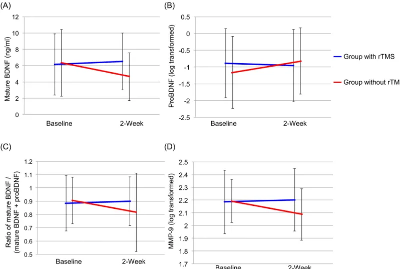
Serum proBDNF levels were compared before and after treatment in 50 patients of the rTMS plus rehabilitation group and 26 patients before and 22 patients of the rehabilitation only group since serum levels of proBDNF in the other patients were below the minimum detectable concentration of the kit. Two-way repeated measures ANOVA of BDNF data showed a signifi-cant group-by-time interaction (group: F = 2.1, df = 1,93, p = 0.152, time: F = 1.7, df = 1,93, p = 0.193, interaction (group x time): F = 4.1, df = 1,93, p = 0.047) (Fig 2A). There were no sig-nificant effects and interaction in serum proBDNF levels (group: F = 0.2, df = 1,66, p = 0.685, time: F = 0.1, df = 1,66, p = 0.730, interaction (group x time): F = 2.6, df = 1,66, p = 0.110) (Fig 2B). Furthermore, two-way repeated measures ANOVA of the ratio of BDNF to total BDNF (BDNF plus proBDNF) showed a significant group-by-time interaction (group: F = 0.3, df = 1,66, p = 0.580, time: F = 2.6, df = 1,66, p = 0.109, interaction (group x time): F = 6.4, df = 1,66, p = 0.014) (Fig 2C).
Two-way repeated measures ANOVA of MMP-9 data showed a significant group-by-time interaction (group: F = 1.5, df = 1,93, p = 0.220, time: F = 3.9, df = 1,93, p = 0.053, interaction (group x time): F = 7.0, df = 1,93, p = 0.010) (Fig 2D).
Fig 1. Effect of rehabilitation with and without 2-week rTMS on FMA and WMFT log performance time. (A) FMA: There was a significant interaction between group and time (F = 7.4, df = 1,93, p = 0.008). (B) WMFT log performance time: There was a significant interaction between group and time (F = 4.6, df = 1,93, p = 0.035).
Correlations with clinical evaluation of upper limb motor function
Baseline BDNF and MMP-9 serum levels did not correlate with changes in FMA and WMFT log performance time in both treatment groups. Furthermore, proBDNF serum levels did not correlate with the increase in FMA score in both groups. Interestingly, there was a significant negative (ρ = -0.422, p = 0.002) correlation between proBDNF serum levels and decrease in WMFT log performance time in the rTMS plus rehabilitation group (n = 50), but not in the rehabilitation group (n = 26) (Fig 3).
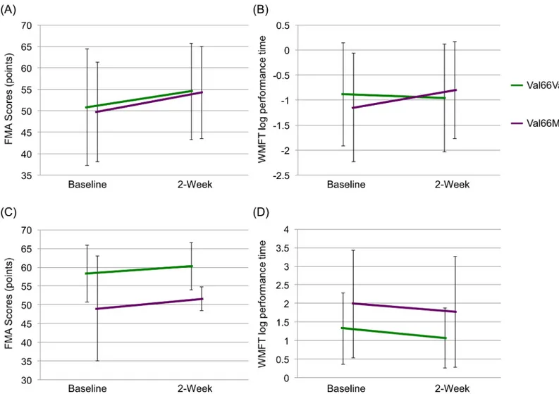
Effect of BDNF Val66Met polymorphism on improvement in motor
function
Based on the small number of patients with met allele, we divided the patients into two groups (val66val vs. val66met and met66met). Two-way repeated measures ANOVA of FMA data showed statistical significance in the rTMS plus rehabilitation group (gene: F = 0.05, df = 1,60, p = 0.828, time: F = 87.0, df = 1,60, p<0.001, interaction (gene x time): F = 1.0, df = 1,60, p = 0.333) (Fig 4A), as well as in the rehabilitation group (gene: F = 4.8, df = 1,31, p = 0.036,
Fig 2. Effects of rehabilitation with and without 2-week rTMS on serum levels of biomarkers. (A) Mature BDNF: There was a significant interaction between group and time (F = 4.1, df = 1,93, p = 0.047). (B) ProBDNF: There was no significant interaction between group and time (F = 2.6, df = 1,65, p = 0.110). (C) BDNF/total BDNF (BDNF and proBDNF) ratio: There was a significant interaction between group and time (F = 6.4, df = 1,66, p = 0.014). (D) MMP-9: There was a significant interaction between group and time (F = 7.0, df = 1,93, p = 0.010).
time: F = 21.0, df = 1,31, p<0.001, interaction (gene x time): F = 0.4, df = 1,31, p = 0.513) (Fig 4B).
Two-way repeated measures ANOVA of WMFT log performance time data showed statisti-cal significance in the rTMS plus rehabilitation group (gene: F = 0.1, df = 1,60, p = 0.740, time: F = 45.3, df = 1,60, p<0.001, interaction (gene x time): F = 2.7, df = 1,60, p = 0.108) (Fig 4C), as well as in the rehabilitation monotherapy group (gene: F = 2.5, df = 1,31, p = 0.127, time: F = 7.2, df = 1,31, p = 0.012, interaction (gene x time): F = 0.1, df = 1,31, p = 0.798) (Fig 4D).
Discussion
This is the first report to show that 2-week low-frequency rTMS increases serum levels of mature BDNF and MMP-9 in stroke patients. Given the key role of BDNF-TrkB signaling in neuroplasticity, it is likely that high serum levels of mature BDNF play at least some role in the beneficial actions of rTMS on motor function in the affected upper limb.
Accumulating evidence suggests that rTMS is effective treatment for poststroke hemiparesis [1]. Rehabilitation with low-frequency rTMS resulted in greater improvement of upper limb hemiparesis than rehabilitation without rTMS. In the present study, the rTMS plus rehabilita-tion group showed better improvement in FMA and WMFT than the rehabilitarehabilita-tion alone group, as reported previously by Abo et al. [29].
Fig 3. Correlation between baseline serum proBDNF levels and decrease in WMFT log performance time. There was a significant correlation between serum proBDNF levels and decrease in WMFT log performance time in patients treated with rehabilitation and rTMS (n = 50,ρ = -0.422, p = 0.002) but not in those treated with rehabilitation only.
The rTMS treatment is reported to result in significant increases in serum levels of total BDNF (mature BDNF + proBDNF) in patients with depression [11]. However, another study showed no such changes in serum levels of total BDNF after rTMS combined with rehabilitation [30]. In the above two studies, the BDNF ELISA kit did not recognize BDNF (mature form) and proBDNF [15]. One possibility for the discrepancy between the studies is the use of ELISA kit. In the present study, we found that serum levels of mature BDNF increased after 2-week rTMS plus rehabilitation, but decreased after 2-week rehabilitation only. Furthermore, serum levels of MMP-9 increased after 2-week rTMS plus rehabilitation whereas they decreased after 2-week rehabilitation only. Considering the role of MMP-9 in the conversion of proBDNF to mature BDNF, it is interesting to note the high levels of serum mature BDNF after 2-week rTMS.
Accumulating evidence indicates that both proBDNF and mature BDNF play important roles in various physiological functions, eliciting opposite effects via the p75NTRand TrkB receptors, respectively [7,8,14]. These two parameters have not so far been measured accurately due to the limited specificity of the BDNF antibody. Luckily, the newly available ELISA Kit allowed us to measure separately the levels of both BDNF (mature form) and proBDNF [15].
Fig 4. Effects of BDNF Val66Met gene polymorphism on FMA and WMFT log performance time. Effects of BDNF Val66Met gene polymorphism on (A) FMA and (B) WMFT log performance time in patients who received rehabilitation plus rTMS combination therapy. Effects of rehabilitation monotherapy on FMA (C) and WMFT log performance time (D) in Val66Val and Val66Met patients.
Our results showed that rTMS plus rehabilitation slightly reduced proBDNF serum levels, whereas rehabilitation alone resulted in a slight increase in proBDNF serum levels. Based on the above changes, rehabilitation alone resulted in reduction in the ratio of mature BDNF/total BDNF, whereas it increased slightly in patients of the rTMS plus rehabilitation group. These findings suggest that low-frequency rTMS can activate the conversion of proBDNF to mature BDNF, and that such change enhances the effects of rehabilitation therapy on improvement of upper limb hemiparesis.
The results also showed a negative correlation between baseline serum proBDNF and improvement in WMFT, reflecting improvement in upper limb motor function. This prelimi-nary result suggests that high baseline level of serum proBDNF could be used to predict poor improvement of motor function in the affected upper limbs, or alternatively, used as a bio-marker of the response to rehabilitation in poststroke patients with hemiparesis. Further stud-ies of large samples are needed to confirm these conclusions.
Several studies have reported thatBDNF gene val66met polymorphism has a negative effect on the outcome of rTMS therapy [13,31]. However, the present results indicate that the out-come of rTMS therapy is not altered by this polymorphism, although we acknowledge the small number of patients included in the study. Nonetheless, our results demonstrate that rTMS combined with rehabilitation improves motor function in the affected limb after stroke irrespective ofBDNF gene polymorphism. Whereas we did not determine the reason for the above differences between the two studies, we believe it relates to differences in the methodol-ogy; the rTMS sessions applied in the present study outnumbered those used in the previous study [13].
Limitations
The present study has several limitations. First, it was not a randomized controlled trial, and rTMS treatment was directly applied to the patient. In addition, the sample size was too small to allow firm conclusions. Further randomized controlled studies of larger population are needed. Second, the sensitivity of the human proBDNF ELISA kits used in this study is less than ideal; in fact, serum levels of proBDNF in some patients were not detected, whereas they were detected in all subjects who participated in a Swedish study [32] and Israeli study [33], suggesting ethnic differences in serum proBDNF levels. The development of highly sensitive proBDNF ELISA kits is desirable for accurate measurement of proBDNF serum levels.
Conclusions
Two-week low-frequency rTMS plus rehabilitation therapy resulted in greater improvement of upper limb motor function than rehabilitation alone, and this effect was mediated, at least in part, through rises in serum levels of mature BDNF and MMP-9 in poststroke patients. Mature BDNF, proBDNF, and MMP-9 could be potentially useful biomarkers of the response to ther-apy in poststroke patients with upper limb motor deficit.
Acknowledgments
The authors gratefully acknowledge the support and the participation of the patients in the study.
Author Contributions
Conceived and designed the experiments: MN KH MA. Performed the experiments: MN SM TI. Analyzed the data: MN KH RM. Contributed reagents/materials/analysis tools: MN RM. Wrote the paper: MN KH WK RM.
References
1. Lefaucheur JP, André-Obadia N, Antal A, Ayache SS, Baeken C, Benninger DH, et al. Evidence-based guidelines on the therapeutic use of repetitive transcranial magnetic stimulation (rTMS). Clin Neurophy-siol. 2014; 125: 2150–2206. doi:10.1016/j.clinph.2014.05.021PMID:25034472
2. Lüdemann-Podubecká J, Bösl K, Nowak DA. Repetitive transcranial magnetic stimulation for motor recovery of the upper limb after stroke. Prog Brain Res. 2015; 218: 281–311. doi:10.1016/bs.pbr.2014. 12.001PMID:25890143
3. Müller-Dahlhaus F, Vlachos A. Unraveling the cellular and molecular mechanisms of repetitive mag-netic stimulation. Front Mol Neurosci. 2013; 6: 50. doi:10.3389/fnmol.2013.00050PMID:24381540 4. Nestler EJ, Barrot M, DiLeone RJ, Eisch AJ, Gold SJ, Monteggia LM. Neurobiology of depression.
Neu-ron. 2002; 34: 13–25. PMID:11931738
5. Hashimoto K, Shimizu E, Iyo M. Critical role of brain-derived neurotrophic factor in mood disorders. Brain Res Brain Res Rev. 2004; 45: 104–114. PMID:15145621
6. Duman RS, Monteggia LM. A neurotrophic model for stress-related mood disorders. Biol Psychiatry. 2006; 59: 1116–1127. PMID:16631126
7. Hashimoto K. Brain-derived neurotrophic factor as a biomarker for mood disorders: an historical over-view and future directions. Psychiatry Clin Neurosci. 2010; 64: 341–357. doi:10.1111/j.1440-1819. 2010.02113.xPMID:20653908
8. Hashimoto K. Sigma-1 receptor chaperone and brain-derived neurotrophic factor: emerging links between cardiovascular disease and depression. Prog Neurobiol. 2013; 100: 15–29. doi:10.1016/j. pneurobio.2012.09.001PMID:23044468
9. Castrén E. Neurotrophins and psychiatric disorders. Handb Exp Pharmacol. 2014; 220: 461–479. doi: 10.1007/978-3-642-45106-5_17PMID:24668483
10. Yukimasa T, Yoshimura R, Tamagawa A, Uozumi T, Shinkai K, Ueda N, et al. High-frequency repetitive transcranial magnetic stimulation improves refractory depression by influencing catecholamine and brain-derived neurotrophic factors. Pharmacopsychiatry. 2006; 39: 52–59. PMID:16555165
11. Zanardini R, Gazzoli A, Ventriglia M, Perez J, Bignotti S, Rossini PM, et al. Effect of repetitive transcra-nial magnetic stimulation on serum brain derived neurotrophic factor in drug resistant depressed patients. J Affect Disord. 2006; 91: 83–86. PMID:16448701
12. Wang HY, Crupi D, Liu J, Stucky A, Cruciata G, Di Rocco A, et al. Repetitive transcranial magnetic stim-ulation enhances BDNF-TrkB signaling in both brain and lymphocyte. J Neurosci. 2011; 31: 11044– 11054. doi:10.1523/JNEUROSCI.2125-11.2011PMID:21795553
13. Chang WH, Bang OY, Shin YI, Lee A, Pascual-Leone A, Kim YH. BDNF polymorphism and differential rTMS effects on motor recovery of stroke patients. Brain Stimul. 2014; 7: 553–558. doi:10.1016/j.brs. 2014.03.008PMID:24767962
14. Lu B, Pang PT, Woo NH. The yin and yang of neurotrophin action. Nat Rev Neurosci. 2005; 6: 603– 614. PMID:16062169
15. Yoshida T, Ishikawa M, Iyo M, Hashimoto K. Serum levels of mature brain-derived neurotrophic factor (BDNF) and its precursor proBDNF in healthy subjects. Open Clin Chem J. 2012; 5: 7–12.
16. Hashimoto K. Serum brain-derived neurotrophic factor as a predictor of incident dementia. JAMA Neu-rol. 2014; 71: 653.
17. Hashimoto K. BDNF and proBDNF as biomarkers for bipolar disorder. Br J Psychiatry. 2014; 205: 410. 18. Hashimoto K. Brain-derived neurotrophic factor (BDNF) and its precursor proBDNF as diagnostic
bio-markers for major depressive disorder and bipolar disorder. Eur Arch Psychiatry Clin Neurosci. 2015; 265: 83–84. doi:10.1007/s00406-014-0557-xPMID:25362578
19. Yoshida T, Ishikawa M, Niitsu T, Nakazato M, Watanabe H, Shiraishi T, et al. Decreased serum levels of mature brain-derived neurotrophic factor (BDNF), but not its precursor proBDNF, in patients with major depressive disorder. PLoS One. 2012; 7: e42676. doi:10.1371/journal.pone.0042676PMID: 22880079
20. Maeda F, Keenan JP, Tormos JM, Topka H, Pascual-Leone A. Modulation of corticospinal excitability by repetitive transcranial magnetic stimulation. Clin Neurophysiol. 2000; 111: 800–805. PMID: 10802449
21. Pascual-Leone A, Amedi A, Fregni F, Merabet LB. The plastic human brain cortex. Annu Rev Neurosci. 2005; 28: 377–401. PMID:16022601
22. Ward NS, Cohen LG. Mechanisms underlying recovery of motor function after stroke. Arch Neurol. 2004; 61: 1844–1848. PMID:15596603
23. Takeuchi N, Chuma T, Matsuo Y, Watanabe I, Ikoma K. Repetitive transcranial magnetic stimulation of contralesional primary motor cortex improves hand function after stroke. Stroke. 2005; 36: 2681–2686. PMID:16254224
24. Villardita C. Raven's colored Progressive Matrices and intellectual impairment in patients with focal brain damage. Cortex. 1985; 21: 627–634. PMID:2419033
25. Fugl-Meyer AR, Jääskö L, Leyman I, Olsson S, Steglind S. The post-stroke hemiplegic patient. 1. a method for evaluation of physical performance. Scand J Rehabil Med. 1975; 7: 13–31. PMID:1135616 26. Wolf SL, Catlin PA, Ellis M, Archer AL, Morgan B, Piacentino A. Assessing Wolf motor function test as
outcome measure for research in patients after stroke. Stroke. 2001; 32: 1635–1639. PMID:11441212 27. Itoh K, Hashimoto K, Kumakiri C, Shimizu E, Iyo M. Association between brain-derived neurotrophic
factor 196 G/A polymorphism and personality traits in healthy subjects. Am J Med Genet B Neuropsy-chiatr Genet. 2004; 124B: 61–63. PMID:14681916
28. Itoh K, Hashimoto K, Shimizu E, Sekine Y, Ozaki N, Inada T, et al. Association study between brain-derived neurotrophic factor gene polymorphisms and methamphetamine abusers in Japan. Am J Med Genet B Neuropsychiatr Genet. 2005; 132B: 70–73. PMID:15459944
29. Abo M, Kakuda W, Momosaki R, Harashima H, Kojima M, Watanabe S, et al. Randomized, multicenter, comparative study of NEURO versus CIMT in poststroke patients with upper limb hemiparesis: the NEURO-VERIFY Study. Int J Stroke. 2014; 9: 607–612. doi:10.1111/ijs.12100PMID:24015934 30. Mirowska-Guzel D, Gromadzka G, Seniow J, Lesniak M, Bilik M, Waldowski K, et al. Association
between BDNF-196 G>A and BDNF-270 C>T polymorphisms, BDNF concentration, and rTMS-sup-ported long-term rehabilitation outcome after ischemic stroke. NeuroRehabilitation. 2013; 32: 573–582. doi:10.3233/NRE-130879PMID:23648611
31. Cheeran B, Talelli P, Mori F, Koch G, Suppa A, Edwards M, et al. A common polymorphism in the brain-derived neurotrophic factor gene (BDNF) modulates human cortical plasticity and the response to rTMS. J Physiol. 2008; 586: 5717–5725. doi:10.1113/jphysiol.2008.159905PMID:18845611 32. Södersten K, Pålsson E, Ishima T, Funa K, Landén M, Hashimoto K, et al. Abnormality in serum levels
of mature brain-derived neurotrophic factor (BDNF) and its precursor proBDNF in mood-stabilized patients with bipolar disorder: a study of two independent cohorts. J Affect Disord. 2014; 160: 1–9. doi: 10.1016/j.jad.2014.01.009PMID:24709015
33. Levin R, Dor-Abarbanel AE, Edelman S, Durrant AR, Hashimoto K, Javitt DC, et al. Behavioral and cog-nitive effects of the N-methyl-D-aspartate receptor co-agonist D-serine in healthy humans: initial find-ings. J Psychiatr Res. 2015; 61: 188–195. doi:10.1016/j.jpsychires.2014.12.007PMID:25554623