P R O O F
Brain Behav Evol 488 DOI: 10.1159/0000XXXXXQuantitative Aspects of the Spatial Distribution
and Morphological Characteristics of the Sea
Bass (
Dicentrarchus labrax
L.; Teleostei,
Serranidae) Trunk Lateral Line Neuromasts
Karine Faucher
a, bJean-Paul Lagardère
bAnne Aubert
aaLaboratoire de Biologie et Environnement Marins, CNRS-Université de La Rochelle, La Rochelle, bCentre de Recherche sur les Ecosystèmes Marins et Aquacoles, CNRS-Ifremer, l’Houmeau, France
Received: July 22, 2004
Returned for revision: September 12, 2004 Accepted after revision: November 11, 2004 Published online: $$$
Key Words
FishW TeleostW Sea bassW Trunk lateral lineW NeuromastW
Hair cell
Abstract
The results presented herein report quantitative data rel-ative to the distribution and morphological characteris-tics of both types of neuromasts encountered on the trunk lateral line of the sea bass (Dicentrarchus labrax, L.). These data were obtained from scanning electron micrographs. They indicate that, as expected, each mod-ified scale of the sea bass possessed a single canal neu-romast with long axis oriented parallel to the fish’s long axis. In contrast to several fish species, two thirds of superficial neuromasts observed herein were oriented perpendicular to the fish’s long axis. However, whatever the main orientation of superficial neuromasts, two thirds of their hair bundles were oriented parallel to the long axis of the animal with approximately half of them in the direction of the head. Similar ratios were observed for canal neuromasts whatever the area of the maculae: central or peripheral. For both types of neuromasts it was not possible to clearly distinguish a paired organiza-tion of hair bundles with opposing polarities. Superficial
neuromasts on each trunk canal scale were located on either the dorsal or ventral side of the canal and ap-peared to be distributed along the trunk lateral line with a higher probability to be encountered closer to the oper-culum. The frequency of presence and the average num-ber of superficial neuromasts per scale increased with fish size. We observed a size gradient for canal neuro-masts between the operculum and caudal peduncle. This gradation was correlated with a reduction of the width of the central area of the canal segment. Canal neuromasts were always localized in the larger portions of the canal segments. Taken together, these results point out some specific features associated with the sea bass trunk later-al line. With the previous report, they establish the first full description of the trunk lateral line of sea bass and will be useful for upcoming experiments regarding the function of the two types of neuromasts.
Copyright © 2005 S. Karger AG, Basel
Introduction
The lateral line system is present in all fishes and aquatic amphibians [Dijkgraff, 1962; Blaxter, 1987; Lan-noo, 1987; Coombs et al., 1989; Webb, 1989a; Northcutt,
BOIS:BBE:ZBRAI488XA.91 FF: ZUP9 E1:
P R O O F
1992]. In fishes, this sensory system detects near field water movements caused by the animal’s own movements and water currents [Denton and Gray, 1983; Bleckmann, 1993; Montgomery et al., 1995; Coombs et al., 1996]. This sensitivity to hydrodynamic stimuli enables the fish to locate congeners, predators, preys and other stationary or moving objects [Dijkgraff, 1962; Partridge and Pitcher, 1980; Blaxter and Batty, 1985; Hoekstra and Janssen, 1986; Montgomery, 1989; Bleckmann, 1993; Janssen et al., 1995, 1999; Montgomery and Hamilton, 1997; Coombs, 1999]. The lateral line system also plays a major role in rheotaxis [Bleckmann, 1993; Pavlov and Tyuryu-kov, 1993; Montgomery et al., 1997; Northcutt, 1997; Baker and Montgomery, 1999; Coombs et al., 2001] which allows fish to travel and intercept food carried downstream, potentially reducing energetic costs of dis-placements, food search and capture [Montgomery et al., 1997; Baker and Montgomery, 1999]. Based on these observations, it has been shown that the involvement of a lateral line system is essential for fish living in environ-ments where visibility is significantly reduced [Schellart and Wubbels, 1998] such as in estuaries. The sea bass,
Dicentrarchus labrax, an economically important fish
spe-cies for fisheries along the French Atlantic coast, is known to potentially use its lateral line system under turbid or turbulent conditions. This fish inhabits the littoral zone and is found near rocky coasts or sandy beaches with high wave energy in addition to estuaries, lagoons and occa-sionally rivers [Barnabé, 1978; Quéro, 1984]. Because the coastal or estuarine habitats of the sea bass are also known to be sites exposed to metal ion pollution [Elbaz-Poulichet and Martin, 1987; Latouche, 1988; Jouanneau et al., 1990; Lapaquellerie et al., 1996; Michel et al., 2000] which can temporarily block its lateral line system [Karl-sen and Sand, 1987] and thus affect its survival [Jans[Karl-sen, 2000], this potential threat to the continuation of the spe-cies motivated us to make a detailed analysis of the func-tional anatomy of the lateral line system.
The functional units of the lateral line system are called the neuromasts. They are distributed on the head, trunk and tail [Coombs et al., 1989; Northcutt, 1992, 1997; Bleckmann, 1993]. These mechanoreceptors are divided into two types according to their location. Superficial neu-romasts are present on the epidermis of modified scales and the canal neuromasts are enclosed in canals below the epidermis [Coombs et al., 1989]. Morphological data on lateral line systems have been reported for several fish species. These studies indicated that among teleosts there is a diverse set of morphological features and distribution patterns [e.g., Münz, 1979, 1989; Coombs et al., 1989;
Song and Northcutt, 1991; Coombs and Montgomery, 1994]. Similar assertions could be described for non-tele-ost fishes [Webb and Northcutt, 1997; Maruska and Tri-cas, 1998; Maruska, 2001; Peach, 2001, 2003]. Few data are available for the sea bass lateral line system [Diaz et al., 2003; Faucher et al., 2003]. Most of the studies listed above are qualitative and to date little is known about the number, the size or orientation of either type of neuro-masts and their associated sensory cells along the fish trunk lateral line [Münz, 1979, 1989; Coombs et al., 1989; Song and Northcutt, 1991; Coombs and Montgomery, 1994; Peach, 2003]. An accurate anatomical description of this sensory system is necessary to improve our under-standing of the functioning of each type of neuromast as well as suggesting how evolutionary processes contributed to the orientation and directional sensitivity of lateral line sense organs in fish species living in turbulent environ-ments.
Materials and Methods
Animals and Tissue Processing
For this study, modified scales isolated from the trunk lateral lines of thirty-eight specimens of sea bass, Dicentrarchus labrax, were examined. The fish averaged 20 cm in length and 120 g in weight. The fish were obtained from a commercial source (Ferme des Ba-leines, Ile de Ré, France). All specimens were housed in 240-liter tanks filled with filtered seawater and kept under natural photoper-iod and constant temperature (16° C). They were fed twice a week
with live molluscs and crustaceans.
Fig. 1. Electron micrographs which illustrate the different quantita-tive parameters measured on neuromasts of the sea bass trunk lateral line. Superficial neuromasts were observed on the epidermis of trunk lateral line scales (A, scale bar = 500 Ìm), whereas canal neuromasts were located inside the segment canal of these scales (B, scale bar = 500 Ìm). For each neuromast, different parts were delimited: the whole neuromast (non-sensory area and macula), its macula and when possible the base of its cupula. The morphological limits taken into account for all measurements (length, width, perimeter and sur-face area) performed in the cupula (C, E), the macula and the whole neuromast (D, F) are indicated on the micrographs (dotted and dashed lines). The lengths and widths of the neuromasts, cupulae and maculae were measured at their largest place based on the limits pre-viously set. The diameter of the canal segment was estimated by mea-suring the length of a line drawn through the width of the canal seg-ment at the level of three specific marks: the central axis of the canal neuromast (2 in B) estimated from its morphological limits and the presence of its wing-like extensions, and the two extremities of the subdermal tube, the nearest possible to suprascalar (1 in B) and infrascalar pores (3 in B).
BOIS:BBE:ZBRAI488XA.91 FF: ZUP9 E1:
P R O O F
Prior to sacrifice, the fish were anaesthetized with 75 mg/l MS 222 (3-aminobenzoic acid ethyl, Sigma) and measured in standard length: from the tip of the snout up to the indentation of the caudal fin. For each fish, both entire trunk lateral lines were sampled in nat-ural seawater. They consisted of a single row of modified scales, dif-fering from the others by the presence of superficial neuromasts and the canal tube containing canal neuromasts, running within the mid-section of each flank from the operculum up to the tail. No superficial or canal neuromasts were observed on the trunk outside these spe-cific areas [Faucher et al., 2003]. Tissue samples were dissected in an artificial solution (composition in mM: NaCl: 150; KCl: 5; CaCl2 W
2H2O: 3; MgCl2 W6H2O: 1.5; HEPES: 10) for which the pH was
adjusted to 8 with NaOH. Each tissue sample consisted of two to three consecutive scales. Some were left intact in order to observe superficial neuromasts. The roof of the canal segment of other scales was carefully removed from the suprascalar to infrascalar pores to allow visualization of canal neuromast (fig. 1A, B).
Tissue samples were fixed in 4% glutaraldehyde (Fisher Scientific Labosi) in sodium cacodylate buffer (0.4 M, pH 7.2), dehydrated in graded acetone concentrations and critical point-dried using liquid CO2 (BALTEC CPD 030). They were then mounted on brass
sup-ports and sputter-coated with gold (Cressington Sputter Coat). Ob-servations were performed with a JEOL JSM-5410LV scanning elec-tron microscope. All quantitative measures were obtained using the image analysis software Biocom Visiol b 200.
Morphological Data
To allow comparisons between scales and/or fishes, an identi-fying number (I.D. #) was assigned for each modified scale that com-posed the trunk lateral line. The I.D. # 1 was given to the first scale located just after the operculum and sequentially until the limit of the caudal fin. A minimum of five observations per scale I.D. # was per-formed. In addition, according to the total number of modified scales, the trunk lateral lines were divided into three equal segments: anterior (S1), middle (S2) and posterior (S3). This allowed us to
account for the different number of modified scales (between 51 and 78) per trunk lateral line. Thus, depending on the fish examined, each segment was composed of 17 to 26 scales.
The distribution of superficial or canal neuromasts along the trunk lateral line was assessed through the determination, per scale and then per segment, of the average number of neuromasts and of the average frequency of neuromast occurrence. In order to do that, the number of modified scales examined was recorded. This led to the total number of scales observed per segment (Nobs). Then, the
number of positive observations (xSi), that is the number of scales for
which one or more canal or superficial neuromasts were observed, was estimated. Finally, the total number of neuromasts observed per segment was determined and noted as nSi.
These data were then used to calculate the frequency of neuro-mast occurrence (fSi) and the average number of neuromasts per scale
(nSi) for the different trunk segments according to equations 1 and 2.
fSi = ™ xSi/Nobs (1)
(nSi = ™ nSi/Nobs (2)
Each neuromast can be considered either as a whole or as constit-uent parts, i.e., the non-sensory and sensory (macula) epithelia and the cupula. The image analysis software Biocom Visiol b 200 allowed us to outline these different areas (fig.1C–F) and quantify some relat-ed metrics such as their respective length (l), width (w), perimeter (p)
and surface area (a). For a given neuromast, according to the pres-ence or not of the cupula, we can measure at the same time either the surface area of the whole neuromast and of the cupula or the surface area of the whole neuromast and of the macula.
Then, additional information can be deduced for each parameter previously mentioned. As an example, for a given type of neuromast the average surface area (a) was calculated (equation 3), according to the total number of neuromasts observed (nm). Likewise, for each type of neuromast the average surface area per segment (aSi) was
cal-culated using equation 4, in which nmSi corresponded to the number
of neuromasts per segment from which data were obtained.
a = ™ a/nm (3)
aSi = ™ a/nmSi (4)
The values obtained for fSi, nSi and aSi were then averaged according
to the corresponding number of fish examined (between 30 and 38) and noted as FSi, NSi and ASi, respectively.
Image analysis software Biocom Visiol b 200 was also used to determine, as much as possible, the width of the canal segment at the level of three specific marks: the center of the neuromast and the two extremities of the subdermal tube, the nearest possible to infrascalar and suprascalar pores (fig. 1B).
Orientation of the Neuromasts and Their Hair Bundles
When possible, the orientations of the two types of neuromasts and their hair bundles were examined. The orientation of the neuro-mast was determined by the angle established between the main axis of the neuromast, that is its length, and a reference axis (0–180°)
which corresponded to the long axis of the animal with 0° on the
head side. Results were expressed as percentages of the total number of neuromasts observed. Round superficial neuromasts (with length ! 1.25 times the width), for which no major axis could be deter-mined, were not accounted for in this part of the study.
In order to study the possibility of a preferential axis of sensitivity for the neuromasts, the orientations of the hair bundles were exam-ined. The angle, from 0 to 360°, between the axis of polarity of the
hair bundles, conferred by the relative position of the kinocilium in relation to the stereocilia, and the long axis of the animal was deter-mined. Results were expressed as percentages of the total number of hair bundles observed per angular intervals of 45°.
Finally, the percentage of hair bundles associated in pairs of opposing polarities within a given neuromast was calculated. For this, the polarity of each hair bundle was compared with the polarity of all adjacent hair bundles, on the rostral, caudal, dorsal and ventral sides. In order to be classified as opposite, the difference between the polarities of adjacent hair bundles should be equal to 180 B 20°.
Then, the percentage of observed pair-wise hair bundles was calcu-lated relative to the number of possible pair-wise hair bundles inside the macula of each neuromast.
Statistical Analyses
All quantitative data were expressed as the mean B SEM (stan-dard error of the mean). The statistical analyses were performed with the statistical software XLSTAT-Pro 6.0. According to the essence of the parameters and the relationships to be tested different statistical tests were used (· = 0.05). The value of the test was noted: H for a Kruskal-Wallis test, U for a Mann-Whitney test, rs for a Spearman’s
rank correlation procedure, F˘1, ˘2 for a linear regression test and ¯2
P R O O F
ResultsNeuromast Distribution along the Trunk Lateral Lines
The distribution of superficial and canal neuromasts along the trunk lateral lines was examined through the determination of their frequency of occurrence and their number within each segment Si (table 1).
The figure 2A represents the average frequency of occurrence (FSi) of superficial neuromasts calculated for each segment. This distribution was heterogeneous along the trunk lateral line (H = 7.136, p = 0.028; table 1). The probability of encountering these neuromasts was higher at the beginning (S1) than at the end (S3) of the trunk later-al line (U = 756.000, p = 0.005). The figure 2B shows that the frequency of occurrence of superficial neuromasts in a fish body tended to increase with the animal’s size (F 1,16 = 7.275, p = 0.016, R² = 0.313). Nevertheless, the distribu-tion of frequency of occurrence was homogeneous among the three segments (Si) of the fish body regardless of fish size.
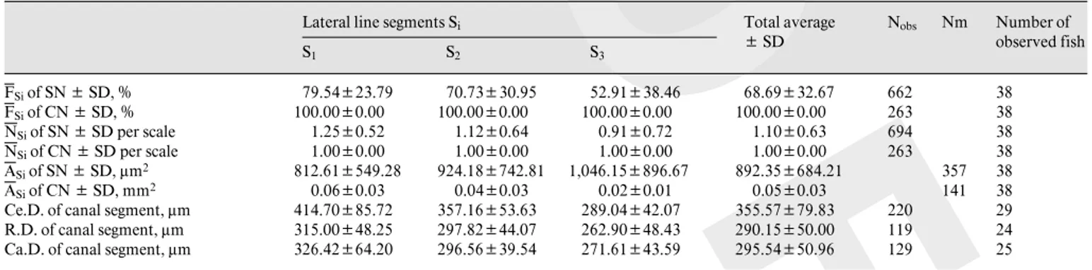
The average number of superficial neuromasts per scale in each segment, Si (NSi), was also examined (fig. 3A). No significant difference among these three seg-ments was observed (H = 4.388, p = 0.111; table 1). In addition, an examination of their position on the scale revealed that they were located indiscriminately on the dorsal or the ventral side of the canal segment. As for the frequency of occurrence, the average number of superfi-cial neuromasts increased with fish size (fig. 3B; F1,16 = 5.427, p = 0.033, R² = 0.253). There were no significant differences among segments Si of the fish body in the average number of superficial neuromasts for fish of all sizes.
Fig. 2. Distribution of the frequency of occurrence of superficial neu-romasts (SN) along the sea bass trunk lateral line and their relation-ship with the fish size. A Average frequency of occurrence of SN cal-culated for each segment Si (FSi). Frequencies are expressed in
per-centage and vertical bars represent the standard error of the mean. SN were more frequently seen at the beginning of the trunk lateral line (S1). B Relationship between the frequency of occurrence of SN per
fish (fSi) and the fish size in each segment Si of the trunk lateral line.
Frequencies are expressed in percentage. The frequency of occurrence of SN increased with fish size (Y = 3.4898x – 8.1544, R² = 0.3126).
Table 1. Quantitative data concerning the distribution and the morphology of both types of neuromasts Lateral line segments Si
S1 S2 S3 Total average B SD Nobs Nm Number of observed fish FSi of SN B SD, % 79.54B23.79 70.73B30.95 52.91B38.46 68.69B32.67 662 38 FSi of CN B SD, % 100.00B0.00 100.00B0.00 100.00B0.00 100.00B0.00 263 38 NSi of SN B SD per scale 1.25B0.52 1.12B0.64 0.91B0.72 1.10B0.63 694 38 NSi of CN B SD per scale 1.00B0.00 1.00B0.00 1.00B0.00 1.00B0.00 263 38 ASi of SN B SD, Ìm2 812.61B549.28 924.18B742.81 1,046.15B896.67 892.35B684.21 357 38 ASi of CN B SD, mm2 0.06B0.03 0.04B0.03 0.02B0.01 0.05B0.03 141 38
Ce.D. of canal segment, Ìm 414.70B85.72 357.16B53.63 289.04B42.07 355.57B79.83 220 29 R.D. of canal segment, Ìm 315.00B48.25 297.82B44.07 262.90B48.43 290.15B50.00 119 24 Ca.D. of canal segment, Ìm 326.42B64.20 296.56B39.54 271.61B43.59 295.54B50.96 129 25
FSi = average occurrence; NSi = average number; ASi = average surface area; Ce.D. = central diameter, R.D. = rostral diameter; Ca.D. = caudal diameter; SN =
superficial neuromasts; CN = canal neuromasts; SD = standard deviation of the mean; Nobs = number of scales observed; Nm = number of neuromasts
BOIS:BBE:ZBRAI488XA.91 FF: ZUP9 E1:
P R O O F
Fig. 3. Distribution of the average number of superficial neuromasts (SN) per scale along the sea bass trunk lateral line and their relation-ship with fish size. A Average number of SN per scale in each seg-ment Si (NSi). Vertical bars represent standard error of the mean. The
data did not support a specific pattern of distribution. B Relationship between the average number of SN per scale per fish (nSi) and the fish
size in each segment Si of the trunk lateral line. The average number
of SN per scale increased with fish size (Y = 0.0609x – 0.2432, R² = 0.2533).
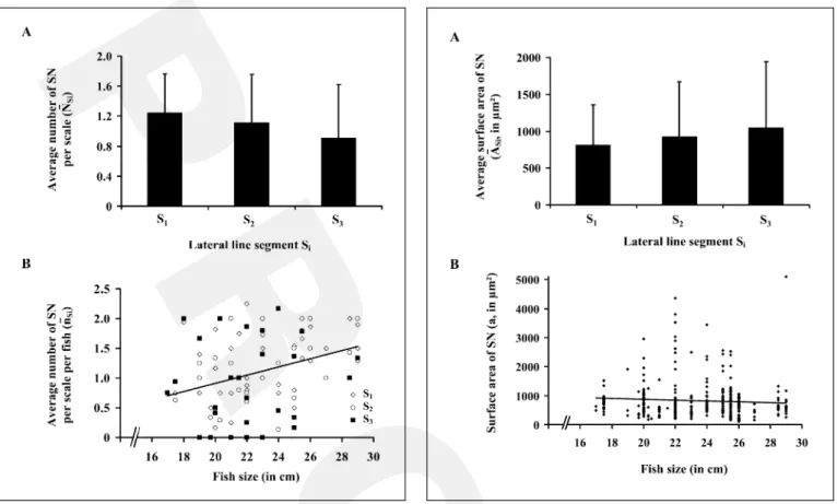
Fig. 4. Distribution of the size of superficial neuromasts (SN) along the sea bass trunk lateral line and their relationship with the fish size.
A Average surface area (ASi) by segment Si (expressed in Ìm2).
Verti-cal bars represent standard error of the mean. No specific pattern of distribution could be established. B Relationship between the surface area (a) of SN and the fish size. Data show that the size of these sensory organs varied according to individuals but not with the fish size (Y = –14.256x + 1150.3, R² = 0.0046).
In contrast to superficial neuromasts, only one canal neuromast could be observed on each modified scale (n = 263; table 1).
Morphological Data
Among the different measures performed on neuro-masts of each type and their constituent parts we have elected to consider the surface area of the whole neuro-mast as the parameter of reference for comparisons (ta-ble 1). This choice was based on several observations. First, this parameter incorporated aspects of all other metrics (perimeter, length and width). Second, analyses indicated that for both types of neuromasts the whole sur-face area was significantly correlated with the sursur-face area at the base of the cupula (rs = 0.940, p ! 0.0001, n = 111
for superficial neuromasts and rs = 0.850, p ! 0.0001, n = 53 for canal neuromasts) and the surface area of the macu-la (rs = 0.874, p ! 0.0001, n = 74 for superficial neuro-masts and rs = 0.978, p ! 0.0001, n = 69 for canal neuro-masts).
The average surface area of superficial neuromasts
within a given segment Si ranged from 230 to 3,600 Ìm2
which corresponded to an average surface area of 892.35 B 684.21 Ìm² (n = 357). This difference in size was also observed between superficial neuromasts present on the same scale. Along the trunk lateral line, the average sur-face area (ASi) of superficial neuromasts was examined (fig. 4A). Statistical analyses indicated that there was no significant difference among the three segments (H = 0.672, p = 0.715; table 1). The relation between the
sur-P R O O F
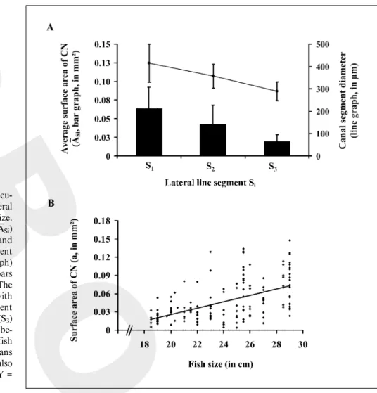
Fig. 5. Distribution of the size of canal neu-romasts (CN) along the sea bass trunk lateral line and their relationship with the fish size.
AEvolution of the average surface area (ASi)
of the CN (expressed in Ìm2; bar graph) and
of the average diameter of the canal segment (in Ìm, measured at CN location; line graph) along the trunk lateral line. Vertical bars represent standard error of the mean. The size of the CN decreased significantly with the central diameter of the canal segment between the beginning (S1) and the end (S3)
of the trunk lateral line. B Relationship be-tween the surface area (a) of CN and the fish size. The average size of these sensory organs varied according to individuals and was also positively correlated with the fish size (Y = 0.0053x – 0.0799, R² = 0.2869).
face area of superficial neuromasts and the fish size, illus-trated in figure 4B, showed that the former varied among individuals but was not correlated with the fish size (F1,333= 1.544, p = 0.215, R² = 0.005).
Similar studies applied to canal neuromasts indicated that within each segment (Si) the average surface areas (a) could also be variable: from 0.005 to 0.130 mm² for an average surface area of 0.05 B 0.03 mm² (n = 141). Along the trunk lateral line, the average surface area of these neuromasts was significantly larger in the anterior part (S1) of the trunk lateral line than in the posterior part (S3; U = 446.000, p ! 0.0001; fig. 5A; table 1). Statistical anal-yses also revealed that canal neuromasts were significant-ly larger (0.05 B 0.03 mm², n = 141) than superficial ones (892.35 B 684.21 Ìm², n = 357; U = 5,110.000; p ! 0.0001).
The diameter of the canal segment was measured, when possible, in its rostral, central and caudal regions
(table 1). Data showed that average canal segment diame-ters measured in its rostral (290.15 B 50.00 Ìm, n = 119) and caudal (295.54 B 50.96 Ìm, n = 129) ends were not significantly different (U = 8,080.500, p = 0.473). Thus, these peripheral diameters could be pooled and compared to the diameter of the canal segment in its central part (355.57 B 79.83 Ìm, n = 220). A Mann-Whitney test indicated that the central diameter of the canal segment, where canal neuromasts were located, was significantly greater in diameter at their peripheral ends (U = 41,764.000, p ! 0.0001). As observed for the size of canal neuromasts, the central diameter of the canal segment
decreased significantly between S1 (414.70 B 85.72 Ìm,
n = 73) and S3 (289.04 B 42.07 Ìm, n = 67; H = 31.498,
p ! 0.0001; fig. 5A; table 1). Finally, the surface area of the canal neuromasts was positively correlated with the fish size (F 1,146 = 58.731, p ! 0.0001, R² = 0.287; fig. 5B).
BOIS:BBE:ZBRAI488XA.91 FF: ZUP9 E1:
P R O O F
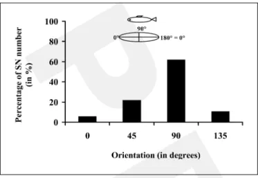
Fig. 6. Orientations of superficial elliptical neuromasts (SN) in regard to the canal segment main axis. The reference axis (0–180°
line) was parallel to the canal axis. The insert shows the multiple orientations of the SN observed (0 and 180° were not distinguished).
The 100 % value corresponded to the total number of SN observed (n = 378). The majority of SN was oriented perpendicularly to the canal axis.
Neuromast Orientations
The orientation of superficial neuromasts was only examined for the elliptical type. For the round ones (length inferior to 1.25 times the width), it was not possi-ble to determine a major axis. Figure 6 shows that among the 378 elliptical superficial neuromasts observed, two thirds were oriented perpendicular to the animal’s long
axis, whereas 22% of the neuromasts were oriented at 45°
and 10.5% at 135° (¯² = 134.075, p ! 0.0001). Only 5.5%
were parallel to the fish’s long axis.
In contrast to superficial neuromasts, the major axis of all canal neuromasts observed was oriented parallel to the animal’s long axis which corresponded also to the axis of the canal segment (n = 263).
Hair Bundle Orientations
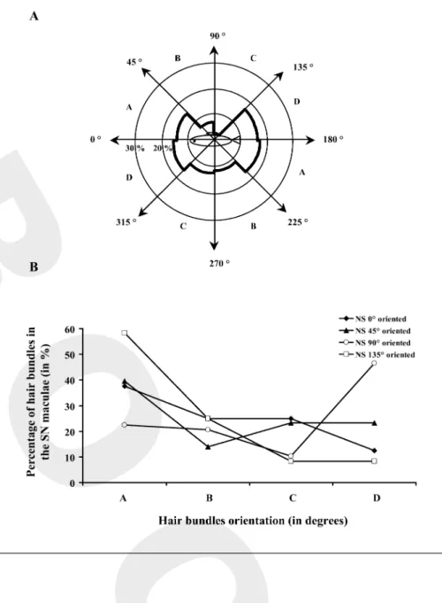
When possible, the orientation of hair bundles within the maculae of each type of neuromast was also examined. In the maculae of superficial neuromasts, elliptical and round, hair bundles presented a preferential orientation (fig. 7A). Indeed, among the 121 hair bundles observed, nearly two-thirds (65.3%) were oriented according to the fish’s long axis (¯² = 11.314, p = 0.000). Among these hair bundles 32.2% were more or less oriented towards the
head (between 316 and 45°) and 33.1% towards the tail
(between 136 and 225°). The last third could be divided
into hair bundles which were dorsally oriented (10.8%
between 46 and 135°) and hair bundles which were
ven-trally oriented (24.0% between 226 and 315°). The
orien-tation of hair bundles in regard to the fish’s long axis was also examined according to the orientation of superficial neuromasts. In this analysis, distributions of the hair bun-dle orientations within the maculae of superficial neuro-masts oriented at 0, 45, 90 and 135° were compared
(fig. 7B). Whatever the main orientation of these neuro-masts, differences between the orientations of their hair bundles were not significant (¯² = 0.154, p = 0.985, n = 32), they were preferentially oriented according to the 0– 180° axis, which corresponded to the animal’s long axis.
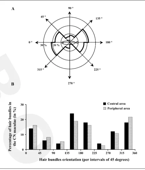
In order to examine the orientation of hair bundles within the maculae of canal neuromasts, 79 hair bundles were observed. They were preferentially oriented along the fish’s long axis (75.6%; ¯² = 23.405, p = 0.000, n = 79; fig. 8A). Among these hair bundles 38.1% were more or
less oriented towards the head (between 316 and 45°) and
37.5% towards the tail (between 136 and 225°). The
remainder of the hair bundles were dorsally (14.3%
be-tween 46 and 135°) or ventrally (10.1 % between 226 and
315°) oriented. The possibility of a different orientation
pattern between the central and peripheral regions of the maculae was also considered (fig. 8B). In the central region, hair bundles were indiscriminately oriented
to-ward the rostral (32% between 316 and 45°) or the caudal
(42% between 136 and 225°) side of the fish. Analogous
values were obtained for the peripheral area with 38%
oriented toward the rostral (between 316 and 45°) versus
35% toward the caudal side (between 136 and 225°) of the
fish. A ¯2 test showed that this distribution of orientations inside the maculae of canal neuromasts was homogeneous between the central and the peripheral areas (¯² = 0.465, p = 0.496, n = 64).
Very few hair bundles organized in pairs with their kinocilia facing in opposite directions were observed in superficial (19.0%) or canal neuromasts (9.9%).
Discussion
The efficiency of the lateral line organs in response to stimulation is dependent on (1) the distribution of hair bundle polarities within the maculae together with (2) the scattering of neuromasts on the flanks of fish. Flock and Wersäll [1962] were the first to demonstrate the relation-ship between hair bundle polarization, given by the eccen-tric position of the kinocilium, and the directional sensi-tivity of hair cells in response to stimulation. In sea bass, hair bundles within the maculae of superficial neuromasts
P R O O F
Fig. 7. Repartition of the hair bundle orien-tations within the maculae of superficial neuromasts (SN) and their relationship with the SN main orientation. The polarity of each hair bundle was defined by the line drawn from the smallest stereocilia towards the kinocilium. Each value was expressed in degree based on a 360-degree reference plane, 0° towards the rostral side and 180 °
towards the caudal side of the trunk lateral line. Results were expressed as percentages of the total number of hair bundles observed.
A Orientation of the hair bundles within the maculae of SN in a 360-degree reference plan with respect to a lateral view of the fish. Most were oriented along the canal axis eith-er towards the rostral or the caudal side of the trunk lateral line. BRelationship be-tween hair bundle orientations (A: bebe-tween 0 and 45°, then 181 and 225 °; B: between 46
and 90°, then 226 and 270 °; C: between 91
and 135°, then 271 and 315 °; D: between
136 and 180°, then 316 and 360 °) within the
maculae of SN and main orientation of SN (0, 45, 90 or 135°). Hair bundle orientations
were constant regardless of the orientation of SN.
were predominantly (70%) oriented parallel to the ani-mal’s long axis, regardless of the main orientation of the neuromast. Half of the superficial neuromast hair bundles were directed towards the head. The remainder exhibited various orientations. Similar ratios were observed within canal neuromasts, whatever the hair bundles’ location in the central or the peripheral area of the maculae [Faucher et al., 2003]. The observation of a major pattern of bipolar orientation parallel to the animal’s long axis for both types of neuromasts agrees with most reports on teleosts [Flock, 1965; Yamada and Hama, 1972; Münz, 1979; Denton and Gray, 1983; Webb, 1989c; Rouse and Pickles, 1991; Bleckman, 1993; Coombs and Montgomery, 1994;
Tsuka-moto et al., 1995; Montgomery et al., 2001]. However, in contrast with these authors, we could not clearly distin-guish a paired organization of hair bundles. Taken togeth-er, these observations strongly suggest that the trunk later-al line of sea bass is essentilater-ally sensitive to stimuli applied along the fish’s long axis. This was expected for canal neu-romasts, due to physical constraints set by the canal seg-ment, in which these receptors were enclosed [Coombs et al., 1989; Webb, 1989b]. However, such limits should not have applied to superficial neuromasts, which were free-standing on the skin and thus might potentially be the tar-get of stimuli coming from any direction. At first, this assumption appeared to be supported by two facts: (1) the
BOIS:BBE:ZBRAI488XA.91 FF: ZUP9 E1:
P R O O F
Fig. 8. Repartition of the hair bundle orien-tations within the maculae of canal neuro-masts (CN) and their relationship with their macula localization (in peripheral or central area). The polarity of each hair bundle was defined by a line drawn from the smallest stereocilia towards the kinocilium. Each val-ue was expressed in degree based on a 360-degree reference plane, 0° towards the
ros-tral side and 180° towards the caudal side of
the trunk lateral line. Results were expressed as percentages of the total number of hair bundles observed. A Orientation of the hair bundles within the CN maculae in a 360-degree reference plan with respect to a lateral view of the fish. They were mainly oriented parallel to the axis of the canal segment, indiscriminately towards the rostral or the caudal side of the trunk lateral line. B Rela-tionship between hair bundle orientation and location within the CN macula. The pre-dominant bi-directional orientation of the hair cell bundles within the maculae of CN was constant regardless of area: peripheral or central.
observation that both round and elliptical superficial neu-romasts exhibited circular maculae [Faucher et al., 2003] and (2) the presence of one third of the hair bundles pre-senting various orientations. However, as mentioned above, the observation that most hair bundles within the maculae of superficial neuromasts were oriented along the longest axis of the animal body contradicts the possibility of a multi-directional sensitivity. Thus, at this point, the significance of the presence of elliptical superficial neuro-masts oriented perpendicularly to the long axis of the fish, as previously observed in other species [Song and North-cutt, 1991; Harvey et al., 1992; Peach and Rouse, 2000], can only be a matter of speculation. To conclude, hair bun-dle orientations within the maculae of sea bass superficial and canal neuromasts support the assumption of a single axis of best sensitivity parallel to the long axis of the fish.
Pertaining to the receptive field, spatial distribution of both types of neuromasts along the sea bass trunk lateral line was typical of actinopterygian fishes [Münz, 1979, 1989; Coombs et al., 1989; Puzdrowski, 1989; Webb, 1989a, b, 1990; Song and Northcutt, 1991; Tsukamoto et al., 1995; Webb and Northcutt, 1997; Northcutt et al., 2000]. A closer look at the distribution of superficial neu-romasts showed that their frequency of occurrence per modified scale was higher closer to the operculum. Com-paratively, this segment of the trunk lateral line also corre-sponded to the area where canal neuromasts were the big-gest. Such gradients in spatial distribution of the neuro-masts were previously observed by Dijkgraaf [1962] and Schellart and Wubbels [1998]. It can therefore be suggest-ed that there might be a gradient of sensory capacity from the beginning to the end of the trunk lateral line. It has
P R O O F
been shown that unlike most teleost fishes [e.g., Flock,1965; Münz, 1979; Appelbaum and Schemmel, 1983; Coombs et al., 1989; Webb, 1990; Harvey et al., 1992; Kroese and Schellart, 1992], the modified scales of sea bass trunk lateral lines did not present a direct connection between the interior of the canal segment and the external medium [Faucher et al., 2003]. Moreover, the overlap of the pores between contiguous scales appeared to result in the formation of a quasi-continuous tube starting at the operculum and ending at the caudal peduncle. Thus, it is likely that for this species the surface of contact between the canal fluid and the external medium should be greatly reduced and might essentially occur at the beginning of the trunk lateral line. Remember that canal neuromasts are sensitive to: (1) water currents relative to fish move-ments [Abdel-Latif et al., 1990], (2) prey and predators in the presence of unidirectional water flow [Engelmann et al., 2000] and (3), like many predator fishes, sea bass always swim against the current [Barnabé, 1978]. In this case, the fish’s sensitivity will be more efficient in the anterior part of the animal where water turbulence is low-er [Webb, 1978]. Thus, it was not surprising that this ante-rior area corresponds with the greatest canal neuromast development and the frequency of superficial neuromasts occurrence.
As observed in most species [Münz, 1979; Webb, 1989c; Song and Northcutt, 1991; Maruska and Tricas, 1998; Northcutt et al., 2000] the size of the sea bass trunk lateral line canal neuromasts and that of their maculae were significantly greater than those of their superficial counterparts. Although their sensory contribution is sup-posed to be different, it appears that if we consider both types of neuromasts, this size difference could be compen-sated for by the relevant number of superficial neuro-masts combined with their higher hair cell densities [Faucher et al., 2003]. Still regarding the size of canal neu-romasts, we observed that it decreased significantly and progressively between the first and the last modified scale of the trunk lateral line. This phenomenon was correlated with a reduction in the width of the canal. It is the first time that a decrease of the canal segment diameter along the trunk lateral line has been reported. This observation constitutes a preliminary finding that requires further substantiation. Previous reports [Coombs et al., 1989; Tarby and Webb, 2003] suggested that the development of canal neuromasts is subordinate to the mechanical lim-its set by the growth of the dermal bone. Accordingly, it was not surprising to notice that, as observed in Cottidae [Janssen et al., 1987] and Cichlidae [Münz, 1989; Tarby and Webb, 2003], the size of canal neuromasts was
posi-tively correlated with fish size. Consequently, and in agreement with Coombs et al. [1989] and Webb [1989b], the size of superficial neuromasts, which are free from morphological constraints, presents a wide range of values and, at the same time, their number increases with fish size [Blaxter et al., 1983; Blaxter, 1987; Prié-Granié; 1988; Harvey et al., 1992; Higgs and Fuiman, 1996; Appelbaum and Riehl, 1997; Poling and Fuiman, 1997; Webb and Shirey, 2003]. Noting that the size of canal neu-romasts increases with animal size, whereas superficial neuromasts increase in number during the post-larval growth [Janssen et al., 1987; Münz, 1989], we can suggest, in accordance with the previous authors, that these phe-nomena allow the fish to maintain a constant density of receptors during development.
Finally, canal neuromasts were always located in the larger width of the spindle-shaped ducts which contrasted with most observations [Jakuboswki, 1967; Coombs et al., 1989; Webb, 1989a; Gibbs, 1999]. According to Schem-mel [1977], these constrictions might amplify the canal neuromasts’ sensitivity, as their presence will increase the velocity of fluid flowing in the canal ducts. In turn, Mont-gomery et al. [1995] showed that these constrictions were responsible for an attenuation of low frequencies and an amplification of higher frequency stimuli. In the sea bass trunk lateral line, it thus appears that these effects do not take place at the level of the neuromast, but at both ends. Might this be an adaptation for a species living in turbu-lent waters?
Acknowledgments
Supported by funding from the Ministère de la Recherche et des Nouvelles Technologies and the Contrat de Plan Etat Région – IFREMER. Karine Faucher was the recipient of a doctoral fellow-ship from the Conseil Général de Charente-Maritime. Thanks to the Ferme des Baleines for providing the animals and to the director and staff of the Aquarium of La Rochelle for housing and feeding them. We would also like to thank the Centre Commun d’Analyses (CCA), University of La Rochelle, for allowing the use of the Scanning Elec-tron Microscope and two anonymous reviewers for critically reading the manuscript.
BOIS:BBE:ZBRAI488XA.91 FF: ZUP9 E1:
P R O O F
References
Abdel-Latif H, Hassan ES, von Campenhausen C (1990) Sensory performance of blind Mexican cave fish after destruction of the canal neuro-masts. Naturwissenschaften 77:237–239. Appelbaum A, Riehl R (1997) Scanning electron
microscopic observations of the chemo- and mechanoreceptors of carp larvae (Cyprinus car-pio) and their relationship to early behaviour. Aquat Living Res 10:1–12.
Appelbaum A, Schemmel Ch (1983) Dermal sense organs and their significance in the feeding behaviour of the common sole Solea vulgaris. Mar Eco Prog Ser 13:29–36.
Baker CF, Montgomery JC (1999) Lateral line me-diated rheotaxis in the antarctic fish, Pagothe-nia borchgrevinki. Polar Biol 21:305–309. Barnabé G (1978) Etude dans le milieu naturel et
en captivité de l’éco-éthologie du loup Dicen-trarchus labrax (L.) (Poisson Serranidae) à l’aide de nouvelles techniques. Ann Sci Nat Zool 20:423–502.
Blaxter JHS (1987) Structure and development of the lateral line. Biol Rev 62:471–514. Blaxter JHS, Batty RS (1985) Herring behaviour in
the dark: responses to stationary and contin-uously vibrating obstacles. J Mar Biol Assoc UK 65:1031–1049.
Blaxter JHS, Gray JAB, Best ACG (1983) Structure and development of the free neuromasts and lateral line system of the herring. J Mar Biol Assoc UK 63:247–260.
Bleckmann H (1993) Role of the lateral line in fish behaviour. In: Behaviour of Teleost Fishes (Pitcher TJ, ed) pp 201–246. London: Chap-man & Hall.
Coombs S (1999) Signal detection theory, lateral-line excitation patterns and prey capture be-haviour of mottled sculpin. Anim Behav 58: 421–430.
Coombs S, Montgomery J (1994) Function and evolution of superficial neuromasts in an ant-arctic notothenioid fish. Brain Behav Evol 44: 287–298.
Coombs S, Braun CB, Donovan B (2001) The orienting response of Lake Michigan mottled sculpin is mediated by canal neuromasts. J Exp Biol 204:337–348.
Coombs S, Hastings M, Finneran J (1996) Mod-eling and measuring lateral line excitation pat-terns to changing dipole source locations. J Comp Physiol A 178:359–371.
Coombs S, Janssen J, Webb JF (1989) Diversity of lateral line systems: evolutionary and function-al considerations. In: Sensory Biology of Aquatic Animals (Atema J, Fay RR, Popper AN, Tavolga WN, eds) pp 553–593. New York: Springer-Verlag.
Denton EJ, Gray JAB (1983) Mechanical factors in the excitation of clupeid lateral lines. Proc R Soc Lond B 218:1–26.
Diaz JP, Prié-Granié M, Kentouri M, Varsamos S, Connes R (2003) Development of the lateral line system in the sea bass. J Fish Biol 62:24– 40.
Dijkgraaf S (1962) The functioning and signifi-cance of the lateral line organs. Biol Rev 38:51– 105.
Elbaz-Poulichet F, Martin JM (1987) Dissolved Cd behaviour in some selected French and Chi-nese estuaries. Consequences on Cd supply to the ocean. Mar Chem 22:125–136.
Engelmann J, Hanke W, Mogdans J, Bleckmann H (2000) Hydrodynamic stimuli and the fish lat-eral line. Nature 408:51–52.
Faucher K, Aubert A, Lagardère JP (2003) Spatial distribution and morphological characteristics of the trunk lateral line neuromasts of the sea bass (Dicentrarchus labrax, L.; Teleostei, Ser-ranidae). Brain Behav Evol 62:223–232. Flock A (1965) Electron microscopic and
electro-physiological studies on the lateral line canal organ. Acta Oto-Laryngol Suppl 199:1–90. Flock A, Wersäll J (1962) A study of the orientation
of the sensory hairs of the receptor cells in the lateral line organ of fish, with special reference to the function of the receptors. J Cell Biol 15: 19–27.
Gibbs MA (1999) Lateral line morphology and cra-nial osteology of the rubynose brotula, Catae-tyx rubrirostris. J Morphol 241:265–274. Harvey R, Blaxter JHS, Hoyt RD (1992)
Develop-ment of superficial and lateral line neuromasts in larvae and juveniles of plaice (Pleuronectes platessa) and sole (Solea solea). J Mar Biol Assoc UK 72:651–668.
Higgs DM, Fuiman LA (1996) Ontogeny of visual and mechanosensory structure and function in atlantic menhaden Brevoortia tyrannus. J Exp Biol 199:2619–2629.
Hoekstra D, Janssen J (1986) Lateral line receptivi-ty in the mottled sculpin (Cottus bairdi). Co-peia 1986:91–96.
Jakubowski M (1967) Cutaneous sense organs of fishes. VIII. The structure of the system of lat-eral-line canal organs in the Percidae. Acta Biol Cracov Ser Zool 10:69–81.
Janssen J (2000) Toxicity of Co2+: implications for
lateral line studies. J Comp Physiol A 186:957– 960.
Janssen J, Coombs S, Hoekstra D, Platt C (1987) Anatomy and differential growth of the lateral system of the mottled sculpin, Cottus bairdi (Scorpaeniformes: Cottidae). Brain Behav Evol 30:210–229.
Janssen J, Jones WR, Whang A, Oshel PE (1995) Use of the lateral line in particulate feeding in the dark by juvenile alewife (Alosa pseudohar-engus). Can J Fish Aquat Sci 52:358–363. Janssen J, Sideleva V, Biga H (1999) Use of the
lat-eral line for feeding in two Lake Baikal scul-pins. J Fish Biol 54:404–416.
Jouanneau JM, Boutier B, Chiffoleau JF, Latouche C, Philipps I (1990) Cadmium in the Gironde fluvioestuarine system: behaviour and flow. Sci Total Environ 97/98:465–479.
Karlsen HE, Sand O (1987) Selective and reversible blocking of the lateral line in freshwater fish. J Exp Biol 133:249–262.
Kroese ABA, Schellart NAM (1992) Velocity- and acceleration-sensitive units in the trunk lateral line of the trout. J Neurophysiol 68:2212– 2221.
Lannoo MJ (1987) Neuromast topography in uro-dele amphibians. J Morphol 191:247–263. Lapaquellerie Y, Maillet N, Jouanneau JM,
Coak-ley JP, Latouche C (1996) Flux de matières en suspension et de cadmium dans le Lot. Hydro-écol Appl 8:173–191.
Latouche C (1988) La pollution en cadmium de l’estuaire de la Gironde. Bull Inst Géol Bassin d’Aquitaine, Bordeaux 44:15–21.
Maruska KP (2001) Morphology of the mechano-sensory lateral system in elasmobranch fishes: ecological and behavioral considerations. Envi-ron Biol Fish 60:47–75.
Maruska KP, Tricas TC (1998) Morphology of the mechanosensory lateral line system in the At-lantic stingray, Dasyatis sabina: the mechano-tactile hypothesis. J Morphol 238:1–22. Michel P, Boutier B, Chiffoleau JF (2000) Net
fluxes of dissolved arsenic, cadmium, copper; zinc, nitrogen and phosphorus from the Gi-ronde estuary (France): seasonal variations and trends. Est Coast Shelf Sci 51:451–462. Montgomery JC (1989) Lateral line detection of
planktonic prey. In: The Mechanosensory Lateral Line. Neurobiology and Evolution (Coombs S, Görner P, Münz H, eds) pp 561– 574. New York: Springer-Verlag.
Montgomery JC, Hamilton AR (1997) Sensory contribution to nocturnal prey capture in the dwarf scorpion fish (Scopaena papillosus). Mar Fresh Behav Physiol 30:209–223.
Montgomery JC, Baker CF, Carton AG (1997) The lateral line can mediate rheotaxis in fish. Na-ture 389:960–963.
Montgomery JC, Coombs S, Baker CF (2001) The mechanosensory lateral line sensory of the hy-pogean form of Astyanax fasciatus. Environ Biol Fish 62:87–96.
Montgomery JC, Coombs S, Halstead M (1995) Biology of the mechanosensory lateral line in fishes. Rev Fish Biol Fisher 5:399–416. Münz H (1979) Morphology and innervation of the
lateral line system in Sarotherodon niloticus (L.) (Cichlidae, Teleostei). Zoomorphology 93: 73–86.
Münz H (1989) Functional organization of the lat-eral line periphery. In: The Mechanosensory Lateral Line. Neurobiology and Evolution (Coombs S, Görner P, Münz H, eds) pp 285– 297. New York: Springer-Verlag.
Northcutt RG (1992) Distribution and innervation of lateral line organs in the axolotl. J Comp Neurol 325:95–123.
Northcutt RG (1997) Swimming against the cur-rent. Nature 389:915–916.
Northcutt RG, Holmes PH, Albert JS (2000) Dis-tribution and innervation of lateral line organs in the channel catfish. J Comp Neurol 421: 570–592.
Partridge BL, Pitcher TJ (1980) The sensory basis of fish schools: relative roles of lateral line and vision. J Comp Physiol 135:315–325. Pavlov DS, Tyuryukov SN (1993) The role of
later-al-line organs and equilibrium in the behavior and orientation of the dace, Leuciscus leucis-cus, in a turbulent flow. J Ichthyol 33:71–77.
P R O O F
Peach MB (2001) The dorso-lateral pit organs ofthe Port Jackson shark contribute sensory in-formation for rheotaxis. J Fish Biol 59:696– 704.
Peach MB (2003) Inter- and intraspecific variation in the distribution and number of pit organs (free neuromasts) of sharks and rays. J Morphol 256:89–102.
Peach MB, Rouse GW (2000) The morphology of the pit organs and lateral line canal neuromasts of Mustelus antarcticus (Chondrichthye: Tria-kidae). J Mar Biol Assoc UK 80:155–162. Poling KL, Fuiman LA (1997) Sensory
develop-ment and concurrent behavioural changes in Atlantic croaker larvae. J Fish Biol 51:402– 421.
Prié-Granié M (1988) Etude des organes chémoré-cepteurs et mécanoréchémoré-cepteurs chez le loup adulte Dicentrarchus labrax L. et au cours de son développement larvaire. Thèse, Université des Sciences et Techniques du Languedoc. Puzdrowski RL (1989) Peripheral distribution and
central projections of the lateral-line nerves in goldfish, Carassius auratus. Brain Behav Evol 34:110–131.
Quéro JC (1984) Les poissons de mer des pêches françaises. Grancher, Paris, pp 1–394.
Rouse GW, Pickles JO (1991) Paired development of hair cells in neuromasts of the teleost lateral line. Proc R Soc Lond B 246:123–128. Schellart NAM, Wubbels RJ (1998) The auditory
and mechanosensory lateral line system. In: The Physiology of Fishes (Evans DH, ed) pp 283–312. Boca Raton FL: CRC Press LLC. Schemmel C (1977) Zur Morphologie und
Func-tion der Sinnesorgane von Typhliasina pearsei (Hubbs) (Ophidioidea, Teleostei). Zoomorpho-logy 87:191–202.
Song J, Northcutt RG (1991) Morphology, distri-bution and innervation of the lateral-line recep-tors of the Florida gar, Lepisosteus platyrhin-cus. Brain Behav Evol 37:10–37.
Tarby ML, Webb JF (2003) Development of the supraorbital and mandibular lateral line canals in the cichlid, Archocentrus nigrofasciatus. J Morphol 255:44–57.
Tsukamoto Y, Tateyama H, Oohigashi S (1995) Architecture of the lateral line organ of the sea eel Conger myriaster. Okajimas Folia Anat Jpn 72:51–58.
Webb PW (1978) Hydrodynamique et énergétique de la propulsion des poissons. Bulletin de l’of-fice des recherches sur les pêcheries du Canada. 190 F. 1–160.
Webb JF (1989a) Developmental constraints and evolution of the lateral line system in teleost fishes. In: The Mechanosensory Lateral Line. Neurobiology and Evolution (Coombs S, Gör-ner P, Münz H, eds) pp 79–97. New York: Springer-Verlag.
Webb JF (1989b) Gross morphology and evolution of the mechanoreceptive lateral-line system in teleost fishes. Brain Behav Evol 33:34–53. Webb JF (1989c) Neuromast morphology and
lat-eral line trunk canal ontogeny in two species of cichlids; an SEM study. J Morphol 202:53–68. Webb JF (1990) Ontogeny and phylogeny of the trunk lateral line system in cichlid fishes. J Zool 221:405–418.
Webb JF, Northcutt RG (1997) Morphology and distribution of pit organs and canal neuromasts in non-teleost bony fishes. Brain Behav Evol 50:139–151.
Webb JF, Shirey JE (2003) Postembryonic devel-opment of the cranial lateral line canals and neuromasts in zebrafish. Devel Dyn 228:370– 385.
Yamada Y, Hama K (1972) Fine structure of the lateral-line organ of the common eel, Anguilla japonica. Z Zellforsch 124:454–464.