DEVELOPMENT OF A DETECTION METHOD OF SOME
CLASSICAL STAPHYLOCOCCAL ENTEROTOXINS IN
MEAT USING ULTRA PERFORMANCE LIQUID
CHROMATOGRAPHY AND TANDEM MASS
SPECTROMETRY : A CONTRIBUTION
MARIE DAVIN
TRAVAIL DE FIN D’ÉTUDES PRÉSENTÉ EN VUE DE L’OBTENTION DU DIPLÔME DE MASTER BIOINGÉNIEUR EN CHIMIE ET BIO-INDUSTRIES
ANNÉE ACADÉMIQUE 2012-2013
"Toute reproduction du présent document, par quelque procédé que ce soit, ne peut être
réalisée qu'avec l'autorisation de l'auteur et de l'autorité académique1 de Gembloux Agro-Bio Tech."
“Any reproduction of this documentby any means whatsoevercan only be achieved with the consentof the authorand Academicauthority2Gembloux Agro-Bio Tech.”
"Le présent document n'engage que son auteur."
"This document only reflects the author."
1 Dans ce cas, l'autorité académique est représentée par le(s) promoteur(s) membre(s) du personnel enseignant de GxABT.
DEVELOPMENT OF A DETECTION METHOD OF SOME
CLASSICAL STAPHYLOCOCCAL ENTEROTOXINS IN
MEAT USING ULTRA PERFORMANCE LIQUID
CHROMATOGRAPHY AND TANDEM MASS
SPECTROMETRY : A CONTRIBUTION
MARIE DAVIN
TRAVAIL DE FIN D’ÉTUDES PRÉSENTÉ EN VUE DE L’OBTENTION DU DIPLÔME DE MASTER BIOINGÉNIEUR EN CHIMIE ET BIO-INDUSTRIES
ANNÉE ACADÉMIQUE 2012-2013
Acknowledgments
I would like to thank Mr Joris VAN LOCO, director of the “Direction Opérationnelle Alimentation, médicaments et sécurité du consommateur” for allowing me to realize my Master Thesis at the “Institut Scientifique de Santé Publique (WIV-ISP).
I would like to thank Mrs Mirjana ANDJELKOVIC, responsible for the service “Résidus toxiques et pharmacologiques”, for her supervision, knowledge and support.
Thank you to Sandra COSIJNS, PhD student in the service “Résidus toxiques et pharmacologiques”, for her patience, support, and encouragements as my supervisor.
Thank you to my Professors from Gembloux Agro Bio-Tech, Micheline VANDENBOL and Georges LOGNAY, for their advice and support.
For their time, knowledge, patience, support and advice, thank you to Tim REYNS, Stéphanie FRASELLE, Eric DECONINCK and Bart DESMEDT.
I thank you, Sébastien, for your love and chemistry.
Finally, I express my gratitude to my brother, family, friends, Jay and thank my parents for their endless support, patience and love.
Résumé
Le but initial de ce travail était d'adapter une méthode de détection et de quantification des entérotoxines de staphylocoques développée dans le lait à une matrice différente mais aussi complexe °: la viande. Deux entérotoxines parmi les plus impliquées dans l'intoxication alimentaire staphylococcique étaient ciblées °: SEA et SEB. Après l'optimisation du protocole sur la viande, le second objectif était de caractériser ses performances par une validation et d'analyser des échantillons réels, dans lesquels les entérotoxines ont déjà été recherchées.
Le protocole s'articule autour de l'utilisation de filtres de porosités différentes afin d'extraire, de purifier et de concentrer les entérotoxines hors de la matrice. La détection et la quantification est réalisée par chromatographie liquide ultra performante couplée à la spectrométrie de masse en tandem (UPLC-MS/MS). L'optimisation du protocole a été entreprise en plusieurs étapes.
La première partie a été la sélection des peptides selon plusieurs critères spécifiques et à l’aide de bases de données. Après la sélection des peptides, les paramètres LC et MS ont été déterminés afin d'assurer l'identification et la quantification adéquates.
La deuxième partie est l'adaptation du protocole d'extraction du lait à la viande. Le principe général a été suivi, et plusieurs points ont été optimisés. Cette adaptation a été réalisée à l'aide de deux outils supplémentaires. Le premier est un outil immunologique basé sur le principe ELFA qui a été utilisé pour détecter la présence des entérotoxines de staphylocoques à plusieurs étapes de l'extraction, et le second est la méthode électrophorétique du SDS-PAGE, utilisée pour évaluer l'efficacité de la procédure de purification.
La troisième partie est l'étape de digestion qui fait la transition entre les deux parties précédentes. En effet, après l'extraction et la purification des entérotoxines, elles doivent être protéolysées en peptides, les analytes détectés par UPLC-MS/MS. L'efficacité de cette étape a été évaluée en comparant les rendements de digestion, calculés grâce aux facteurs de réponse des signaux obtenus.
Finalement, après avoir travaillé sur toutes les parties séparément, un test final a eu lieu en rassemblant les optimisations différentes afin d'estimer l'efficacité globale du protocole.
En raison de la grande complexité de la matrice et de retards causés par des problèmes techniques au niveau de l'appareil, le deuxième objectif (validation et essais sur des échantillons réels) n'a pas été atteint.
Plusieurs améliorations importantes ont été portées dans l'adaptation à la viande de la méthode.
Le protocole final est le suivant : Les entérotoxines sont extraites de la viande à l'aide d'un solvant aqueux composé de1,5 % de NaCl en solution dans un tampon acétate à pH 4. Le dichlorométhane est utilisé pour extraire les graisses des échantillons et la phase aqueuse est récupérée par centrifugation.
Une série de filtrations, (un filtre seringue en PTFE 0,1 µm comme premier filtre de purification ; un filtre de centrifugeuse 50 kDa MWCO PES comme deuxième filtre de purification et un filtre de centrifugeuse MWCO PES à 5 kDa comme filtre de concentration) est appliquée aux échantillons. Les toxines concentrées sont ensuite préparées pour la digestion à l'aide de dithiothréitol et d’iodoacetamide (respectivement un agent réducteur et un agent d’alkylation) et soumis à une protéolyse (digestion) en solution. La digestion est effectuée par une trypsine modifiée. La digestion se déroule dans un tampon de Trishydroxyméthylaminométhane (Tris) et de chlorure de calcium (CaCl2).
Les peptides résultants de la digestion sont analysés par UPLC-MS/MS avec SPE en ligne. Des standards internes à marqueurs isotopiques sont utilisés pour la détection et la quantification.
Summary
The initial goal of this work was to adapt a detection and quantification method of Staphylococcal enterotoxins developed in milk to a different but also complex matrix°: meat. Two enterotoxins among the most implied in staphylococcal food poisoning were targeted°: SEA and SEB. After optimizing the protocol on meat, the second objective was to characterize its performances by a validation and to analyse real samples, already tested for Staphylococcal enterotoxins.
The protocol is articulated around the use of several size filters to extract, purify and concentrate the toxins out of the matrix. The detection and quantification takes place using Ultra Performance Liquid Chromatography coupled to Tandem Mass Spectrometry (UPLC-MS/MS). The optimisation of the protocol was undertaken in several parts.
The first part was the selection of peptides according to several specific criteria and databases. After the selection of the peptides, MS and LC parameters were determined in order to ensure proper identification.
The second part was the adaptation of the extraction protocol from milk to meat. The general principle was followed and several points were optimized. This adaptation was evaluated using two additional tools. The first is an immunological tool based on the ELFA principle used to detect the presence of Staphylococcal enterotoxins at several steps of the extraction, and the second is SDS-PAGE, an electrophoretic method, used to evaluate the efficiency of the purification steps.
The third part was the digestion step which makes the transition between the two previous parts. Indeed, after the extraction and purification of the enterotoxins, they have to be broken down into peptides, the analytes detected by UPLC-MS/MS. The efficiency of this step was assessed by comparing digestion yields, calculated with the peak areas and response ratios.
Finally, after working on all those parts separately, a final testing took place by bringing all the different optimizations together in order to estimate the efficiency of the global protocol.
Due to the high complexity of the matrix and to delays caused by successive technical issues of the device, the second objective (validation and testing of real samples) was not achieved.
Several important improvements have been brought in the adaptation of the method to meat.
The final protocol goes as follows. Enterotoxins are extracted from meat using an aqueous solvent composed with 1.5% NaCl in pH 4 acetate buffer solution. Dichloromethane is used as a fat extraction solvent and the aqueous phase is recovered by centrifugation.
A series of filtrations, implying a 0.1 µm PTFE syringe filter as first purification filter; a 50 kDa MWCO PES centrifuge filter as a second purification filter and a 5 kDa MWCO PES centrifuge filter as a concentration filter, is applied on the samples. The concentrated toxins are then prepared for digestion using dithiothreitol and iodoacetamid (respectively a reducing and alkylating agent) and submitted to in-solution proteolysis (digestion) by a modified trypsin. The digestion takes place in a buffer made of Trishydroxyméthylaminométhane (Tris) and calcium chloride (CaCl2).
The peptides resulting from the digestion are analysed by online SPE-UPLC-MS/MS and isotopically marked internal standards are used for proper identification and quantification.
Table of Contents
Acknowledgments ... 5 Résumé ... 6 Summary ... 8 List of figures ... 12 List of tables ... 15 List of abbreviations ... 17 1 Introduction ... 19 2 Staphylococcus aureus ... 20 2.1Staphylococcal enterotoxins ... 212.1.1 Description and Classification ... 21
2.1.2 Staphylococcal Enterotoxins and Staphylococcal Enterotoxin-like toxins .... 21
2.1.3 Nomenclature ... 23
2.1.4 Structure ... 23
2.1.5 Genes ... 25
2.1.6 Clinical Manifestations ... 25
2.2Investigating Staphylococcal Food Poisoning Outbreaks ... 29
2.2.1 Bioassays ... 29
2.2.2 Molecular tools ... 30
2.2.3 Immunological assays ... 30
2.2.4 Detection, identification and quantification of SEs ... 33
2.3Objectives of the thesis ... 40
2.3.1 SETTECT Project ... 40
2.3.2 SETTECT Strategy ... 40
2.3.3 Objectives of the thesis ... 41
2.3.4 Milk protocol ... 42
3 Material and methods ... 43
3.1Materials ... 43
3.2Methods ... 48
3.2.1 Identification by UPLC-MS/MS ... 48
3.2.2 Extraction, purification and concentration ... 54
3.2.4 Additional tools ... 72
3.2.5 Full method evaluation ... 75
4 Results and discussions ... 78
4.1Identification by UPLC-MS/MS ... 78
4.1.1 Pre-selection of peptides ... 78
4.1.2 LC-MS/MS parameters ... 78
4.2Extraction, purification and concentration ... 83
4.2.1 Extraction and first steps of purification°: choice of aqueous solvent, precipitation mode, extraction efficiency, and organic solvent ... 83
4.2.2 Second part of purification and concentration°: choice of filters ... 90
4.3Digestion ... 94
4.4Full method evaluation ... 97
5 Conclusions and perspectives ... 102
5.1Conclusions on the achieved work ... 102
5.2Future work ... 104 5.3Perspectives ... 105 6 References ... 107 6.1Articles ... 107 6.2Books ... 109 6.3Web references ... 109 6.4Academics ... 110 6.5Official documents ... 110 Annexes ... 111
List of figures
Figure 1. 3D structures of various staphylococcal enterotoxins (Hennekinne et al., 2011). Figure 2. Dendrogram of staphylococcal SAgs (in Popoff’s Comprehensive Sourcebook of Bacterial Protein Toxins, pg 832).
Figure 3. Model for the structure of the complex MHC class II and T-cell receptor. (A) Conventional antigen. (B) Superantigen. The model shows the processed antigen peptide presented by MHC class II which attracts specific T-cell bearing antigen specific T-cell receptor (TCR) variable chain. In contrast, superantigens bind directly to the outside of the MHC molecule and cross-link it to variable chain, which initiates non-specific activation of the cell (Balaban & Rasooly, 2000).
Figure 4. Isotope dilution strategies for MS-based absolute quantification of proteins. Three types of internal standards are available°: (1) The PSAQ protein standard (“Protein Standard Absolute Quantification”) is an isotope-labelled version of the target protein which is directly added into the sample; (2) The QconCAT (“Quantification concatamer”) standard is a chimerical protein containing one/several isotope-labelled proteotypic peptide(s) of the targeted protein. This concatamer is added before the digestion step so that the standard peptide(s) is/are released in the samples; (3) The AQUA peptides (“Absolute Quantification”) are synthetic isotope-labelled copies of the target proteotypic peptides. They are generally added to the samples before LC-MS analysis (Brun et al., 2009).
Figure 5. SETTECT (Staphylococcal EnteroToxin DeTECTion) detection and quantification strategy.
Figure 6. General protocol for Staphylococcal Enterotoxins detection in meat. Green°: extraction, purification and concentration optimization; Grey°: Digestion optimization; Orange°: UPLC-MS/MS optimization.
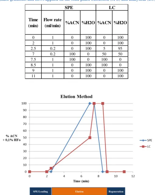
Figure 7. Mobile phase gradients applied on the SPE and LC columns. The analytical steps are represented under the time axis.
Figure 8. Scheme of the manipulations applied to each series of samples tested in the experiment “Choice of an aqueous solvent for toxins extraction and of a precipitation mode (part 1/2)”.
Figure 9. Scheme of the manipulations applied to each series of samples tested in the experiment “Choice of an aqueous solvent for toxins extraction and of a precipitation mode (part 2/2)”.
Figure 10. Theoretical purification and concentration of the toxins when using ultrafiltration centrifuge filters.
Figure 11. Scheme of the manipulations applied to each sample tested in the experiment “Microfiltration with a syringe filter” aiming at the choice of a syringe filter.
Figure 12. Scheme of the manipulations applied to each sample tested in the experiment “Ultrafiltration with centrifuge filters” aiming at the choice of the purification centrifuge filters.
Figure 13. Scheme of the manipulations applied to each sample tested in the experiment “Ultrafiltration with centrifuge filters” aiming at the choice of the concentration centrifuge filters.
Figure 14. Scheme of the manipulations applied to each sample of the experiment “digestion” aiming at the choice of the digestion buffer.
Figure 15. Scheme of the manipulations applied to the series of the experiment “full method evaluation” aiming at the estimation of toxins losses in the meat protocol.
Figure 16. Chromatograms of the internal standards injected in 0.1% formic acid. Each signal is labelled by the internal standard and corresponding endogenous peptide name. The analytical steps are represented under the time axis.
Figure 17. Visual examinations in the testing of extraction solvent. This picture presents the states of the samples after the addition of the aqueous extraction solvent, eventual pH adjustments and vortexing.
Figure 18. Visual examinations in the testing of phase separation. This picture presents the states of the samples after extraction with an aqueous solvent, precipitation, addition of the organic extraction solvent, vortexing, and centrifugation.
Figure 19. Protein contents of the samples after toxins extraction and meat precipitation by solutions of 0.9%, 1.5% and 3% NaCl in pH4 acetate buffer, obtained by SDS-PAGE.
Figure 20. Left°: Protein contents of the samples after toxins extraction and meat
precipitation by solutions of 0.9%, 1.5% and 3% NaCl in pH4 acetate buffer, fat extraction by CH2Cl2 and centrifugation, obtained by SDS-PAGE.
Right°: Protein contents of the samples after toxins extraction and meat precipitation by solutions of 0.9%, 1.5% and 3% NaCl in pH4 acetate buffer, fat extraction by CH2Cl2, centrifugation and 0.1 µm syringe filtration, obtained by SDS-PAGE.
Figure 21. Protein contents of the samples after toxins extraction and meat precipitation by solutions of 0.9%, 1.5% and 3% NaCl in pH4 acetate buffer, fat extraction by CH2Cl2, centrifugation, 0.1 µm syringe filtration, and 50 kDa MWCO centrifuge filtration, obtained by SDS-PAGE.
Figure 22. Protein contents of the samples after toxins extraction and meat precipitation by solutions of 0.9%, 1.5% and 3% NaCl in pH4 acetate buffer, fat extraction by CH2Cl2,centrifugation, 0.1 µm syringe filtration, 50 kDa and 5 kDa MWCO centrifuge filtration, obtained by SDS-PAGE. Left°: 5 kDa MWCO filters filtrates. Right°: 5 kDa MWCO filters residues.
Figure 23. Visual examination of fat removal from the tested samples by two organic solvents (dichloromethane on the left and n-hexane on the right).
Figure 24. Protein contents of the samples after toxins extraction by PBS solution, meat precipitation by lowering the pH with pure acetic acid, fat extraction by CH2Cl2,centrifugation, syringe filtration, 50 kDa and 5 kDa MWCO centrifuge filtration, obtained by SDS-PAGE. Left°: Testing of the 0.2 µm PVDF filter. Right°: Testing of the 0.1 µm PTFE filter. “SP” are protein contents of samples extracted with the reference method (PBS solution and pH lowering by hand, with acetic acid), extracted with CH2Cl2 and centrifuged (3200 X g; 20 min) “0.2; 50;5” and “0.1; 50; 5” are the samples after the filtration steps.
Figure 25. Testing the specificity of ultra-filters by VIDAS SET2.
Figure 26. Estimation of the toxins losses throughout the meat protocol. Each graph represents the toxins recoveries for each series of samples (spiked at a key point of the meat protocol), according to one of the peptides followed transitions.
Figure 27. Comparison of the estimated toxins digestion yields for digestion in presence or absence of meat.
List of tables
Table 1. Factors affecting growth and enterotoxin production by Staphylococcus aureus (Hennekine et al., 2011).
Table 2. Major characteristics of staphylococcal enterotoxins (Le Loir et al., 2003).
Table 3. Percentage of amino acid identity in different staphylococcal enterotoxins (Le Loir et al., 2003).
Table 4. Genetic support of some staphylococcal toxins (Hennekinne et al., 2010).
Table 5. Causative agents of food-borne disease outbreaks recorded in France between 1999 and 2000. Frequencies of each type of agent are given in per cent (Le Loir et al., 2003).
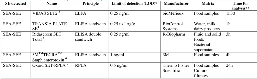
Table 6. Commercial kits based on immunoassays for detection of staphylococcal enterotoxins.
Table 7. Overview of methods for Staphylococcal enterotoxins (SEs) detection and quantification.
Table 8. Mobile phase gradients and flows applied on the solid phase extraction (SPE) and analytical (LC) columns.
Table 9. Identification of the aliquots withdrawn during the experiment “Choice of an aqueous solvent for toxins extraction and of a precipitation mode (part 1/2)”. The aliquots highlighted in orange were submitted to SDS-PAGE analysis.
Table 10. Identification of the aliquots withdrawn during the experiment “Choice of an aqueous solvent for toxins extraction and of a precipitation mode (part 2/2)”.
Table 11. Identification of the aliquots withdrawn during the experiment “Microfiltration with a syringe filter” aiming at the choice of syringe filter.
Table 12. Series tested in the experiment "full method evaluation" and their identification. Table 13. Pre selection of unique and stable peptides related to each Staphylococcal Enterotoxin SEA and SEB.
Table 14. List of the peptides related to Staphylococcal Enterotoxins SEA and SEB. The table lists for each peptide, its name, sequence, the different transitions followed by the MS/MS for the endogenous peptide and for the matching internal standard, and some MS/MS parameters (cone voltages and collision energies). The peptides highlighted in red were eliminated after injection on column of a pure mix.
Table 15. Repartition of the followed transitions into three MRM functions. For each peptide transition, start and end times of the functions are exposed, along with their retention and dwell times, cone voltages and collision energies.
Table 16. Experimental set-up of the micro-filter selection.
Table 17. VIDAS results on the testing of the different purification and concentration filters.
Table 18. Estimated digestion yields calculated for each followed transition. The yields are sorted by tested digestion buffer, in presence of the aqueous extraction solvent or not. Red values are the digestion yields obtained for all SEA related transitions. Green values are the best obtained values for the final transitions selection, in the presence of aqueous buffer.
Table 19. Estimation of the toxins recoveries obtained for each series of samples spiked with toxins at key steps of the meat protocol. The recoveries are sorted by selected transition.
List of abbreviations
AB All blue Standard Molecular Proteins
ACN Acetonitrile
ANSES French Agency for Food, Environmental and Occupational Health & Safety APC Antigen Presenting Cells
AQUA Absolute Quantification
C Cysteine
CNS Coagulase Negative Staphylococci CPS Coagulase Positive Staphylococci
DAD Diode Array Detection
DB Dual color Standard Molecular Proteins
DTT Dithiothreitol
E Glutamic acid
Eh Redox Potential
ELFA Enzyme-Linked Fluorescent Assay ELISA Enzyme-Linked Immunosorbent Assay ESI ElectroSpray Ionization
ES+ Positive Electrospray Ionization
EU-RL European Union - Reference Laboratory
FBDs Foodborne Diseases
G Glycine
HAc Acetic Acid
HFO Formic Acid
HPLC High Performance Liquid Chromatography
IAA Iodoacetamid Ig Immunoglobulin IS Internal Standard K Lysine LC Liquid Chromatography M Methionine
m/z Mass to charge ratio
MALDI Matrix-assisted laser desorption/ionization MHC Major Histocompatibility Complex
MRM Multi-residual monitoring
mRNA Messenger RNA (ribonucleic acid) MRSA Methicillin-Resistant S. aureus
MS Mass Spectrometry
MS/MS Tandem mass spectrometry
MW Molecular Weight
MWCO Molecular Weight Cut Off
N Asparagine
PBS Phosphate Buffer Saline (solution)
PEG Polyethylene glycol
PES Polyethersulfone
PSAQ Protein Standard Absolute Quantification
PT Pyrogenic exotoxins
PTFE Polytetrafluoroethylene PVDF Polyvinylidene Difluoride
Q Glutamine
QconCAT Quantification concatamer
QIT Quadrupole Ion Trap
QqQ Triple Quadripole
qTOF Quadripole Time of Flight
R Arginine
RPLA Reverse Passive Latex Agglutination RT-PCR Reverse Transcriptase PCR
RT-qPCR Real time PCR
SAgs Superantigens
SDS-PAGE Sodium dodecyl sulphate polyacrylamide gel electrophoresis
SE Staphylococcal Enterotoxin
se Gene coding for a staphylococcal enterotoxin SEA, SEB,… Staphylococcal Enterotoxin A, B,…
SEl Staphylococcal Enterotoxins-like toxin SETTECT Staphylococcal Enterotoxin Detection
SFP(O)s Staphylococcal Food Poisonning (Outbreaks) SPE Solid Phase Extraction
speA Streptococcal pyrogenic exotoxin A ssa Streptococcal superantigen
T Threonine
TCR T-cell receptor
Tris Trishydroxyméthylaminométhane TSS(T) Toxic Shock Syndrome (Toxin)
UPLC-MS/MS Ultra Performance Liquid Chromatography coupled to Tandem Mass Spectrometry
1 Introduction
Food poisoning caused by ingestion of Staphylococcus aureus enterotoxins is one of the most common foodborne diseases. Staphylococcus aureus is a well-studied, omnipresent bacterium which is not only found in the environment but is also part of the commensal mammalian flora. S. aureus produces enterotoxins which can cause gastro-enteritis, emesis or act as superantigen.
This work is a contribution to the development of a new method for the rapid detection and quantification of staphylococcal enterotoxins using online Solid Phase Extraction and Ultra Performance Liquid Chromatography coupled to Tandem Mass Spectrometry (SPE-UPLC-MS-MS).
S. aureus and its toxins have been thoroughly studied in the past decades and many
descriptions of this micro-organism can be found in the literature. However a rapid review of its characteristics will help identify the specificities of this bacterium, its toxins and why they cause so much concern.
Afterwards, a review of different existing strategies that were developed in order to characterize staphylococcal food poisoning outbreaks will be described, along with the benefits that would be brought by the development of a new method involving UPLC-MS/MS.
Finally, the objectives of the present work will be presented, followed by the methodologies employed to try and achieve them, the results, and the conclusions and perspectives that can be drawn from it.
2 Staphylococcus aureus
Staphylococcus aureus belongs to the Staphylococcus genus which is part of the
Staphylococcaceae family and accounts more than 50 species and subspecies. Species are classified in two groups: the coagulase positive Staphylococci (CPS), including S. aureus, S.
intermedius and S. delphini, and coagulase negative Staphylococci (CNS), such as S. epidermidis, S. haemolyticus and S. capitis. Some species may also present either a coagulase
positive or negative phenotype like S. hyicus (Gaebler Vasconcelos et al., 2010).
Biologically, S. aureus is described as a gram-positive, non-sporulated, catalase positive, facultative anaerobic, chemoorganotrophic and non-motile bacterium. Cells are spherical (cocci) and can be single, paired or form grape-like clusters (as staphylo means grape in greek). These organisms possess a respiratory and fermentative metabolism (Le Loir et al., 2003).
The growth of S. aureus is influenced by environmental factors such as water activity (aw), pH, redox potential, temperature… S. aureus is capable of growing in a temperature range from 7 to 48.5°C, with an optimum between 30 and 37°C. It is also very tolerant towards pH, as it can grow between pH 4.2 and 9.3 with an optimum between pH 7 and 7.5. Staphylococci are known for their resistance towards harsh environmental conditions, such as sodium chloride concentrations up to 10-15% (w/v), and have the ability to recover from non-physiological environments. This large tolerance exposed in Table1 makes S. aureus a ubiquitous organism that can be found in the air, dust, sewage, water, environmental surfaces but also animals and humans. Indeed S. aureus is part of the normal flora found on the skin and mucous membranes of mammals and birds (Hennekinne et al., 2011).
Staphylococcus aureus, just as many microbial pathogens, have great capabilities when it
comes to colonizing and infecting their hosts. These organisms, described as opportunistic, adhere very strongly to epithelial cells, colonize catheters or other devices and form biofilms. They easily reach the bloodstream and cause infections to high risk patients (intensive care unit patients, pre-term new-borns, cancer and transplanted patients…) (Gaebler Vasconcelos
et al., 2010).
Table 1. Factors affecting growth and enterotoxin production by Staphylococcus aureus (Hennekine et al., 2011).
Organism growth Staphylococcal enterotoxin production
Factor Optimum Range Optimum Range
Temperature (°C) 37 7-48 37-45 10-45
pH 6-7 4-10 7-8 4-9.6
Water activity (aw) 0.98 0.830.99* 0.98 0.850.99**
NaCl (%) (w/v) 0 0-20 0 0-10
Redox potential (Eh) > + 200 mV < -200 mV to > +200mV > + 200 mV < -100 mV to > +200mV
Atmosphere Aerobic Anaerobic-aerobic Aerobic (5-20%
dissolved O2)
Anaerobic-aerobic
*Aerobic (anaerobic 0.90 0.99) **Aerobic (anaerobic 0.92 0.99)
2.1 Staphylococcal enterotoxins
2.1.1 Description and Classification
S. aureus is a pathogen capable of producing various toxins. Staphylococci in general and S. aureus in particular are capable of producing toxins named pyrogenic toxic superantigens,
including the toxic-shock syndrome toxin (e.g. TSST-1) and staphylococcal enterotoxins (SEs). Many authors have reported the production of one or several enterotoxins by other
Staphylococcus species such as S. cohnii, S. xylosus, S. haemolyticus and S. epidermidis
(Ortega et al., 2010).
SEs are part of a large group of pyrogenic exotoxins (PT). This group includes SEs, two groups of Toxic Shock-Syndrome Toxins (TSSTs), exfoliatins A and B, and the streptococcal pyrogenic exotoxins. An interesting characteristic all those toxins share, besides their functional effects, is their common phylogenetic relationships, structure and sequence homology (see Balaban & Rasooly, 2000for a review).
SEs are remarkably stable to factors that easily destroy the bacteria such as heat treatment, freezing, drying, low pH and most proteolytic enzymes (pepsin, trypsin, chymotrypsin, rennin, papain…), except for the TSST-1 toxin. An interesting property of those toxins is that while inactivation through heat varies according to SE type, SE concentration, pH and matrix, some inactivation can be reversed under alkaline pH. Furthermore, heating can in many cases cause a loss of serologic activity but not of biological activity. This means that those toxins are undetectable with antibodies because they lost their serological recognition but remain active (Hennekinne et al., 2010).
2.1.2 Staphylococcal Enterotoxins and Staphylococcal Enterotoxin-like toxins
Staphylococcal Enterotoxins A and B (SEA and SEB) were the first described SEs. For a long time, only five SEs designated SEA to SEE were reported in the literature and because they all were discovered when some major food poisoning outbreaks occurred, all SEs were described as emetic substances, some being stronger than others (Ortega et al., 2010).
Since then, different SEs have been described, bringing the actual number to 23. Many of the new toxins were only predicted by genotyping, from the study of the classic enterotoxins (SEA-SEE) genes sequences or egc locus (Pocsfalvi et al., 2008). As they were studied, it was discovered that their common property is named superantigenic effect. This effect will be described later on. Because of this, two groups can be identified among those superantigens (SAgs). The toxins named “SE”, standing for Staphylococcal Enterotoxins, possess an emetic property while the “SEl” toxins (Staphylococcal Enterotoxins-like) either do not induce emesis or have not yet been studied for this property. (Lina et al., 2004).
Table 2. Major characteristics of staphylococcal enterotoxins (Le Loir et al., 2003).
SE type ORF length (bp) Precursor length (aa) Mature SE length (aa) Molecular mass (kDa) pI A 774 257 233 27,100 7.3 B 801 266 239 28,336 8.6 C1 801 266 239 27,531 8.6 C2 801 266 239 27,531 7.8 C3 801 266 239 27,563 8.1 C (bovine) NA NA NA 27,618 7.6 C (sheep) NA NA NA 27,517 7.6 C (goat) NA NA NA 27,600 7.0 D 777 258 228 26,360 7.4 E 774 257 230 26,425 7.0 G 777 258 233 27,043 5.7 H 726 241 218 25,210 ND I 729 242 218 24,928 ND J 806 268 245 28,565 8.65 K 729 242 219 25,539 6.5 L 723 240 215 24,593 8.66 M 722 239 217 24,842 6.24 N* 720 258 227 26,067 6.97 O* 783 260 232 26,777 6.55
*Named SEK and SEL in Jarraud et al., 2001, renamed SEN and SEO, respectively, in a correction note published in J.
Immunol. 166: 4260 (2001)
NA: not available ND: not determined
2.1.3 Nomenclature
So far, 23 serologically distinct staphylococcal SAgs have been described and include TSST-1, SEs A-E, G-J and the SEl K-R, U, U2 and V (Ortega et al., 2010). Because of the distinctions that exist between staphylococcal superantigens as regards their emetic activity, the International Nomenclature Committee for Staphylococcal Superantigens introduced in 2004 a new nomenclature for the naming of all these rapidly discovered or predicted proteins. Only staphylococcal superantigens causing emesis to primates after oral administration should be designated as staphylococcal enterotoxins. Other SAgs that either do not exhibit emetic properties or have not yet been tested should be designated as staphylococcal enterotoxin-like toxins (SEl) type X (Gaebler Vasconcelos et al., 2010). Letters from A to V simply identify toxins in the chronological order they were described, SElV being the last discovered toxin. SEF is the only exception as it was later renamed TSST-1(Lina et al., 2004)
2.1.4 Structure
SEs (and SEls) are secreted proteins with a mature length of approximately 220–240 amino acids and low-molecular weights ranging from 24 to 30 kDa, and are soluble in water and saline solutions (Sospedra et al., 2013). Their major characteristics are listed in Table 2. Their sequences are rich in lysine, aspartic acid, glutamic acid and tyrosine residues. Crystallographic studies show similar three-dimensional SEs structures. The common structural description of SEs is a “small N-terminal α-helix connected to a β-folded sheet known as domain B or oligosaccharide-binding fold (O/B). Such O/B fold is connected to a wall of β-folded sheets by a central diagonal α-helix forming domain A” (Gaebler Vasconcelos et al., 2010, pg.36). Several models are exposed in Figure 1.
Slight differences may be observed from one toxin to another, amongst which the cysteine fold is probably the most notable. As many SEs contain a cysteine loop it is believed that this structure is involved in their emetic activity. Interestingly, such fold has also been observed in streptococcal pyrogenic exotoxin A (speA). This added to the observation that high sequence homologies exist between SEs, the streptococcal superantigen (ssa) and speA supports the hypothesis of Staphylococcus aureus and Streptococcus pyogenes toxins to share an ancestral toxin gene or that horizontal gene transfer took place in the evolution of these species (Gaebler Vasconcelos et al., 2010).
Finally, the high similarities between SEs sequences, which are exposed in Table 3, allow a classification into five groups according to their homologies. A representation of this classification is schematised in Figure2. Note that only 15% of the amino acid residues are completely conserved throughout all SEs groups (Le Loir et al., 2003; Ortega et al., 2010).
Table 3. Percentage of amino acid identity in different staphylococcal enterotoxins (Le Loir et al., 2003).
Toxin SEA SEB SEC1 SED SEE SEG SEH SEI SEJ SEM SEN SEO
SEA 100 33 30 50 83 27 37 39 64 35 39 37 SEB 100 68 35 32 43 33 31 33 29 32 36 SEC1 100 31 29 41 27 26 30 26 29 33 SED 100 52 27 35 33 51 41 38 39 SEE 100 27 35 35 63 37 39 37 SEG 100 34 28 29 28 31 30 SHE 100 33 35 38 34 31 SEI 100 34 31 31 57 SEJ 100 38 42 33 SEM 100 28 31 SEN 100 42 SEO 100
Figure 2. Dendrogram of staphylococcal SAgs (in Popoff’s Comprehensive Sourcebook of Bacterial Protein Toxins, pg 832).
2.1.5 Genes
SAgs are accessory proteins meaning they are not necessary for growth and multiplication. Some of their corresponding genes are located on accessory movable genetic elements. A non-exhaustive list of SE genes and their supports is illustrated in Table 4 (Le Loir et al., 2003). Because enterotoxin genes are located on movable elements, there is an uneven distribution of SEs between S. aureus strains. About 77% of S. aureus strains are positive for one or several enterotoxin genes, and it has been observed that some genes tend to co-exist (i.e. sei and seg or sej and sed). Besides genes horizontal transfer takes place between strains, which constitutes an important part of their pathogenicity evolution (Ortega et al., 2010).
Table 4. Genetic support of some staphylococcal toxins (Hennekinne et al., 2010).
Toxin type Genetic location
SEA Prophage
SEB Chromosome, plasmid, pathogenicity island
SEC 1-2-3 Plasmid
SED Plasmid (pIB485)
SEE Prophage
SEG Enterotoxin gene cluster (egc), chromosome
SEH Transposon
SEI egc, chromosome
SElJ Plasmid (pIB485)
SEK Pathogenicity island
SElL Pathogenicity island
SElM egc, chromosome
SElN egc, chromosome
SElO egc, chromosome
SElP Prophage (Sa3n)
SElQ Pathogenicity island
SER Plasmid (pIB485)
SES Plasmid (pIB485)
SET Plasmid (pIB485)
SElU egc, chromosome
SElU2 egc, chromosome
SElV egc, chromosome
2.1.6 Clinical Manifestations
S. aureus is considered a major public health issue because it can cause many infections
that range from mild to severe or fatal, either on humans or animals. It is responsible for infecting superficial lesions (abscesses, wound infections…), causing systemic infections (septicaemia, endocarditis, and osteomyelitis) and toxin-mediated diseases like the Toxic Shock Syndrome, Kawasaki’s Disease and staphylococcal food poisoning. Concern in nosocomial bacteraemia has recently increased the interest in Staphylococcus species, known for causing community- and hospital-acquired infections. There are multiple clinical manifestations because each strain produces a combination of toxins causing virulence and invasiveness (Pocsfalvi et al., 2008; Ortega et al., 2010). Besides, the increase of antibiotic resistance has led to the apparition of methicillin-resistant S. aureus (MRSA) strains considered by the American National Nosocomial Infections Surveillance System as being among the most common causes of healthcare-associated infections (Normanno et al., 2007; Kuehnert et al, 2010).
a) Food poisoning and SEs emetic effect
S. aureus is among the leading causes of food-borne diseases (FBDs) worldwide for two
reasons. First S. aureus is often present in food contaminated by humans. As an estimated 30-50% of human population carries S. aureus, mainly in the nasopharynx or on the hands where the organisms can persist without causing any damage, simple coughing, sneezing or food handling combined with poor hygiene may cause contamination, especially when occurring after heat treatment. When it comes to raw foods, contamination from animal origins is more frequent (i.e. mastitis). The second reason is that S. aureus is capable of growing and producing toxins in a wide variety of foods (milk and milk-transformed products, meat, salads, cooked meals…) (Ortega et al., 2010).
FBDs are defined by the World Health Organization as “diseases of infectious or toxic nature caused by or thought to be caused by the consumption of food or water”. FBDs include food-borne infections, which are caused by pathogens that contaminate foods, and food-borne poisoning, caused by substances present in food (Le Loir et al., 2003).
Staphylococcal food poisoning (SFP) belongs to food-borne poisoning as it is caused by the ingestion, through food, of preformed Staphylococcal Enterotoxins (Gaebler Vasconcelos
et al., 2010). The exact implication of S. aureus in foodborne diseases (FBDs) is difficult to
assess because available data is incomplete and thus not very representative (Ortega et al., 2010; Lecture Analyse de la Qualité, Pr. M. Sindic, ULg GxABT). Table 5 gives an insight of its implication in FBDs.
The most commonly observed symptoms of SFPs are abdominal cramps, nausea, vomiting and diarrhoea. Those symptoms can appear 1-4 hours after eating and usually disappear after 24-48 hours (Dupin H., 1992).
Historical association between SEs and food-borne poisoning is the reason why SEs were originally described as emetic substances. Strains isolated from foods involved in SFPs produce mainly SEA and to a lesser extent SEB, SEC and SED (Dupuis et al., 2008). In France, SEA is involved in 65% of SFPOs and SEB in 20% (Dupin H., 1992).
The infective dose required to induce SFP to humans remains uncertain. The first reason is that the infective dose depends on the patient’s sensitivity (Le Loir et al., 2003). The second is that bioassays, consisting feeding a suspected food to a monkey, have shown that the amount triggering the food-poisoning symptoms is lower for humans than it is for monkeys. For instance, the 50% effective dose of SEA is 1 µg in humans and 5 µg in monkeys (Ikeda et
al., 2005), but it has also been reported that the ingestion of doses as low as 20–100 ng of SEs
are susceptible to cause food-poisoning (Rodriguez-Caturla et al., 2012). Finally, the knowledge on infective doses is limited by the sensitivity of the methods used to detect and quantify the enterotoxins. Therefore, it is important to lower the detection and quantification limits of the methods in order to establish properly this infective dose.
The physiopathology of SFPs and the emetic function of enterotoxins are very partially known and still the object of many hypothesis and researches (see Ortega et al., 2010; Gaebler Vasconcelos et al., 2010 for recent researches). A strong hypothesis is that SEs stimulate the emetic centre and the gut transit because they affect the vague nerve and the intestinal epithelium. More precisely, it is believed that the enterotoxins increase the permeability of the intestinal mucosal cells to chloride ions. This activation of the membrane pores leads to secretory diarrhoea.
At first, the cysteine loop, common to the first SEs, was suspected to be implied in the emesis mechanism but the fact that some SEs (like SEI and SEK) lack that specific structure while presenting the emetic property, even though it is significantly weaker than for other SEs, questions this hypothesis (Ortega et al., 2010).
Table 5. Causative agents of food-borne disease outbreaks recorded in France between 1999 and 2000. Frequencies of each type of agent are given in per cent (Le Loir et al., 2003).
Causative agents Outbreaks
(N=530) Cases (N=6451) Hospitalizations (N=872) Death (N=7)
Salmonella sp. (Enteritidis, Typhimurium,
Heidelberg, and other serotypes)
63.8 47.7 16.8 100
Staphylococcus aureus 16 25.6 17.1 0
Clostridium perfrigens 5.1 12.3 0.5 0
Bacillus cereus 2.8 3.7 10.0 0
Histamine 3.8 1.4 30.4 0
Other pathogens (Campylobacter sp.,
Dinophysis, Clostridium botulinum, Shigella
sp., Calcivirus, HAV, Vibrio sp., E. coli, etc.)
8.5 9.2 7.6 0
b) Superantigenic effect
Historically, SEs were only described as emetic substances. It is only later, with the extensive study of their properties, that SEs have been described as SAgs.
Superantigens are defined as “microbial antigens with the common capacity to activate and induce uncontrolled mitosis on T-lymphocytes presenting any specific variable region” (Gaebler Vasconcelos et al., 2010, pg 34).
This term was suggested after a series of independent studies demonstrated that SEs and streptococcal pyrogenic exotoxins share two properties: (i) the ability to directly bind the class II Major Histocompatibility Complex (MHC) of the antigen presenting cells (APC) and (ii) the ability to bind to the T-cell receptors (TCR) β-chain in another way than the usual peptide recognition mechanism (Ortega et al., 2010). The mechanisms involved in the interactions between SAgs, TCR and APC have been characterized (Balaban & Rasooly, 2000).
In an usual immune response, as schematised on Figure 3a, the antigens are internalized and processed by APC then they are presented to TCR in the form of peptides bound to molecules of class-I and class-II MHC which themselves are proteins bound to the membranes of the APC. The binding between TCR and usual antigens requires the recognition of all five variable elements of TCR (Vβ, Dβ, Jβ, Vα and Jα). This recognition of the antigen is a primary step in the cellular immune response and makes the specificity of the immune response.
On the contrary, superantigens affect the immune system by binding directly with the TCR and the MHC of antigen-presenting cells, as exposed in Figure 3b. This binding only requires the recognition of specific Vβ chains of the TCR, which induces a non-specific polyclonal immune response as T-cells are activated at orders of magnitude higher than the antigen-specific activation. Indeed, SAgs can stimulate about 20% of all T-cells, against 0.01% for the conventional antigens.
This nonspecific and exaggerated activation results in a proliferation of T-cells and a massive secretion of interleukines, various cytokines and lymphokines. These compounds are the ones responsible for severe outcomes of superantigens. They act as capillary vasodilators, leading to fever, hypotension, systemic toxicity (shock), which can all cause death (Le Loir et
al., 2003).
Figure 3. Model for the structure of the complex MHC class II and T-cell receptor. (A) Conventional antigen. (B) Superantigen. The model shows the processed antigen peptide presented by MHC class II which attracts specific T-cell bearing antigen specific T-T-cell receptor (TCR) variable chain. In contrast, superantigens bind directly to the outside of the MHC molecule and cross-link it to variable chain, which initiates non-specific activation of the cell (Balaban & Rasooly, 2000).
c) Emesis and superantigenicity: two functions
A precision about superantigens in general and staphylococcal toxins in particular is to be highlighted. SEs and SEls do not present the same properties. All of them share superantigenic activity but only a few are emetic. A toxin is classified as enterotoxin only due to its ability to cause emesis when orally administrated to monkeys. All superantigens are thus not emetic (or one might say all superantigens are not enterotoxins). SAgs are actually a family of several groups of proteins, SEs and SEls being part of them.
The interconnection of superantigenic and emetic activities is uncertain. The activities are located on different domains of the proteins but in most cases a correlation can be observed as a decreased superantigenicity often results in decreased emetic activity (Ortega et al., 2010).
d) TSST-1
TSST-1 is secreted by some S. aureus strains. It is not emetic, probably because of its sensibility to proteolytic enzymes, but is capable of crossing mucosal barriers. This toxin is responsible for the Toxic Shock Syndrome (TSS) which affects the whole organic system and causes fever, hypotension, rash, vomiting, circulatory failure, organ failure… If not treated properly, a fatal shock may develop in less than 24h. This toxin’s production is most known to happen to young women during their menstruation but also to post-op patients or in association with other infections (Ortega et al., 2010).
TSST-1 was first named SEF because it shows structural and functional similarities with other SEs. It was renamed after it was proved to show a different behaviour than other enterotoxins and to share less gene sequence homology with genes coding for SEs than these toxins usually do with one another (Gaebler Vasconcelos et al., 2010). To avoid any further confusion, the SEF appellation was chosen not to be attributed again to name an enterotoxin (Lina et al., 2004).
For all the reasons previously mentioned, staphylococcal enterotoxins constitute a significant threat to Public Health. The American Center for Disease Control has even registered the staphylococcal enterotoxin B (SEB) as a potential warfare contaminant of food and water supplies (Brun et al., 2007). A rapid overview of the existing strategies used to detect S. aureus and its toxins (or to confirm SFP) will show the need for the development of more efficient, reliable, fast and most important allowing quantification methods. It will show the interest of this work.
2.2 Investigating Staphylococcal Food Poisoning Outbreaks
As previously mentioned, SEs are responsible for a large part of FBDs. Because of this implication, most detection tools were developed in the purpose of confirming and identifying SFPOs and find applications in the field of alimentation. However, as will be developed later on, a tool allowing the accurate identification and quantification of staphylococcal toxins could find applications in other fields, such as health.
All existing tools developed to investigate SFPOs are based on the research of either the microorganism responsible for the outbreak, the gene coding for the toxin, the intermediate to the toxin production (the translated messenger RNA (mRNA)) or the toxin itself.
Their general principles are exposed along with advantages and drawbacks.
2.2.1 Bioassays
The first ways of detecting food contaminations were by biological methods. Bioassays are based on the capacity of a suspected food to induce food poisoning symptoms when fed to a monkey. This type of method is not only outdated because it faces serious ethical issues, but also because it does not provide any indication on the nature and severity of the contamination. Besides, as mentioned in the clinical manifestation, section 2.1.6, symptoms of SFP only appear with bioassays at higher doses than those involved in human food poisoning (Hennekine et al., 2010).
2.2.2 Molecular tools
Molecular tools usually involve Polymerase Chain Reaction (PCR), used for the detection of enterotoxin genes and the characterization of S. aureus strains involved in SFPOs, or Reverse Transcriptase PCR (RT-PCR), an intermediate to the detection of se genes and the actual detection of the SEs because it can prove gene expression by detecting mRNA sequences, responsible for the toxin’s production (see reviews Hennekine et al., 2011 and Gaebler Vasconcelos et al., 2010 for details).
However, methods involving molecular tools cannot be used to confirm SFPOs (i.e. to link a SE to a food source because SFP can only be confirmed if the presence in food of one or several SEs has been demonstrated). Indeed, methods involving PCR can only detect the presence of genes encoding for SEs, which does not mean the enterotoxin has been secreted.
Duquenne (2010) developed an alternative method based on real time RT-PCR (RT-qPCR) to try and estimate the corresponding level of transcript toxins, but it still does not prove the SEs presence.
PCR and RT-PCR can however bring valuable information. For this reason the European Union - Reference Laboratory (EU-RL) for CPS decided in 2005 to use PCR procedure in an integrated approach to improve SFPO characterization (Hennekine et al., 2010).
Because molecular tools cannot be used to link a SE to a food source and because SFP can only be confirmed if the presence in food of one or several SEs has been demonstrated, alternative methods for detecting the toxins, the real causes of SFPs, have to be used.
2.2.3 Immunological assays
Immunological tools constitute the official method for detection of SEs in food. These assays rely on the recognition of an enterotoxin epitope by a specific antibody, either monoclonal or polyclonal (Dupuis et al., 2008). A wide range of commercial kits have been developed for the simultaneous detection of several enterotoxins (SEA to SEE). This development was made possible by the improvement of techniques that helped obtaining pure toxins and producing SEs antibodies (Gaebler Vasconcelos et al., 2010; Sospedra et al., 2013).
Commercial methods, referenced in Table 6, use various principles such as the enzyme-linked immunosorbent assay (ELISA), the enzyme-enzyme-linked fluorescent assay (ELFA) or reverse passive latex agglutination (RPLA). ELISA and ELFA couple antibody-antigen recognition to an enzymatic reaction that liberates a colour compound. The visible compound is a signal measured by spectroscopy. In RPLA tests, the cross-linking of the latex particles by the specific antigen/antibody reaction results in a visible latex agglutination (Gaebler Vasconcelos et al., 2010; Sospedra et al., 2013; Thermoscientific).
The double antibody sandwich ELISA test is the most frequent because it is available both for screening and specific identification. Indeed commercial tests are divided in two categories. The tests that detect SEA to SEE as a whole and informs on the total SEs (screening), and the ones that can differentiate six or seven types of SEs (SEA, SEB, SEC1, SEC2, SEC3, SED and/or SEE), or serotyping tests. However, none of those kits yet allows detection of SEG to SElV (Hennekinne et al, 2009).
Immunoassays all present the advantage of being simple, rapid and highly sensitive. Several drawbacks and limitations must however be mentioned.
Manipulations frequently require time-consuming incubation periods.
The development of a new specific antibody is expensive (~1 million dollar) and takes over a year.
Not all toxins can yet be detected due to lack of available antibodies.
High sequence and structural homologies exist between SEs and only a few specific antibodies are available. Therefore the techniques are less sensitive to small variations and not suited to the identification of SEs presenting antigenic similarities.
Matrices like foods are complex and may lead to false positives when the antibody reacts with unrelated antigens or endogenous peroxides. For example, the Immunoglobulin G (IgG)-binding staphylococcal protein A is co-secreted in food with SE and is well-known for interfering with assays (Dupuis et al., 2008).
If the enterotoxin epitope is damaged, for instance by heating, the enterotoxin is serologically but not biologically inactivated and the immunoassay leads to a false negative. Also, heat-treated enterotoxins may aggregate, reducing their reactivity with antibodies.
The main drawback of methods based on specific monoclonal or polyclonal antibodies remains its high cost, preventing it from being used in routine tests. Therefore, immunoassays are reserved to serious epidemiology issues (Gaebler Vasconcelos et al., 2010; Dupuis et al., 2008; Sospedra et al., 2013).
For information, a list of applications and studies based on immuno-assays and using some of the kits referenced in Table 6 is available in a review written by Sospedra et al. (2013).
So far, the official method for the detection of staphylococcal enterotoxins types SEA to SEE in all types of food matrices (milk and milk products and other food matrices) is based on the use of the VIDAS SET2 and the RIDASCREEN SET TOTAL commercial kits, as mentioned by the “European Screening Method of the European Union – Reference Laboratory for Coagulase Positive Staphylococci, including Staphylococcus aureus (EU-RL for CPS)”, in the Version of September 5th, 2010 (ANSES).
Table 6. Commercial kits based on immunoassays for detection of staphylococcal enterotoxins.
SE detected Name Principle Limit of detection (LOD)* Manufacturer Matrix Time for
analysis**
SEA-SEE VIDAS SET2 3 ELFA 0.25 ng/ml bioMérieux Food samples 1h30
SEA-SEE TRANSIA PLATE
SE4
ELISA sandwich 0.25 to 1 ng/g BioControl Systems
Water, milk, dairy products
1h SEA-SEE Ridascreen SET
Total 5
ELISA double sandwich
0.25 ng/ml R-Biopharm Fluid and solid
foods Bacterial supernatants 3h SEA-SEE 3MTMTECRATM Staph enterotoxin 6
ELISA sandwich 1 ng/ml 3M Food samples 4h
SEA-SED Oxoid SET-RPLA 7 RPLA 0.5 ng/ml Thermo Fisher
Scientific
Food samples Culture filtrates
24h
*Limits of detection indicated in kits manual
**Time of extraction is not included in time for analysis
Note: A commercial kit named TSST-RPLA has also been developed for the specific detection of TSST (Thermo scientific).
3Biomérieux 4 Biocontrol 5R-Biopharm 63M 7Thermo scientific
2.2.4 Detection, identification and quantification of SEs
The methods previously exposed are not suitable for preventing outbreaks or to properly identify them. Indeed, they are too laborious, too long or too expensive to be applied in preventive analysis and it is usually after several FBD cases have been reported that research is done to identify the responsible foodstuff and the associated pathogen or toxin. Besides, none of those methods allow unambiguous identification, let along quantification, as molecular tools are inefficient at proving the existence of the toxins in foods and immunoassays are not specific enough, not suitable for quantification and more importantly limited in the range of toxins they can identify.
Recently, researchers have started to explore proteomics approaches in an attempt to develop fast and specific methods to detect and quantify SEs. The main matrixes investigated are foods, as SEs are mainly investigated for food-poisoning.
The development of mass spectrometers and more specifically of two soft ionization methods has revolutionized the analysis of biomolecules. Electrospray ionization (ESI) and matrix-assisted laser desorption/ionization (MALDI) allowed the development of new analysis strategies (Hennekine et al., 2010).
Mass spectrometry (MS) is a very sensitive technique that provides specific and rapid results. New detection and quantification strategies were thus developed around MS for protein and peptide mixtures analysis. Besides, MS-based methods allow multiplex analyses (Brun et al., 2009), which is a real advantage for the confirmation and characterization of SFPOs, as several SEs may be involved.
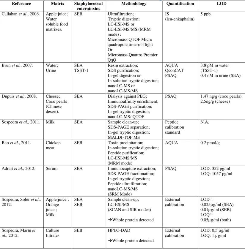
Several detection and quantification strategies have been explored, all articulated around the use of mass spectrometry. There are several variations but also common points to the recently developed methods. In food analysis, extraction and purification steps are critical for SEs detection and quantification and must be carefully optimized. Those parts of the protocols tend to differ, depending on the food, for instance. On the other hand, many of the developed strategies lead to the proteolysis (or digestion) of the extracted enterotoxins into specific peptides, even though some strategies also aim the detection of whole proteins. Enzymatic digestion is about reducing the target protein into smaller peptides, yielding to a large number of peptides in solution. Those peptides can be separated by Liquid Chromatography (LC). Afterwards, their molecular mass is determined by ElectroSpray Ionization Mass Spectrometry (ESI-MS) or Matrix-Assisted Laser Desorption/Ionization Time Of Flight Spectrometry (MALDI-TOF).The most widely used proteolytic enzyme for protein cleavage is trypsin because of its high specificity and ability to digest insoluble or adsorbed protein (Bao et al., 2011). Finally, because foods often result into strong matrix effects, several quantification strategies using internal calibration, with or without isotope-labelled internal standards were developed.
The following section will be dedicated to the three proteins quantification strategies that use isotope-labelled internal standards. Next, a rapid overview of protocols using some of these quantification strategies along with the extraction, purification, concentration, and eventual LC and Mass spectrometry MS tools employed for SEs detection and quantification will be exposed.
a) Quantification methods – Internal Standards
Nowadays, mass spectrometry permits the simultaneous characterization of several proteins in a very specific and rapid way. The actual challenge lays in the accurate quantification of these proteins.
The internal standard calibration strategy is a very useful quantification tool for analysing samples with strong matrix effects, like foods. More specifically isotope dilution strategies, which are part of the large family of internal calibration, have been developed to provide correct identification and absolute quantification. Three kinds of quantification strategies using this isotope dilution principle are exposed in the literature°: Protein Standard Absolute Quantification (PSAQ), using intact labelled proteins, Absolute quantification (AQUA) peptides, using chemically synthesised labelled peptides or Quantification concatamer (QconCAT), using concatamers of tryptic labelled peptides in an artificial protein. All their principles are schematised in Figure 4 (Brun et al., 2009).
AQUA peptides
The AQUA peptide strategy uses chemically synthesized isotope-labelled peptides. Those peptides are spiked in known quantities into the samples either before the digestion step or just before MS analysis.
The AQUA method is fast and easy to use because a large range of AQUA peptides are commercially available and affordable compared to other isotope standards.
However, several drawbacks must be mentioned.
First, this strategy but does not take any of the sample preparation step into account. If those standards were injected at early stages of the protocol, all extraction, purification and concentration steps could significantly decrease the AQUA peptides recovery, and consequently the quantification accuracy. Besides, as those standards are peptides, they are not submitted to digestion and the efficiency of this step is not taken into account either. So if this approach is chosen, special attention must be taken in evaluating the digestion yield and the recovery of the pre-fractionation steps.
Second, the peptides must be chosen carefully with regards to their sequences because some peptides are more stable than others and some chemical synthesis limits exist. For example, peptides shorter than 15 amino acids are preferred and reactive residues such as tryptophane, methionine, cysteine… or some sequence patterns (N-terminal glutamine, aspartate-glycine…) should be avoided (Polyquant). Also their sequences can affect their conservation as peptides can have a tendency to adhere to certain surfaces, resulting in quantification underestimations. Storage at -80°C and careful over-time monitoring of the quality and concentration are recommended (Brun et al., 2009).
Finally, in case of multiplex analysis, the experiment cost can rapidly increase because very pure AQUA peptides are needed. This is the reason why often, only one carefully selected highly specific of the target protein AQUA peptide is used for quantification.
Studies where AQUA peptides have been successfully employed are listed in Table 7.
Protein Standard Absolute Quantification (PSAQ)
The ideal internal standard for the quantification of a protein is its corresponding full-length isotope-labelled in vitro-synthesized protein.
Here again, this quantification tool presents advantages and drawbacks.
First, those ideal standards can be spiked into the samples at very early stages of the analytical process, as shown on the scheme Figure 4 even if the sample is to undergo intensive pre-treatment, which is often required considering the complexity of food matrices (Brun et al., 2009; Adrait et al., 2011). Because PSAQ standards display the same characteristics and behaviour as the target proteins they will account for possible protein losses and avoid differences in digestion between internal standard and target protein (Dupuis
et al., 2008).
Second, using a full-length marked protein instead of a peptide allows a larger coverage of the protein sequence, increasing specificity and robustness.
A third characteristic of PSAQ standards is that being artificially synthesised, those proteins do not carry the post-translational modifications of the targets. The generation of such proteins is possible but challenging, time consuming and expensive (Polyquant). It is therefore only feasible for a very small number of proteins. Intact protein standards, lacking post-translational modifications, have however been used successfully in absolute quantification experiments. Some are referenced in Table 7.
Quantification concatamer (QconCAT)
QconCAT concatamers are chimerical proteins made of different marked peptides whose sequences originate from different proteins. QconCAT proteins constitute an interesting intermediate to both previously exposed quantification tools (AQUA peptides and PSAQ proteins).
QconCAT concatamers were specifically developed for multiplex absolute quantification of proteins as up to 100 peptides can be included in a structure. Because they are proteins, those concatamers can be spiked into the samples just before the protein digestion step, as exposed in Figure 4. During digestion the isotope-labelled peptides are released by the digestion and serve in MS analysis as standards for quantification (Brun et al., 2009).
But because they are artificial proteins without a three-dimensional structure, they differ in their biological characteristics and behaviour and are digested at higher rates than folded proteins. Therefore, QconCAT proteins cannot be spiked at early stages of the protocol and their different sensitivity to digestion must be estimated, and maybe compensated by surrounding each proteotypic peptide of the concatamer with its native flanking sequences (Kito et al., 2007).
The main advantage of the QconCAT methodology really is that it facilitates multiplex protein quantification. With regards to the cost of those standards, it is probably more economical to use AQUA peptides if only a few proteins are to be quantified. But if a high number of proteins are targeted, using QconCATs is the economical solution (Polyquant).
Figure 4. Isotope dilution strategies for MS-based absolute quantification of proteins. Three types of internal standards are available°:
(1) The PSAQ protein standard (“Protein Standard Absolute Quantification”) is an isotope-labelled version of the target protein which is directly added into the sample;
(2) The QconCAT (“Quantification concatamer”) standard is a chimerical protein containing one/several isotope-labelled proteotypic peptide(s) of the targeted protein. This concatamer is added before the digestion step so that the standard peptide(s) is/are released in the samples;
(3) The AQUA peptides (“Absolute Quantification”) are synthetic isotope-labelled copies of the target proteotypic peptides. They are generally added to the samples before LC-MS analysis (Brun et al., 2009).