HAL Id: inserm-00719461
https://www.hal.inserm.fr/inserm-00719461
Submitted on 19 Jul 2012HAL is a multi-disciplinary open access archive for the deposit and dissemination of sci-entific research documents, whether they are pub-lished or not. The documents may come from teaching and research institutions in France or abroad, or from public or private research centers.
L’archive ouverte pluridisciplinaire HAL, est destinée au dépôt et à la diffusion de documents scientifiques de niveau recherche, publiés ou non, émanant des établissements d’enseignement et de recherche français ou étrangers, des laboratoires publics ou privés.
Anti-RANKL therapy for bone tumours: Basic,
pre-clinical and clinical evidences
Dominique Heymann
To cite this version:
Dominique Heymann. Anti-RANKL therapy for bone tumours: Basic, pre-clinical and clini-cal evidences. Journal of Bone Oncology, Amsterdam : Elsevier, c2012-, 2012, 1 (1), pp.2-11. �10.1016/j.jbo.2012.03.001�. �inserm-00719461�
Anti-RANKL therapy for bone tumours: basic, pre-clinical and clinical evidences
HEYMANN Dominique a,b,c
a
INSERM, UMR 957, Nantes F-44035, France b
Université de Nantes, Nantes atlantique universités, Laboratoire de Physiopathologie de la Résorption Osseuse et Thérapie des Tumeurs Osseuses Primitives, Nantes F-44035, France c
CHU de Nantes, Nantes F-44035, France
Reprints to :
Prof. Dominique Heymann INSERM UMR-S 957
Pathophysiology of Bone Resorption and Therapy of Primary Bone Tumors
Faculty of Medicine, 1 rue Gaston Veil 44035 Nantes cedex, France
Phone : 33 (0) 272 641 132 ; Fax : 33 (0) 240 412 860
Abstract
Bone remodelling is related to coordinated phases of bone resorption and bone apposition allowing the maintenance of bone integrity, the phosphocalcic homeostasis all along the life and consequently the bone adaptation to mechanical constraints or/and to endocrine fluctuations. Unfortunately, bone is a frequent site of tumour development originated from bone cell lineages (primary bone tumours: bone sarcomas) or from non osseous origins (bone metastases: carcinomas). These tumour cells disrupt the balance between osteoblast and osteoclast activities resulting in a disturbed bone remodelling weakening the bone tissue, in a strongly altered bone microenvironment and consequently facilitating the tumour growth. At the early stage of tumour development, osteoclast differentiation and recruitment of mature osteoclasts are strongly activated resulting in a strong bone matrix degradation and release of numerous growth factors initially stored into this organic/calcified matrix. In turn these soluble factors stimulate the proliferation of tumour cells and exacerbate their migration and their ability to initiate metastases. Because Receptor Activator of NFκB Ligand (RANKL) is absolutely required for in vivo osteoclastogenesis, its role in the bone tumour growth has been immediately pointed out and has consequently allowed the development of new targeted therapies of these malignant diseases. The present review summarizes the role of RANKL in the bone tumour microenvironment, the most recent pre-clinical and clinical evidences of its targeting in bone bone metastases and bone sarcomas. The following paragraphs position RANKL targeted therapy among the other anti-resorptive therapies available and underline the future directions which are currently under investigations.
Key words: bone cancers, bone metastases, bone sarcomas, osteoclasts, RANKL, bone remodeling
Introduction
Bone is a very dynamic tissue resulting from coordinated phases of formation and resorption called bone remodelling. Additionally to its role in phosphocalcic homeostasis, bone remodelling process is necessary for bone growth, for renewal of cellular and extracellular matrix components to adapt bone organisation to the various biological and mechanical constraints [1-3]. Bone remodelling then leads to the renewal of around 10% of total bone mass each year in human. This metabolic process is based on a molecular crosstalk occurring between osteoblasts involved in bone apposition and osteoclasts specialized in bone resorption. Osteoclasts are multinucleated cells and originated from hematopoietic stem cells [4-6] whereas osteoblasts derived from bone marrow mesenchymal stem cells [3, 7, 8]. Osteoblasts control osteoclast differentiation and activation through a very complex network of soluble factors which act in combination with various hormones produced by endocrine system even if contacts between both cell types also strongly contribute to full activation of osteoclasts [9, 10]. Reciprocity between osteoblasts and osteoclasts can be observed as shown by bidirectional signalling limiting osteoclast activities and stimulating osteoblast differentiation [11].
Bone remodelling can be dysregulated by oncologic events originated from bone cells (primary bone tumors: osteosarcoma, chondrosarcoma, Ewing’s sarcoma, etc) or from non osseous origins (bone metastases). Large series revealed that around 0.2% of all neoplasms are bone sarcomas and two new primary bone tumours arise per 100,000 persons a year [12]. Bone tissue is then the most frequent site of their first relapse and consequently, the incidence of bone metastases is relatively high and is dependent of the cancer cell types (i.e. in 70-80% of patients with breast or prostate cancer, in 40% of patients with lung metastases or with kidney cancer). Bone metastases are frequently associated with numerous clinical complications named skeletal-related events (SREs) and have a strong deleterious impact on the quality of life. SREs include pathological fractures or spinal cord compression and
exacerbated bone pains. All bone tumours disrupt the equilibrium between bone apposition and bone resorption leading on the first stop of the tumour development to an osteolytic process followed or not by bone forming lesions. Soluble mediators stored initially into the bone matrix contribute in turn to stimulate the tumor growth and to maintain the vicious cycle between bone and tumour cells [13]. The lost of equilibrium between bone formation and degradation combined with an osteomimetism behavior of cancer cells (cancer cells acquire bone-like properties) explains the diversity of histological features (osteolytic or bone forming tumours) of bone metastases [14]. Additionally, the modulation of bone micro-environment (“niche” concept) by cancer cells is benefit for their proliferation and also contributes to the drug resistance patterns [15].
In the late 1990’s, two research groups in Japan and in USA have identified a truncated TNF receptor-like molecule (named OPG for osteoprotegerin, TNFRSF11B) inducing marked osteopetrosis phenotype when overexpressed in transgenic mice [16, 17]. One year later, RANKL (Receptor Activator of Nuclear Factor kB Ligand or TNFSF11) has been identified as a ligand for OPG [18, 19]. In many years, OPG/RANKL couple is became the principal system regulating osteoclastogenesis and bone resorption and has impressively stimulated the development of OPG/RANKL targeting agents for the treatment of osteolytic disorders in oncologic contexts or not competing with bisphosphonates, a well admitted drug class for the treatment of bone loss [13, 19-23].
In all bone cancers, a strong relationship between tumour cells and bone micro-environment has been then clearly established, facilitating the tumor development and/or the metastatic process. These specific communication pathways have strongly stimulated the research and development programs to design new drugs to treat oncologic bone diseases and have led specifically to the development of therapies targeting RANKL. The present review summarizes the most recent progresses in the treatment of bone cancers based on RANKL
targeting and underlines the future directions which are currently under pre-clinical investigations.
OPG, RANK and RANKL are key protagonists controlling osteoclast biology and bone remodeling
The critical function of OPG in osteoclastogenesis has been initially revealed by the osteopetrotic phenotype of mice overexpressing it [18-19]. In contrast, OPG deficient mice exhibit osteoporotic phenotype which is totally reversed by administration of recombinant OPG [24]. RANKL has been identified as the main ligand of OPG known to bind RANK (TNFRSF11A), a transmembrane receptor of the TNFR superfamily [25]. RANKL transgenic mice and RANKL knockout mice are respectively osteoporotic and osteopetrotic [Figure 1]. In fact, membrane and soluble RANKL produced by osteoblasts interact with RANK expressed on monocyte lineage and osteoclast precursors, induces osteoclast differentiation and consequently activates bone resorption [23, Figure 1]. Discovery of the RANK/RANKL signalling pathway through NFkB in the osteoclast has clearly provided new insights into the mechanisms of osteoclastogenesis and how hormonal networks impact bone remodelling [23, 26]. OPG is the third protagonists and acts as a decoy receptor, binds to RANKL, inhibits RANK-RANKL interactions and in fine is a strong anti-resorptive agent. The balance between bone resorption and bone apposition consequently depends on the ratio OPG/RANKL (Figure 1). For instance, the relative equilibrium between OPG and RANKL levels results to a stable bone mass, and in contrast for instance to RANKL knockout where bone remodeling is in favor of excessive bone formation due a marked reduction of osteoclastogenesis (Figure 1). Similarly, a clear relationship has been established between RANKL/OPG ratio and the severity of osteolysis in oncologic diseases as in benign diseases [27]. It is now admitted that RANKL is absolutely required for osteoclastogenesis in vivo
even if RANKL can be substituted in vitro by other ligands such as TNFα [28]. As the other TNF members, OPG, RANK and /RANKL exhibit very complex stoichiometric characteristics. Indeed, OPG is a dimeric molecule, even it can act as a monomer and RANL and RANK are homotrimeric complexes [20, 28, 29]. Additionally, OPG biology is more complex than those initially described and possesses numerous ligands such as other TNF Related Apoptosis Inducing Ligand (TRAIL) [31], proteoglycans [32] and glycosaminoglycans [33, 34], von Willebrand factor [35], complex VIII [36] which modulate its own activity.
OPG, RANK and RANKL contribute to the vicious cycle established between tumour cells and bone microenvironment: evidences for sarcomas and carcinomas
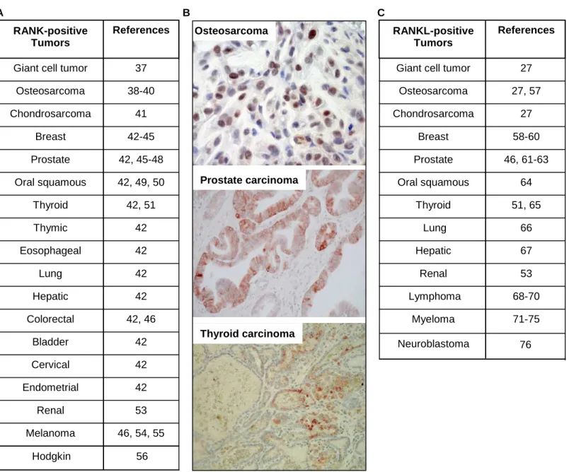
In bone microenvironment, OPG/RANK/RANKL molecular triad is not solely expressed by bone cells. Whether OPG is considered as an ubiquitary receptor [20], membrane RANK is expressed by various tumour cells originate from primary bone tumours or bone metastases, from mesenchymal and epithelial origin (Figure 2A, B). A recent study showed that more than 80% of bone metastases from solid tumours are RANK positive as revealed by immunohistochemistry [42-43]. RANK is also expressed in more than 50% of human osteosarcoma specimens, with preferential expression in osteosarcomas that develop in pathological bone and bad responders to chemotherapy [39].These observations then identify tumour cells as potential RANKL targets. Additionally to its expression by osteoblasts and bone marrow stromal cells, RANKL is produced similarly to RANK by numerous cancer cell types from various origins (Figure 2C). RANKL expression is modulated by a lot of cytokines, hormones [20] and by hypoxia dysregulating bone remodelling, a common feature of malignant tumors [77]. Indeed, the invasion of bone tissue by a primary or metastatic tumour cell precociously affects the balance between bone resorption and bone formation. According the tumour entities, tumour-derived factors (IGF, BMP, etc) can stimulate
osteoblast differentiation and activation and lead to tumour associated osteoblastic lesions (Figure 3) or in contrast, RANKL released by tumour cells can activate osteoclastogenesis and the recruitment of mature osteoclasts resulting in osteolytic lesions. The co-existence of both phenomenons leads to the formation of mixed osteoblastic/osteoclastic lesions. In turn, dysregulated bone cells-released extracellular matrix components and soluble mediators (TGFβ, etc) initially trapped in the bone matrix and stimulate proliferation of tumour cells and then the growth of tumour mass (Figure 3). This mechanism is defined as the osteoclast-dependent role of RANK/RANKL axis in tumorigenesis. However, RANK/RANKL axis influences tumorigenesis through osteoclast-independent pathway (Figure 3). RANKL produced by bone microenvironment constitutes a fertile soil for RANK-positive tumour cells. Initially proposed by Paget at the end of 1900th the concept of the seed and soil for
primary and secondary bone tumours has been strengthened by the discovery of RANKL and RANKL partly explains why various tumours preferentially metastasize to bone. RANKL released by osteoblasts and bone marrow stromal cells creates a cytokine gradient between bone site and extraosseous sites and triggers the migration of RANK-positive tumours cells. Interestingly, numerous tumour cells expressed functional RANK as shown by the signal transduction (P-ERK1/2, P-P38, P-IkB, etc) induced by RANKL [39, 47, 50]. The first evidence of this mechanism has been established by Jones et al. [45] and has been now described prostate carcinoma [45, 47, 48], breast carcinoma [45], oral squamous carcinoma [50], lung cancer cells [52] and melanoma [45]. The implication of RANK/RANKL axis in tumour cell migration has been confirmed by exploration in human samples. Indeed, the levels of RANK expressed by primary tumour cells are directly related to the occurrence of bone metastases in solid tumours and more specifically in breast, prostate and melanoma [42-44]. Furthermore, RANK expression could be considered as an independent predictor of poor prognosis in breast cancer patients with bone metastasis in contrast with visceral metastasis
for which no correlation has been shown [77]. Similarly, increased RANKL expression is related to the migration of renal carcinoma [53]. Whether the role of functional RANK expression has been clarified for carcinomas, its role in the pathogenesis of sarcomas is not fully understood. Indeed, it has been shown that osteosarcoma cells express the RANK protein [39]. RANK signalling under the action of RANKL, results in the modulation of a panel of more than 70 specific genes demonstrating that osteosarcoma cells are therefore RANKL targets [40]. Wittrant et al. [38] showed that RANKL directly induces BMP-2 expression in RANK positive osteosarcoma cells and may contribute by this way to the osteoblastic lesions characteristic of osteosarcomas. More recently, Lee et al. observed that RANKL expression is correlated to clinical behaviour of patients suffering from high-grade osteosarcoma [57].
RANK/RANKL axis is involved in the tumorigenic process
RANK/RANKL axis is not only associated with the bone metastastic process, and several arguments point out its involvement in the tumorigenicity process itself. RANK/RANKL may participate to the initial oncogenic program as shown by high expression of RANK on melanoma-initiating cells compared to the other melanoma cells [54]. Epithelial mesenchymal transition (EMT) is the first step allowing the extravasation and migration of carcinoma cells and RANKL appears clearly involved in this process. Indeed, Yamada et al. show that RANKL promotes EMT and induces angiogenesis independently of VEGF in a human head and neck squamous carinoma [78]. RANKL has also a strong impact on normal epithelial cells as shown by its effect on mammary gland development evidenced by a lactation defect in RANKL knockout mice [79, 80]. In fact, RANKL promotes the proliferation and survival of mammary epithelial cells [79-82] and RANK expression increases during the gestation more specifically at ductal branch points [82]. More
interestingly, whether RANKL or RANK overexpression in the mammary epithelial cells results in aberrant proliferation and hyperplasia of mammary glands, it directly correlates with preneoplasias and the development of spontaneous mammary tumours [83, 84]. Several authors hypothesized that RANKL may act as a paracrine factor for mammary stem cells [85, 86]. Consequently, blockade of RANKL significantly reduces the occurrence of mammary tumours [84]. Overall, these data give clear evidences of the RANK/RANKL axis contributes to the initial steps of tumorigenesis at least for mammary glands, to the dissemination process of carcinoma cells and to the establishment of bone metastases.
Therapies targeting RANK/RANKL axis for patients suffering from bone tumours: pre-clinical and pre-clinical arguments
Given this context, targeting of RANKL signalling with its decoy receptor OPG or with a soluble form of its membranous receptor RANK (RANK-Fc) inhibits tumour associated osteolysis in several experimental bone tumour models, including rat and mouse primary bone tumours and bone metastases. Indeed, OPG and RANK-Fc administered by non viral gene transfer or as recombinant molecules are effective in preventing the formation of osteolytic lesions associated with osteosarcoma development and in reducing the tumour incidence leading to a significant increase of animal survival [87, 88]. Moreover, recent experiments demonstrated that RNA interference strategy targeting RANKL improved the tumour response to chemotherapy in a murine model of osteosarcoma [89]. Similarly, administration of recombinant OPG-Fc or RANK-Fc has been investigated in numerous murine models of bone metastases [20, 23, 79] and confirm that blockade of the RANK/RANKL axis is extremely efficient in preclinical assessment to prevent tumour-induced osteolysis, to reduce tumour growth and to improve the survival rate. According these pre-clinical proofs of concept, recombinant OPG (OPG-Fc) has been evaluated in
postmenopausal [90] and in patients suffering from myeloma and osteolytic bone metastases (Table 1).Results demonstrated that OPG was well tolerated and demonstrated the efficacy of a single injection of OPG, which strongly reduced bone turnover for a sustained period and suppressed bone resorption as indicated by the decrease of bone resorption markers (urinary NTX/creatinine); these effects were comparable to those obtained with pamidronate. However, due to the risk of immune modulation of OPG through its binding to TRAIL [31] and other ligands [32-36], a fully human monoclonal antibody (IgG2) specifically targeting soluble and membrane RANKL has been developed [92-93]. Clinical data in osteoporotic patients revealed that denosumab was well tolerated with no related serious adverse events occurred and that a single-dose (0.01 mg/kg to 3.0 mg/kg) resulted in a dose-dependent sustained decrease from baseline in bone turnover [92-93]. This antibody, named denosumab only recognizes the human protein and its nonhuman-primate homolog and its administration in chimeric mice expressing murine/human leads to a strong inhibition of bone resorption concomitantly to an increase of the bone mineral density [94].
Numerous clinical trials (phase II and phase III) have been then designed to evaluate the efficacy of denosumab in oncology mainly in breast and prostate bone metastases (Table 1). These studies revealed that denosumab reduced significantly bone turnover markers similarly to osteoporotic patients. More specifically, it reduced levels of uNTX/Cr as well as serum TRAP5b thus showing a marked inhibition of osteoclastogenesis. According the results obtained, the recommendations for the use of denosumab are 120 mg s.c. every 4 months in oncology. Using this dose, bone resorption markers are suppressed around 90% in most patients independently of the tumour types [103]. Various studies have been set up to compare denosumab versus bisphosphonate treatment mainly zoledronic acid [95-97, 103, 109]. Single dose of pamidronate for instance (90 mg i.v.) reduced in a similar intensity the levels of bone resorption markers but the effects of denosumab were more sustained [95]. Phase III study
demonstrated that denosumab significantly delayed the time of first SRE (Skeletal Related Event) but also the risk of multiple SRE and whether zoledronic acid showed similar effects, statistical analyses are in favour of superiority for denosumab (Table 1). The time of disease progression and the overall survival rate were similar between anti-RANKL treatment and bisphosphonate. Zoledronic acid treatment requires a strict monitoring of kidney function due to its toxicity in contrast to denosumab even if a greater hypocalcemia requiring specific monitoring has been classically observed after denosumab treatment [111]. Additionally to the phase acute phase reaction observed in patients after the first administration of zoledronic acid, osteonecrosis of the jaw occurred infrequently after long-term treatment by nitrogen-bisphosphonates, in around 2% of patients [97, 108, 109, 112-115]. This incidence appears similar in bisphosphonate- and denosumab-treated patients. Consequently, the establishment of meticulous oral hygiene and surgical procedures prior to the administration of bisphosphonates and denosumab is the best method for preventing osteonecrosis of the jaw, prevention being better than treatment. In all studies, denosumab was well tolerated with the convenience of a subcutaneous administration and no requirement for renal monitoring. Overall, these clinical trials demonstrated that denosumab represents a potential treatment option economically viable for patients with bone metastases [116]. Very recently, a novel anti-RANKL antibodies derived from camelidae has been assessed in postmenopausal patients [117]. The results from this Phase I trial, including the one year follow-up information, indicate that ALX-0141 is well tolerated and can be administered safely over a wide range of doses. ALX-0141 exhibited a strong and sustained inhibitory effect on bone resorption markers.
The other anti-resorptive in therapies of bone cancer
Bisphosphonates have been used successfully for many years to treat the skeletal complications associated with the benign and malignant bone diseases [118-121]. Bisphosphonates became progressively a standard treatment for cancer-associated hypercalcemia and to control metastatic bone pain. Bisphosphonates are chemical compounds based on a phosphorus-carbon-phosphorus template and are characterized by their strong affinity for bone hydroxyapatite crystals and their anti-resorptive potency. Three families of bisphosphonate has been produced: the first possesses simple substituents attached to the central carbon and inhibits weakly the bone resorption; the second family possesses an aliphatic side chain containing a single nitrogen atom and exerts a more potent anti-resorptive activity; the third generation contain a heterocyclic substituent with one or two nitrogen atoms and are powerful bone resorption inhibitors and anti-tumour agents [118, 119]. The members of the first family which do not contain nitrogen atom are metabolised in cytotoxic analogues of ATP leading to cell death. Nitrogen-containing bisphosphonates inhibit the activity of two enzymes involved in the mevalonate pathway: farnesyl diphosphate synthase (FPP) and geranylgeranyl diphosphate synthase (GGPP). This inhibition results in osteoclast apoptosis by the strong reduction of the prenylation process, the loss of osteoclatic ruffled border and modifications of cell cytoplasmic actin ring [118, 119]. Additionally, nitrogen-containing bisphosphonates exert direct activities on tumour cells (breast, prostate, lung renal carcinoma, osteosarcoma, chondrosarcoma, etc) through the inhibition of prenylation mechanim which induces tumour-cell apoptosis, inhibits of cell proliferation, modulates tumour-cell adhesion and inhibits tumour-cell dissemination [118-124]. Thus, bisphosphonates inhibit the development of bone tumors through direct activity on tumour cells and indirect activity on osteoclasts. Pre-clinical revealed the therapeutic benefit of bisphosphonates for the treatments of primary bone tumours and bone metastases alone and combined with chemotherapy or
signalling pathway inhibitors [126-133]. Clinical trials have clearly confirmed their therapeutic interests [Table 1].
Since many decades, bone tumours have stimulated imagination of researchers and numerous therapeutic alternatives have been proposed [134, 135, Figure 4]. The better knowledge of OPG/RANK/RANKL system leads to the development of peptides mimicking OPG and blocking RANK-RANKL interactions [136-139]. Inhibitors of NF-KB signalling showed interesting anti-resorpive activities [140, 143]. The targeting of integrins more specifically αvβ3 strongly reduced the osteolytic process and the development of bone tumours [144-150]. Specific blockade of enzymatic activities has been envisaged with a great success. MMP9 involved in osteoclast migration and its targeting blocked by antisense oligodeoxyribonucleotide strongly affects osteoclast migration and resoption [151]. Cathepsin K, a key cysteine-proteinase related to osteolytic process [152, 153] stimulates a huge enthusiasm in the world of bone research. Several companies have then developed chemical inhibitors of cathepsin K to treat malignant and non-malignant bone loss with interesting results [154-157]. Thus, forty three women suffering from breast metastatic disease has been recently randomized in double-blind study to evaluate the impact of an oral cathepsin K inhibitors [odanacatib 5 mg daily for 4 weeks or 4 mg zoledronic acid i.v] on bone resorption markers [157]. Odanacatib appeared generally safe and well tolerated and has suppressed osteolytic markers similarly to zoledronic acid after 4 weeks of treatment. These results strengthen the therapeutic interest of cathepsin K for oncologic bone loss.
Conclusions
Bone tissue attracts massively tumour cells where they find a favorable environment to maintain the stem cell dormancy and where they find a fertile ground for their development. This “fatal attraction” linked to the specific bone niche, has boosted therapeutic innovations
targeting the tumour cells and/or their microenvironment [158]. During the last past decade, RANK/RANKL axis emerged in bone biology as predominant protagonists of bone remodeling and as therapeutic targets of bone loss diseases. Better knowledge of RANK/RANKL biology will better define their relevance as biomarkers in bone oncology, and a complete cartography of RANK expression will be very useful to predict good responders to anti-RANKL therapies. Although anti-RANKL therapy progressively competes with approaches by bisphosphonates, a lot of prospect including signal transduction inhibitors, peptides or enzymatic inhibitors has been already identified and pre-clinical data as well as clinical trials allow personalized therapies in bone oncology.
References
[1] Dempster DW. Anatomy and functions of the adult skeleton. In: Primer of the metabolic bone diseases and disorders of mineral metabolism, Ed. M.J. Favus, American Society for Bone and Mineral Research Publication Office, Durham, 6th Ed.; 2006, p. 7-11.
[2] Favus MJ, Bushinsky DA, Lemann Jr J. Regulation of calcium, magnesium, and phosphate metabolism. In: Primer of the metabolic bone diseases and disorders of mineral metabolism, Ed. M.J. Favus, American Society for Bone and Mineral Research Publication Office, Durham, 6th Ed; 2006, p. 76-83.
[3] Deschaseaux F, Sensebe L, Heymann D. Mechanisms of bone repair and regeneration. Trends Mol Med 2009;15:417-29
[4] Ross FP. Osteoclast biology and bone resorption. In: Primer of the metabolic bone diseases and disorders of mineral metabolism, pp 30-35, Murray J. Favus (Ed.), American Society for Bone and Mineral Research Publication Office, Durham, 6th Ed; 2006, p. 30-35.
[5] Rousselle AV, Heymann D. Osteoclastic acidification during bone resorption. Bone 2002; 30:533-40.
[6] Filgueira L. Osteoclast differentiation and function. In Bone Cancer, Ed. Heymann D, Academic Press; 2010, p. 59-66.
[7] Billiard J, et al. Regulation of osteblast differentiation and bone cancers by Wnt and PTH signaling pathways. In Bone Cancer, Ed. Heymann D, Academic Press; 2010, p. 47-58.
[8] Aubin JE, Lian JB, Stein GS. Bone formation maturation and functional activities of osteoblast lineage cells. In: Primer of the metabolic bone diseases and disorders of mineral metabolism, pp 20-29, Murray J. Favus (Ed.), American Society for Bone and Mineral Research Publication Office, Durham, 6th Ed; 2006, p. 20-29.
[9] Takahashi N, et al. Osteoblastic cells are involved in osteoclast formation. Endocrinology 1988;123:2600-2.
[10] Jimi E, et al. Osteoclast function is activated by osteoblastic cells through a mechanism involving cell-to-cell contact. Endocrinology 1996;137:2187-90.
[11] Mundy GR, Elefteriou F. Boning up on ephrin signaling. Cell 2006; 126:441-3.
[12] Hauben EI, Hogendoorn PCW. Epidemiology of primary bone tumors and economical aspects of bone metastases. In Bone Cancer, Ed. Heymann D, Academic Press; 2010, p. 3-8.
[13] Wittrant Y, et al. RANKL/RANK/OPG: new therapeutic targets in bone tumours and associated osteolysis. Biochim Biophys Acta 2004;1704:49-57.
[14] Koeneman KS, Yeung F, Chung LW. Osteomimetic properties of prostate cancer cells: a hypothesis supporting the predilection of prostate cancer metastasis and growth in the bone environment. Prostate 1999; 39:246-61.
[15] David E. The bone niche of chondrosarcoma: a sanctuary for drug resistance, tumour growth and also a source of new therapeutic targets. Sarcoma, 2011, ID 932451.
[16] Simonet WS, et al. Osteoprotegerin: a novel secreted protein involved in the regulation of bone density. Cell 1997; 89: 309-19.
[17] Tsuda E, et al. Identity of osteoclastogenesis inhibitory factor (OCIF) and osteoprotegerin (OPG): a mechanism by which OPG/OCIF inhibits osteoclastogenesis in vitro. Endocrinology 1998; 139: 1329-37.
[18] Lacey DL, et al. Osteoprotegerin ligand is a cytokine that regulates osteoclast differentiation and activation. Cell 1998; 93: 165-76.
[19] Yasuda H, et al. Osteoclast differentiation factor is a ligand for osteoprotegerin/osteoclastogenesis-inhibitory factor and is identical to TRANCE/RANKL. Proc Natl Acad Sci. USA 1998; 95: 3597-602.
[20] Theoleyre S, Wittrant Y, Kwan Tat S, Fortun Y, Redini F, Heymann D. The molecular triad OPG/RANK/RANKL: Involvement in the orchestration of pathophysiological bone remodeling. Cytokine Growth Factor Rev 2004;15:457-75.
[21] Heymann D, Fortun Y, Rédini F, Padrines M. Osteolytic bone diseases: physiological analogues of bone resorption effectors as alternative therapeutic tools to the standard bisphosphonates. Drug Discov Today 2005;10:242-47.
[22] Mori K, Ando K, Heymann D, Redini F. Receptor activator of nuclear factor-kappa B ligand (RANKL) stimulates bone-associated tumours through functional RANK expressed on bone associated cancer cells? Histol Histopathol 2009; 24:235-42.
[23] Baud'huin M, Lamoureux F, Duplomb L, Rédini F, Heymann D. RANKL, RANK, osteoprotegerin: key partners of osteoimmunology and vascular diseases. Cell Mol Life Sci. 2007;64:2334-50.
[24] Min H, et al. Osteoprotegerin reverses osteoporosis by inhibiting endosteal osteoclasts and prevents vascular calcification by blocking a process resembling osteoclastogenesis. J Exp Med 2000,192:463-74.
[25] Anderson DM, et al. A homologue of the TNF receptor and its ligand enhance T-cell growth and dendritic-cell function. Nature 1997; 390:175-9.
[26] Boyle WJ, Simonet WS, Lacey DL. Osteoclast differentiation and activation. Nature 2003;423:337-42.
[27] Grimaud E, et al. Receptor Activator of Nuclear Factor kB Ligand (RANKL)/Osteoprotegerin (OPG) ratio is increased in severe osteolysis. Am J Pathol 2003;163:2021-31.
[28] Kwan Tat S, Padrines M, Theoleyre S, Heymann D, Fortun Y. IL-6, RANKL, TNF-alpha/IL-1: interrelations in bone resorption pathophysiology. Cytokine Growth Factor Rev 2004;15:49-60.
[29] Liu C, et al. Structural and functional insights of RANKL-RANK interaction and signaling. J Immunol 2010;184:6910-9.
[30] Ito S, Hata T. Crystal structure of RANK ligand involved in bone metabolism. Vitam Horm 2004;67:19-33.
[31] Emery JG, et al. Osteoprotegerin is a receptor for the cytotoxic ligand TRAIL. J Biol Chem 1998; 273:14363-7.
[32] Standal T, et al. Osteoprotegerin is bound, internalized, and degraded by multiple myeloma cells. Blood 2002;100:3002-7.
[33] Theoleyre S, et al. Cellular activity and signaling induced by osteoprotegerin in osteoclasts: involvement of Receptor Activator of Nuclear Factor kB Ligand and MAPK. Biochim Biophys Acta Mol Cell Res, 2004, 1644:1-7.
[34] Lamoureux F, et al. Glycosaminoglycans as potential regulators of osteoprotegerin therapeutic activity in osteosarcoma. Cancer Res 2009 ;69:526-36.
[35] Zannettino AC, et al. Osteoprotegerin (OPG) is localized to the Weibel-Palade bodies of human vascular endothelial cells and is physically associated with von Willebrand factor. J Cell Physiol 2005;204:714-23.
[36] Baud’huin M, Duplomb L, Télétchéa S, Charrier C, Maillasson M, Fouassier M, Heymann D. Factor VIII/von Willebrand factor complex controls RANKL-induced osteoclastogenesis and cell survival. J Biol Chem, 2009, 264: 31704-13.
[37] Atkins GJ, et al. RANK Expression as a cell surface marker of human osteoclast precursors in peripheral blood, bone marrow, and giant cell tumors of bone. J Bone Miner Res 2006;21:1339-49.
[38] Wittrant Y, Mori K, Riet A, Kamijo A, Heymann D, Rédini F. RANKL directly induces bone morphogenetic protein-2 expression in RANK-expression POS-1 osteosarcoma cells. Int J Oncol 2006;28:261-9.
[39] Mori K, et al. Human osteosarcoma cells express functional Receptor Activator of Nuclear Factor-kappa B. J Pathol 2007; 211: 555-62.
[40] Mori K, et al. Receptor Activator of Nuclear Factor-kB Ligand (RANKL) directly modulates gene expression profile of RANK-positive Saos-2 human osteosarcoma cells. Oncol Rep, 2007;18:1365-71.
[41] Hsu CJ, et al. Involvement of integrin up-regulation in RANKL/RANK pathway of chondrosarcomas migration. J Cell Biochem 2010;111:138-47
[42] Santini D, et al. Receptor activator of NF-kB (RANK) expression in primary tumors associates with bone metastasis occurrence in breast cancer patients. PLoS One 2011;6:e19234.
[43] Santini D, et al. Expression pattern of receptor activator of NFkB (RANK) in a series of primary solid tumors and related metastases. J Cell Physiol 2011;226:780-784.
[44] Bhatia P, Sanders MM, Hansen MF. Expression of receptor activator of nuclear factor-kappaB is inversely correlated with metastatic phenotype in breast carcinoma. Clin Cancer Res 2005;11:162-5.
[45] Jones DH, et al. Regulation of cancer cell migration and bone metastasis by RANKL. Nature 2006;440:692-6.
[46] Chen G, Sircar K, Aprikian A, Potti A, Goltzman D, Rabbani SA. Expression of RANKL/RANK/OPG in primary and metastatic human prostate cancer as markers of disease stage and functional regulation.Cancer 2006;107:289-98.
[47] Mori K, Le Goff B, Charrier C, Battaglia S, Heymann D, Rédini F. DU145 human prostate cancer cells express functional receptor activator of NFkappaB: new insights in the prostate cancer bone metastasis process. Bone 2007;40:981-90.
[48] Armstrong AP, Miller RE, Jones JC, Zhang J, Keller ET, Dougall WC. RANKL acts directly on RANK-expressing prostate tumor cells and mediates migration and expression of tumor metastasis genes. Prostate 2008;68:92-104.
[49] Chuang FH, Hsue SS, Wu CW, Chen YK. Immunohistochemical expression of RANKL, RANK, and OPG in human oral squamous cell carcinoma. J Oral Pathol Med 2009;38:753-8.
[50] Shin M, Matsuo K, Tada T, Fukushima H, Furuta H, Ozeki S, Kadowaki T, Yamamoto K, Okamoto M, Jimi E. The inhibition of RANKL/RANK signaling by osteoprotegerin suppresses bone invasion by oral squamous cell carcinoma cells. Carcinogenesis 2011; 32:1634-40.
[51] Heymann MF, Riet A, Le Goff B, Battaglia S, Paineau J, Heymann D.OPG, RANK and RANK ligand expression in thyroid lesions. Regul Pept 2008;148:46-53.
[52] Chen LM, et al. RANKL increases migration of human lung cancer cells through intercellular adhesion molecule-1 up-regulation. J Cell Biochem 2011;112:933-41.
[53] Mikami S, et al. Increased RANKL expression is related to tumour migration and metastasis of renal cell carcinomas. J Pathol 2009;218:530-9.
[54] Kupas V, et al. RANK is expressed in metastatic melanoma and highly upregulated on melanoma-initiating cells. J Invest Dermatol 2011;131:944-55.
[55] Mori K, Ando K, Lezot F, Heymann D. RANK/RANKL Axis in Melanoma. In Breakthroughs in melanoma research, Ed. Tanaka Y, InTech Publisher, chapter 27, 2011.
[56] Fiumara P, et al. Functional expression of receptor activator of nuclear factor kappaB in Hodgkin disease cell lines. Blood 2001;98:2784-90.
[57] Lee JA, et al. RANKL expression is related to treatment outcome of patients with localized, high-grade osteosarcoma. Pediatr Blood Cancer 2011;56:738-43.
[58] Rucci N, et al. Receptor activator of NF-kappaB ligand enhances breast cancer-induced osteolytic lesions through upregulation of extracellular matrix metalloproteinase inducer/CD147. Cancer Res 2010;70:6150-60.
[59] Cross SS, et al. Expression of receptor activator of nuclear factor kappabeta ligand (RANKL) and tumour necrosis factor related, apoptosis inducing ligand (TRAIL) in breast cancer, and their relations with osteoprotegerin, oestrogen receptor, and clinicopathological variables.J Clin Pathol 2006;59:716-20.
[60] Van Poznak C, Cross SS, Saggese M, Hudis C, Panageas KS, Norton L, Coleman RE, Holen Expression of osteoprotegerin (OPG), TNF related apoptosis inducing ligand (TRAIL), and receptor activator of nuclear factor kappaB ligand (RANKL) in human breast tumours. J Clin Pathol 2006;59:56-63.
[61] Sabbota AL, Kim HR, Zhe X, Fridman R, Bonfil RD, Cher ML. Shedding of RANKL by tumor-associated MT1-MMP activates Src-dependent prostate cancer cell migration. Cancer Res 2010;70:5558-66.
[62] Odero-Marah VA, et al. Receptor activator of NF-kappaB Ligand (RANKL) expression is associated with epithelial to mesenchymal transition in human prostate cancer cells. Cell Res 2008;18:858-70.
[63] Yuvaraj S, Griffin AC, Sundaram K, Kirkwood KL, Norris JS, Reddy SV. A novel function of CXCL13 to stimulate RANK ligand expression in oral squamous carcinoma cells. Mol Cancer Res 2009;7:1399-407.
[64] Penno H, Nilsson O, Brändström H, Winqvist O, Ljunggren O. Expression of RANK-ligand in prostate cancer cell lines. Scand J Clin Lab Invest 2009;69:151-5.
[65] Sood SK, Balasubramanian S, Higham S, Fernando M, Harrison B. Osteoprotegerin (OPG) and related proteins (RANK, RANKL and TRAIL) in thyroid disease. World J Surg 2011;35:1984-92.
[66] Nakamura ES, et al. RANKL-induced CCL22/macrophage-derived chemokine produced from osteoclasts potentially promotes the bone metastasis of lung cancer expressing its receptor CCR4. Clin Exp Metastasis 2006;23:9-18.
[67] Sasaki A, et al. Receptor activator of nuclear factor-kappaB ligand (RANKL) expression in hepatocellular carcinoma with bone metastasis. Ann Surg Oncol 2007;14:1191-9.
[68] Barcala V, et al. RANKL expression in a case of follicular lymphoma. Eur J Haematol 2003;70:417-9.
[69] Shibata H, et al. Malignant B-lymphoid cells with bone lesions express receptor activator of nuclear factor-kappaB ligand and vascular endothelial growth factor to enhance osteoclastogenesis. Clin Cancer Res 2005;11:6109-15.
[70] Shu ST, Martin CK, Thudi NK, Dirksen WP, Rosol TJ. Osteolytic bone resorption in adult T-cell leukemia/lymphoma. Leuk Lymphoma 2010;51:702-14.
[71] Sezer O, Heider U, Jakob C, Eucker J, Possinger K. Human bone marrow myeloma cells express RANKL. J Clin Oncol 2002;20:353-4.
[72] Roux S, et al. RANK (receptor activator of nuclear factor-kappaB) and RANKL expression in multiple myeloma. Br J Haematol 2002;117:86-92.
[73] Farrugia AN, et al. Receptor activator of nuclear factor-kappaB ligand expression by human myeloma cells mediates osteoclast formation in vitro and correlates with bone destruction in vivo. Cancer Res 2003;63:5438-45.
[74] Heider U, et al. Expression of receptor activator of NF-kappaB ligand (RANKL) mRNA in human multiple myeloma cells. J Cancer Res Clin Oncol 2004;130:469-74.
[75] Buckle CH, Neville-Webbe HL, Croucher PI, Lawson MA. Targeting RANK/RANKL in the treatment of solid tumours and myeloma. Curr Pharm Des 2010;16:1272-83.
[76] Granchi D, et al. In vitro blockade of receptor activator of nuclear factor-kappaB ligand prevents osteoclastogenesis induced by neuroblastoma cells. Int J Cancer 2004;111:829-38. [77] Tang ZN, Zhang F, Tang P, Qi XW, Jiang J. Hypoxia induces RANK and RANKL expression by activating HIF-1α in breast cancer cells. Biochem Biophys Res Commun 2011;48:411-6.
[78] Zhang L, et al. Receptor activator for nuclear factor kB expression predicts poor prognosis in breast cancer patients with bone metastasis but not in patients with visceral metastasis. J Clin Pathol 2012; 65:36-40.
[79] Dougall WC. Molecular pathways: osteoclast-dependent and osteoclast-independent roles of the RANKL/RANK/OPG pathway in tumorigenesis and metastasis. Clin Cancer Res 2012; 18:326-335.
[80] Fata JE, et al. The osteoclast differentiation factor osteoprotegerin-ligand is essential for mammary gland development. Cell 2000;103:41-50.
[81] Mukherjee A, et al. Targeting RANKL to a specific subset of murine mammary eptithelial cells induces ordered branching morphogenesis and alveologenesis in the absence of progesterone receptor expression. FASEB 2010;24:4408-19.
[82] Fernandez-Valdivia R et al. The RANKL signaling axis is sufficient to elicit ductal sidebranching and alveologenesis in the mammary gland of the virgin mouse. Dev Biol 2009;328:127-39.
[83] Gonzalez-Suarez E, Branstetter D, Armstrong A, Dinh H, Blumberg H, Dougall WC. RANK overexpression in transgenic mice with mouse mammary tumor virus promoter controlled RANK increases proliferation and impairs alveolar differentiation in the mammary epithelia and disrupts lumen formation in cultured epithelial acini. Mol Cell Biol 2007;27:1442-54.
[84] Gonzalez-Suarez E, et al. RANK ligand mediates progestin-induced mammary epithelial proliferation and carcinogenesis. Nature 2010;468:103-37.
[85] Joshi PA, et al. Progesterone induces adult mammary stem cell expansion. Nature 2010;465:803-7.
[86] Asselin-Labat ML, et al. Control of mammary stem cell function by steroid hormone signalling. Nature 2010;465:798-802.
[87] Lamoureux F, et al. Therapeutic relevance of osteoprotegerin gene therapy in osteosarcoma: prevention of bone resorption, inhibition of tumor progression, increase of animal survival. Cancer Res 2007;67:7308-18
[88] Lamoureux F, et al. Therapeutic efficacy of soluble receptor activator of NF-kB delivered by non viral gene transfer in a mouse model of osteolytic osteosarcoma. Mol Cancer Ther 2008;7:3389-98.
[89] Rousseau J, et al. Formulated siRNAs targeting RANKL prevent osteolysis and enhance chemotherapeutic response in osteosarcoma models. J Bone Miner Res 2011;26:2452–62
[90] Bekker PJ, et al. The effect of a single dose of osteoprotegerin in postmenopausal women. J Bone Miner Res 2001;16:348-60.
[91] Body JJ, et al. A phase I study of AMGN-0007, a recombinant osteoprotegerin construct, in patients with multiple myeloma or breast carcinoma related bone metastases. Cancer 2003;97:887-892.
[92] Bekker PJ, et al. A single-dose placebo-controlled study of AMG162, a fully human monoclonal antibody to RANKL, in postmenopausal women. J Bone Miner Res 2004;19:1059-66.
[93] McClung MR, et al. Denosumab in postmenopausal women with low bone mineral density. N England J Med 2006;354:821-31.
[94] Kostenuik PJ, et al. Denosumab, a fully human monoclonal antibody to RANKL, inhibits bone resorption and increases BMD in knock-in mice that express chimeric (murine/human)
[95] Body JJ, et al. A study of the biological receptor activator of nuclear factor-kappaB ligand inhibitor, denosumab, in patients with multiple myeloma or bone metastases from breast cancer. Clin Cancer Res 2006;12:1221-8.
[96] Henry DH, et al. Randomized, double-blind study of denosumab versus zoledronic acid in the treatment of bone metastases in patients with advanced cancer (excluding breast and prostate cancer) or multiple myeloma. J Clin Oncol 2011;29:1125-32.
[97] Stopeck AT, et al. Denosumab compared with zoledronic acid for the treatment of bone metastases in patients with advanced breast cancer: a randomized, double-blind study. J Clin Oncol 2010;28:5132-39.
[98] Body JJ, et al. Effects of denosumab in patients with bone metastases with or without previous bisphosphonate exposure. J Bone Miner Res 2010;25:440-6.
[99] Lipton A, et al. Extended efficacy and safety of denosumab in breast cancer patients with bone metastases not receiving prior bisphosphonate therapy. Clin Cancer Res 2008;14:6690-6.
[100] Lipton A, et al. Randomized active-controlled phase II study of denosumad efficacy and safety in patients with breast cancer-related bone metatases. J Clin Oncol 2007;45:4431-7.
[101] Ellis GK, et al. Randomized trial of denosumab in patients receiving adjuvant aromatase inhibitors for nonmetastatic breast cancer. J Clin Oncol 2008;26:4875-82.
[102] Ellis GK, et al. Effect of denosumab on bone and mineral density in women receiving adjuvant aromatase inhibitors for non-metastatic breast cancer: subgroup analyses of a phase 3 study. Breast Cancer Res Treat 2009;118:81-7.
[103] Fizazi K, et al. Randomized phase II trial of denosumab in patients with bone metastases from prostate cancer, breast cancer, or other neoplasms after intravenous bisphosphonates. J Clin Oncol 2009;27:1564-71.
[104] Vij R, et al. An open-label, phase 2 trial of denosumab in the treatment of relapsed or plateau-phase multiple myeloma. Am J Jematol 2009;84:650-6.
[105] Fizazi K, et al. Denosumab treatment of prostate cancer with bone metastases and increased urine N-telopeptide levels after therapy with intravenous bisphosphonates: results of a randomized phase II trial. J Urol 2009;182:509-15.
[106] Smith MR, et al. Denosumab in mem receiving androgen-deprivation therapy for prostate cancer. N Engl J Med 2009;361:745-55.
[107] Smith MR, et al. Effects of denosumab on bone mineral density in men receiving androgen deprivation therapy for prostated cancer. J Urol 2009; 182:2670-5.
[108] Smith MR, et al. Denosumab and bone-metastasis-free survival in men with castration-resistant prostate cancer: results of a phase 3, randomized, placebo-controlled trial. Lancet 2012; 379:39-46.
[109] Fizazi K, et al. Denosumab versus zoledronic acid for treatment of bone metastases in men with castration resistant prostate cancer: a randomized, double-blind study. Lancet 2011;377:813-22.
[110] Thomas D, et al. Denosumab in patients with giant-cell tumour of bone: an open-label, phase 2 study. Lancet Oncol 2010;11:275-80.
[111] Jamal SA, et al. Effects of denosumab on fracture and bone mineral density by level of kidney function. J Bone Miner Res 2011;26:1839-35.
[112] Coleman R, et al. Zoledronic acid. Expert Opin Drug Saf 2011;10:133-45.
[113] Migliorati CA, Epstein JB, Abt E, Berenson JR. Osteonecrosis of the jaw and bisphosphonates in cancer: a narrative review. Nat Rev Endocrinol 2011;7:34-42.
[114] Filleul O, Crompot E, Saussez S. Bisphosphonate-induced osteonecrosis of the jaw: a review of 2,400 patient cases 2010;136:1117-24.
[115] Heymann D. Bisphosphonates and bone diseases: past, present and future. Curr Pharm Des 2010;16:2948-9.
[116] Xie J, et al. Economic evaluation of denosumab compared with zoledronic acid in hormone-refractory prostate cancer patients with bone metastases. J Manag Care Pharm 2011;17:621-43.
[117] Van de Wetering de Rooij L, et al. Safety, pharmacokinetics and efficacy of anti-RANKL nanobody ALX-0141 in healthy postmenopausal wome. Ann Rheum Dis 2011;70(suppl 3):136.
[118] Heymann D, Ory B, Gouin F, Green J, Rédini F. Bisphosphonates: new therapeutic agents for the treatment of bone tumors. Trends Mol Med, 2004, 10: 337-343.
[119] Ory B, Moriceau G, Rédini F, Heymann D. mTOR inhibitors (rapamycin and derivatives) and nitrogen-bisphosphonates: bi-functional compounds for the treatment of bone tumors. Current Med Chem, 2007, 14, 1381-1387.
[120] Coleman RE, McCloskey EV. Bisphosphonates in oncology. Bone 2011;49:71-6.
[121] Coleman R. The use of bisphosphonates in cancer treatment. Ann N Y Acad Sci 2011;1218:3-14.
[122] Ory B, et al. Zoledronic acid suppresses lung metastases and extends overall survival of osteosarcoma of osteosarcoma-bearing mice. Cancer, 2005, 104: 2522-2529.
[123] Gouin F, Rédini F, Ory B, Heymann D. Zoledronic acid slows down rat chondrosarcoma progression, increases overall survival and delays tumor recurrence after intralesional curettage. Int J Cancer, 2006, 119:980-984.
[124] Ory B, Blanchard F, Battaglia S, Gouin F, Rédini F, Heymann D. Zoledronic acid activates the DNA S phase checkpoint and induces osteosarcoma cell death characterized by
AIF and EndoG translocation in dependently of p53 and Rb status. Mol Pharmacol, 2007, 71: 333-343.
[125] Holen I, Coleman RE. Bisphosphonates as treatment of bone metastases. Curr Pharm Des 2010;16:1262-71.
[126] Lamoureux F, et al. Relevance of a new rat syngenic model of osteoblastic metastases from prostate carcinoma for pre-clinical studies using zoledronic acid. Int J Cancer, 2008, 122: 751-760.
[127] Moriceau G, et al. Therapeutic approach of primary bone tumors by bisphosphonates. Current Pharm Des 2010;16:2981-87.
[128] Ory B, Moriceau G, Redini F, Heymann D. mTOR inhibitors (rapamycin and its derivatives) and nitrogen containing bisphosphonates: bi-functional compounds for the treatment of bone tumours. Curr Med Chem 2007;14:1381-7.
[129] Heymann D, et al. Enhanced tumor regression and tissue repair when zoledronic acid is combined with ifosfamide in rat osteosarcoma. Bone, 2005, 37: 74-86.
[130] Moriceau G, et al. Zoledronic acid potentiates mTOR inhibition and abolishes the resistance of osteosarcoma cells to RAD001 (Everolimus): pivotal role of the prenylation process. Cancer Res 2010, 70:10329-39.
[131] Battaglia S, et al. Impact of oncopediatric dosing regimen of zoledronic acid on bone growth: preclinical studies and case report of an osteosarcoma pediatric patient. J Bone Miner Res 2011,26: 2439-51.
[132] Daubiné F, Le Gall C, Gasser J, Green J, Clézardin P. Antitumor effects of clinical dosing regimens of bisphosphonates in experimental breast cancer bone metastasis. J Natl Cancer Inst 2007;99:322-30.
[133] Ottewell PD, Mönkkönen H, Jones M, Lefley DV, Coleman RE, Holen I. Antitumor effects of doxorubicin followed by zoledronic acid in a mouse model of breast cancer. J Natl Cancer Inst 2008;100:1167-78.
[134] Heymann D, Fortun Y, Redini F, Padrines M. Osteolytic bone diseases:physiological analogues of bone resorption effectors as alternative therapeutic tools Drug Discov Today 2005; 10:242-7
[135] Heymann D. Novel targeted therapies of bone tumors and future directions. Future Medicine, E book, “Bone Metastasis management”, Ed. P. Picci, P Ruggieri. In press.
[136] Engleman VW, et al. A peptidomimetic antagonist of the alpha (v)beta3 inhibits bone resorption in vitro and prevents osteoporosis in vivo. J Clin Invest 1997;99:2284–92.
[137] Cheng X, et al. Disabling of RANK receptor complex by novel osteoprotegerin like peptidomimetics restores bone loss in vivo. J Biol Chem 2004;279:8269–77
[138] Aoki K, et al. A TNF receptor loop peptide mimic blocks RANK ligand-induced signaling, bone resorption, and bone loss. J Clin Invest 2006;116:1525-34.
[139] Heath DJ, et al. An osteoprotegerin-like peptidomimetic inhibits osteoclastic bone resorption and osteolytic bone disease in myeloma. Cancer Res 2007;67:202-8.
[140] Penolazzi L, et al. Peptide nucleic acid-DNA decoy chimeras targeting NF-kappa B transcription factors: induction of apoptosis in human primary osteoclasts. Int J Mol Med 2004;14:145–52
[141] Penolazzi L., et al. Decoy oligonucleotides targeting NF-κB transcription factors: induction of apoptosis in human primary osteoclasts. Biochem Pharmacol 2003; 66:1189–98
[142] Clohisy JC, et al. NF-κB signaling blockade abolishes implant particle-induced osteoclastogenesis. J Orthop Res 2004;22:13–20.
[143] Clohisy JC, et al. Direct inhibition of NF-kappa B blocks bone erosion associated with inflammatory arthritis. J Immunol 2003;171:5547–53
[144] Carron CP, et al. Peptidomimetic antagonists of alphavbeta3 inhibit bone resorption ny inhibiting osteoclast bone resorptive activity,not osteoclast adhesion to bone. J Endocrinol 2000;165:587–98.
[145] Bakewell SJ, et al. Platelet and osteoclast beta3 integrins are critical for bone metastasis. Proc Natl Acad Sci U S A 2003;100:14205-10.
[146] Harms JF, et al. A small molecule antagonist of the alpha(v)beta3 integrin suppresses MDA-MD-435 skeletal metastasis. Clin Exp Metastasis 2004;21:119–28.
[147] van der Horst G, et al. Targeting of α(v)-integrins in stem/progenitor cells and supportive microenvironment impairs bone metastasis in human prostate cancer. Neoplasia 2011;13:516-25
[148] Zhao Y, et al. Tumor alphavbeta3 integrin is a therapeutic target for breast cancer bone metastases. Cancer Res 2007;67:5821-30.
[149] Wadas TJ, Deng H, Sprague JE, Zheleznyak A, Weilbaecher KN, Anderson CJ. Targeting the alphavbeta3 integrin for small-animal PET/CT of osteolytic bone metastases. J Nucl Med 2009 ;50:1873-80.
[150] Harms JF, et al. A small molecule antagonist of the alpha(v)beta3 integrin suppresses MDA-MB-435 skeletal metastasis. Clin Exp Metastasis 2004;21:119-28.
[151] Ishibashi O, Niwa S, Kadoyama K, Inui T. MMP-9 antisense oligodeoxynucleotide exerts an inhibitory effect on osteoclastic bone resorption by suppressing cell migration. Life Sci 2006 ;79:1657-60.
[152] Costa AG, Cusano NE, Silva BC, Cremers S, Bilezikian JP. Cathepsin K: its skeletal actions and role as a therapeutic target in osteoporosis. Nat Rev Rheumatol 2011;7:447-56.
[153] Sturge J, Caley MP, Waxman J. Bone metastasis in prostate cancer: emerging therapeutic strategies. Nat Rev Clin Oncol 2011;8:357-68.
[154] Ochi Y, et al. Effects of ONO-5334, a novel orally-active inhibitor of cathepsin K, on bone metabolism. Bone 2011;49:1351-6.
[155] Le Gall C, et al. A cathepsin K inhibitor reduces breast cancer induced osteolysis and skeletal tumor burden. Cancer Res 2007;67:9894-902.
[156] Xiang A, et al. Changes in micro-CT 3D bone parameters reflect effects of a potent cathepsin K inhibitor (SB-553484) on bone resorption and cortical bone formation in ovariectomized mice. Bone 2007;40:1231-7.
[157] Jensen AB, et al. The cathepsin K inhibitor odanacatib suppresses bone resorption in women with breast cancer and established bone metastases: results of a 4-week, double-blind, randomized, controlled trial. Clin Breast Cancer 2010 ;10:452-8.
[158] Weilbaecher KN, Guise T, Mc Cauley LK. Cancer to Bone: a fatal attraction. Nat Cancer Rev 2011;11:411-425.
Figure Legends:
Figure 1: RANKL is absolutely required for osteoclast differentiation in vivo as revealed by the bone phenotype exhibited by RANKL knockout mice. (A) Osteopetrotic phenotype exhibited by RANKL knockout (RANKL-/-) compared to wild type (WT) C57BL6 mice analyzed by µCT (skyscan 1076). (B) Osteoblasts produced RANKL (membrane and soluble forms) which binds to membrane RANK expressed by osteoclast precursors, OPG synthesized by osteoblasts acts as a decoy receptor, blocks the interactions RANKL/RANK and then inhibits bone resorption. The lack of RANKL results in a disturbed bone remodeling characterized by an excessive bone formation and a reduced bone resorption compared to the control mice.
Figure 2: RANK is expressed by numerous tumour cell type. (A) Main tumour cell types expressing RANK; (B) RANK immunostaining on osteosarcoma [39], prostate carcinoma and thyroid carcinoma [51]. In all cases, RANK is expressed by cancer cells.
Figure 3: Direct and indirect role of RANKL in bone tumour development. RANKL contributes to the development of bone tumours via the activation of osteoclastogenesis and bone resorption defining an osteoclast-dependent pathway. RANKL also can bind directly RANK-expressing tumour cells, stimulating epithelial mesenchymal transition, cell migration and then identifying an osteoclast-independent. Tumour cells dysregulate the balance between osteoblasts and osteoclasts, resulting in osteolytic, osteoblastic or osteoblastic. osteoclastic mixed lesions according the tumour cell type.
Figure 4: Therapeutic arsenals currently used or in development targeting osteoclast lineage to treat bone tumours. These therapeutic approaches targets osteoclast the differentiation and/or their activation by blocking RANKL binding to RANK, signal transduction, cell adhesion and migration or enzymatic activities.
Table 1: Summary of the main clinical trials in oncology assessing anti-RANKL therapies Clinical trials drug assessed Cancer Number of patients included Doses References Phase I OPG recombinant Bone metastases (Breast) Myeloma 26 28 s.c. 0.1-3 mg/Kg 91
Phase I, Denosumab Bone metastases (Breast) Myeloma 29 25 Denosumab s.c. 0.1-3 mg/Kg Pamidronate 90m g i.v. 95 Randomized, double blind Denosumab versus zoledronate Bone metastases (excluding breast, prostate and myeloma) 886 890 Denosumab s.c. 120 mg monthly Zoledronate i.v. 4 mg monthly 96 Randomized, double blind Denosumab versus zoledronate Bone metastases (Breast) 1,026 1,020 Denosumab s.c. 120 mg monthly Zoledronate i.v. 4 mg monthly 97
Phase II, Denosumab with and without bisphosphonate exposure
Bone metastases 366 Denosumab
s.c. 60 or 180 mg every 12 weeks Denosumab s.c. 30, 120, or 180 mg every 4 weeks 98-100 Denosumab vs placebo and adjuvant aromatase inhibitors Non-metastatic breast cancer 127 (treated) 125 (placebo) Denosumab s.c. 60 mg every 6 weeks 101, 102
Phase II, randomized trial
Denosumab after i.v. bisphosphonates
Bone metastases (prostate, breast cancers and other neoplasms) 111 Denosumab s.c. 180 mg every 4 or 12 weeks 103 Phase II Denosumab Myeloma 96 Denosumab s.c.120 mg on days 1, 8, and 15 (loading doses) of cycle 1 (28 days), and then on study day 29 (day 1 of cycle 2) and on day 1 of every cycle (28 days) thereafter 104 Phase II, Randomized Denosumad after i.v. bisphosphonates Bone metastases (Prostate) 111 Denosumab s.c. 180 mg every 4 or 12 weeks 105
Denosumad and androgen-deprivation s.c. 60 mg every 6 months Double-blind study Denosumab and androgen-deprivation
Prostate cancer 734 per group Denosumab s.c. 60 mg every 6 months 107 Phase III Denosumab in castration-resistant patients
Prostate cancer 716 per group Denosumab
s.c. 120 mg every 4 weeks 108 Phase III, Denosumab versus zoledronate in castration-resistant patients
Prostate cancer 950 per group Denosumab s.c. 120 mg or zoledronate 4 mg i.v. every 4 weeks 109 Phase II Denosumab Giant cell tumours of bone 37 Denosumab s.c. 120 mg monthly 110
X
Bone formation Bone resorption RANKL OPG RANKOsteoblasts Osteoblasts Osteoclast
precursors Bone formation Bone resorption
A
B
Figure 1 WT RANKL -/-OsteoclastsFigure 2 Osteosarcoma Prostate carcinoma Thyroid carcinoma B 46, 54, 55 Melanoma 53 Renal 42 Endometrial 42 Cervical 42 Bladder 42, 46 Colorectal 42 Hepatic 42 Lung 42 Eosophageal 42 Thymic 42, 51 Thyroid 42, 49, 50 Oral squamous 42, 45-48 Prostate 42-45 Breast 41 Chondrosarcoma 38-40 Osteosarcoma 37 Giant cell tumor
References RANK-positive Tumors A 71-75 Myeloma 68-70 Lymphoma 53 Renal 67 Hepatic 66 Lung 51, 65 Thyroid 64 Oral squamous 46, 61-63 Prostate 58-60 Breast 27 Chondrosarcoma 27, 57 Osteosarcoma 27 Giant cell tumor
References RANKL-positive
Tumors C
RANK-expressing tumour cells RANK-expressing osteoclast precursors and osteoclasts RANKL RANKL Tumour-derived factors Osteoblasts and bone marrow stromal cells
Tumour growth and metastatic development
Extracellular matrix components and factors stored into the bone matrix
RANKL Osteolytic lesions Mixed osteoblastic/osteoclastic lesions Figure 3
Mature and active osteoclast Osteoclast precursors Quiescent osteoclast Fusion and differentiation Survival and adhesion Anti-RANKL antibody OPG, RANK-Fc Blocking peptides Bisphosphonates Integrin inhibitors MMP9 inhibitors (migration) Cathepsin K inhibitors (resorption) Antisense oligodeoxyribonucleotides
NFκκκκB inhibitors Tyrosine kinase inhibitors Signal tranduction inhibitors