THE
JOURNAL • RESEARCH • www.fasebj.org
Perinatal short-chain fructooligosaccharides program
intestinal microbiota and improve enteroinsular axis
function and inflammatory status in high-fat diet-fed
adult pigs
Cindy Le Bourgot,*,† St´ephanie Ferret-Bernard,†Emmanuelle Apper,* Bernard Taminiau,‡Armelle Cahu,† Laurence Le Normand,†Fr´ed´erique Respondek,* Isabelle Le Hu¨erou-Luron,†and Sophie Blat†,1
*Tereos, Marckolsheim, France;†INRA, INSERM, Univ Rennes, Nutrition Metabolisms and Cancer, NuMeCan, Rennes, France; and ‡University of Liege, Liege, Belgium
ABSTRACT:Perinatal nutrition programs physiologic and metabolic functions, with consequences on the suscepti-bility to develop metabolic diseases in adulthood. The microbiota represents a key factor of such programming. We investigated whether perinatal prebiotic [short-chain fructooligosaccharides (scFOS)] supplementation improved adult metabolic health in association with microbiota changes in pigs used as human model. Sows were supple-mented with scFOS or not during the end of gestation and the entire lactation, and offspring received scFOS accordingly during 1 mo after weaning. Pigs were then fed a standard diet for 5 mo, followed by a high-fat diet for 3 mo once adults. Perinatal scFOS supplementation induced a persistent modulation of the composition of the fecal microbiota in adulthood, notably by increasing thePrevotella genus. Meanwhile, scFOS animals displayed im-proved capacity to secrete glucagon-like peptide-1 and imim-proved pancreas sensitivity to glucose without any changes in peripheral insulin sensitivity. Perinatal scFOS supplementation also increased ileal secretory IgA se-cretion and alkaline phosphatase activity and decreased TNF-a expression in adipose tissue. Inconclusion, perinatal scFOS supplementation induced long-lasting modulation of intestinal microbiota and had beneficial consequences on the host physiology in adulthood. Our results highlight the key role of perinatal nutrition on later microbiota and host metabolic adaptation to an unbalanced diet.—Le Bourgot, C., Ferret-Bernard, S., Apper, E., Taminiau, B., Cahu, A., Le Normand, L., Respondek, F., Le Hu¨erou-Luron, I., Blat, S. Perinatal short-chain fructooligosaccharides pro-gram intestinal microbiota and improve enteroinsular axis function and inflammatory status in high-fat diet-fed adult pigs. FASEB J. 33, 000–000 (2019). www.fasebj.org
KEY WORDS:nutritional programming • prebiotic • GLP-1 • Prevotella
The developmental origins of health and disease concept, based on substantial epidemiologic evidences and exper-imental data (1, 2), stipulates that adult metabolic health may be programmed by environmental factors, including nutrition, during perinatal life. The early stages of life,
from the fetal life to the end of early childhood in humans (2 yr old), are a critical window of sensitivity to such programming.
The role of the microbiota in programming physiology and metabolism has recently emerged. Exposure to anti-biotics in infancy, for example, has long-lasting conse-quences on the intestinal microbiota composition (3, 4) and on overweight and metabolic status (5–7). The underlying mechanisms are not fully understood but may involve the known crosstalk between the microbiota and host physi-ology. For instance, bacterial metabolites, such as short-chain fatty acids (SCFAs), namely acetate and propionate, enhance the release of glucagon-like peptide-1 (GLP-1) by L-cells of the distal small intestine and proximal colon through the activation of the G-protein-coupled free fatty acid receptor 2 (8). GLP-1, also termed incretin, is an in-testinal hormone known to potentiate the glucose-induced insulin secretion, and interestingly, it also stimulates b-cell neogenesis and proliferation and inhibits their apoptosis, ABBREVIATIONS: AUC, area under the curve; ConA, concanavalin A;
CTRL, control; FCS, fetal calf serum; GLP-1, glucagon-like peptide-1; HF, high fat; HOMA-IR, homeostasis model assessment insulresistance in-dex; IAP, intestinal alkaline phosphatase; INSR, insulin receptor; IVGTT, intravenous glucose tolerance test; MLN, mesenteric lymph node; OF, oligofructose; OTU, operational taxonomic unit; PND, postnatal day; R1/ 2, first/second replicate; S2, insulin-sensitivity index 2; SCFA, short-chain fatty acid; scFOS, short-chain fructooligosaccharide; SI, insulin-sensitivity index; sIgA, secretory IgA; SREBP1C, sterol regulatory element-binding transcription factor 1C
1Correspondence: INRA, INSERM, Univ Rennes, NuMeCan, 16 Le Clos, F-35590 Saint-Gilles, France. E-mail: sophie.blat@inra.fr
doi: 10.1096/fj.201800108R
This article includes supplemental data. Please visit http://www.fasebj.org to obtain this information.
thereby increasing b-cell mass and insulin-secretion ca-pacity (9). This specific communication between the gut and the pancreas, which is mandatory to maintain glucose homeostasis, has been integrated under the concept of the enteroinsular axis. Intestinal microbiota, influencing this enteroinsular communication, can therefore modulate host glucose homeostasis.
The infant microbiota is shaped by early environment as delivery mode and maternal dietary habits in humans and animal models (10–12). In fact, it is very influenced by the microbiota of its mother, from passage through the placenta and from the birth canal to breastfeeding and skin contact (13). The favoring of the colonization of a beneficial microbiota in infancy, generating, for instance, beneficial metabolites for the host, could be a good way to promote health later in life.
Prebiotic fibers are good candidates to modulate microbiota positively. They are selectively fermented in-gredients that allow specific changes of the intestinal microbiota, both in composition and metabolism, confer-ring benefits on the host well-being and health (14). The impact of the consumption of a diet supplemented with prebiotics during pregnancy and lactation on offspring health has not been studied extensively but deserves attention as a result of the influence of prebiotics on microbiota composition and activity. Short-chain fruc-tooligosaccharides (scFOS) are highly interesting prebiotic fibers, obtained from sucrose and consisting of 2–4 fruc-tose units linked to 1 glucose molecule, which are selec-tively fermented by the gut microbiota, promoting growth of Lactobacillus, Bifidobacterium, Akkermensia, and Blautia coccoides mainly, as well as SCFA production (15–18). The high degree of similarity between the gut microbiota of mothers supplemented with scFOS and that of their off-spring in mice, 2 wk after birth (19), suggests that im-provement of the profile of maternal gut microbiota does enhance early colonization of the offspring gut through bacterial transfer in utero, at birth, and during suckling. We previously demonstrated in pigs that maternal scFOS supplementation induced an increased fermentative ac-tivity of the offspring microbiota during lactation with a higher butyrate production after weaning (20), com-pared with offspring of nonsupplemented sows. We also observed a long-lasting effect of the perinatal scFOS supplementation on microbiota metabolite production, .2 mo after the end of supplementation (21), suggesting a persistent effect of early scFOS supplementation on microbiota composition. Consequently, it seems possible to modulate the early colonization of the gut microbiota by a maternal prebiotic supply with a persistent effect on the microbiota composition and SCFA distal gut content. However, to our knowledge, the consequences of such a microbiota modulation on the physiology and metabolism of the host have not been investigated. Our hypothesis is that the establishment of a favorable microbiota in neo-nates by perinatal consumption of prebiotics could have a positive impact on the developmental trajectory of the intestine and pancreas, enabling individuals to cope met-abolically better with an unbalanced diet as adults. Therefore, we investigated whether perinatal scFOS sup-plementation could impact the gut microbiota and the host
physiology, enteroinsular axis, and glucose homeostasis in adult offspring.
For such purpose, we supplemented sows with scFOS during the last third of gestation and the entire lactation and went on the supplementation of their offspring for 1 mo after weaning. Given evidence that the developmental programming effects of early nutrition can be latent, adult pigs received an unbalanced diet during 3 mo to reveal the potential effects of early scFOS supplementation on pro-gramming of their enteroinsular axis and metabolic health. MATERIALS AND METHODS
Animals, diets, and experimental design
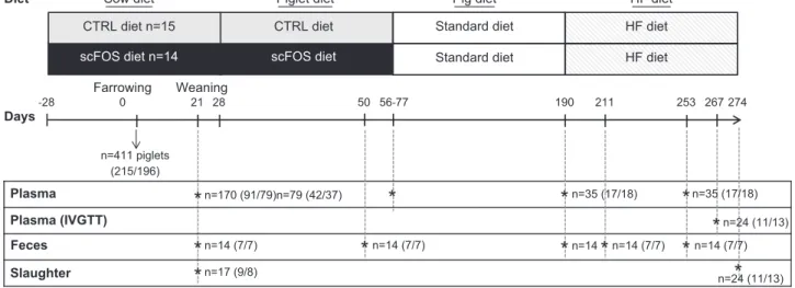
The experimental protocol was designed in compliance with legislations of the European Union (directive 86/609/EEC) and France (decree 2001-464 29/05/01) for the care and use of laboratory animals (agreement for animal housing number C-35-275-32). The regional Ethics Committee in Animal Experiments approved the procedure described herein (2016020217308570). Twenty-nine sows (large white3 land-race) and their piglets [(large white3 landrace) 3 Pietrain] from the INRA (Saint-Gilles, France) experimental herd were used in 2 replicates [n = 17 in the first replicate (R1) and n = 12 in the second replicate (R2)]. From 28 d before the presumed day of farrowing, sows were fed a standard diet (Cooperl, Lamballe, France), supplemented with either scFOS (95% of scFOS with molecular chain length between 3 and 5 mono-meric units; Profeed P95; Beghin-Meiji, Marckolsheim, France; scFOS group; n = 14) or maltodextrin as a control [Maldex; Tereos Starch & Sweeteners Europe, Marckolsheim, France; control (CTRL) group; n = 15; Fig. 1]. Sows were given 3 kg d21feed during gestation and were fed ad libitum during lactation, resulting in an approximate daily intake of 10 g scFOS over the experimental gestation and lactation periods, as detailed previously (21). Before weaning, sow-reared pig-lets had no access to creep feed. At weaning [postnatal day (PND) 28], piglets were fed ad libitum a commercial starter diet, supplemented with 0.15% scFOS (n = 37) or maltodextrin (CTRL; n = 42), according to the maternal diet, during 4–7 wk. This early postweaning period corresponds to the onset of food diversification in humans; this is why we decided to cover this period with scFOS supplementation too, in addition to maternal supplementation. Then, pigs were randomly se-lected to be used further in the experiment [n = 17 CTRL (10 from R1 and 7 from R2) and n = 18 scFOS (11 from R1 and 7 from R2)] and reared on a commercial growing diet until 6 mo of age, followed by a high-fat (HF), high-energetic diet, for-mulated at INRA to provide 22.6% of energy from lipids, until 9 mo of age (PND 274; Fig. 1). Offspring body weight was measured weekly until weaning and then every 2 wk during the supplementation period. From PND 155 (5 wk before the introduction of the HF diet), pigs were weighed weekly until the end of the experiment. Pigs were monitored daily for food intake, as well as for fever or diarrhea. No medication or an-tibiotic treatment was administered throughout the experi-mental protocol. Composition of sow diets and pig diets is provided in Supplemental Table 1.
Fecal and blood sample collection
Fecal and blood samples were collected at different stages of development during the experiment. Fecal samples were collected from a subset of piglets at PND 21, 50, 190, 211, and 253 to analyze microbiota composition (PND 21, n = 6 per group; PND 190, n = 6 per group; PND 253, n = 6 per group)
and to determine the evolution of SCFA production (n = 7 per group and per age; Fig. 1). For microbiota analysis, sub-samples of feces were stored immediately at 280°C after collection without any treatment, whereas for SCFA analy-sis, 1 ml 0.5% ortho-phosphoric acid solution per gram of feces was added, and samples were centrifuged at 1700 g for 15 min at 4°C. Supernatants were then stored at220°C for later analysis. Blood was collected in a subset of piglets at PND 21 (n = 91 CTRL and n = 79 scFOS) and after weaning at PND 77 (n = 42 CTRL and n = 37 scFOS), PND 190 (n = 17 CTRL and n = 18 scFOS), and PND 253 (n = 17 CTRL and n = 18 scFOS) in tubes containing EDTA. After centrifugation at 2500 g for 10 min at 4°C, plasma samples were stored at 220°C for further analysis of glucose, insulin, and inflam-matory marker concentrations.
16S rRNA high-throughput sequencing of fecal microbiota
Genomic DNA was extracted from feces using a QIAamp DNA Stool Mini Kit (Qiagen, Hilden, Germany), according to the manufacturer’s instructions, including a bead-beating step. PCR amplification of the V1–V3 region of the 16S rDNA and library preparation was performed as previously described (22). In brief, PCR amplification of the V1–V3 region of the 16S rDNA and library preparation was performed with the following primers (with Illumina overhand adapters): forward (59-GAGAGTTT-GATYMTGGCTCAG-39) and reverse (59-ACCGCGGCTGCT-GGCAC-39). Each PCR product was purified with the Agencourt AMPure XP beads kit (Beckman Coulter, Pasadena, CA, USA) and submitted to a second PCR round for indexing, using the Nextera XT index primers 1 and 2. After purification, PCR products were quantified using the Quant-IT PicoGreen (Thermo Fisher Scientific, Waltham, MA, USA) and diluted to 10 ng/ml. A final quantification, by quantitative PCR, of each sample in the library was performed using the KAPA SYBR FAST qPCR Kit (Kapa Biosystems, Wilmington, MA, USA) before normalization, pooling, and sequencing on a MiSeq sequencer using v3 reagents (Illumina, San Diego, CA, USA).
The 16S rRNA gene reads were processed with the mothuR package (23). The quality of all sequence reads was denoised us-ing the Pyronoise algorithm implemented in mothuR and filtered with the following criteria: minimal length of 425 bp, an exact match to the barcode, and 1 mismatch allowed to the proximal primer. The sequences were checked for the presence of chime-ric amplifications using Uchime. The resultant read sets were compared with a reference dataset of aligned sequences of the corresponding region derived from the SILVA database (v1.19) of full-length rRNA gene sequences implemented in mothuR (24). The final reads were clustered into operational taxonomic units (OTUs) using the nearest neighbor algorithm using mothuR with a 0.03 distance unit cutoff. A taxonomic identity was attributed to each OTU by comparison with the SILVA database (80% homo-geneity cutoff). As mothuR is not dedicated to the taxonomic assignment beyond the genus level, all unique sequences for each OTU were compared with the SILVA dataset 111 using BLASTN algorithm. For each OTU, a consensus-detailed taxonomic iden-tification has been given based on the identity (,1% of mismatch with the aligned sequence) and the metadata associated with the best hit (validated bacterial species or not).
SCFA measurement in feces
SCFA assay was performed by gas chromatography in super-natant of ortho-phosphoric acid-treated feces as previously de-scribed (25).
Tissue sample collection
At PND 21 and 274, 17 (n = 9 CTRL and n = 8 scFOS) and 24 (n = 11 CTRL and n = 13 scFOS) pigs, respectively, were euthanized in our experimental slaughterhouse by electrical stunning and exsanguination (Fig. 1). After opening the intestinal cavity, por-tal blood was collected (PND 21) in tubes containing the dipeptidylpeptidase-IV inhibitor (10 ml/ml blood; EMD Millipore, Billerica, MA, USA) and EDTA. Mesenteric lymph
Days-28 0 21 28 56-77 190 274 Farrowing Plasma Plasma (IVGTT) Feces Slaughter Weaning n=170 (91/79)n=79 (42/37) n=35 (17/18) 267 n=24 (11/13) n=17 (9/8) n=14 n=14 (7/7) 211 253 n=14 (7/7)
*
*
n=24 (11/13)*
n=14 (7/7)*
*
*
*
*
*
*
*
n=35 (17/18) n=411 piglets (215/196) n=14 (7/7) 50*
scFOS diet n=14 Diet scFOS diet CTRL diet n=15Sow diet Piglet diet
CTRL diet Standard diet
Standard diet HF diet HF diet t e i d F H t e i d g i P
Figure 1. Overview of the study design. Twenty-nine sows and their piglets were used in 2 replicates (n = 17 in R1 and n = 12 in R2). From 28 d before the presumed day of farrowing, sows were fed either a control diet (n = 15, CTRL group) or scFOS-supplemented diet (n = 14, scFOS group) until the end of the lactation. Plasma was collected in suckled piglets (n = 170) at PND 21 and in weaned pigs at PND 77 (n = 79), PND 190 (n = 35), and PND 253 (n = 35) to analyze glucose homeostasis at different stages. An IVGTT was performed at the end of the experiment at PND 267 (n = 24) to evaluate the glucose homeostasis. Fecal contents were collected at PND 21, PND 190, PND 211, and PND 253 (n = 14/age of collect) to determine the SCFA production, reflective of the fermentative activity of the microbiota. Seventeen piglets were euthanized at PND 21 to investigate the development of the enteropancreatic axis, and 24 pigs were euthanized at the adult stage to evaluate the adaptation of the enteropancreatic axis to 3 mo of the HF diet.
nodes (MLNs) were removed and placed immediately in HBSS (MilliporeSigma, Saint-Quentin Fallavier, France), supple-mented with 2% fetal calf serum (FCS), 100 IU/ml penicillin, and 100 mg/ml streptomycin (MilliporeSigma) for mononuclear im-mune cell isolation. Ileal biopsies (with and without the Peyer’s patch) were rinsed with PBS containing 1% DTT (MilliporeSigma) and 1% FCS and then placed in medium made with 74% PBS, 25% DMEM (MilliporeSigma), and 1% FCS, supplemented with 5 g/ml gentamicin (MilliporeSigma) and 1.2 mg/ml amphotericin B (MilliporeSigma) for immediate explant cul-ture. Intestinal tissues from distal ileum (10 cm proximal to the ileocecal junction), cecum (distal part, i.e., bottom of the organ), and proximal colon (at the end of the first third of colon; PDN 21 and 274) were collected, rinsed with cold PBS, and stored at280°C until GLP-1 extraction and assay. For later immunohisto-chemical analyses, samples of 1 cm3were dissected from cecum and from the pancreas body and fixed in 4% paraformalde-hyde. Another sample of the pancreas body was collected and stored immediately at280°C for insulin extraction. In addition, at PND 274, a 5 cm ileal segment was rinsed with cold PBS, and collected ileal lavage was stored at220°C until secretory IgA (sIgA) analysis. Mucosa was scrapped from a 10 cm ileal segment for further intestinal alkaline phosphatase (IAP) assay. Samples of dorsal longissimus muscle and visceral and dorsal subcutaneous adipose tissue were frozen in liquid nitrogen and stored at280°C until further molecular biology analysis.
Intravenous glucose tolerance test
At PND 253, under general anesthesia, a catheter was inserted into 1 external jugular vein of the animals (26) (n = 11 CTRL and n = 13 scFOS). One week after surgery and overnight food withdrawal, the intravenous glucose tolerance test (IVGTT) was performed, consisting in multiple blood samplings: 2 basal samples were taken at 30 and 15 min before the intravenous glucose injection (0.5 g/kg body weight) and at 0 (just after the end of glucose injection), 3, 6, 10, 15, 20, 25, 30, 35, 40, 50, 60, and 75 min after glucose injection. Blood was collected in tubes con-taining EDTA (or EDTA plus a dipeptidylpeptidase-IV inhibitor, as described previously in Tissue sample collection). After cen-trifugation at 2500 g for 10 min at 4°C, plasma samples were stored at220°C (or 280°C for GLP-1 assay-dedicated samples) for later analyses. Insulin and glucose assays were performed on all plasma samples. GLP-1 assays were only performed on basal samples. The incremental area under the curve (AUC) was cal-culated over 75 and 30 min for glucose and insulin. The acute insulin response (=mean insulin concentration above basal val-ues for the first 6 min) and the rate of glucose disappearance (KG;
=the negative slope of the regression line obtained with the log-transformed plasma glucose values from 3 to 30 min, in percentage per minute) were also calculated. Insulin sensitivity was evaluated from fasting basal values of glucose and insulin using the homeo-stasis model assessment insulin-resistance index [HOMA-IR; = (basal value of glucose3 basal value of insulin)/22.5] and by cal-culating the insulin-sensitivity index 2 (S2), described to be well
correlated to the insulin-sensitivity index obtained by the gold standard euglycemic hyperinsulinemic clamp (27) and taking into account AUC0–30 minof insulin and KG. The glucose and insulin
responses were also integrated using the minimal model, described by Bergman (28), to obtain the insulin-sensitivity index SI, as well as
the glucose efficiency index (SG).
Hormone, glucose, lipid, and inflammatory marker assays
Insulin content was extracted from the pancreas in 20 ml etha-nol acid solution (1.5% HCl 12 M, 75% absolute ethaetha-nol, 23.5% H2O; Polytron 3100; Kinematica, Bohemia, NY, USA; 24,000 rpm,
23 20 s). Insulin concentrations were measured on plasma and pancreas extracts (dilution 1:3000) by a commercially available radioimmunoassay kit using iodinated porcine insulin (Insulin-CT; Cisbio Bioassays, Codolet, France). GLP-1 content was extracted from ileal mucosa, cecum, and colon by homogeniza-tion of 1 g tissue in 5 ml ethanol acid soluhomogeniza-tion (1% HCl 12 M, 74% absolute ethanol, 25% H2O; Polytron 3100; Kinematica; 24,000
rpm, 23 20 s). Then, a GLP-1 concentration was measured in plasma and intestinal samples (dilutions 1:1000, 1:200, and 1:250 for ileal mucosa, cecum, and colon, respectively) using a com-mercially available GLP-1 (active) ELISA kit (EMD Millipore).
Plasma glucose, nonesterified fatty acids, triglycerides, cho-lesterol, and haptoglobin were assessed by an automated spec-trophotometric method (Konelab 20i; Thermo Fisher Scientific, Illkirsh, France) using specific commercial kits for each marker (BioM´erieux, Bruz, France). Plasma LPS was measured using the porcine LPS ELISA kit (MyBioSource, San Diego, CA, USA). The concentration of bioactive phosphatase alkaline was deter-mined in ileal mucosa with a commercial kit (Sensolyte; Anaspec, San Jose, CA, USA) using paranitrophenyl phosphate as the substrate. Alkaline phosphatase activity concentrations were expressed per milligram of soluble protein.
sIgA was measured in ileal washes. Once collected, samples of ileal washes were homogenized and centrifuged for 10 min at 4°C at 500 g, and supernatants were collected and stored at220°C until analysis of the total sIgA level using the swine IgA ELISA Quantitation Kit (Bethyl Laboratories, Montgomery, AL, USA).
Finally, MLN cells were isolated by mechanical dissociation before purification over a density gradient (Histopaque, density of 1.077 g/ml; MilliporeSigma) and cultured for 72 h at 37°C under an atmosphere containing 5% CO2, as we previously
de-scribed (21). Culture of both types of ileal biopsies (with and without the Peyer’s patch) were processed, as already described (29), and incubated for 20 h. Three different conditions of culture were performed for MLN cells and biopsies: unstimulated con-dition, in the presence of 5 mg/ml concanavalin A [ConA; from Canavalia ensiformis (Jack bean); MilliporeSigma], or in the pres-ence of 10 mg/ml LPS (ultrapure LPS, from Escherichia coli 0111: B4 strain; InvivoGen, Toulouse, France) for MLN cells and 25 mg/ ml LPS (from E. coli 055:B5 strain; MilliporeSigma) for biopsies. Supernatants were collected and stored at220°C for later cytokine analysis by ELISA (R&D Systems Europe, Lille, France). Concen-trations were expressed in picograms per milliliter of supernatants for MLN cells (IFN-g, TNF-a, IL-10) and picograms per milligram of tissue for explant cultures (IFN-g, TNF-a, IL-10, IL-8).
Immunohistochemical analyses
After fixation for 24 h in 4% paraformaldehyde, pancreas and cecum samples were cryoprotected overnight at 4°C in PBS containing 30% sucrose, embedded in the optimum cutting temperature compound (TissueTek; Sakura Finetek Europe, Zoeterwoude, The Netherlands), frozen in isopentane, and sec-tioned (10 mm) using a cryostat-microtome. Immunohistochem-ical analysis of pancreas was processed, as previously described (30), to obtain the number, area of islets, and percentage of en-docrine tissue. Cecum sections were incubated with PBS con-taining 4% normal horse serum and 0.5% Triton X-100 for 30 min. Sections were then exposed overnight to a rabbit anti-GLP-1 antibody (1:500; Abcam, Cambridge, United Kingdom). After washing with PBS, they were incubated with an anti-rabbit an-tibody conjugated with FITC (1:200; Jackson ImmunoResearch Laboratories, West Grove, PA, USA) for 3 h. Sections were washed again with PBS, coverslipped with Vectashield (Vector Laboratories, Burlingame, CA, USA), and examined with a fluorescence microscope (Eclipse E400; Nikon Instruments France, Champigny-Sur-Marne, France) attached to a digital camera (Digital Still DXM 1200; Nikon Instruments France). The whole section of the tissue was scanned using a digital slide
scanner (Nanozoomer 2.0-RS; Hamamatsu Photonics France, Massy, France). Recorded files were analyzed using the software NDPI (Hamamatsu Photonics France) for determination of the number of GLP-1-secreting cells per area of mucosa.
RNA extraction and real-time quantitative PCR To characterize sensitivity of peripheral tissues to insulin, we analyzed mRNA levels of GLUT-4 and insulin receptor (INSR) in long back muscle, subcutaneous and visceral adi-pose tissues, plus sterol regulatory element-binding tran-scription factor 1C (SREBP1C) in both adipose tissues—the level of these mRNA being well correlated to protein ex-pression in these tissues (31, 32). Total RNA from adipose tissue and muscle samples was extracted via the Trizol method (Thermo Fisher Scientific, Waltham, MA, USA). Extracted RNA samples were quantified using a NanoDrop ND-1000 spectrophotometer (Thermo Fisher Scientific, Wal-tham, MA, USA). The RNA quality was verified using the Agilent RNA 6000 Nano kit and an Agilent 2100 Bioanalyzer (Agilent Technologies France, Massy, France). All samples met quality criteria. All extracted RNA exhibited an RNA Integrity Number.7. Total RNA (2 mg) was used for reverse transcription, performed according to the manufacturer protocol (High Capacity Complementary DNA Reverse Transcription Kit; Thermo Fisher Scientific, Waltham, MA, USA). Real-time PCR was performed with the StepOnePlus real-time PCR machine using SyberGreen master mix (Thermo Fisher Scientific, Waltham, MA, USA) for detection. For each primer pair, efficiencies of PCR were measured by the slope of a standard curve using serial dilutions of a pool of cDNA from the present experiment. In this experiment, they ranged from 95 to 100%, with R2. 0.99. The relative quantity of target gene transcripts was normalized against the geo-metric mean of 3 or 2 housekeeping genes and analyzed using the DDCtmethod (33). In this experiment, Ribosomal Protein
L4 (RPL4), Glyceraldehyde-3-Phosphate Dehydrogenase (GAPDH), and Tyrosine 3-Monooxygenase/Tryptophan 5-Monooxygenase Activation Protein Zeta (YWHAZ) were used as housekeeping genes for muscle and visceral adipose tissue samples, whereas GAPDH and Hypoxanthine phos-phoribosyltransferase 1 (HPRT1) were identified as stable reference genes for subcutaneous adipose tissue samples, as they exhibit high-stability values among samples and were not significantly affected by dietary treatments (Supple-mental Table 2). Relative quantification of genes of interest was obtained comparing the treated group (scFOS) with the untreated group (CTRL).
Statistical analysis
With the consideration of microbiota analysis, statistical differ-ences of population abundance between groups were assessed with ANOVA, corrected for multitesting (Benjamini-Hochberg False Discovery Rate) using STAMP software (http://kiwi.cs.dal. ca/Software/STAMP) (34). Statistical differences of specific bacterial populations between the same age groups were assessed by 2-way ANOVA and Tukey-Kramer post hoc test using GraphPad Prism software (v.6.04 for Windows; GraphPad Software, La Jolla, CA, USA; www.graphpad.com). All other data, not relating to the composition of the microbiota, were subjected to ANOVA using R Core Team (2013; R Foundation for Statis-tical Computing, Vienna, Austria; http://www.R-project.org/). Two-way ANOVA, with diet (CTRL vs. scFOS), replicate (R1 vs. R2), and their interaction as factors, was performed for body weight, body composition, and metabolic and inflammatory parameters at each age. Adult pig body weight and energy in-take during the HF diet period were subjected to ANOVA with repeated measurements, including diet (CTRL vs. scFOS), time (weeks), replicate (R1 vs. R2), and the interactions between diet and time and diet and replication. When diet effect or interaction between diet and time was significant, differences between di-etary groups at each time were further measured by Tukey post hoc test. Insulin and glucose responses to IVGTT were analyzed by ANOVA with repeated measurements, including diet (CTRL vs. scFOS), time (minutes after glucose injection), replication (R1 vs. R2), and the interactions between diet and time and diet and replication, followed by Tukey post hoc test. Correlations among all parameters were evaluated using Spearman’s Rho (R) test. All data are presented as means6SEM. Statistical significance was defined as P # 0.05, and trend was reported as P # 0.10.
RESULTS
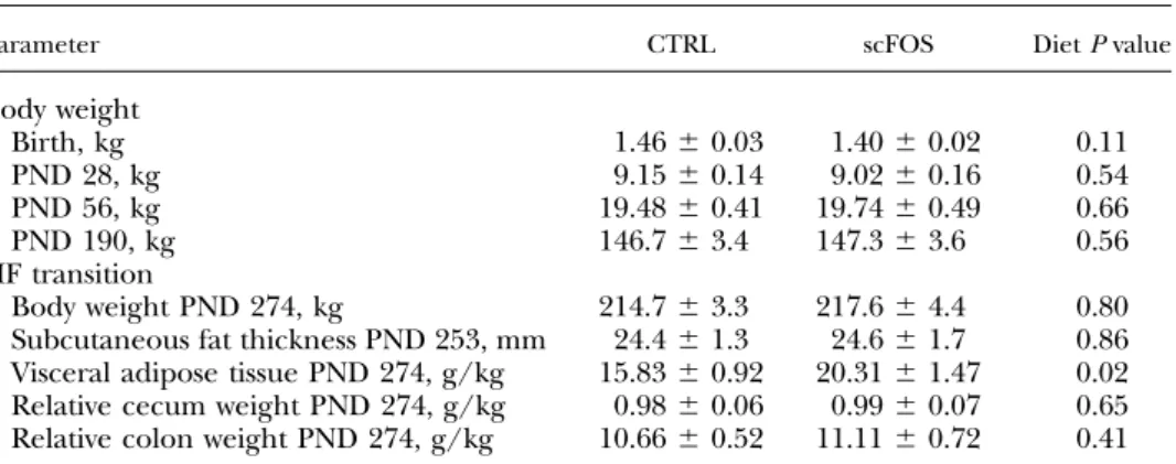
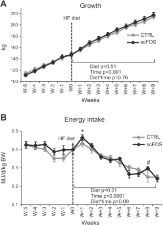
Early scFOS supplementation did not modify body weight at any stage of development (until PND 190; Table 1). The HF diet did not induce any further growth differences between scFOS and CTRL pigs (Fig. 2A). The energy in-take of the HF diet decreased continuously with time, ex-cept during the first week, with a peak of energy intake (Fig. 2B) being higher for the scFOS group than for CTRL pigs (P = 0.02) but without differences on growth between both groups (Fig. 2A). Moreover, scFOS pigs tended to ingest more energy during the eighth week (P = 0.07) of the HF diet (Fig. 2B).
TABLE 1. Growth performance from birth to PND 274 and body composition (PND 253–274) of pigs born from sows supplemented with scFOS or not during the perinatal life
Parameter CTRL scFOS Diet P value
Body weight Birth, kg 1.466 0.03 1.406 0.02 0.11 PND 28, kg 9.156 0.14 9.026 0.16 0.54 PND 56, kg 19.486 0.41 19.746 0.49 0.66 PND 190, kg 146.76 3.4 147.36 3.6 0.56 HF transition Body weight PND 274, kg 214.76 3.3 217.66 4.4 0.80
Subcutaneous fat thickness PND 253, mm 24.46 1.3 24.66 1.7 0.86 Visceral adipose tissue PND 274, g/kg 15.836 0.92 20.316 1.47 0.02 Relative cecum weight PND 274, g/kg 0.986 0.06 0.996 0.07 0.65 Relative colon weight PND 274, g/kg 10.666 0.52 11.116 0.72 0.41
Mean values6SEM. CTRL, perinatal control diet; HF, high-fat diet; PND, postnatal day; scFOS, perinatal short-chain fructooligosaccharide supplemented diet.
Early scFOS intake modified fecal microbiota composition and fecal SCFA content and impacted the glucose metabolism of suckling piglets
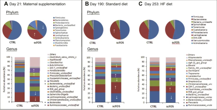
In 21-day-old piglets nursed by sows supplemented with scFOS, the abundance of bacteria belonging to Bacter-oidetes phylum was increased (P, 0.001), whereas that of bacteria from Firmicutes phylum was reduced (P, 0.001; Fig. 3A). At the genus level, maternal scFOS supplemen-tation induced a higher relative proportion of Prevotella (P,0.001),Bacteroidales_unclassifed(P,0.01),andTreponema (P, 0.001) and a reduction of Bacteroides (P , 0.001) and Ruminococcaceae_unclassified (P, 0.001; Fig. 3A). The highest proportion of Treponema was specifically linked to 1 piglet. The fecal SCFA content was also modified with an increase in total SCFA concentration in feces of suckling piglets whose mothers received scFOS supplementation (P = 0.001; Table 2), more particularly, acetate, propionate, valerate, and caproate (P , 0.05). Yet, these differences in fecal SCFA concentrations were not maintained after weaning at the end of the scFOS supplementation (PND 50; Table 2).
Plasma insulin concentration of scFOS piglets at PND 21 was reduced (P = 0.02) with no difference in glycemia, resulting in a lower insulin/glucose ratio (P = 0.02; Table 3). In addition, plasma GLP-1 concentration tended to decrease at PND 21 in scFOS piglets (P = 0.09) without modulation of the density of GLP-1-secretingL-cells in the cecal part of the intestine (Table 3). The proportion of pancreatic endocrine tissue was significantly decreased in scFOS piglets (P = 0.01), as well as the pancreatic content of insulin (P = 0.05; Table 3). In addition, the macronutrient composition of sow’s milk, measured 3 wk after farrow-ing, indicated that scFOS-supplemented sows produced milk with higher dry matter (P = 0.004), lipid (P = 0.01), and protein (P = 0.09) contents, resulting in a higher energy value (P = 0.005; Supplemental Table 3).
The early changes in fecal microbiota were maintained in older pigs independently of the diet
At PND 190, while they were fed a standard diet, pigs from the scFOS group displayed a higher proportion of Bac-teroidetes phylum (P , 0.001), as a consequence of the increase of the Prevotella genus abundance (P, 0.001; Fig. 3B). A higher proportion of the Lachnospiraceae_unclassified genus (P, 0.001) was also observed in the scFOS group. In addition, the early scFOS supplementation induced a de-crease in the proportion of Ruminococcaceae_unclassified and Bacteroidales_unclassified genuses (P, 0.05; Fig. 3B). However, the fecal SCFA concentrations were not differ-ent between groups (P = 0.76; Table 2).
Three weeks after the beginning of the HF diet (at PND 211), scFOS pigs tended to display a higher acetate con-centration in feces (P = 0.09), but it did not persist 6 wk later (at PND 253; Table 2). However, the microbiota composi-tion differed between CTRL and scFOS groups at PND 253. Adult scFOS pigs, under an HF diet, displayed an increased proportion of Proteobacteria phylum (P , 0.01) without any changes in the 2 main phyla (Firmicutes and Bacter-oidetes; Fig. 3C). At the genus level, the proportion of Pre-votella was still higher in the scFOS group (P, 0.001; Fig. 3C). Interestingly, the Prevotella genus abundance was positively correlated to total SCFA concentration in fecal content (P, 0.05; R = 0.48).
Early scFOS supplementation programmed the enteroinsular axis and glucose
metabolism in adult pigs
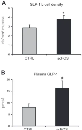
At PND 253, the GLP-1-secretingL-cell density in the ce-cum was higher (+33%) in the scFOS group compared with CTRL pigs (P = 0.03; Fig. 4A). Concomitantly, fasting plasma GLP-1 concentration tended to increase approxi-mately twice in the scFOS group compared with the CTRL group (P = 0.09; Fig. 4B). Interestingly, these 2 parameters were positively correlated (P = 0.05, R = 0.47). In the pan-creas, no difference was measured on the proportion of endocrine tissue (0.976 0.16% for CTRL and 0.96 6 0.09% for the scFOS group) nor on the insulin content (6.706 1.61 mUI/g for CTRL and 7.206 1.57 mUI/g for the scFOS group). However, the pancreatic insulin content was
Figure 2. Body weight and food intake in adult pigs. A) Growth of adult pigs (n = 17 CTRL and n = 18 scFOS). Growth increased significantly with time (P , 0.001), whatever the perinatal diet. B) Energy intake expressed in mega joules per day per kilogram of body weight (BW; n = 17 CTRL and n = 18 scFOS). Energy intake decreased with time (P, 0.001), and a tendency to an interaction between perinatal diet and time (P = 0.09) was observed. CTRL, perinatal control diet; HF, high-fat; scFOS, perinatal short-chain fructooligosaccharide supplemented diet.; W, weeks after the beginning of the HF diet. Mean values6SEM. *P, 0.05 vs. CTRL, #P , 0.10 vs. CTRL.
positively correlated to the density of cecal GLP-1L-cells (P = 0.001, R = 0.65).
Plasma fasting glucose and insulin concentrations were not different between groups, neither before (PND 190) nor after the HF diet period (PND 253; Table 4), nor were the lipid concentrations (triglycerides, free fatty acids, and total cholesterol; data not shown). When the glucose ho-meostasis was challenged by an intravenous injection of
glucose (IVGTT), scFOS animals secreted more insulin than CTRL animals with no difference in glucose profile (Fig. 5). Calculated indexes from insulin and glucose profiles resulted in a tendency to a higher insulin AUC between 0 and 30 min after glucose injection in scFOS pigs (P = 0.07), as well as a reduction in S2(P = 0.09) and in SI (P = 0.02) indices (Table 4). A negative correlation was established between glucose AUC0–30 min and the cecal GLP-1-producingL-cells (P = 0.001, R =20.62).
Interestingly, the HOMA-IR index was negatively correlated to the relative abundance of Prevotella (R = 20.64), but positively to the Ruminococcaceae_unclassified genus (R = 0.66). Conversely, plasma GLP-1 concentra-tion was negatively correlated to the proporconcentra-tion of the Ruminococcaceae_unclassified genus (R =20.60) but positively to total SCFA (R = 0.66), acetate (R = 0.67), propionate (R = 0.53), and butyrate (R = 0.54) in feces.
Early scFOS intake did not modify insulin sensitivity but reduced proinflammatory cytokine expression in visceral adipose tissue No difference in subcutaneous adiposity but an increase in visceral adipose tissue weight was noticed in scFOS pigs (P = 0.02). No other difference in relative organ weight was recorded between groups at slaughter (Table 1). None of the studied genes representative of insulin sensitivity in muscle and adipose tissues displayed differences in mRNA levels between CTRL and scFOS groups at PND 273 (Table 5). TNF-a and IL-10 expression was not different between scFOS and CTRL animals in the sub-cutaneous adipose tissue, but TNF-a expression signifi-cantly decreased in the visceral adipose tissue of scFOS pigs compared with CTRL pigs (P = 0.02; Table 5). Plasma
Figure 3. Fecal microbiota composition analysis by 16S profiling. Mean phylotype distribution (phylum and genus levels) expressed as cumulated relative abundance mean (n = 6 per group) at PND 21 during suckling (A), PND 190 after standard diet feeding (B), and at PND 253 after a 3 mo period of HF diet feeding (C ). CTRL, perinatal control diet; scFOS, perinatal short-chain fructooligosaccharide supplemented diet; HF, high-fat. *↑↓P , 0.05 between groups at the same age.
TABLE 2. SCFA concentration in feces (millimoles per kilogram) of pigs supplemented perinatally with scFOS or not
SCFA CTRL scFOS Diet P value
PND 21 Total SCFA 16.56 4.6 33.46 2.8 0.001 Acetate 10.06 2.7 20.56 1.5 0.003 Propionate 1.96 0.6 4.96 0.6 0.005 Butyrate 1.76 0.6 3.16 1.0 0.37 Valerate 0.376 0.12 1.036 0.28 0.04 Caproate 0.0086 0.007 0.1876 0.091 0.004 PND 50 Total SCFA 138.76 24.2 127.36 19.1 0.70 PND 190 Total SCFA 101.36 12.4 106.06 8.3 0.76 HF transition PND 211 Total SCFA 90.16 6.1 114.16 13.2 0.12 Acetate 52.36 3.3 67.26 7.5 0.09 Propionate 18.96 1.3 23.06 2.4 0.16 Butyrate 10.76 1.3 13.76 2.4 0.28 PND 253 Total SCFA 91.46 16.1 87.56 7.1 0.83
Mean values6SEM. CTRL, perinatal control diet; PND, postnatal day; scFOS, perinatal short-chain fructooligosaccharide supplemented diet; HF, high-fat diet.
haptoglobin and LPS concentrations were not modified by early scFOS supplementation (Table 6).
Early scFOS intake improved intestinal protection in adult pigs
Adult scFOS pigs displayed a tendency for an increased IAP level in the ileal mucosa (P = 0.098) and a significant
increase in sIgA concentration in ileal washes (P = 0.03) compared with CTRL pigs (Table 6). The concentration of sIgA in ileal washes was positively correlated to the IAP level (P = 0.03, R = 0.46) and negatively to the plasmatic LPS (P = 0.02, R =20.48). In vitro stimulation of MLN cells from scFOS animals with ConA mitogen secreted more IFN-g than those from CTRL animals (P = 0.003), but there was no difference when MLN cells were stimulated with LPS (Table 6). The secretion of the other measured cyto-kines (TNF-a and IL-10) was not different between groups (Table 6) in both conditions (except LPS-induced IL-10 secretion, which tended to be lower in scFOS; P = 0.06). Cytokine secretions (TNF-a, IL-10, IFN-g, and IL-8), after in vitro culture of ileal explants, with and without the Peyer’s patch, were not modified by early scFOS supple-mentation (Supplemental Table 4).
DISCUSSION
Accumulating evidences suggest that nutrition during fetal and early postnatal life programs microbial, meta-bolic, and immune development in offspring, with later consequences on their susceptibility to metabolic diseases. We showed that scFOS supplementation, known to well balance the gut microbiota during perinatal life, programs the intestinal endocrine function with increased pan-creas sensitivity to glucose in a GLP-1-dependent manner through microbiota changes, as well as the resistance to inflammation. This scFOS-induced programming may be beneficial to cope with an unbalanced diet in adulthood.
We first analyzed the microbiota and metabolic profile of suckling piglets whose mothers were supplemented with scFOS or not. Our results demonstrated that it is possible to modulate the microbiota establishment in suckling piglets by maternal scFOS supplementation during the perinatal period. Indeed, the bacterial compo-sition of scFOS offspring displayed a higher proportion of Bacteroidetes phylum and more particularly, an in-crease in the Prevotella genus compared with the non-supplemented CTRL group. Total SCFA content, notably acetate, was increased in feces of suckling scFOS piglets.
TABLE 3. Glucose homeostasis and enteroinsular axis in suckling piglets (PND 21) born from sows supplemented in scFOS or not during the perinatal life
Parameter CTRL scFOS Diet P value
Plasma Glucose, mM 7.556 0.12 (n = 90) 7.296 0.16 (n = 79) 0.50 Insulin, mIU/ml 9.546 0.58 (n = 91) 8.246 0.61 (n = 79) 0.02 Insulin/glucose 1.266 0.08 (n = 90) 1.086 0.08 (n = 78) 0.02 GLP-1, pM 7.186 1.74 (n = 8) 3.406 0.43 (n = 7) 0.09 Cecum GLP-1-secretingL-cell, nb/mm2mucosa 1.736 0.20 (n = 5) 1.376 0.41 (n = 5) 0.46 Pancreas Endocrine tissue proportion, % 2.876 0.20 (n = 6) 1.996 0.15 (n = 5) 0.01 Insulin content, IU/g 13.626 0.60 (n = 8) 11.436 0.84 (n = 6) 0.05
Mean values6SEM. Parameters were measured 1 h after breastfeeding. PND, postnatal day; CTRL, perinatal control diet; scFOS, perinatal short-chain fructooligosaccharide supplemented diet.
Figure 4. GLP-1-secretingL-cell density in cecum and plasma GLP-1 concentration at PND 273. A) GLP-1-secreting L-cell density in cecum (n = 11 CTRL and n = 13 scFOS). B) Plasma GLP-1 concentration after an overnight food withdrawal (n = 11 CTRL and n = 13 scFOS). CTRL, perinatal control diet; scFOS, perinatal short-chain fructooligosaccharide supple-mented diet. Mean values6SEM.*P, 0.05, #P , 0.10.
This is consistent with the increase of Prevotella, known to ferment complex carbohydrates and to produce high amounts of SCFA (35). In connection with these early microbiota modifications, the maternal scFOS supple-mentation induced an improvement of the glucose toler-ance in suckling piglets, as the amount of insulin required to return to the basal glycemia after suckling was dimin-ished. Maternal scFOS supplementation did not modify the intestinal endocrine function, with the same L-cell density and GLP-1 tissue content between both groups, but reduced the development of the endocrine pancreas, objectified by a reduction in endocrine tissue percentage and insulin secretion by the pancreas, in association with a tendency to a decreased plasma GLP-1 concentration. The difference of maternal milk composition, with an increase in lipid content and energy value and a tendency for more proteins with scFOS supplementation, could explain such effects. In fact, a modulation of the diet during the perinatal life (via the maternal diet or directly by the postnatal diet) can induce significant effects on endocrine pancreas de-velopment and glucose tolerance (36–38). In addition, the lower development of endocrine pancreas, a potential consequence of a lower request of its endocrine function, could be a result of an improved insulin sensitivity of the peripheral tissues, as observed in rats fed by mothers supplemented with 21.6% of oligofructose (OF)/inulin, with an overexpression of uncoupling protein 1 and peroxi-some proliferator-activated receptor-g coactivator 1a in adi-pose tissue (39).
Afterward, we analyzed the programming effects of such early modifications in adulthood. There was no difference in fecal SCFA concentration between scFOS and CTRL groups after weaning or in adulthood, con-trary to our previous results showing an increase bu-tyrate content in the distal intestine in 2 (20)- and 3-mo-old pigs (21). However, early scFOS-induced differences in microbiota composition persisted until
adulthood, particularly the higher relative proportion of the Prevotella genus. Accordingly, in another study in rats, microbiota modulation in early life by maternal OF/inulin intake (21.6% from conception to weaning) induced persistent modifications with an increase of Bifidobacterium spp. and Clostridium coccoides in adult rat offspring (40).
In association with these microbiota changes, metabolic parameters of the host were modulated by early scFOS intake. Actually, a higher fasting GLP-1 concentration in adult pigs that received early scFOS supplementation correlated to a significant increased GLP-1-secretingL-cell density in the cecum, indicating an increased capacity of the scFOS adult offspring to secrete GLP-1. Adult rats, following a perinatal OF supplementation, did not display changes in postprandial GLP-1 level but showed an in-crease in gastric inhibitory peptide concentration, another incretin hormone, secreted by more proximal K cells (41). Moreover, when ingested directly by healthy adults or adults displaying deleterious metabolic conditions (as a result of an HF diet or obesity), prebiotics increased the number of intestinal GLP-1-producing L-cells, GLP-1 content, and circulating levels of GLP-1 in humans, rats, and mice (42–49), which might, in turn, improve their metabolic status (50). SCFAs, produced by the microbiota after scFOS intake, increase the production and release of GLP-1 by enteroendocrineL-cells in the intestine and favor L-cell differentiation in crypts (47). In our study, perinatal scFOS consumption displayed similar effects on L-cells, 7 mo after the end of supplementation. One explanation could be the maintenance over time of a favorable micro-biota, notably with high fermentative activity, established during early life. This is supported by the positive correlation observed between the SCFA production and plasmatic GLP-1 concentration. Therefore, our data demonstrated a programming of the intestinal endocrine function, probably mediated by the early establishment
TABLE 4. Glycemia, insulinemia, and IVGTT indices before (PND 190) and after (PND 253) the 3 mo of an HF diet in adult pigs supplemented perinatally with scFOS or not
Parameter CTRL scFOS Diet P value
PND 190 (standard diet)
Fasting glucose, mM 4.496 0.12 4.466 0.15 0.629
Fasting insulin, mIU/ml 18.706 1.29 18.176 2.08 0.436
HOMA-IR 3.806 0.32 3.806 0.64 0.440 PND 253 (HF diet) Glucose, mM 4.756 0.12 4.846 0.12 0.872 Insulin, mIU/ml 17.546 2.47 18.156 1.97 0.878 HOMA-IR 3.776 0.55 4.036 0.53 0.956 IVGTT indices
AUCGlucose 0–30 min 503.56 21.8 521.86 14.8 0.483
AUCInsulin 0–30 min 4235.16 339.5 5316.56 430.8 0.068
AIRInsulin 0–6 min 114.56 9.5 136.66 11.6 0.164
KG(%/min) 4.196 0.42 4.06 0.27 0.695
S2[min21× (mIU × ml21)21] 3.726 0.61 2.666 0.23 0.085
SI[104× min21× (mIU × ml21)21] 6.896 0.81 5.316 0.84 0.019
SG(102× min21) 2.516 0.24 2.046 0.21 0.109
Mean values6SEM. AIR, acute insulin response; AUC, area under the curve; CTRL, perinatal control diet; HOMA-IR, homeostasis model assessment of insulin resistance; KG, rate of glucose disappearance; PND, postnatal day; scFOS, perinatal short-chain fructooligosaccharide supplemented diet; S2and SI, insulin sensitivity indexes; SG, glucose efficiency index.
of a beneficial microbiota composition and fermentative activity, which persisted into adulthood.
Once released from intestinalL-cells, GLP-1 stimulates insulin secretion and has a trophic effect on the pancreas by
regulating islet cell proliferation and differentiation (9). No difference was observed in pancreatic anatomy and in-sulin content between scFOS and CTRL adult pigs, but the scFOS group tended to secrete more insulin in response to an intravenous glucose stimulus with no modification of the glucose profile. This increased insulin release high-lighted a higher pancreas sensitivity to glucose in scFOS pigs, as they displayed no insulin resistance in their pe-ripheral tissues. This could be a result of their higher basal GLP-1 concentration, as suggested by Chan et al. (51), who demonstrated that the increase in doses of GLP-1 stimu-lated insulin release during IVGTT in mice. The positive correlation between GLP-1-secreting L-cell density and pancreatic insulin content underlies such a role of GLP-1 on pancreas insulin-secretion capacity in our study. This was in accordance with data in HF-fed diabetic mice, in which OF supplementation, during 4 wk, was efficient to reduce starvation and postprandial glycemia in associa-tion with increased levels of plasma and pancreatic insulin. These results were obtained in a GLP-1-dependent man-ner through an increased plasma GLP-1 concentration and total GLP-1 content in the proximal colon (44). In our study, we did not observe changes of the glucose profile during the IVGTT, but the negative correlation seen be-tween AUCGlucoseand the GLP-1-producingL-cells sup-ported the role of intestinal endocrine function in the regulation of glucose homeostasis. Interestingly, the ob-served negative correlation between Prevotella abundance and the HOMA-IR index may suggest that the increased Prevotella proportion could be a key factor to prevent de-velopment of insulin resistance.
The programming of the intestinal immune function, which is also dependent of the microbiota composition and its fermentative activity, was investigated in adult pigs. Perinatal scFOS supplementation induced a benefi-cial long-lasting effect on intestinal sIgA secretion and IAP concentration (higher values of both parameters in scFOS pigs), with no significant difference in plasma LPS. The expression of IAP has been shown to be controlled by the gut microbiota (52), as well as the sIgA release in the
TABLE 5. Expression of genes related to insulin sensitivity and inflammation in muscle and adipose tissues in adult pigs supplemented perinatally with scFOS or not
Gene CTRL scFOS Diet P value
Muscle
GLUT4 1.016 0.04 1.006 0.08 0.67
INSR 1.026 0.06 1.116 0.06 0.28
Subcutaneous adipose tissue
GLUT4 1.116 0.16 0.836 0.08 0.12
INSR 1.056 1.00 0.866 0.08 0.14
SREBP1C 1.036 0.09 0.836 0.08 0.10
IL-10 1.126 0.15 1.456 0.24 0.66
TNF-a 1.106 0.13 1.226 0.18 0.59
Visceral adipose tissue
GLUT4 1.066 0.23 0.766 0.09 0.23
INSR 1.026 0.17 0.856 0.06 0.27
SREBP1C 1.016 0.12 0.766 0.09 0.20
TNF-a 1.036 0.20 0.506 0.09 0.02
Values are expressed as the ratio of the relative expression of the scFOS group to the CTRL group. CTRL, perinatal control diet; scFOS, perinatal short-chain fructooligosaccharide supplemented diet.
Figure 5. Glucose and insulin responses to an IVGTT at PND 267. A) Insulin profile (n = 11 CTRL and n = 13 scFOS). B) Glucose profile (n = 11 CTRL and n = 13 scFOS). CTRL, perinatal control diet; scFOS, perinatal short-chain fructooligosaccharide supplemented diet. Mean values6SEM. *P, 0.05 vs. CTRL, #P , 0.10 vs. CTRL.
intestinal lumen (53), in accordance with the persistent microbiota modulation induced by perinatal scFOS sup-plementation in adulthood. The increased sIgA secretion could be linked to the modification of bacteria populations and more particularly, to the higher relative abundance of Prevotella, as suggested by Mach et al. (54), showing a positive correlation between Prevotella and sIgA luminal concentration in weaned pigs. Intestinal sIgA secretion is essential to prevent inflammatory responses by down-regulating the expression of inflammatory cytokines, such as TNF-a and IL-6 (55). Another regulator of intestinal inflammation is IAP, which controls several gut biologic processes, such as LPS detoxification, reducing its toxicity for epithelial cells, inflammation, and bacterial trans-location to the lymphoid organs (56). An increased IAP activity may help to decrease chronically plasma LPS concentration and thereby, may reduce metabolic endo-toxemia, which contributes to HF diet-induced metabolic disorders (50). Although there was no significant differ-ence in plasma LPS, numeric values were 37% lower in scFOS pigs, and once again, TNF-a expression in their visceral adipose tissue was significantly decreased com-pared with CTRL pigs. Accordingly, Hallam et al. (40) showed a reduction of plasma LPS in adult rats sup-plemented with prebiotics in early life, linked to a higher abundance of Bifidobacterium spp., suggested to be mediated by milk oligosaccharides. In our study, maternal scFOS supplementation induced macronutrient composi-tion differences in sow milk, which could contribute to modulate the composition and the metabolism of the microbiota colonizing the neonatal gut and consequently, their long-lasting effects (40). Finally, there was a posi-tive correlation between sIgA and IAP activity and a negative one between sIgA and plasma LPS concentra-tion, underlying a possible link between these different inflammatory parameters. Overall, our data suggested that scFOS adult offspring may be more resistant to inflammation.
The pattern of growth was not affected by perinatal scFOS supplementation nor was the body weight gain during the HF challenge, even if scFOS pigs tended to ingest more energy. Both groups regulated their energy intake after the first week of HF diet transition and did not
develop hyperphagia. scFOS pigs displayed an increased visceral adiposity at PND 274, but as adiposity is pre-dominantly located in subcutaneous adipose tissue in pigs (;65%), whereas internal adiposity represents only 5% of total body fat (57), this reflected only a very slight quan-titative effect on global adiposity. It had indeed no effect on the systemic inflammatory status and on the insulin sen-sitivity of peripheral tissues. Interestingly, even if heavier, the visceral tissue of scFOS pigs expressed less TNF-a compared with the CTRL group. Hallam and Reimer (41) showed a reduction in the percentage of total body fat in adult rats fed an HF high-sucrose diet whose mothers had been supplemented with prebiotics. This discrepancy could be explained by a species variability but also by the higher dose of prebiotic and the different period of supplementa-tion used in their study (21.6% during the whole gestasupplementa-tion) compared with ours (0.33% during the last one-third of gestation and 0.15% for lactation and post weaning), as well as the different diet composition in adulthood (41).
In summary, our study demonstrated that the intestinal endocrine function of adult pigs and their susceptibility to inflammation is submitted to a nutritional programming that may participate to the metabolic imprint. This pro-gramming is highly connected to the microbiota. These results are of high interest to understand nutritional pro-gramming in humans and thus, to develop nutritional strategies to prevent metabolic disorders. Indeed, pigs used as a model for humans are considered to be more relevant than rodents, especially because there is higher similarity in anatomy, physiology, metabolism, and microbiota be-tween humans and pigs (58). Among nutritional strategies, perinatal scFOS supplementation, known to well balance the microbiota durably, seems to be an interesting way to prevent metabolic disorders and susceptibility to inflam-mation later in life, notably when confronted with a dele-terious nutritional environment.
ACKNOWLEDGMENTS
The authors thank the technical staff of NuMeCan for its expert assistance during the course of this project. They also acknowledge all of the staff of Rennes Porcine Experimental TABLE 6. Plasma inflammatory, ileal defense markers, and stimulated MLN cell cytokine secretions in adult pigs supplemented perinatally with scFOS or not
Parameter CTRL scFOS Diet P value
PND 260
Plasma haptoglobin, mg/ml 2.176 0.21 2.506 0.31 0.45
Plasma LPS, ng/ml 17.006 3.73 10.636 2.31 0.64
PND 274
Ileal alkaline phosphatase, mg/g protein 0.366 0.04 0.486 0.05 0.10
Ileal washes sIgA, mg/g protein 25.86 10.0 61.16 24.0 0.03
ConA-induced MLN cell IFN-g secretion, pg/ml 271.36 93.1 502.16 178.5 0.003
LPS-induced MLN cell IFN-g secretion, pg/ml 47.76 23.9 25.56 12.9 0.78
ConA-induced MLN cell TNF-a secretion, pg/ml 439.76 84.5 394.16 76.9 0.69
LPS-induced MLN cell TNF-a secretion, pg/ml 119.26 31.4 86.26 28.9 0.40
ConA-induced MLN cell IL-10 secretion, pg/ml 1167.26 117.5 868.26 103.0 0.14
LPS-induced MLN cell IL-10 secretion, pg/ml 74.86 14.3 44.16 13.8 0.06
Facilities (UEPR) for animal care and feeding. This study received funding from Tereos, a company producing scFOS. F.R. and E.A. are employed by Tereos. A French patent application has beenfiled (FR 17/0035). C.L.B., S.F.B., B.T., A.C., L.L.N., I.L.H.L., and S.B. declare no conflict of interest.
AUTHOR CONTRIBUTIONS
C. Le Bourgot, I. Le Hu¨erou-Luron, and S. Blat designed research; C. Le Bourgot, S. Ferret-Bernard, B. Taminiau, A. Cahu, L. Le Normand, I. Le Hu¨erou-Luron, and S. Blat conducted research and analyzed data; E. Apper and F. Respondek contributed reagents/materials/analysis; C. Le Bourgot, S. Ferret-Bernard, E. Apper, I. Le Hu¨erou-Luron, and S. Blat wrote the manuscript; and all authors read and approved thefinal manuscript and agreed to be accountable for all aspects of the work in ensuring that questions related to the accuracy or integrity of any part of the work are appropriately investigated and resolved.
REFERENCES
1. Gluckman, P. D., Hanson, M. A., and Beedle, A. S. (2007) Early life events and their consequences for later disease: a life history and evolutionary perspective. Am. J. Hum. Biol. 19, 1–19
2. Godfrey, K. M., Gluckman, P. D., and Hanson, M. A. (2010) Developmental origins of metabolic disease: life course and intergenerational perspectives. Trends Endocrinol. Metab. 21, 199–205 3. Bedford Russell, A. R., and Murch, S. H. (2006) Could peripartum antibiotics have delayed health consequences for the infant? BJOG 113, 758–765
4. Schokker, D., Zhang, J., Vastenhouw, S. A., Heilig, H. G., Smidt, H., Rebel, J. M., and Smits, M. A. (2015) Long-lasting effects of early-life antibiotic treatment and routine animal handling on gut microbiota composition and immune system in pigs. PLoS One 10, e0116523 5. Ajslev, T. A., Andersen, C. S., Gamborg, M., Sørensen, T. I., and Jess, T.
(2011) Childhood overweight after establishment of the gut microbiota: the role of delivery mode, pre-pregnancy weight and early administration of antibiotics. Int. J. Obes. 35, 522–529
6. Cox, L. M., Yamanishi, S., Sohn, J., Alekseyenko, A. V., Leung, J. M., Cho, I., Kim, S. G., Li, H., Gao, Z., Mahana, D., Z´arate Rodriguez, J. G., Rogers, A. B., Robine, N., Loke, P., and Blaser, M. J. (2014) Altering the intestinal microbiota during a critical developmental window has lasting metabolic consequences. Cell 158, 705–721
7. Trasande, L., Blustein, J., Liu, M., Corwin, E., Cox, L. M., and Blaser, M. J. (2013) Infant antibiotic exposures and early-life body mass. Int. J. Obes. 37, 16–23
8. Tolhurst, G., Heffron, H., Lam, Y. S., Parker, H. E., Habib, A. M., Diakogiannaki, E., Cameron, J., Grosse, J., Reimann, F., and Gribble, F. M. (2012) Short-chain fatty acids stimulate glucagon-like peptide-1 secretion via the G-protein-coupled receptor FFAR2. Diabetes 61, 364–371
9. Baggio, L. L., and Drucker, D. J. (2007) Biology of incretins: GLP-1 and GIP. Gastroenterology 132, 2131–2157
10. De Filippo, C., Cavalieri, D., Di Paola, M., Ramazzotti, M., Poullet, J. B., Massart, S., Collini, S., Pieraccini, G., and Lionetti, P. (2010) Impact of diet in shaping gut microbiota revealed by a comparative study in children from Europe and rural Africa. Proc. Natl. Acad. Sci. USA 107, 14691–14696
11. Dominguez-Bello, M. G., Costello, E. K., Contreras, M., Magris, M., Hidalgo, G., Fierer, N., and Knight, R. (2010) Delivery mode shapes the acquisition and structure of the initial microbiota across multiple body habitats in newborns. Proc. Natl. Acad. Sci. USA 107, 11971–11975 12. Penders, J., Thijs, C., Vink, C., Stelma, F. F., Snijders, B., Kummeling, I., van den Brandt, P. A., and Stobberingh, E. E. (2006) Factors influencing the composition of the intestinal microbiota in early infancy. Pediatrics 118, 511–521
13. Pannaraj, P. S., Li, F., Cerini, C., Bender, J. M., Yang, S., Rollie, A., Adisetiyo, H., Zabih, S., Lincez, P. J., Bittinger, K., Bailey, A., Bushman, F. D., Sleasman, J. W., and Aldrovandi, G. M. (2017) Association
between breast milk bacterial communities and establishment and development of the infant gut microbiome. JAMA Pediatr. 171, 647–654
14. Roberfroid, M., Gibson, G. R., Hoyles, L., McCartney, A. L., Rastall, R., Rowland, I., Wolvers, D., Watzl, B., Szajewska, H., Stahl, B., Guarner, F., Respondek, F., Whelan, K., Coxam, V., Davicco, M. J., L´eotoing, L., Wittrant, Y., Delzenne, N. M., Cani, P. D., Neyrinck, A. M., and Meheust, A. (2010) Prebiotic effects: metabolic and health benefits. Br. J. Nutr. 104 (Suppl 2), S1–S63
15. Pan, X. D., Chen, F. Q., Wu, T. X., Tang, H. G., and Zhao, Z. Y. (2009) Prebiotic oligosaccharides change the concentrations of short-chain fatty acids and the microbial population of mouse bowel. J. Zhejiang Univ. Sci. B 10, 258–263
16. Respondek, F., Gerard, P., Bossis, M., Boschat, L., Bruneau, A., Rabot, S., Wagner, A., and Martin, J. C. (2013) Short-chain fructo-oligosaccharides modulate intestinal microbiota and metabolic pa-rameters of humanized gnotobiotic diet induced obesity mice. PLoS One 8, e71026
17. Swanson, K. S., Grieshop, C. M., Flickinger, E. A., Bauer, L. L., Chow, J., Wolf, B. W., Garleb, K. A., and Fahey, G. C., Jr. (2002) Fructooligosaccharides and Lactobacillus acidophilus modify gut microbial populations, total tract nutrient digestibilities and fecal protein catabolite concentrations in healthy adult dogs. J. Nutr. 132, 3721–3731
18. Liu, T. W., Cephas, K. D., Holscher, H. D., Kerr, K. R., Mangian, H. F., Tappenden, K. A., and Swanson, K. S. (2016) Nondigestible fructans alter gastrointestinal barrier function, gene expression, histomorphology, and the microbiota profiles of diet-induced obese C57BL/6J mice. J. Nutr. 146, 949–956
19. Fujiwara, R., Watanabe, J., and Sonoyama, K. (2008) Assessing changes in composition of intestinal microbiota in neonatal BALB/c mice through cluster analysis of molecular markers. Br. J. Nutr. 99, 1174–1177
20. Le Bourgot, C., Le Normand, L., Formal, M., Respondek, F., Blat, S., Apper, E., Ferret-Bernard, S., and Le Hu¨erou-Luron, I. (2017) Maternal short-chain fructo-oligosaccharide supplementation in-creases intestinal cytokine secretion, goblet cell number, butyrate concentration and Lawsonia intracellularis humoral vaccine response in weaned pigs. Br. J. Nutr. 117, 83–92
21. Le Bourgot, C., Ferret-Bernard, S., Le Normand, L., Savary, G., Menendez-Aparicio, E., Blat, S., Appert-Bossard, E., Respondek, F., and Le Hu¨erou-Luron, I. (2014) Maternal short-chain fructooligo-saccharide supplementation influences intestinal immune system maturation in piglets. PLoS One 9, e107508
22. Bindels, L. B., Neyrinck, A. M., Salazar, N., Taminiau, B., Druart, C., Muccioli, G. G., François, E., Blecker, C., Richel, A., Daube, G., Mahillon, J., de los Reyes-Gavil´an, C. G., Cani, P. D., and Delzenne, N. M. (2015) Non digestible oligosaccharides modulate the gut microbiota to control the development of leukemia and associated cachexia in mice. PLoS One 10, e0131009
23. Schloss, P. D., Westcott, S. L., Ryabin, T., Hall, J. R., Hartmann, M., Hollister, E. B., Lesniewski, R. A., Oakley, B. B., Parks, D. H., Robinson, C. J., Sahl, J. W., Stres, B., Thallinger, G. G., Van Horn, D. J., and Weber, C. F. (2009) Introducing mothur: open-source, platform-independent, community-supported software for describing and comparing microbial communities. Appl. Environ. Microbiol. 75, 7537–7541
24. Quast, C., Pruesse, E., Yilmaz, P., Gerken, J., Schweer, T., Yarza, P., Peplies, J., and Gl¨ockner, F. O. (2013) The SILVA ribosomal RNA gene database project: improved data processing and web-based tools. Nucleic Acids Res. 41, D590–D596
25. Jouany, J. P., Zainab, B., Senaud, J., Groliere, C. A., Grain, J., and Thivend, P. (1981) Role of the rumen ciliate protozoa Polyplastron multivesiculatum, Entodinium sp. and Isotricha prostoma in the digestion of a mixed diet in sheep. Reprod. Nutr. Dev. 21, 871–884
26. Mosnier, E., Le Floc’h, N., Etienne, M., Ramaekers, P., S`eve, B., and P`ere, M. C. (2010) Reduced feed intake of lactating primiparous sows is associated with increased insulin resistance during the peripartum period and is not modified through supplementation with dietary tryptophan. J. Anim. Sci. 88, 612–625
27. Christoffersen, B., Ribel, U., Raun, K., Golozoubova, V., and Pacini, G. (2009) Evaluation of different methods for assessment of insulin sensitivity in Gottingen minipigs: introduction of a new, simpler method. Am. J. Physiol. Regul. Integr. Comp. Physiol. 297, R1195–R1201 28. Bergman, R. N. (2005) Minimal model: perspective from 2005. Horm.
29. Chatelais, L., Jamin, A., Gras-Le Guen, C., Lall`es, J. P., Le Hu¨erou-Luron, I., and Boudry, G. (2011) The level of protein in milk formula modifies ileal sensitivity to LPS later in life in a piglet model. PLoS One 6, e19594
30. Blat, S., Morise, A., Sauret, A., Louveau, I., Mac´e, K., Le Hu¨erou-Luron, I., and S`eve, B. (2012) The protein level of isoenergetic formulae does not modulate postprandial insulin secretion in piglets and has no consequences on later glucose tolerance. Br. J. Nutr. 108, 102–112
31. Atkinson, B. J., Griesel, B. A., King, C. D., Josey, M. A., and Olson, A. L. (2013) Moderate GLUT4 overexpression improves insulin sensitivity and fasting triglyceridemia in high-fat diet-fed transgenic mice. Di-abetes 62, 2249–2258
32. Kampmann, U., Christensen, B., Nielsen, T. S., Pedersen, S. B., Ørskov, L., Lund, S., Møller, N., and Jessen, N. (2011) GLUT4 and UBC9 protein expression is reduced in muscle from type 2 diabetic patients with severe insulin resistance. PLoS One 6, e27854
33. Livak, K. J., and Schmittgen, T. D. (2001) Analysis of relative gene expression data using real-time quantitative PCR and the 2(2Delta Delta C(T)) method. Methods 25, 402–408
34. Parks, D. H., and Beiko, R. G. (2010) Identifying biologically relevant differences between metagenomic communities. Bioinformatics 26, 715–721
35. Chen, T., Long, W., Zhang, C., Liu, S., Zhao, L., and Hamaker, B. R. (2017) Fiber-utilizing capacity varies in Prevotella- versus Bacteroides-dominated gut microbiota. Sci. Rep. 7, 2594
36. Delamaire, E., Parnet, P., Coup´e, B., Hoebler, C., Blat, S., Poupeau, G., Boquien, C. Y., Champ, M., and Darmaun, D. (2012) Long term metabolic impact of high protein neonatal feeding: a preliminary study in male rat pups born with a low birth weight. Clin. Nutr. 31, 741–748
37. Guo, F., and Jen, K. L. (1995) High-fat feeding during pregnancy and lactation affects offspring metabolism in rats. Physiol. Behav. 57, 681–686
38. Petrik, J., Reusens, B., Arany, E., Remacle, C., Coelho, C., Hoet, J. J., and Hill, D. J. (1999) A low protein diet alters the balance of islet cell replication and apoptosis in the fetal and neonatal rat and is associated with a reduced pancreatic expression of insulin-like growth factor-II. Endocrinology 140, 4861–4873
39. Maurer, A. D., and Reimer, R. A. (2011) Maternal consumption of high-prebioticfibre or -protein diets during pregnancy and lactation differentially influences satiety hormones and expression of genes involved in glucose and lipid metabolism in offspring in rats. Br. J. Nutr. 105, 329–338
40. Hallam, M. C., Barile, D., Meyrand, M., German, J. B., and Reimer, R. A. (2014) Maternal high-protein or high-prebiotic-fiber diets affect maternal milk composition and gut microbiota in rat dams and their offspring. Obesity (Silver Spring) 22, 2344–2351
41. Hallam, M. C., and Reimer, R. A. (2013) A maternal high-protein diet predisposes female offspring to increased fat mass in adulthood whereas a prebioticfibredietdecreasesfatmassinrats.Br.J.Nutr.110,1732–1741 42. Cani, P. D., Dewever, C., and Delzenne, N. M. (2004) Inulin-type fructans modulate gastrointestinal peptides involved in appetite reg-ulation (glucagon-like peptide-1 and ghrelin) in rats. Br. J. Nutr. 92, 521–526
43. Cani, P. D., Hoste, S., Guiot, Y., and Delzenne, N. M. (2007) Dietary non-digestible carbohydrates promote L-cell differentiation in the proximal colon of rats. Br. J. Nutr. 98, 32–37
44. Cani, P. D., Knauf, C., Iglesias, M. A., Drucker, D. J., Delzenne, N. M., and Burcelin, R. (2006) Improvement of glucose tolerance and
hepatic insulin sensitivity by oligofructose requires a functional glucagon-like peptide 1 receptor. Diabetes 55, 1484–1490
45. Cani, P. D., Possemiers, S., Van de Wiele, T., Guiot, Y., Everard, A., Rottier, O., Geurts, L., Naslain, D., Neyrinck, A., Lambert, D. M., Muccioli, G. G., and Delzenne, N. M. (2009) Changes in gut microbiota control inflammation in obese mice through a mechanism involving GLP-2-driven improvement of gut permeabil-ity. Gut 58, 1091–1103
46. Delm´ee, E., Cani, P. D., Gual, G., Knauf, C., Burcelin, R., Maton, N., and Delzenne, N. M. (2006) Relation between colonic proglucagon expression and metabolic response to oligofructose in high fat diet-fed mice. Life Sci. 79, 1007–1013
47. Kaji, I., Karaki, S., Tanaka, R., and Kuwahara, A. (2011) Density distribution of free fatty acid receptor 2 (FFA2)-expressing and GLP-1-producing enteroendocrine L cells in human and rat lower intestine, and increased cell numbers after ingestion of fructo-oligosaccharide. J. Mol. Histol. 42, 27–38
48. Parnell, J. A., and Reimer, R. A. (2012) Prebiotic fibres dose-dependently increase satiety hormones and alter Bacteroidetes and Firmicutes in lean and obese JCR:LA-cp rats. Br. J. Nutr. 107, 601–613: 49. Everard, A., Lazarevic, V., Derrien, M., Girard, M., Muccioli, G. G., Neyrinck, A. M., Possemiers, S., Van Holle, A., François, P., de Vos, W. M., Delzenne, N. M., Schrenzel, J., and Cani, P. D. (2011) Responses of gut microbiota and glucose and lipid metabolism to prebiotics in genetic obese and diet-induced leptin-resistant mice. [Erratum: Diabetes (2011) 60:3307.] Diabetes 60, 2775–2786
50. Everard, A., and Cani, P. D. (2013) Diabetes, obesity and gut microbiota. Best Pract. Res. Clin. Gastroenterol. 27, 73–83
51. Chan, H. M., Jain, R., Ahr´en, B., Pacini, G., and D’Argenio, D. Z. (2011) Effects of increasing doses of glucagon-like peptide-1 on insulin-releasing phases during intravenous glucose administration in mice. Am. J. Physiol. Regul. Integr. Comp. Physiol. 300, R1126–R1133 52. Bates, J. M., Akerlund, J., Mittge, E., and Guillemin, K. (2007)
Intestinal alkaline phosphatase detoxifies lipopolysaccharide and prevents inflammation in zebrafish in response to the gut microbiota. Cell Host Microbe 2, 371–382
53. Ohland, C. L., and Jobin, C. (2015) Microbial activities and intestinal homeostasis: a delicate balance between health and disease. Cell. Mol. Gastroenterol. Hepatol. 1, 28–40
54. Mach, N., Berri, M., Estell´e, J., Levenez, F., Lemonnier, G., Denis, C., Leplat, J. J., Chevaleyre, C., Billon, Y., Dor´e, J., Rogel-Gaillard, C., and Lepage, P. (2015) Early-life establishment of the swine gut micro-biome and impact on host phenotypes. Environ. Microbiol. Rep. 7, 554–569
55. Boullier, S., Tanguy, M., Kadaoui, K. A., Caubet, C., Sansonetti, P., Corth´esy, B., and Phalipon, A. (2009) Secretory IgA-mediated neu-tralization of Shigellaflexneri prevents intestinal tissue destruction by down-regulating inflammatory circuits. J. Immunol. 183, 5879–5885 56. Lall`es, J. P. (2010) Intestinal alkaline phosphatase: multiple biological
roles in maintenance of intestinal homeostasis and modulation by diet. Nutr. Rev. 68, 323–332
57. Mourot, J. (2004) Control of the adiposity in breeding animals. Bull. Acad. Vet. Fr. 157, 29–34
58. Roura, E., Koopmans, S. J., Lall`es, J. P., Le Huerou-Luron, I., de Jager, N., Schuurman, T., and Val-Laillet, D. (2016) Critical review evaluating the pig as a model for human nutritional physiology. Nutr. Res. Rev. 29, 60–90
Received for publication January 18, 2018. Accepted for publication June 18, 2018.