2-Aminopropane-1,2,3-tricarboxylic acid: Synthesis and co-crystallization
with the class A b-lactamase BS3 of Bacillus licheniformis
Joséphine Beck
a, Eric Sauvage
b, Paulette Charlier
b, Jacqueline Marchand-Brynaert
a,*a
Unité de Chimie Organique et Médicinale, Université catholique de Louvain, Bâtiment Lavoisier, Place Louis Pasteur 1, B-1348 Louvain-la-Neuve, Belgium
bCentre d’Ingénierie des Protéines, Institut de physique B5a, Université de Liège, B-4000 Sart- Tilman, Belgium
a r t i c l e
i n f o
Article history: Received 31 March 2008 Revised 8 May 2008 Accepted 9 May 2008 Available online 16 May 2008 Keywords:
2-Aminopropane-1,2,3-tricarboxylic acid Glycine bis-alkylation
b-Lactamase inhibitors Aminocitrate-BS3 complex X-ray diffraction analysis
a b s t r a c t
The title compound 4 has been prepared in four steps from ethylglycinate in 63% overall yield. This amino analog of citric acid has been co-crystallized with the class A b-lactamase BS3 of Bacillus licheniformis and the structure of the complex fully analyzed by X-ray diffraction. Tris-ethyl aminocitrate 3 and the free tris-acid 4 have been tested against a member b-lactamase from all distinct subgroups. They are novel inhibitors of class A b-lactamases, still modest but more potent than citrate and isocitrate.
Ó 2008 Elsevier Ltd. All rights reserved.
The production of b-lactamases represents the most widespread
and often the most efficient mechanism devised by bacteria to
es-cape the lethal action of b-lactam antibiotics.
1Our research focuses
on the synthesis and the biochemical evaluation of potential
inhib-itors of b-lactamases. Numerous b-lactamase inhibinhib-itors have been
reported in the literature.
2However, most of these drugs are
b-ltam derivatives, and when exposed to such molecules, bacteria
ac-quire resistance. To disrupt this vicious circle, non b-lactam
inhibitors may be an alternative. The search of novel structures
considered as ‘hits’ in medicinal chemistry proceeds from different
strategies, such as the mechanism-based design and the screening
of various chemical libraries, but also from serendipity. Our
pres-ent work is precisely based on a fortuitous observation related to
protein crystallography, when using 100 mM sodium citrate buffer
in the crystallization protocols.
Citrate has been recently shown, by X-ray diffraction analysis,
to perfectly fit into the active site of the Bacillus licheniformis BS3
b-lactamase, and to behave as a modest inhibitor of this serine
en-zyme with a micromolar K
ivalue.
3We speculated that the
replace-ment of the hydroxyl function of citrate with an amine function
could enhance the affinity for the target enzyme. Moreover, the
amine function could be used to introduce different side chains
susceptible to modulate the interactions within the active site.
In this paper, we propose a simple method for preparing the
amino analog of citric acid, namely
2-aminopropane-1,2,3-tricar-boxylic acid, and we compare the structures of complexes formed
between citrate or aminocitrate and the BS3 b-lactamase. The
citrate regio-isomer, that is, isocitrate, has been also involved in
the structural and biochemical studies. Our aim was to pave the
route towards the discovery of novel anti-b-lactamase compounds
(
Fig. 1
).
The previous syntheses of 2-aminopropane-1,2,3-tricarboxylic
acid are based on two methods: (i) the hydrolysis of hydantoin
prepared from diethyl or diisobutylacetone-dicarboxylate
4,5and
(ii) the double alkylation of nitro-acetate anion with haloesters
followed by reduction and ester hydrolysis.
6,7Here we disclose
an alternative, more practical method.
Fully protected 2-amino-propane-1,2,3-tricarboxylic acid
deriv-ative 2 was prepared by bis-alkylation of the glycine imine
precur-sor 1 (
Scheme 1
), a methodology originally developed by
O’Donnell, and currently applied for the synthesis of particular
amino acids.
8–10The majority of glycine mono-alkylation reactions
0960-894X/$ - see front matter Ó 2008 Elsevier Ltd. All rights reserved. doi:10.1016/j.bmcl.2008.05.045
* Corresponding author. Tel.: +32 10 47 27 40; fax: +32 10 47 41 68. E-mail address:jacqueline.marchand@uclouvain.be(J. Marchand-Brynaert).
HO
CO
2H
HO
2C
CO
2H
1 2 3 4HO
*CO
2H
*HO
2C
CO
2H
1 2 3 4H
2N
CO
2H
HO
2C
CO
2H
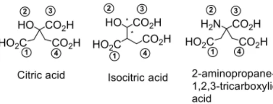
1 2 3 4Citric acid
Isocitric acid
2-aminopropane-1,2,3-tricarboxylic
acid
Figure 1. Structures of compounds of interest and numbering of their functional groups.
Contents lists available at
ScienceDirect
Bioorganic & Medicinal Chemistry Letters
makes use of benzophenone-derived imines, while the
bis-alkyla-tions are more readily performed on
m-chlorobenzaldehyde-de-rived imines.
11,12Thus, we prepared the Schiff base 1 from
m-chlorobenzaldehyde and glycine ethyl ester hydrochloride. Crude
imine 1 was then reacted with two equivalents of ethyl
bromoace-tate and an excess of potassium carbonate, under phase transfer
conditions (Method A).
13The completion of bis-alkylation required
a very long reaction time, about 1–2 days of heating in acetonitrile
at 50 °C. Crude compound 2 was quantitatively recovered.
We then considered alternative protocols versus the original
O’Donnell’s method. Using sodium hydride as base in
dimethyl-formamide solution, we performed the deprotonation at 0 °C
and the alkylation at 20 °C; the best results were collected by
addition of the reagents in two successive fractions of one
equiv-alent (Method B).
14Lastly, we used lithium diisopropylamide
(LDA, 2 equiv) for imine 1 deprotonation at low temperature
( 78 °C) in tetrahydrofurane solution.
15After 1 h, two
equiva-lents of ethyl bromoacetate were added and the mixture was
warmed to room temperature (Method C). This protocol
fur-nished similar results to Method B. However, the Method C
was preferred because it allowed working on larger quantities,
always with good and reproducible yields, most probably due
to the fact that the reactive medium remains homogeneous.
Crude 2 was directly submitted to a smooth acidic hydrolysis
followed by a basic work-up to furnish the free amine 3 which
was purified by column chromatography on silica gel with 50%
yield. This yield corresponds to pure product isolated after three
steps of reaction from glycine ester.
16The amine 3 is
character-ized in
1H NMR by two doublets corresponding to the
symmet-rical methylenic protons (4H) around 2.66–2.88 ppm (J = 16 Hz,
AB pattern).
Tris-ester 3 could be fully deprotected by treatment with 6 N
HCl at reflux; the amino analog of citric acid was isolated as the
hydrochloride salt 4 with 90% yield.
17The sequence of reaction
we propose now constitutes the best route towards this non
natu-ral amino acid (four steps from ethylglycinate with only one
chro-matographic purification; overall yield = 63%).
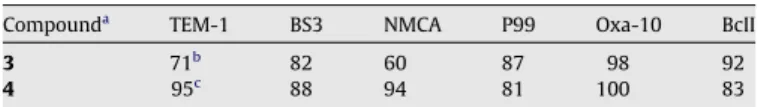
The biochemical activity of tris-ester 3 and tris-acid 4 has been
evaluated at pH 7.2 against representative b-lactamases of class A
(TEM-1,
18BS3,
3NMCA
19), class C (P99
20), class D (Oxa-10
21), and
class B (BcII
22a); all are serine enzymes, except the last one which
is a zinc enzyme working with two Zn(II) in the active site.
22bThe
enzymes (1–100 nM) were incubated with the tested compounds
during 30 min at 37 °C. Then a chromogenic substrate (nitrocefine)
was added and the hydrolysis rate of this substrate was followed
by spectrophotometry at 482 nm.
23Results of
Table 1
(residual
activity) are expressed as percentages of b-lactamases initial
activ-ity. Aminocitrate derivatives 3 and 4 are modest inhibitors of class
A and class C b-lactamases; they are inactive against class D
b-lac-tamase and slightly active as inhibitors of zinc b-lacb-lac-tamase.
The K
ivalues have been measured in the cases of BS3 and
TEM-1 enzymes for aminocitrate 4, citrate and isocitrate. As shown in
Table 2
, the amino analog is the more active compound.
This result stimulates our interest in comparative
crystallo-graphic studies of BS3-4 and BS3-isocitrate complexes with the
known BS3-citrate complex.
3,24The novel complexes were
ob-tained by a similar method.
25Among the residues with a demonstrated role in the catalytic
process of b-lactam hydrolysis by class A b-lactamases, Ser70 and
Ser130 are directly involved in the catalytic mechanism. Thr235
and Arg244 interact with the substrate carboxylate and are thus
in-volved in the positioning of the b-lactam antibiotic in the active
site. Class A b-lactamases are also characterized by the presence
of an oxyanion hole defined by the amide groups of Ser70 and
Ala237, which draws the b-lactam carbonyl oxyanion in the course
of hydrolysis of the b-lactam ring.
26Citrate was firstly observed in
the active site of BS3 enzyme as a co-crystallization product.
3N
CO
2Et
N
CO
2Et
EtO
2C
CO
2Et
H
2N
CO
2Et
EtO
2C
CO
2Et
Cl
Cl
Cl
CHO
Cl
H
3N
CO
2Et
iBr
CO
2Et
2
ii iii ivH
3N
CO
2H
HO
2C
CO
2H
Cl
1
2
3
4
Scheme 1. Synthesis of aminocitrate. Reagents and conditions: (i) CH2Cl2, Et3N,
MgSO4, 20 °C, 24 h; (ii) Method A: K2CO3, Bu4NBr, CH3CN, 50 °C, 1–2 days; Method
B: NaH, DMF, 0–20 °C, two successive additions; Method C: LDA, THF, 78–20 °C; (iii) 1 N HCl, CH3CN, 20 °C, then basic work-up; (iv) 6 N HCl.
Table 1
Inhibition of b-lactamases by aminocitrate derivatives Compounda
TEM-1 BS3 NMCA P99 Oxa-10 BcII
3 71b
82 60 87 98 92
4 95c
88 94 81 100 83
Results expressed as percentages of initial activities.
a
Compounds were tested at pH 7.2 in 50 mM phosphate buffer and at a con-centration of 100 lM, otherwise mentioned.
b 0.2 mM. c 2 mM. Table 2 Kivaluesa(lM) Compound BS3 TEM-1 Citrate 490 730 Isocitrate 2200 1500 4 250 150 a
Compounds were tested at pH 5 in 25 mM acetate buffer.
Figure 2. Inhibition of the class A b-lactamase of B. licheniformis BS3 by citrate. Orange molecular surface shows the active site cleft with a molecule of citrate (in yellow sticks). Residues of the active site interacting with the citrate molecule are shown in green stick. Oxygen atoms are red and nitrogen atoms are blue. Citrate carboxylates are numbered according to theFigure 1. Blue surface indicates the oxyanion hole. Colors are identical forFigures 3 and 4.
As shown in
Figure 2
, an oxygen atom of the carboxylate 1 of
the citrate molecule interacts with the two catalytic serines
(2.5 Å to Ser70 Oc and 2.4 Å to Ser130 Oc). One oxygen atom of
the carboxylate 3 is at 2.8 Å from Thr235 Oc and at 3.2 Å from
Ser130 Oc, whereas the other oxygen atom interacts with Arg244
and Thr235. The hydroxyl group points out of the catalytic cleft
and the carboxylate 4 salt bridges with Arg244. The carboxylate
3 occupies a position identical to the carboxylate group of a
b-lac-tam antibiotic (for example in the acyl-enzyme complex between
BS3 and cefoxitin).
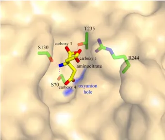
3Although aminocitrate (
Fig. 3
) only differs from citrate by the
substitution of the hydroxyl group with an amine group, its
posi-tion and orientaposi-tion in the active site and the interacposi-tions with
the catalytic cleft residues are different from those of the citrate,
except carboxylate 3 that makes similar interactions. The amine
group of the aminocitrate directly interacts with Ser70 Oc and
Ser130 Oc (3.2 Å and 2.3 Å, respectively) and an oxygen atom of
the carboxylate 4 lies in the oxyanion hole.
In the case of isocitrate (
Fig. 4
), carboxylate 1 interacts with
Ser70 Oc and Ser130 Oc, whereas an oxygen atom of the
carboxyl-ate 4 is in the oxyanion hole.
27Table 3
summarizes the direct interactions between amino acid
residues and the inhibitors functional groups.
Clearly, citrate, isocitrate and aminocitrate adopt different
con-formations in the active site of BS3. However, the main difference
concerns the functional group which interacts with the two
cata-lytic serine residues (Ser70 and Ser130): a carboxylate for citrate
and isocitrate and the amine group in the case of amino-citrate.
This could probably explain the enhanced biochemical activity of
4 compared to (iso)citrate. Modification of the amine group of 4,
as initially proposed, seems now less appropriate than
substitu-tions at the level of the carboxylic funcsubstitu-tions for further
develop-ments. As a matter of fact, the tris-ethyl ester 3 was found to be
slightly more active than 4 in the competitive inhibition tests
ver-sus b-lactamases (
Table 1
).
The interest of using X-ray data for the design of
structure-based small-molecule enzyme inhibitors remains an open
ques-tion, the technique featuring as much advantages as drawbacks.
28Considering citrate to identify new ‘hits’, the situation is quite
ambiguous since the complexes formed with class A and class C
b-lactamases showed different structures.
29In conclusion, we have set up a practical synthesis of
2-amino-1,2,3-tricarboxylic acid (amino analog of citric acid) and shown
that this non natural aminoacid 4 behaves as a modest inhibitor
of several b-lactamases. Beside its potential, but debatable, use as
Figure 3. Inhibition of the class A b-lactamase of B. licheniformis BS3 by aminocitrate.
Figure 4. Inhibition of the class A b-lactamase of B. licheniformis BS3 by isocitrate.
Table 3
Summary of direct interactions between amino acid residues and functional groups 1–4 (seeFig. 1)
Group-atoms BS3-citratea
BS3-aminocitratea
BS3-isocitratea
Carboxylate 1-O1 Ser130 Oc (2.4) — Ser130 Oc (2.1)
Ser70 Oc (2.5) Ser70 Oc (2.6)
Carboxylate 1-O2 — — Thr235 Oc (2.8)
Hydroxyl 2/amine 2 — Ser130 Oc (2.3) —
Ser70 Oc (3.2)
Carboxylate 3-O1 Ser130 Oc (3.2) Thr235 Oc (2.3) Thr235 Oc (3.1)
Thr235 Oc (2.8) Arg244 NH1 (3.2)
Carboxylate 3-O2 Arg244 NH1 (3.1) — Thr235 Oc (2.8)
Thr235 Oc (3.1)
Carboxylate 4-O1 Arg244 NH1 (2.8) Ala237 NH (2.9) Ala237 NH (2.8)
Ser70 (2.9) Ser70 (3.0)
Carboxylate 4-O2 — — —
a
novel ‘hit’ towards anti-b-lactamases, aminocitrate 4 has been
al-ready recognized as a valuable compound for cation complexation.
Applications in the field of heavy metal sequestering agents have
been
disclosed,
30that
should
benefit
of
our
convenient
synthesis.
Acknowledgments
The biochemical evaluations have been performed in the
labo-ratories of professors J. M. Frère and M. Galleni (University of
Liège). We thank R. Herman for protein crystallization work, and
the staff of beamline BM30a at ESRF for assistance in X-ray data
collection. This work was supported in part by the Belgian Program
on Interuniversity Poles of Attraction initiated by the Belgian State,
Prime Minister’s Office, Science Policy Programming (PAI 5/33 and
PAI 6/19), by the Fonds National de la Recherche Scientifique (IISN
4.4505.00, FRFC 9.45/9.99, FRFC 2.4.508.01.F, FRFC 9.4.538.03.F,
FRFC 2.4.524.03) and the University of Liège (Fonds spéciaux,
Cré-dit classique). J.M.-B. is senior research associate of the FRS-FNRS
(Belgium).
Supplementary data
Supplementary data associated with this article can be found, in
the online version, at
doi:10.1016/j.bmcl.2008.05.045
.
References and notes
1. Fisher, J. F.; Meroueh, S. O.; Mobashery, S. Chem. Rev. 2005, 105, 395. 2. Sandanayaka, V. P.; Prashad, A. S. Curr. Med. Chem. 2002, 9, 1145.
3. Fonze, E.; Vanhove, M.; Dive, G.; Sauvage, E.; Frere, J-M. J.-M.; Charlier, P. Biochemistry 2002, 41, 1877.
4. Durnow, A.; Rombusch, K. Chem. Ber. 1955, 88, 1334.
5. Connors, T. A.; Mauger, A. B.; Peutherer, M. A.; Ross, W. C. J. J. Chem. Soc. 1962, 4601.
6. Kaji, E.; Zen, S. Bull. Chem. Soc. Jpn 1973, 46, 337.
7. Fu, Y.; Hammarstrom, L. G.; Miller, T. J.; Franczek, F. R.; Mc Laughlin, M. L.; Hammer, R. P. J. Org. Chem. 2001, 66, 7118.
8. O’Donnell, M. J. Aldrichim. Acta 2001, 34, 3.
9. O’Donnell, M. J.; Polt, R. L. J. Org. Chem. 1982, 47, 2663.
10. Allwein, S. P.; Secord, E. A.; Martins, A.; Mitten, J. V.; Nelson, T. D.; Kress, M. H.; Dolling, H. Synlett 2004, 2489.
11. (a) O’Donnell, M. J.; Wu, S.; Huffman, J. C. Tetrahedron 1994, 50, 4507; (b) O’Donnell, M. J. Acc. Chem. Res. 2004, 37, 506.
12. Kitamura, M.; Shirakawa, S.; Maruoka, K. Angew. Chem., Int. Ed. 2005, 44, 1549. 13. O’Donnell, M. J.; Wojciechowski, K.; Ghosez, L.; Navarro, M.; Sainte, F.; Antoine,
J. P. Synthesis 1984, 4, 313.
14. Park, K.-H.; Abbate, E.; Olmstead, M. M.; Kurth, M. J.; Najdi, S. Chem. Commun. 1998, 16, 1679.
15. Laue, K. W.; Haufe, G. Synthesis 1998, 10, 1453. 16. Preparation of compound 3.
Step i: hydrochloride salt of ethyl glycinate (1 g, 7.16 mmol) and triethylamine (1 equiv, 1 mL, 7.16 mmol) were dissolved in dichloromethane (10 mL) and stirred for 6 h at 20 °C. Magnesium sulfate (1.5 g) and 3-chlorobenzaldehyde (0.9 equiv, 730 lL, 6.44 mmol) were then added and the mixture was further stirred overnight. After filtration, the organic solution was washed three times with brine, dried (MgSO4) and
concentrated under vacuum to give 1 as yellow oil (1.45 g, quantitative yield). HRMS (ESI): 226.0645 (calculated for C11H12ClNO2+H: 226.0635);1H
NMR (CDCl3, 300 MHz): d ppm, 1.32 (t, 3H, CH3, J = 7.14 Hz), 4.26 (q, 2H,
CH2, J = 7.14 Hz), 4.41 (s, 2H, CH2), 7.33–7.45 (m, 2H, Ar), 7.63 (d, 1H, Ar,
J = 7.5 Hz), 7.82 (s, 1H, Ar), 8.25 (s, 1H, NCH).13
C NMR (CDCl3, 125 MHz): d
ppm, 14.36 (CH3), 61.35 (CH2), 62.07 (CH2), 127.00 (CHAr), 128.23 (CHAr),
130.02 (CHAr), 131.31 (CHAr), 134.98 (CAr), 137.47 (CAr), 164.03 (NCH), 170.01 (CO2Et).
Step ii (Method C): to the crude imine 1 (580 mg, 2.6 mmol) dissolved in dry THF (12 mL) and cooled at 78 °C, was added under argon atmosphere LDA (commercial solution in THF, 2.1 equiv, 2.7 mL, 5.42 mmol). After 1h at 78 °C, ethyl bromoacetate was added (2.1 equiv, 570 lL, 5.42 mmol) and the mixture was allowed to slowly warm up to 20 °C, overnight, under stirring. After addition of water, the solution was concentrated under vacuum. The oily residue was dissolved in ether and the organic layer was washed successively with aqueous NaHCO3and brine. Drying over MgSO4
and concentration under vacuum furnished 2 as a brown oil (1 g, quantitative yield). 1
H NMR (CDCl3, 500 MHz): d ppm, 1.24 (t, 6H, CH3,
J = 7.14 Hz), 1.29 (t, 3H, CH3, J = 7.16 Hz), 3.13 (d, 2H, CH2, J = 15.79 Hz), 3.29
(d, 2H, CH2, J = 15.77 Hz), 4.14 (q, 4H, CH2, J = 7.10 Hz); 4.26 (q, 2H, CH2,
J = 7.14 Hz), 7.34 (dd, 1H, Ar, J = 8 Hz and J = 7.6 Hz), 7.40 (ddd, 1H, Ar, J = 8 Hz, J = 2.17 Hz and J = 1.14 Hz), 7.57 (dt, 1H, Ar, J = 7.6 Hz and J = 1.3 Hz), 7.76 (s, 1H, Ar), 8.28 (s, 1H, NCH).13C NMR (CDCl
3, 125 MHz):
dppm, 14.37 (CH3), 40.83 (CH2), 60.79 (CH2), 61.94 (CH2), 67.86 (C), 127.24
(CHAr), 128.19 (CHAr), 130.02 (CHAr), 131.39 (CHAr), 135.00 (CAr), 137.85 (CAr), 159.63 (N@CH), 170.65 (CO2Bn), 171.00 (CO2Me).
Step iii: the crude imine 2 (857 mg, 2.15 mmol) was dissolved in acetonitrile (3 mL) and 1 N HCl was added (1.5 equiv, 3.2 mL, 3.22 mmol). After 30 min at room temperature, the solution was concentrated under vacuum, and the aqueous phase was extracted three times with ether. The aqueous phase was then neutralized with NaHCO3and basified to pH 10 with 1 N NaOH.
This was extracted five times with CH2Cl2. The organic layers were collected
and washed with brine. After drying (MgSO4) and concentration under
vacuum, the residue was purified by column chromatography on silica gel to furnish 3 as yellow oil (296 mg, 50% yield). Rf= 0.5 (CH2Cl2/EtOAc, 1:1);
HRMS (ESI): 276.1450 (calculated for C12H21NO6+1: 276.1447); 1H NMR
(CDCl3, 250 MHz): d ppm, 1.30 (t, 9H, J = 7.13 Hz, CH3), 2.56 (s, 2H, NH2),
2.66–2.72 (d, 2H, J = 16.0 Hz, CH2), 2.82–2.88 (d, 2H, J = 16.0 Hz, CH2), 4.15
(q, 4H, J = 7.13 Hz, CH2);13C NMR (CDCl3, 62.5 MHz): d ppm, 14.3 (CH3) 43.6
(CH2), 55.8 (C), 61.0 (CH2), 61.9 (CH2), 170.5 (CO2Et), 175.0 (CO2Et).
17. Preparation of compound 4. Amine 3 (100 mg) was treated with 6 N HCl (10 mL) at reflux for 1 night. After extraction with CH2Cl2, the aqueous
phase was concentrated under vacuum and the residue was dried under high vacuum to furnish 4 as a white solid (75 mg, 90% yield). HRMS (ESI): 192.0503 (calculated for C6H9NO6+1: 192.0508);1H NMR (D2O, 250 MHz): d
ppm, 2.98 (s, 4H);13C NMR (D
2O, 62.5 MHz): d ppm, 39.9 (CH2), 59.3 (C),
173.7 (CO2H).
18. Fonze, E.; Charlier, P.; To’th, Y.; Vermeire, M.; Raquet, X.; Dubus, A.; Frere, J. M. Acta Crystallogr., Sect. D: Biol. Crystallogr. 1995, D51, 682.
19. Swaren, P.; Maveyraud, L.; Raquet, X.; Cabantous, S.; Duez, C.; Pedelacq, J.-D.; Mariotte-Boyer, S.; Mourey, L.; Labia, R.; Nicolas-Chanoine, M.-H.; Nordmann, P.; Frere, J.-M.; Samama, J.-P. J. Biol. Chem. 1998, 273, 26714.
20. Dubus, A.; Ledent, P.; Lamotte-Brasseur, J.; Frere, J. M. Proteins 1996, 25, 473. 21. (a) Paetzel, M.; Danel, F.; De Castro, L.; Mosimann, S. C.; Page, M. G. P.;
Strynadka, N. C. J. Nat. Struct. Biol. 2000, 7, 918; (b) Golemi, D.; Maveyraud, L.; Vakulenko, S.; Tranier, S.; Ishiwata, A.; Kotra, L. P.; Samama, J.-P.; Mobashery, S. J. Am. Chem. Soc. 2000, 122, 6132.
22. (a) Carfi, A.; Duee, E.; Galleni, M.; Frere, J.-M.; Dideberg, O. Acta Crystallogr., Sect. D: Biol. Crystallogr. 1998, D54, 313; (b) Llarrull, L. I.; Tioni, M. F.; Kowalski, J.; Bennett, B.; Vila, A. J. J. Biol. Chem. 2007, 282, 30586.
23. Determination of biochemical activity. The enzymes were produced and purified as previously described.18–22
The enzymes (1–100 nM) were incubated with the tested compounds (100 lM, otherwise mentioned) and the chromogenic substrate nitrocefine (100 lM) in a phosphate buffer (50 mM, pH 7.2). The hydrolysis rate of this substrate was followed by spectrophotometry at 482 nm. The residual activity was obtained by comparison with the variation of the absorbance of a reference (sample without inhibitor) and indicated inTable 1. Results are expressed as % of initial activities; variations of results are within ±5%. Plot V/VI versus inhibitor
concentration (ratios of hydrolysis in the absence and in the presence of inhibitors) gave the inhibition constant indicated inTable 2. All experiments were performed three times.
24. Ledent, P.; Duez, C.; Vanhove, M.; Lejeune, A.; Fonze, E.; Charlier, P.; Rhazi-Filali, F.; Thamm, I.; Guillaume, G.; Samyn, B.; Devreese, B.; Van Beeumen, J.; Lamotte-Brasseur, J.; Frere, J.-M. FEBS Lett. 1997, 413, 194.
25. BS3 b-lactamase purification, crystallization, and data collection. The expression, purification and initial crystallization conditions of the BS3 enzyme (BS3-citrate) were described previously.3,24
Typically, monoclinic crystals were obtained using the hanging drop vapor diffusion method with drops containing 5 lL of a protein solution (at a concentration of 40 mg/mL in 50 mM NaCl, 10 mM Tris buffer, pH 7.2) and 5 lL of 10% PEG 6000 in 100 mM sodium citrate buffer (pH 3.4), equilibrated against 1 mL of the latter solution at 20 °C. The BS3-isocitrate crystals were obtained in the same conditions, by replacing the citrate buffer by a 100 mM sodium isocitrate buffer at the same pH. The BS3-aminocitrate crystals were grown in drops containing 5 lL of a protein solution (at a concentration of 38 mg/ mL in 50 mM NaCl, 10 mM Tris buffer, pH 7.2), 4 lL of 8% PEG 6000 in 100 mM sodium aminocitrate buffer (pH 3.4) plus 1 lL of 0.1 M urea additive, equilibrated against 1 mL of a 20% PEG 6000 solution, at 20 °C. X-ray diffraction experiments were carried out under cryogenic conditions (100 K) after transferring the crystals into a reservoir solution supplemented with 50% glycerol. The diffraction data for the BS3-isocitrate crystal were measured at ESRF (Grenoble, France) on the FIP-BM30a beamline (k = 1.0 Å) using a MarResearch 165 mm CCD detector. Data for the BS3-aminocitrate crystal were collected with a Rigaku RU-200 rotating anode generator operating at 40 kV and 100 mA and a MarResearch Mar345 Imaging Plate (k = 1.5418 Å). Intensities were indexed and integrated using MOSFLM version 6.01. The scaling of the intensity data was accomplished with SCALA of the CCP4 program suite and all corresponding statistics are available asSupplementary data. The atomic coordinates are available at the Protein Data Bank under the codes 1I2S (citrate), 1W7F (isocitrate) and 3B3X (aminocitrate).
26. Matagne, A.; Lamotte-Brasseur, J.; Frere, J.-M. Biochem. J. 1998, 330, 581. 27. One diastereoisomer, which configuration is (2R,3S), was found in the enzymic
cavity: Heretsch, P.; Thomas, F.; Aurich, A.; Krautscheid, H.; Sicker, D.; Giannis, A. Angew. Chem., Int. Ed. 2008, 47, 1958.
28. Besong, G. E.; Bostock, J. M.; Stubbings, W.; Chopra, I.; Roper, D. I.; Lloyd, A. J.; Fishwick, C. W. G.; Johnson, A. P. Angew. Chem., Int. Ed. 2005, 39, 6403. 29. In the class C b-lactamase CMY2 of Klebsiella pneumoniae (b-lactamase
structurally close to P99 enzyme), the citrate molecule seems to make a 180° rotation in the active site, in comparison with its position in the BS3 catalytic cleft (seeSupplementary data). The atomic coordinates are available at the Protein Data Bank under the code 1ZC2.
30. (a) Brown, M. R.; Shankar, R.; Sallis, J. D. J. Labelled. Compd. Radiopharm. 1984, 21, 905; (b) Tsao, J. W.; Schoen, F. J.; Shankar, R.; Sallis, J. D.; Levy, R. J. Biomaterials 1988, 9, 393; (c) Shankar, R.; Crowden, S.; Sallis, J. D. Atherosclerosis 1984, 52, 191; (d) Shankar, R.; Brown, M. R.; Wong, L. K.; Sallis, J. D. Experientia 1984, 40, 265; (e) Yamamoto, H.; Koide, S.; Takayanagi, Y. PCT Int. Appl. WO 9635662, 1996.; (f) Takahashi, M. Jpn Kokai Tokkyo Koho JP 09059162, 1997.