HAL Id: dumas-03157969
https://dumas.ccsd.cnrs.fr/dumas-03157969
Submitted on 3 Mar 2021HAL is a multi-disciplinary open access archive for the deposit and dissemination of sci-entific research documents, whether they are pub-lished or not. The documents may come from teaching and research institutions in France or abroad, or from public or private research centers.
L’archive ouverte pluridisciplinaire HAL, est destinée au dépôt et à la diffusion de documents scientifiques de niveau recherche, publiés ou non, émanant des établissements d’enseignement et de recherche français ou étrangers, des laboratoires publics ou privés.
The value of electrocardiogram and echocardiography to
distinguish Fabry disease from sarcomeric hypertrophic
cardiomyopathy
Nicolas Junqua
To cite this version:
Nicolas Junqua. The value of electrocardiogram and echocardiography to distinguish Fabry disease from sarcomeric hypertrophic cardiomyopathy. Human health and pathology. 2020. �dumas-03157969�
UNIVERSITÉ de CAEN NORMANDIE
---
UFR SANTÉ
FACULTÉ de MÉDECINE
Année 2019/2020
THÈSE POUR L’OBTENTION
DU GRADE DE DOCTEUR EN MÉDECINE
Présentée et soutenue publiquement le : lundi 7 septembre 2020
par
M JUNQUA Nicolas
Né (e) le 18/11/1992 à Châtenay-Malabry (92)
TITRE DE LA THÈSE
:
Valeur de l’électrocardiogramme couplé à
l’échocardiographie pour différencier une maladie de Fabry
d’une cardiomyopathie hypertrophique sarcomérique
Président :
Monsieur le Professeur MILLIEZ Paul
Membres :
Monsieur LABOMBARDA Fabien
Monsieur LEGALLOIS Damien
Monsieur DE BOYSSON Hubert
Année Universitaire 2019/2020
DoyenProfesseur Emmanuel TOUZÉ Assesseurs
Professeur Paul MILLIEZ (pédagogie) Professeur Guy LAUNOY (recherche)
Professeur Sonia DOLLFUS & Professeur Evelyne EMERY (3ème cycle)
Directrice administra9ve Madame Sarah CHEMTOB PROFESSEURS DES UNIVERSITÉS - PRATICIENS HOSPITALIERS
M.
AGOSTINI Denis
Biophysique et médecine
nucléaire
M.
AIDE Nicolas
Biophysique et médecine
nucléaire
M.
ALLOUCHE Stéphane
Biochimie et biologie moléculaire
M.
ALVES Arnaud
Chirurgie digesRve
M.
AOUBA Achille
Médecine interne
M. BABIN Emmanuel
Oto-Rhino-Laryngologie
M.
BÉNATEAU Hervé
Chirurgie maxillo-faciale et
stomatologie
M.
BENOIST Guillaume
Gynécologie - Obstétrique
M.
BERGER Ludovic
Chirurgie vasculaire
M.
BERGOT Emmanuel
Pneumologie
M.
BIBEAU Frédéric
Anatomie et cytologie
pathologique
Mme BRAZO Perrine
Psychiatrie d’adultes
M.
BROUARD Jacques
Pédiatrie
M.
BUSTANY Pierre
Pharmacologie
Mme CHAPON Françoise
Histologie, Embryologie
Mme CLIN-GODARD Bénédicte
Médecine et santé au travail
M.
DAMAJ Ghandi Laurent
Hématologie
U N I V E R S I T É D E C A E N · N O R M A N D I E
M.
DAO Manh Thông
Hépatologie-Gastro-Entérologie
M.
DEFER Gilles
Neurologie
M.
DELAMILLIEURE Pascal
Psychiatrie d’adultes
M.
DENISE Pierre
Physiologie
Mme DOLLFUS Sonia
Psychiatrie d'adultes
M.
DREYFUS Michel
Gynécologie - Obstétrique
M.
DU CHEYRON Damien
RéanimaRon médicale
Mme ÉMERY Evelyne
Neurochirurgie
M.
ESMAIL-BEYGUI Farzin
Cardiologie
Mme FAUVET Raffaèle
Gynécologie – Obstétrique
M.
FISCHER Marc-Olivier
Anesthésiologie et réanimaRon
M.
GÉRARD Jean-Louis
Anesthésiologie et réanimaRon
M.
GUILLOIS Bernard
Pédiatrie
Mme GUITTET-BAUD Lydia
Epidémiologie, économie de la
santé et prévenRon
M.
HABRAND Jean-Louis
Cancérologie opRon
Radiothérapie
M.
HAMON Mar9al
Cardiologie
Mme HAMON Michèle
Radiologie et imagerie médicale
M.
HANOUZ Jean-Luc
Anesthésie et réa. médecine
péri-opératoire
M.
HULET Christophe
Chirurgie orthopédique et
traumatologique
M.
ICARD Philippe
Chirurgie thoracique et
cardio-vasculaire
M.
JOIN-LAMBERT Olivier
Bactériologie - Virologie
Mme JOLY-LOBBEDEZ Florence
Cancérologie
M.
JOUBERT Michael
Endocrinologie
M.
LAUNOY Guy
Epidémiologie, économie de la
santé et prévenRon
M.
LE HELLO Simon
Bactériologie-Virologie
M. LOBBEDEZ Thierry
Néphrologie
M.
LUBRANO Jean
Chirurgie viscérale et digesRve
M.
MAHE Marc-André
Cancérologie
M.
MANRIQUE Alain
Biophysique et médecine
nucléaire
M.
MARCÉLLI Chris9an
Rhumatologie
M.
MARTINAUD Olivier
Neurologie
M. MAUREL Jean
Chirurgie générale
M.
MILLIEZ Paul
Cardiologie
M.
MOREAU Sylvain
Anatomie/Oto-Rhino-Laryngologie
M.
MOUTEL Grégoire
Médecine légale et droit de la
santé
M.
NORMAND Hervé
Physiologie
M.
PARIENTI Jean-Jacques
BiostaRsRques, info. médicale et tech. de communicaRonM.
PELAGE Jean-Pierre
Radiologie et imagerie médicale
Mme PIQUET Marie-Astrid
NutriRon
M.
QUINTYN Jean-Claude
Ophtalmologie
Mme RAT Anne-Chris9ne
Rhumatologie
M.
RAVASSE Philippe
Chirurgie infanRle
M.
REPESSE Yohann
Hématologie
M.
REZNIK Yves
Endocrinologie
M. ROD Julien
Chirurgie infanRle
M.
ROUPIE Eric
Médecine d’urgence
Mme THARIAT JulieZe
Radiothérapie
M.
TILLOU Xavier
Urologie
M.
TOUZÉ Emmanuel
Neurologie
M.
TROUSSARD Xavier
Hématologie
Mme VABRET Astrid
Bactériologie - Virologie
M.
VERDON Renaud
Maladies infecReuses
Mme VERNEUIL Laurence
Dermatologie
PROFESSEURS ASSOCIÉS DES UNIVERSITÉS A MI-TEMPS
M. DE LA SAYETTE Vincent
Neurologie
Mme DOMPMARTIN-BLANCHÈRE Anne
Dermatologie
M. GUILLAUME Cyril
Médecine palliaRve
M. LE BAS François
Médecine Générale
M. SABATIER Rémi
Cardiologie
PRCE
Mme LELEU Solveig
Anglais
PROFESSEURS EMERITES
M.
HURAULT de LIGNY Bruno
Néphrologie
Mme KOTTLER Marie-Laure
Biochimie et biologie moléculaire
M.
LE COUTOUR Xavier
Epidémiologie, économie de la
santé et prévenRon
M. LEPORRIER Michel
Hématologie
Année Universitaire 2019/2020
DoyenProfesseur Emmanuel TOUZÉ Assesseurs
Professeur Paul MILLIEZ (pédagogie) Professeur Guy LAUNOY (recherche)
Professeur Sonia DOLLFUS & Professeur Evelyne EMERY (3ème cycle)
Directrice administra9ve Madame Sarah CHEMTOB
MAITRES DE CONFERENCES DES UNIVERSITÉS - PRATICIENS HOSPITALIERS
M.
ALEXANDRE Joachim
Pharmacologie clinique
Mme BENHAÏM Annie
Biologie cellulaire
M.
BESNARD Stéphane
Physiologie
Mme BONHOMME Julie
Parasitologie et mycologie
M.
BOUVIER Nicolas
Néphrologie
M.
COULBAULT Laurent
Biochimie et Biologie moléculaire
M.
CREVEUIL Chris9an
BiostaRsRques, info. médicale et
tech. de communicaRon
M. DE BOYSSON Hubert
Médecine interne
Mme DINA Julia
Bactériologie - Virologie
Mme DUPONT Claire
Pédiatrie
M.
ÉTARD Olivier
Physiologie
M.
GABEREL Thomas
Neurochirurgie
M.
GRUCHY Nicolas
GénéRque
M.
GUÉNOLÉ Fabian
Pédopsychiatrie
M.
HITIER Mar9n
Anatomie - ORL Chirurgie
Cervico-faciale
M.
ISNARD Christophe
Bactériologie Virologie
M.
JUSTET Aurélien
Pneumologie
Mme KRIEGER Sophie
Pharmacie
U N I V E R S I T É D E C A E N · N O R M A N D I E
M.
LEGALLOIS Damien
Cardiologie
Mme LELONG-BOULOUARD Véronique
Pharmacologie fondamentale
Mme LEVALLET Guénaëlle
Cytologie et Histologie
M.
MITTRE Hervé
Biologie cellulaire
M.
SESBOÜÉ Bruno
Physiologie
M.
TOUTIRAIS Olivier
Immunologie
M.
VEYSSIERE Alexis
Chirurgie maxillo-faciale et
stomatologie
MAITRES DE CONFERENCES ASSOCIÉS DES UNIVERSITÉS A MI-TEMPS
Mme ABBATE-LERAY Pascale
Médecine générale
M. COUETTE Pierre-André
Médecine générale
Mme NOEL DE JAEGHER Sophie
Médecine générale
M. PITHON Anni
Médecine générale
M. SAINMONT Nicolas
Médecine générale
Mme SCHONBRODT Laure
Médecine générale
MAITRES DE CONFERENCES EMERITES
Mme DEBRUYNE Danièle
Pharmacologie fondamentale
Mme DERLON-BOREL Annie
Hématologie
Remerciements
A Monsieur le professeur Paul Milliez,
Merci de l’honneur que vous me faites d’assister à ma soutenance de thèse en tant que président de jury et de venir juger ce travail de thèse. Merci à vous pour l’enseignement reçu au sein de votre service.
A Fabien,
Je te remercie pour ton immense aide et implicaRon dans ce travail de thèse ainsi que pour le partage de tes connaissance au fil des visites à l’hôpital.
A Damien,
Je te remercie pour ta disponibilité dans ce travail de thèse et pour ton aide précieuse pour les staRsRques.
A Monsieur le docteur Hubert De Boysson
Merci de l’honneur que vous me faites de juger ce travail de thèse.
A mes parents et à ma soeur,
Je vous remercie de m’avoir soutenu tout au long de ces dix longues années d’études et d’avoir su m’aider et m’écouter dans les moments difficiles.
A Kléa,
Je te remercie de ton souRen incondiRonnel, de ton aide de tous les instants et d’être à mes côtés au quoRdien.
A mes amis rencontrés sur Cherbourg,
Un grand merci pour nos soirées passées ensemble et nos bons moments qui ne se comptent plus.
A mes co-internes de cardiologie
Merci à vous d’avoir été là au fil des stages de cet internat qui sera passé bien trop vite (et à nos SIRC de folie).
Abbrevia9ons
ACE: angiotensin-converRng enzyme BSA: body surface area
ESC: European Society of Cardiology Gb3: globotriaosylceramide
HCM: hypertrophic cardiomyopathy LVH: leg ventricular hypertrophy LAH: leg anterior fascicular block LBBB: leg bundle block branch LPH: leg posterior fascicular block LV: Leg ventricle
LVEF: leg ventricular ejecRon fracRon
LVOTO: leg ventricular ouilow tract obstrucRon NYHA: New-York Heart AssociaRon
RBBB: right bundle block branch RVH: right ventricular hypertrophy
Tables
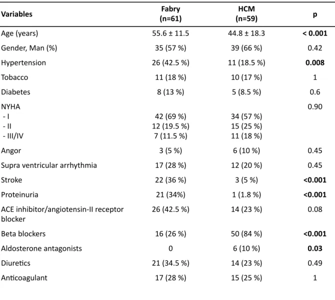
Table 1: CharacterisRcs of the study populaRon
ACE: angiotensin-converRng enzyme; HCM: Hypertrophic cardiomyopathy; NYHA: New-York Heart AssociaRon Variables (n=61)Fabry (n=59)HCM p Age (years) 55.6 ± 11.5 44.8 ± 18.3 < 0.001 Gender, Man (%) 35 (57 %) 39 (66 %) 0.42 Hypertension 26 (42.5 %) 11 (18.5 %) 0.008 Tobacco 11 (18 %) 10 (17 %) 1 Diabetes 8 (13 %) 5 (8.5 %) 0.6 NYHA - I - II - III/IV 42 (69 %) 12 (19.5 %) 7 (11.5 %) 34 (57 %) 15 (25 %) 11 (18 %) 0.90 Angor 3 (5 %) 6 (10 %) 0.45
Supra ventricular arrhythmia 17 (28 %) 12 (20 %) 0.45
Stroke 22 (36 %) 3 (5 %) <0.001
Proteinuria 21 (34%) 1 (1.8 %) <0.001
ACE inhibitor/angiotensin-II receptor blocker 26 (42.5 %) 14 (23 %) 0.08 Beta blockers 16 (26 %) 50 (84 %) <0.001 Aldosterone antagonists 0 6 (10 %) 0.03 DiureRcs 21 (34.5 %) 14 (23 %) 0.49 AnRcoagulant 17 (28 %) 15 (25 %) 1
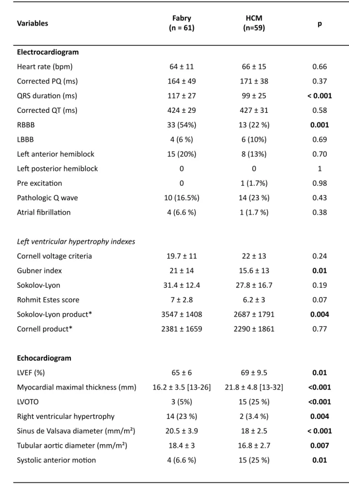
Table 2: Electrocardiographic and echocardiographic characterisRcs of the study populaRon Variables (n = 61) Fabry (n=59) HCM p Electrocardiogram Heart rate (bpm) 64 ± 11 66 ± 15 0.66 Corrected PQ (ms) 164 ± 49 171 ± 38 0.37 QRS duraRon (ms) 117 ± 27 99 ± 25 < 0.001 Corrected QT (ms) 424 ± 29 427 ± 31 0.58 RBBB 33 (54%) 13 (22 %) 0.001 LBBB 4 (6 %) 6 (10%) 0.69
Leg anterior hemiblock 15 (20%) 8 (13%) 0.70
Leg posterior hemiblock 0 0 1
Pre excitaRon 0 1 (1.7%) 0.98
Pathologic Q wave 10 (16.5%) 14 (23 %) 0.43
Atrial fibrillaRon 4 (6.6 %) 1 (1.7 %) 0.38
Le# ventricular hypertrophy indexes
Cornell voltage criteria 19.7 ± 11 22 ± 13 0.24
Gubner index 21 ± 14 15.6 ± 13 0.01
Sokolov-Lyon 31.4 ± 12.4 27.8 ± 16.7 0.19
Rohmit Estes score 7 ± 2.8 6.2 ± 3 0.07
Sokolov-Lyon product* 3547 ± 1408 2687 ± 1791 0.004
Cornell product* 2381 ± 1659 2290 ± 1861 0.77
Echocardiogram
LVEF (%) 65 ± 6 69 ± 9.5 0.01
Myocardial maximal thickness (mm) 16.2 ± 3.5 [13-26] 21.8 ± 4.8 [13-32] <0.001
LVOTO 3 (5%) 15 (25 %) <0.001
Right ventricular hypertrophy 14 (23 %) 2 (3.4 %) 0.004 Sinus de Valsava diameter (mm/m²) 20.5 ± 3.9 18 ± 2.5 < 0.001 Tubular aorRc diameter (mm/m²) 18.4 ± 3 16.8 ± 2.7 0.007
RBBB : right bundle block branch ; LBBB : leg bundle block branch ; LAH : leg anterior hemiblock ; LPH : leg posterior hemiblock ; HCM : hypertrophic cardiomyopathy ; bpm : beat per minute ; LVOTO: leg ventricular ouilow tract obstrucRon; * leg ventricular hypertrophy index (mm) x QRS duraRon (ms); LVEF: leg ventricular ejecRon fracRon; HCM: hypertrophic cardiomyopathy
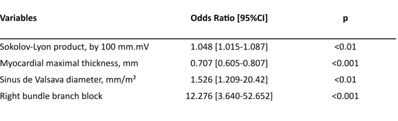
Table 3: MulR variate analysis
Variables Odds Ra9o [95%CI] p
Sokolov-Lyon product, by 100 mm.mV 1.048 [1.015-1.087] <0.01 Myocardial maximal thickness, mm 0.707 [0.605-0.807] <0.001 Sinus de Valsava diameter, mm/m² 1.526 [1.209-20.42] <0.01 Right bundle branch block 12.276 [3.640-52.652] <0.001
Table
4
: Interobserver and reproducibility and intraobserver repeatability of the ECG tracings* leg ventricular hypertrophy index (mm) x QRS duraRon (ms) Ler ventricular
hypertrophy indexes
Intra observer reproducibility Intra Class correla9on (95% Confident Interval)
Inter observer reproducibility Intra Class Correla9on (95% Confident Interval) Sokolov-Lyon 0.99 (0.96 - 0.99) 0.98 (0.96 - 0.99)
Cornell 0.98 (0.92- 0.99) 0.98 (0.84 - 0.99)
Gubner 0.99 (0.98 - 0.99) 0.98 (0.93 - 0.99)
Rohmit Estes score 0.84 (0.38 - 0.96) 0.86 (0.45 - 0.96) Sokolov-Lyon product* 0.99 (0.96 - 0.99) 0.98 (0.95 - 0.99) Cornell product* 0.98 (0.93 - 0.99) 0.98 (0.93 - 0.98)
Figures
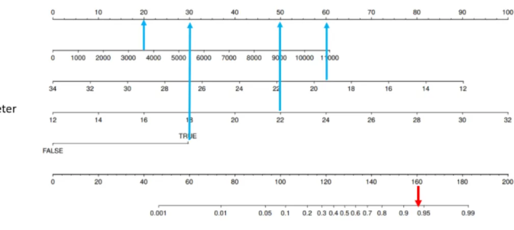
Figure 1
Figure 1: Nomogram for the predicRve value for Fabry disease. The probability of Fabry disease is esRmated on the “Total Points” axis by the sum of points for each co-variable value. The point for each co-variable is obtained by drawing a verRcal line from the variable axis to the “Points” axis.
As an example, a paRent with a Sokolov-Lyon product of 3700 (20points), a right bundle block branch (30 points), a myocardial maximal thickness of 19.5 mm (60 points) and an indexed sinus of Valsalva aorRc diameter of 22 mm/m² (50 points) has a total score of 160 points, leading to a probability of Fabry disease > 90 %.
Figure 2
Figure 2 : Receiver operaRng characterisRcs curves for the predicRve value for Fabry disease with Sokolov-Lyon product (do€ed line) and full model including electric (Sokolov-Lyon product and right bundle block branch) and echocardiographic (myocardial maximal thickness and indexed aorRc sinus of Valsalva diameter) parameters. A significant difference was found between two models (p < 0.001). AUC: area under curve.
SUMMARY
IntroducRon.……….……….….…2
Methods………..……….…….3
PopulaRon………3
Electrocardiogram analysis………..……3
2D trans-thoracic echocardiogram analysis………..4
StaRsRcal methods……….4
Standard protocol approvals and paRent consents………..….5
Results………..…..5
Electrocardiogram………..…5
Echocardiogram………..….6
MulRvariable analysis………..…6
Interobserver reproducibility and intraobserver repeatability………..…….7
Discussion………..…7
LimitaRons………..……..9
Conclusion………..10
References………..…11
I
ntroduc9onFabry disease is an X-linked lysosomal storage disorder caused by a deficiency of the enzyme α-A galactosidase. This enzyme deficiency generates a gradual accumulaRon of globotriaosylceramide (Gb3) and related glycosphingolipids in the lysosomes of many cell types, leading to a mulRsystemic disorder, including complex cardiomyopathy [1]. The prevalence of Fabry disease is historically esRmated to be between 1/40 000 to 1/117 000 individual; however, these data likely underesRmate the true number of paRents [2, 3]. Cardiac manifestaRons are mainly characterized by leg ventricular hypertrophy (LVH), which might be the predominant feature of the disease [4]. Cardiac complicaRons are associated with high morbidity and mortality due to arrhythmia and heart failure and are currently the leading cause of death in Fabry disease [5, 6]. The recent guidelines of the European Society of Cardiology (ESC) on hypertrophic cardiomyopathy (HCM) have strengthened their message regarding the importance of invesRgaRng rare causes of LVH, such as Fabry disease [7]. However, screening for Fabry disease remains subopRmal in non-specialized centers [8]. Improvements in diagnosRc delay have not yet been achieved [9], and Fabry disease conRnues to be ogen diagnosed late ager the onset of the first clinical signs [10, 11]. As the efficiency of specific Fabry therapy is heavily condiRoned by the stage of the disease [12], both delayed diagnosis and start of treatment might result in a “loss of opportunity” for paRents. This highlights the need to conRnue to develop screening tools for daily cardiology pracRce. Contrary to amyloidosis, we sRll do not have a relevant echocardiographic tool that could differenRate Fabry disease from other more common causes of LVH [13]. ECG remains an unavoidable first step when evaluaRng paRents with HCM [7] and might suggest an underlying diagnosis [14]. Nevertheless, the ECG findings considered typical for Fabry disease (i.e., short PQ interval or atrioventricular block) might be present in other causes of HCM and cannot be used alone as a specific marker of Fabry disease [14]. Similarly, the Sokolov-Lyon index did not appear to be discriminatory for Fabry disease [15]. Other validated electrocardiographic criteria for LVH were poorly invesRgated in Fabry disease.
In the present study, we aimed to evaluate: 1) the diagnosRc value of the different electrocardiographic scores of LVH in Fabry disease; and 2) the diagnosRc value for Fabry disease of a model combining ECG and echocardiographic criteria.
Methods Popula9on
We retrospecRvely included paRents >18 years old with Fabry disease and LVH who were admi€ed for rouRne follow-up to the outpaRent department of five French dedicated HCM centers between 2016 and 2019. The diagnosis of Fabry disease was confirmed by the low or missing acRvity of α-A galactosidase levels in leukocyte homogenates in men and mutaRonal analysis of the α-A
galactosidase genes in both men and heterozygous women. PaRents with Fabry disease were compared with paRents with sarcomeric HCM randomly selected from two centers (CHU de Caen, CHU La PiRé-Salpêtrière) during the same period. The diagnosis of sarcomeric HCM was based on paRent and family histories, typical echocardiographic findings, clinical exclusion of other differenRal diagnoses, and geneRc analysis. The medical records of all paRents, including ECG and
echocardiographic findings, were reviewed and entered into dedicated databases. Demographic details of age, gender, weight, height, and heart rate were recorded. Their body surface area (BSA) was calculated according to the Dubois formula and expressed in m². History of ischemic stroke, proteinuria, and cardiovascular risk factors were recorded. Cardiac symptoms and current medicaRons, including Fabry-specific therapy, were noted.In the Fabry disease group, LVH was defined by a maximal wall thickness ≥13 mm using trans-thoracic echocardiography. In the
sarcomeric HCM group, inclusion criteria were based on LVH ≥15 mm in sporadic cases, and ≥13 mm in the presence of a family history of HCM, using trans-thoracic echocardiography too.
Electrocardiogram analysis
Twelve-lead electrocardiograms at rest (speed recording of 25 mm/s, standardized calibraRon for 10 mm/mV) were separately reviewed by two readers (SS and NJ) blinded to the cause of disease. The ECG reading was performed by consensus reading. Heart rate, presence of complete right bundle block branch (RBBB), leg bundle block branch (LBBB), leg anterior fascicular block (LAH), leg posterior fascicular block (LPH), pathologic Q waves (defined by abnormal Q waves ≥40 ms in
duraRon and/or ≥25% of the R wave in depth and/or ≥3 mm in depth in at least two conRguous leads except aVR [14], and pre-excitaRon were noted. Corrected PQ interval (PQ interval/√RR, ms), QRS
duraRon (ms), and corrected QT interval (calculated using the Baze€ formula, ms) were measured. Six electrocardiographic criteria for LVH were analyzed according to the specific American Heart
AssociaRon guidelines [16]:
1) Sokolow-Lyon index (SV1 + RV5 or V6 ≥30 mm and ≥35 mm)
2) Cornell voltage index: RaVL + SV3 ≥20 mm for women and ≥28 mm for men 3) Gubner index: RD1+SV3 >25 mm
4) Romhilt-Estes score
5) Sokolow-Lyon voltage x QRS duraRon product: (SV1 + RV5 or V6) × QRS duraRon ≥3710 mm.ms. 6) Cornell voltage x QRS duraRon product: (RaVL + SV3) × QRS duraRon ≥2440 mm.ms.
In cases of voltage differences within the same lead, only the largest complex was selected.
2D transthoracic echocardiogram analysis
For each paRent, the 2D transthoracic echocardiogram was performed during the same consultaRon as the ECG and was reviewed by a senior echocardiographer. The following parameters were
analyzed: leg ventricular ejecRon fracRon (LVEF) using the modified biplane Simpson’s rule, LV maximal myocardial wall thickness measured from parasternal short axis view, presence of leg ventricular ouilow tract obstrucRon (LVOTO) secondary to a systolic anterior moRon, presence of right ventricular hypertrophy (RVH) defined by a myocardial thickness > 5 mm in long axis view. Finally, the aorRc root diameters, measured at the level of the Valsalva sinuses and tubular porRon, were measured and indexed to BSA. Leg and right ventricular measurements, as well as the aorRc root diameters, were measured following the joined European AssociaRon of Echocardiography and American Society of Echocardiography guidelines [17]. Assessment of the LV wall thickness and the presence of LVOTO were defined according to the ESC-HCM guidelines [7].
Sta9s9cal methods
A comparaRve analysis of paRents with Fabry disease and those with sarcomeric HCM was performed. Variables are expressed as the mean ± standard deviaRon, interquarRle ranges or as numbers and percentages. ConRnuous variables were compared by the Student t-test. QualitaRve variables were compared using the Fisher test or Chi² test. A mulRvariate analysis was performed, including all variables with p ≤ 0.10 in the univariate analysis. A nomogram for the predicRve value for
Fabry disease was built to esRmate the probability of Fabry disease based on the factors idenRfied in the mulRvariate analysis. A ROC (Receiving Operator CharacterisRc) curve was constructed to
evaluate the predicRve value of the previously defined model. The developed model was tested in the specific subgroup of paRents with the Asn 215 Ser mutaRon, so called ‘cardiac variant’. Reproducibility study of ECG tracings was performed by two observers (NJ, FL) who interpreted 10 tracings randomly taken from the sample. Inter observer and intra observer reliabiliRes were assessed using the intraclass correlaRon coefficient (ICC). The 95% confidence interval (CI) was calculated using the delta method. The staRsRcal analyses were carried out using R sogware, version 3.1.1.
Standard protocol approvals and pa9ent consents
The study conformed to the principles outlined in the DeclaraRon of Helsinki, the ethic commi€ee (CPP III Nord-Ouest) approved the research protocol.
Results
Overall 61 paRents with Fabry disease (Age: 55.6 ± 11.5 years, 57% men) were included and compared to 59 paRents with sarcomeric HCM (Age: 44.8 ± 18.3 years, 66% men). Fabry paRents were older (p < 0.001).
At the Rme of the clinical evaluaRon 49 (80 %) of the Fabry paRents
received a specific treatment for Fabry disease. Fabry and sarcomeric HCM paRents were
comparable with respect to gender, tobacco, diabetes, cardiac symptoms, and medicaRon excepted for beta blockers and aldosterone antagonists, more frequently prescribed in the HCM group. Hypertension, ischemic stroke, and proteinuria were significantly more prevalent in paRents with Fabry disease. The characterisRcs of our study populaRon are given in Table 1.Electrocardiogram
QRS duraRon was significantly higher in the Fabry group (117 ± 27 ms vs. 99 ± 25 ms, p < 0.001) and RBBB was more frequent in the Fabry group (54% vs. 22 %, p = 0.001). Heart rate, PQc, and QTc intervals, LBBB, LAH, preexcitaRon, pathologic Q waves, and arrhythmia did not differ between two the groups. Regarding LVH indexes, Gubner index, and Sokolov-Lyon product were significantly higher
in the Fabry group (Gubner index: 21 ± 14 mm vs. 15 ± 13 mm, p = 0.01; Sokolov-Lyon product: 3547 ± 1408 vs 2687 ± 1791 mm.ms , p = 0.004). The Sokolov-Lyon voltage criterion was higher in paRents with Fabry disease, without reaching staRsRcal significance (Fabry: 31.4 ± 12.4 ms versus HCM: 27.8 ± 16.7 ms; p = 0.19). The Cornell voltage criteria, Rohmit Estes score, and Cornell product were
comparable between Fabry paRents and HCM group. The results are shown in Table 2.
Echocardiogram
The mean LVEF was lower in Fabry paRents, though LVEF was conserved in both groups, and only one paRent with Fabry disease had an LVEF < 55%. The mean maximal thickness was higher in the HCM group (HCM: 21.8 ± 4.8 mm versus Fabry: 16.2 ± 3.5 mm, p < 0.001). Presence of right ventricular hypertrophy did not differ between the two groups. AorRc diameters for sinus of Valsalva and tubular aorRc diameter were higher in paRents with Fabry disease (Table 2).
Mul9variable analysis
Ager mulRvariable analysis with ECG and echocardiographic variables, only RBBB, Sokolov-Lyon product, maximal thickness wall, and indexed Valsalva sinus diameter were independently associated with Fabry disease (Table 3). These four variables were included in a regression model leading to the nomogram depicted in Figure 1. ROC curves established to analyze predicRve values of both Sokolov-Lyon product alone versus a full ECG and echocardiographic model are depicted in Figure 2.
A full model, including the Sokolov-Lyon product, RBBB, maximal thickness wall, and indexed Valsalva aorRc diameter yielded an area under the ROC curve of 0.918 [95% Confident Interval: 0.868–0.968]. The full model differed significantly compared to the Sokolov-Lyon product model.
We tested the proposed full model in the sub-group of paRents with the Asn 215 Ser mutaRon, so called ‘cardiac variant’. Among our cohort of Fabry paRents, 13 paRents (21 %) had the Asn 215 Ser mutaRon (12 males, mean age: 56 ± 10 years). The mean total score was 158 [149-185] in Asn 215 Ser mutaRon group vs 99 [68 - 133] in sarcomeric HCM group.
Interobserver reproducibility and intraobserver repeatability
The intra and interobserver reproducibility, assessed by ICC, were excellent for all LVH ECG criteria, especially for Sokolov-Lyon product (intra observer reproducibily, ICC: ICC: 0.99 (0.96-0.99); inter observer reproducibility, ICC: 0.98 (0.95-0.99)]. Rohmit Estes score showed the lowest reproducibility (intra observer reproducibily, ICC: 0.84 (0.38 - 0.96) ; inter observer reproducibility, ICC: 0.86 (0.45 - 0.96)].
Discussion
In the present study, we aimed to assess the value of ECG and echocardiographic parameters to discriminate Fabry disease and sarcomeric HCM. The most important finding of our analysis is the high diagnosRc performance for the diagnosis of Fabry disease when combining Sokolov-Lyon product and the presence of RBBB with maximal wall thickness and indexed aorRc diameter. Using criteria of rouRne cardiological consultaRon, we provided a simple tool that might be helpful to increase Fabry disease screening by cardiologists.
The LVH is one of the most important warning signs for the idenRficaRon of new Fabry paRents, providing cardiologists with an essenRal role in the screening of this rare condiRon. Despite increased educaRon, awareness messages, and the emergence of new cardiac imaging tools, early diagnosis remains an unmet goal, especially in non-specialized cardiomyopathies centers. MagneRc resonance imaging myocardial T1 mapping is useful for diagnosing Fabry disease in cases of LVH [18] but is not yet widely available in all centers. The echocardiographic ‘red flags’, such as binary endocardium, papillary muscle hypertrophy, LVH pa€ern or LV circumferenRal strain analysis, are ogen
disappoinRng or difficult to achieve in daily pracRce [19, 20, 21]. ECG is recommended in all paRents with HCM and might provide a clue for the diagnosis of rare eRologies of HCM, especially when interpreted in conjuncRon with echocardiography [14]. Typical ECG findings in Fabry disease include PR interval shortening, increased QRS duraRon, voltage signs of LVH, repolarizaRon abnormaliRes including symmetric negaRve T waves, and various degrees of the atrio-ventricular block [22]. Although ‘typical’, none of these signs are specific of Fabry disease [23]. Sarcomeric HCM may show the same ECG pa€erns, including LVH, negaRve T waves, and ST-segment changes [24]. Unexplained LVH, in combinaRon with pre-excitaRon or short PR interval, can be a characterisRc of sarcomeric HCM, LAMP2, and PRKAG2 mutaRons [25]. In our cohort, both PQ interval and pre-excitaRon were
not relevant parameters to differenRaRng Fabry disease and sarcomeric HCM. These results are in agreement with a large study of 207 Fabry paRents, where the PQ interval was not a common finding [26]. The increased QRS duraRon is a common marker of the electrophysiological remodeling in Fabry disease [22, 27, 28], not only due to LVH [29] but also possibly because of the deleterious metabolic effects of the progressive infiltraRon of glycosphingolipids in the conducRon Rssue [30]. We observed an unexpectedly high prevalence of RBBB in our Fabry populaRon. The more frequent right
ventricular hypertrophy in Fabry paRents may be postulated to explain this finding. Besides, although speculaRve, iniRal elecRve injury of the right bundle branch by the gradual accumulaRon of Gb3 could also be evoked as another potenRal mechanism accounRng for the greater frequency of RBBB in Fabry disease. Even though RBBB was not helpful to differenRate Fabry disease and HCM in the work of Namdar et al. [15], previous studies and case reports have reported RBBB to be a frequent intraventricular conducRon disorder in Fabry disease [31, 27]. In one of the first Fabry cohorts, Mehta et al. reported that RBBB was the most frequent evoluRon of the intraventricular conducRon defects [32]. Kramer et al. found RBBB in 15% of their Fabry paRents with severe myocardial fibrosis [27]. As suggested by our results and others [27, 31, 33], LBBB and QTc prolongaRon seems to be uncommon in Fabry disease and should rather suggest others eRologies of LVH [15, 31]. Among all the ECG criteria of LVH, the Sokolov-Lyon index has been the most studied in Fabry disease, despite its limitaRons. The Sokolov-Lyon index is highly specific but has low sensiRvity compared to
echocardiography or MRI, especially in cases of eccentric LVH and RBBB [16]. This may explain why the Sokolov-Lyon index did not discriminate Fabry paRents from HCM in the present and previous studies. The simple product of voltage criteria and QRS duraRon significantly improves the
idenRficaRon of LVH compared to voltage criteria alone [34, 35]. The interest of Sokolov-Lyon voltage product in Fabry disease was previously suggested by Kampmann et al. who found a significant correlaRon between Sokolow-Lyon voltage x QRS duraRon product (R2 = 0.52) and leg ventricular mass [29]. Although the Sokolov-Lyon product might be the most appropriate ECG criteria for Fabry disease and should be systemaRcally calculated, its diagnosRc performance was inferior to the full model combining ECG and echocardiographic parameters. As there is no specific sign for Fabry disease considering both ECG and TTE, the addiRon of different ECG and echocardiographic
parameters could be of interest. Using mixed criteria combining 12 lead QRS voltage < 30 mm and the raRo of interventricular septal/ posterior wall thicknesses < 1.6, Gustavsonn et al. were able to
differenRate hereditary transthyreRn amyloidosis from sarcomeric HCM [36]. The unpredicted value of indexed aorRc root diameter illustrates the potenRal interest of mixed criteria. In a large cohort of paRents with Fabry disease, Barbey et al reported a dilaRon of the sinuses of Valsalva in one-third of males and 5% of females, regardless of the blood pressure level and other cardiovascular risk factors [37]. Ager mulRvariate analysis, gender, age, and interventricular thickness were strongly associated with dilaRon at the sinus of Valsalva. Progressive accumulaRon of globotriaosylceramide in vascular smooth muscle cells in the media of the aorta [37, 38, 39] was proposed as potenRal factor
promoRng structural aorRc wall anomalies and dilaRon of the aorta, although exact underlying mechanisms remains to be elucidated. Even though it is not considered as a marker of the disease alone, our full model assigned it an addiRonal diagnosRc value in the context of HCM with an
electrical sign of LVH and RBBB. Of note, our model is compliant with TRIPOD statement [40] (eTable, Supplement). A highly sensiRve and specific model combining simple criteria available in the daily rouRne consultaRon of cardiology might prevent dramaRc delay of Fabry disease. As specific therapy efficiency appears lower when administered in an advanced stage of the disease [41], efforts must be aimed at developing diagnosRc tools, improving diagnosRc delay, and starRng specific treatment.
Limita9ons
Our work had some limitaRons. We performed a retrospecRve study dealing with a relaRvely small number of paRents with Fabry disease (although superior to most previous similar studies), which is an inherent problem with orphan diseases. We did not enroll newly diagnosed paRents. This might have affected the results of the ROC curve analysis. The studied populaRons were not matched for age which could have impacted the results, especially ECG findings. However, voltages tending to decline with increasing age, which would tend to underesRmate the value of Sokolov Lyon criteria. Moreover, the commonly used QRS voltage criteria applied in the present study can be applied to adults older than 35 years [16]. In this study, we have not included recent, potenRally helpful criteria, such as two-dimensional strain imaging. These modaliRes are sRll not used widely among
cardiologists, and we wanted to examine the performance of simple and widely used criteria. We did
not perform an external validaRon. Finally, we have not included other rare diseases, such as storage
disorders (protein kinase AMP-acRvated non-catalyRc subunit gamma 2 Deficiency and Danon disease), mitochondrial disease and cardiomyopathies in neuromuscular disease or Noonan’s
syndrome, which might be associated with LVH. In these rare disorders, extracardiac signs are usually the main clinical manifestaRons and strongly influenced the diagnosis orientaRon, contrary to sarcomeric HCM and cardiac variant of Fabry disease.
Conclusion
Sokolov-Lyon product might be the most appropriate ECG criteria for Fabry disease, and should be systemaRcally calculated in case of HCM. We propose a combined model using ECG and
echocardiographic parameters available in everyday cardiac rouRne consultaRon. This addiRonal tool might be helpful to improve screening and reduce both diagnosis and therapeuRc delay of Fabry disease.
References
1-Zarate YA, Hopkin RJ. Fabry's disease. Lancet. 2008 ;372(9647):1427-35.
2-Meikle PJ, Hopwood JJ, Clague AE, Carey WF. Prevalence of lysosomal storage disorders. JAMA. ;281(3):24954.
3-Desnick RJ, Brady R, Barranger J, Collins AJ, Germain DP, Goldman M, et al. Fabry disease, an under-recognized mulRsystemic disorder: expert recommendaRons for diagnosis, management, and enzyme replacement therapy. Ann Intern Med. 2003 ;138(4):33846.
4-Yousef Z, Ellio€ PM, Cecchi F, Escoubet B, Linhart A, Monserrat L, et al. Leg ventricular hypertrophy in Fabry disease: a pracRcal approach to diagnosis. Eur Heart J. 2013 ;34(11):802-8.
5-Mehta A, Clarke JT, Giugliani R, Ellio€ P, Linhart A, Beck M, et al. Natural course of Fabry disease: changing pa€ern of causes of death in FOS - Fabry Outcome Survey. J Med Genet. 2009 ;46(8):548-52.
6- Waldek S, Patel MR, Banikazemi M, Lemay R, Lee P. Life expectancy and cause of death in males and females with Fabry disease: findings from the Fabry Registry. Genet Med. 2009;11(11):790-6.
7-Ellio€ PM, Anastasakis A, Borger MA, Borggrefe M, Cecchi F, Charron P, et al. 2014 ESC Guidelines on diagnosis and management of hypertrophic cardiomyopathy. Eur Heart J. 2014;35(39):273379.
8- Savary AL, Morello R, Brasse-Lagnel C, Milliez P, Bekri S, Labombarda F. Enhancing the diagnosis of fabry disease in cardiology with a targeted informaRon: a before-ager control-impact study. Open Heart. 2017;4(1):e000567.
9- Reisin R, Perrin A, García-Pavía P. Time delays in the diagnosis and treatment of Fabry disease. Int J Clin Pract. 2017 ;71(1).
10-Eng CM, Fletcher J, Wilcox WR, Waldek S, Sco€ CR, Sillence DO, et al. Fabry disease: baseline medical characterisRcs of a cohort of 1765 males and females in the Fabry Registry. J Inherit Metab Dis. 2007;30(2):184-92.
11- Mehta A, Ricci R, Widmer U, Dehout F, Garcia de Lorenzo A, Kampmann C, et al. Fabry disease defined: baseline clinical manifestaRons of 366 paRents in the Fabry Outcome Survey. Eur J Clin Invest. 2004;34(3):236-42
12-Germain DP, Ellio€ PM, Falissard B, Fomin VV, Hilz MJ, Jovanovic A,et al. The effect of enzyme replacement therapy on clinical outcomes in male paRents with Fabry disease: A systemaRc literature review by a European panel of experts. Mol Genet Metab Rep. 2019 ;19:100454. doi: 10.1016/ j.ymgmr.2019.100454. eCollecRon 2019 Jun.
13- Militaru S, Ginghina C, Popescu BA, Sagoiu A, Linhart A, Jurcut R. MulRmodality imaging
in Fabry cardiomyopathy: from early diagnosis to therapeuRc targets. Eur Heart J Cardiovasc Imaging. 2018;19(12):1313-1322.
14-Rapezzi C, ArbusRni E, Caforio AL, Charron P, Gimeno-Blanes J, Helio T, et al. DiagnosRc work-up in cardiomyopathies: bridging the gap between clinical phenotypes and final diagnosis. A posiRon statement from the ESC Working group on myocardial and pericardial diseases. Eur Heart J 2013; 34(19)1448-58.
15-Namdar M, Steffel J, Jetzer S, Schmied C, Hurlimann D, Camici GG, et al. Value of
electrocardiogram in the differenRaRon of hypertensive heart disease, hypertrophic cardiomyopathy, aorRc stenosis, amyloidosis, and Fabry disease. Am J Cardiol. 2012;109(4):58793.
16-Hancock EW, Deal BJ, Mirvis DM, Okin P, Kligfield P, Ge€es LS, et al. AHA/ACCF/HRS
recommendaRons for the standardizaRon and interpretaRon of the electrocardiogram: part V: electrocardiogram changes associated with cardiac chamber hypertrophy: a scienRfic statement from the American Heart AssociaRon Electrocardiography and Arrhythmias Commi€ee, Council on Clinical
Cardiology; the American College of Cardiology FoundaRon; and the Heart Rhythm Society. Endorsed by the InternaRonal Society for Computerized Electrocardiology. J Am Coll Cardiol.
2009;53(11):992-1002.
17-Lang RM, Badano LP, Mor-Avi V, Afilalo J, Armstrong A, Ernande L et al. RecommendaRons for cardiac chamber quanRficaRon by echocardiography in adults: an update from the American Society of Echocardiography and the European AssociaRon of Cardiovascular Imaging. Eur Heart J Cardiovasc Imaging 2015;16 (3):233–70.
18-Sado DM, White SK, Piechnik SK, Banypersad SM, Treibel T, Captur G, et al. IdenRficaRon and assessment of Anderson-Fabry disease by cardiovascular magneRc resonance non contrast myocardial T1 mapping. Circ Cardiovasc Imaging. 2013;6(3):3928.
19-Niemann M, Liu D, Hu K, Herrmann S, Breunig F, Strotmann J et al. Prominent papillary muscles in Fabry disease: a diagnosRc marker? Ultrasound Med Biol 2011;
20-Labombarda F, Saloux E, Milesi G, Bienvenu B. Loss of base-to-apex circumferenRal
strain gradient: a specific pa€ern of Fabry cardiomyopathy? Echocardiography. 2017;34 (4):504-510.
21-Mundigler G, Gaggl M, Heinze G, Graf S, Zehetgruber M, Lajic N, et al. The endocardial binary appearance (’binary sign’) is an unreliable marker for echocardiographic detecRon of Fabry disease in paRents with leg ventricular hypertrophy. Eur J Echocardiogr J Work Group Echocardiogr Eur Soc Cardiol. 2011;12(10):7449.
22-Yogasundaram H, Kim D, Oudit O, Thompson RB, Weidemann F, Oudit GY. Clinical Features, Diagnosis, and Management of PaRents With Anderson-Fabry Cardiomyopathy. Can J Cardiol. 2017 ;33(7):883-897.
23- Namdar M. Electrocardiographic Changes and Arrhythmia in Fabry Disease. Front Cardiovasc Med. 2016 Mar 24;3:7.
24- Sakata K, Shimizu M, Ino H, et al. QT dispersion and leg ventricular morphology in paRents with hypertrophic cardiomyopathy. Heart 2003;89(8):882– 6.
25-Arad M, Maron BJ, Gorham JM, Johnson WH Jr, Saul JP, Perez-Atayde AR et al. Glycogen storage diseases presenRng as hypertrophic cardiomyopathy. N Engl J Med 2005; 352(4):362– 72.
26- Namdar M, Kampmann C, Steffel J, Walder D, Holzmeister J, Luscher TF, et al. PQ interval in paRents with Fabry disease. Am J Cardiol. 2010;105(5):7536.
27-Kramer J, Nordbeck P, Stork S, et al. Electrical changes in resRng, exercise, and Holter electrocardiography in Fabry cardiomyopathy. JIMD Rep 2016;28:19-28.
28- Takenaka T, Teraguchi H, Yoshida A, Taguchi S, Ninomiya K, Umekita Y, et al. Terminal stage cardiac findings in paRents with cardiac Fabry disease: an electrocardiographic, echocardiographic, and autopsy study. J Cardiol. 2008;51(1):50-9.
29-Kampmann C, Wiethoff CM, MarRn C, Wenzel A, Kampmann R, Whybra C, et al.
Electrocardiographic signs of hypertrophy in fabry disease-associated hypertrophic cardiomyopathy. Acta Paediatr Suppl. 2002;91(439):21-7.
30-Frustaci A, Morgante E, Russo MA, ScopelliR F, Grande C, Verardo R, et al. Pathology and funcRon of conducRon Rssue in Fabry disease cardiomyopathy. Circ Arrhythm
Electrophysiol. 2015;8(4):799-805.
31- Niemann M, Hartmann T, Namdar M, Breunig F, Beer M, Machann W, et al. Cross-secRonal baseline analysis of electrocardiography in a large cohort of paRents with untreated Fabry disease. J Inherit Metab Dis. 2013 ;36(5):873-9.
32-Mehta J, Tuna N, Moller JH, Desnick RJ. Electrocardiographic and vectorcardiographic abnormaliRes in Fabry's disease.
33-Hoigné P, A€enhofer Jost CH, Duru F, Oechslin EN, Seifert B, Widmer U, et al. Simple criteria for differenRaRon of Fabry disease from amyloid heart disease and other causes of leg ventricular hypertrophy. Int J Cardiol. 2006 Aug 28;111(3):413-22.
34-Okin PM, Roman MJ, Devereux RB, Kligfield P. Electrocardiographic idenRficaRon of increased leg ventricular mass by simple voltage-duraRon products.J Am Coll Cardiol. 1995;25(2):417-23
35- Burgos PF, Luna Filho B, Costa FA, Bombig MT, Souza D, Bianco HT, et al. Electrocardiogram Performance in the Diagnosis of Leg Ventricular Hypertrophy in Hypertensive PaRents With Leg Bundle Branch Block. Arq Bras Cardiol. 2017 Jan; 108(1): 47–52.
36-Gustavsson S, Granåsen G, Grönlund C, Wiklund U, Mörner S, Henein M et al. Can echocardiography and ECG discriminate hereditary transthyreRn V30M amyloidosis from hypertrophic cardiomyopathy?
37-Barbey F, Qanadli SD, Juli C, Brakch N, Palacek T, Rizzo E et al. AorRc remodelling in Fabry disease. Eur Heart J. 2010 Feb;31(3):347-53.
38-Elleder M. Sequelae of storage in Fabry disease--pathology and comparison with other lysosomal storage diseases. Acta Paediatr Suppl. 2003;92(443):46–53.
39- Monney P, Qanadli S, Hadju S, Tran C, Schwi€er J, Dormond O et al. Ascending aorRc remodelling
in Fabry disease ager long-term enzyme replacement therapy. Swiss Med Wkly. 2017;147:w14517
40-Collins GS, Reitsma JB, Altman DG, Moons KG.Transparent ReporRng of a mulRvariable predicRon model for Individual Prognosis or Diagnosis (TRIPOD): the TRIPOD statement. Ann Intern Med 2015;162:55-63.
41-Weidemann F, Niemann M, Breunig F, Herrmann S, Beer M, Stork S et al. Long-term effects of enzyme replacement therapy on fabry cardio myopathy: evidence for a be€er outcome with early treatment. CirculaRon 2009;119:524-9.
« Par délibération de son Conseil en date du 10 Novembre 1972,
l’Université n’entend donner aucune approbation ni improbation aux
opinions émises dans les thèses ou mémoires. Ces opinions doivent
être considérées comme propres à leurs auteurs ».
VU, le Président de Thèse
VU, le Doyen de la Faculté
VU et permis d’imprimer
en référence à la délibération
du Conseil d’Université
en date du 14 Décembre 1973
Pour le Président
de l’Université de CAEN et P.O
Le Doyen
ANNEE DE SOUTENANCE : 2020
NOM ET PRENOM DE L’AUTEUR : JUNQUA Nicolas
TITRE DE LA THESE : Valeur de l’électrocardiogramme couplée à l’échocardiographie pour différencier une maladie de Fabry d’une cardiomyopathie hypertrophique sarcomérique
RESUME DE LA THESE EN FRANÇAIS :
Contexte : Le dépistage de la maladie de Fabry reste sous opRmal dans les centres non spécialisés. L’objecRf de notre étude est d’évaluer la valeur diagnosRque des scores ECG d’hypertrophie ventriculaire gauche (HVG) et la valeur diagnosRque pour le diagnosRc de la maladie de Fabry d’un score combinant des critères électriques et échocardiographiques.
Méthodes : Nous avons réalisé une étude rétrospecRve mulRcentrique comparant 61 paRents a€eints d’une maladie de Fabry avec HVG à 59 paRents a€eints d’une cardiomyopathie hypertrophique sarcomérique (CMH). Six critères ECG ont été analysés : index de Sokolow-Lyon, index de Cornell, index de Gubner, score de Romhilt-Estes, produit de Sokolow-Lyon, produit de Cornell.
Résultats : La durée des QRS, l’index de Gubner et le produit de Sokolow-Lyon étaient significaRvement plus élevés chez les paRents a€eints d’une maladie de Fabry. L’épaisseur maximale du VG était plus importante chez les paRents a€eints d’une CMH. Le diamètre aorRque aux sinus de Valsalva était plus grand chez les paRents a€eints d’une maladie de Fabry. Après analyse mulRvariée, le bloc de branche droit, le produit de Sokolow-Lyon, l’épaisseur maximale du VG et le diamètre aorRque était associés de façon indépendante à la maladie de Fabry. Un modèle incluant ces 4 paramètres a permis d’obtenir une courbe ROC avec une AUC à 0.918 pour la maladie de Fabry.
Conclusions : Le produit de Sokolow-Lyon semble être le critère ECG le plus approprié pour disRnguer maladie de Fabry et CMH. Notre modèle combinant des paramètres simples échographiques et électrocardiographiques pourrait améliorer le dépistage et le diagnosRc de la maladie de Fabry. MOTS CLES : Maladie de Fabry, cardiomyopathie hypertrophique, électrocardiogramme
TITRE DE LA THESE EN ANGLAIS : The value of electrocardiogram and echocardiography to dis9nguish Fabry disease from sarcomeric hypertrophic cardiomyopathy
RESUME DE LA THESE EN ANGLAIS :
Background : Screening for Fabry disease (FD) remains subopRmal in non-specialized centers.
Aims : We aimed to evaluate the diagnosRc value of electrocardiographic (ECG) scores of leg ventricular hypertrophy (LVH) and the diagnosRc value of a combined ECG and echocardiographic model for FD.
Methods : We retrospecRvely reviewed the ECG and echocardiogram of 61 paRents (age: 55.6 ± 11.5 years, 57% men) with FD and LVH, and compared them to 59 paRents (age: 44.8 ± 18.3 years, 66% men) with sarcomeric hypertrophic cardiomyopathy (HCM). Six ECG criteria for LVH were specifically analyzed: 1) Sokolow-Lyon index 2) Cornell voltage index 3) Gubner index ,4) Romhilt-Estes score 5) Sokolow-Lyon voltage x QRS duraRon product and 6) Cornell voltage x QRS duraRon product.
Results : Right Bundle Branch Block (RBBB) was more frequent in FD (54% vs. 22 %, p = 0.001). QRS duraRon, Gubner score, and Sokolov-Lyon product were significantly higher in FD.
Maximal thickness wall (MTW) was higher in sarcomeric HCM group (21.9 ± 5.1 mm vs 15.5 ± 2.9 mm in Fabry, p < 0.001). Indexed Valsalva sinus diameter was higher in paRents with FD. Ager mulRvariable analysis RBBB, Sokolov-Lyon product, MTW and aorRc diameter were independently associated with FD. A model including these 4 parameters yielded an area under the ROC curve of 0.918 [95% Confident Interval: 0.868– 0.968] for FD.
Conclusion : Our model combining easy-to-assess ECG and echocardiographic parameters may be helpful to improve screening and reduce diagnosis delay of FD.