Up-Regulation of IL-7 Receptor
␣
1
Denis Franchimont,
2*
‡Je´roˆme Galon,
2,3* Melanie S. Vacchio,
†Samuel Fan,* Roberta Visconti,*
David M. Frucht,* Vincent Geenen,
‡George P. Chrousos,
‡Jonathan D. Ashwell,
§and
John J. O’Shea*
Despite the effects of glucocorticoids on immune function, relatively little is known about glucocorticoid-inducible genes and how their products may regulate lymphocyte function. Using DNA microarray technology to analyze gene expression in PBMC from healthy donors, we identified IL-7R␣ as a glucocorticoid-inducible gene. This observation was confirmed at the mRNA and protein levels. Conversely, TCR signaling decreased IL-7R␣ expression, and the relative strength of signaling between these two receptors determined the final IL-7R␣ levels. The up-regulation of IL-7R␣ by glucocorticoids was associated with enhanced IL-7-mediated signaling and function. Moreover, IL-7-mediated inhibition of apoptosis at increasing concentrations of glucocorticoids is con-sistent with enhanced cell sensitivity to IL-7 following glucocorticoid exposure. These observations provide a mechanism by which glucocorticoids may have a positive influence on T cell survival and function. The Journal of Immunology, 2002, 168: 2212–2218.
E
licitation of a T cell response to a specific self or foreignAg requires the presentation of an Ag/MHC complex by APCs to the TCR (1). This process takes place in second-ary lymphoid and nonlymphoid organs, where it maintains naive and mature T cell responses to foreign Ags, peripheral T cell ho-meostasis, and self-tolerance (2– 4). Alternative concurrent signals regulate TCR-mediated expansion or deletion of peripheral T cells. In fact, glucocorticoids may modulate the nature and the intensity of the immune response (5–7).
The most recognized biologic effect of glucocorticoids at high concentrations (exogenously administrated or endogenously pro-duced in response to stress) is immunosuppression, and these hor-mones are potent inducers of T cell apoptosis (8). Because the glucocorticoid receptor (GR)4
is a transcriptional regulator, the simplest means to account for its effects on cell viability is to postulate that glucocorticoids induce the expression of gene prod-ucts that directly or indirectly cause cell death. To date, however, there are no clear candidates for glucocorticoid-induced genes that cause cell death. Alternatively, glucocorticoids also have positive effects on immune development and function (5). The GR and the TCR independently induce apoptosis, yet together they promote T cell survival (9). Indeed, glucocorticoids antagonize and repress
TCR-mediated signals for cell death (10 –12). This mutual regu-lation for survival not only occurs in the thymus but also exists in the peripheral immune system.
This context-dependent action of the GR led us to identify genes whose expression is modified by glucocorticoids and ask how they might influence T cell function. In this work we report that screen-ing of almost 10,000 genes with DNA microarrays revealed that IL-7R ␣-chain (IL-7R␣) is one of the most prominent gene in-duced by glucocorticoids. Furthermore, the positive regulation of IL-7R␣ by glucocorticoids was associated with enhanced IL-7-mediated signaling and function. These observations provide a mechanism by which glucocorticoids may influence T cell func-tion and immune responsiveness.
Materials and Methods
Cytokines, Abs, and cells
The following reagents were purchased: recombinant human IL-7 (R&D Systems, Minneapolis, MN), anti-phosphotyrosine Ab (4G10; Upstate Bio-technology, Lake Placid, NY), polyclonal rabbit anti-IL-7R␣ Ab (Santa Cruz Biotechnology, Santa Cruz, CA), and monoclonal mouse anti-human CD3 and human CD28 (BD PharMingen, San Diego, CA). The following Abs used for flow cytometry were purchased: PE-conjugated anti-human IL-7R␣ (CD127) mAb (Immunotech, Marseilles, France); FITC-conju-gated anti-human IL-2R␣ (CD25), CD3, CD4, CD8, anti-CD19, and anti-CD14 mAb (BD Biosciences, San Jose, CA); and isotype-matched IgG controls (BD PharMingen). Polyclonal rabbit anti-Stat5a Ab was produced as previously described (13). Polyclonal rabbit anti-IL-7 and anti-IL-7R␣ neutralizing Abs were obtained from R&D Systems. Anti--actin mAb was purchased from Sigma-Aldrich (St. Louis, MO). Strepta-vidin-PE was purchased from Caltag Laboratories (Burlingame, CA). Me-tyrapone was obtained from ICN Biochemicals (Costa Mesa, CA). Human IL-2 was provided by Dr. C. Reynolds (National Cancer Institute, Freder-ick, MD). NK3.3 cells were provided by Dr. J. Kornbluth (St. Louis Uni-versity, St. Louis, MO). PBMC from healthy donors were isolated by Ficoll-Paque (Amersham Pharmacia Biotech, Piscataway, NJ) gradient centrifugation. When peripheral T cells were used, PBMC were activated with PHA (2g/ml) for 72 h and cultured for an additional day in presence of IL-2 (40 IU/ml), as described previously (14). Typically, this resulted in
⬎95% CD3⫹cell purity, and before stimulation cells were washed with
acidified medium (pH 6.4) and rested in RPMI 1640 containing 1% BSA. For TCR cross-linking experiments, PBMC were activated with coated anti-CD3/anti-CD28 (10g/ml) for 3 days and isolated by Ficoll-Paque
*Lymphocyte Cell Biology Section, National Institute of Arthritis and Musculoskel-etal and Skin Diseases,†Laboratory of Immune Cell Biology, National Cancer
Insti-tute,‡Pediatric Endocrinology Branch, National Institute of Child Health and Human
Development, and§Experimental Immunology Branch, National Cancer Institute,
Na-tional Institutes of Health, Bethesda, MD 20892
Received for publication June 28, 2001. Accepted for publication December 21, 2001. The costs of publication of this article were defrayed in part by the payment of page charges. This article must therefore be hereby marked advertisement in accordance with 18 U.S.C. Section 1734 solely to indicate this fact.
1D.F. is supported by the Belgian National Foundation for Scientific Research. 2D.F. and J.G. contributed equally to this work.
3Address correspondence and reprint requests to Dr. Je´roˆme Galon at the current
address: Laboratoire d’Immunologie Cellulaire et Clinique, Institut National de la Sante´ et de la Recherche Me´dicale, Unite´ 255, Institut de Recherches Biome´dicales des Cordeliers, 75270 Paris Cedex 06, France. E-mail address: jerome.galon@ u255.bhdc.jussieu.fr
4Abbreviations used in this paper: GR, glucocorticoid receptor; Dex, dexamethasone;
␥c, common␥ subunit; GILZ, glucocorticoid-induced leucin zipper.
gradient centrifugation. Purified human cord blood CD4⫹T cells and pu-rified human bone marrow stromal cells were purchased from Poietic Tech-nologies (Gaithersburg, MD). Naive CD4⫹ T cells, which express CD45RA (⬎95%), were isolated from cord blood from umbilical blood collected from scheduled cesarean deliveries. Cells were isolated and re-suspended in HBSS containing 0.5% BSA and 5 mM EDTA, and CD4⫹T cells were from cord blood mononuclear cells using negative immunomag-netic selection, and purity was⬎95%.
FACS analysis
Expression of IL-7R␣ was detected using PE-conjugated anti-human IL-7R␣ mAb or PE-conjugated anti-mouse IL-7R␣ Ab. Expression of IL-2R␣ was detected using FITC-conjugated anti-human IL-2R␣ mAb or FITC-conjugated anti-mouse IL-2R␣ (CD25) Ab. The cells were washed, incu-bated with conjugated Abs and isotype-matched IgG-PE/isotype-matched FITC-IgG as controls for 30 min at 4°C, washed three times with PBS containing 0.5% BSA, and then analyzed by flow cytometry. Samples were analyzed on a FACSCalibur flow cytometer (BD Biosciences). Apoptosis was evaluated using propidium iodide/annexin V (Immunotech) staining.
Microarrays
PBMC were prepared as described and stimulated with dexamethasone (Dex; 10⫺7M) for 16 h. RNA extraction was performed (RNAgents; Pro-mega, Madison, WI), and mRNA was purified with oligoTex mRNA iso-lation columns (Qiagen, Valencia CA). Microarray analysis was performed using the GEM microarray (Incyte, St. Louis, MO). The two fluorescent cDNA probes were mixed and simultaneously hybridized to a microarray containing 9182 human expressed genes. The microarray was scanned, and the intensity of the fluorescence at each array element was proportional to the expression level of that gene in the sample. The ratio of the two flu-orescence intensities provided a quantitative measurement of the relative gene expression level in the two cell samples.
RNase protection assay
Human thymocytes and mature T lymphocytes were rested for 4 h, pretreated with Dex at different concentrations for various times, and stimulated with IL-7 (10 ng/ml) for various times. RNA extraction was performed, and mRNA expression was evaluated by RNase protection assay. RNase protection assay was performed as follows:32P-labeled RNA probes were synthesized using
SP6 RNA polymerase or T7 RNA polymerase for the multiprobe template set (Riboquant; BD PharMingen). DNA was digested with DNase I (Roche, In-dianapolis, IN), and RNA probes were extracted with phenol/chloroform and precipitated with ethanol. Labeled RNA probes were hybridized overnight with target RNA (5g) at 56°C and were digested with T1 RNase (Life Technologies, Gaithersburg, MD). The protected mRNA fragment was ex-tracted with phenol and chloroform, precipitated with ethanol, resolved on a 6% denaturing polyacrylamide gel, and subjected to autoradiography. Gene transcripts were identified by the length of the protected fragments. Equal loading of RNA was estimated from the amounts of protected fragments of two housekeeping genes, L32 and GAPDH.
Immunoprecipitation and immunoblotting
NK3.3 cells and peripheral T lymphocytes were pretreated with different concentrations of Dex for various times, resuspended in 1 ml of serum-free medium (2⫻ 106NK3.3 cells), and stimulated with IL-7 (20 ng/ml) for 15
min. Following stimulation cells were washed once in PBS and lysed on ice in a buffer containing 1% Triton X-100, 50 mM Tris-HCl (pH 7.5), 300 mM NaCl, 2 mM EDTA, 200M Na3VO4, 10g/ml aprotinin, 10 g/ml
leupeptin, and 2.5M p-nitrophenyl p-guanidinobenzoate on ice for 30 min. Immunoprecipitation with anti-Stat Ab and subsequent SDS-PAGE were performed as described previously (13) with detection by ECL (LumiGLO; Kirkegaard & Perry Laboratories, Gaithersburg, MD).
Table I. Glucocorticoid-regulated genes of activated PBMCa
GB 0 Dex Rank
Gc up-regulated genes Cell life and death
␦ sleep-inducing peptide, GILZ homolog W63717 546 3357 1 Cell surface receptors/transporters
IL-7R␣ chain AF043129 122 546 5
Scavenger receptor superfamily Z22968 216 1052 3
IL-1R, type II U64094 729 2336 13
Redox and oxidation damage control
Indolamine-pyrrole 2,3 dioxygenase M34455 486 2118 4 Metabolism
Diamine acetyltransferase AA336592 372 1264 10 Dihydroxyvitamin D3-induced protein S73591 370 2300 2
Cell division/proliferation
Metallothionein 1L F26137 1672 6322 6
Metallothionein 1E Y12653 1672 6126 8
Metallothionein IB H72532 1343 4801 9
Metallothionein 3 (growth inhibitory factor) AI418147 2680 8937 12 B cell translocation gene 1, antiproliferative AI925293 163 558 11
Diubiquitin Y12653 193 704 7
Gc down-regulated gene Cell division/proliferation
Epiregulin D30783 146 40 1
Extracellular matrix/cell adhesion
Integrin,␣1 X68742 132 38 3
Carcinoembryonic Ag (member 6) X52378 116 34 4 Cell surface receptors/transporters
Potassium inwardly rectifying channel U65406 145 46 5 Solute carrier family 28 (sodium transporter) U84392 118 37 8 Solute carrier family 26 (sulfate transporter) U14528 170 57 10 Miscellaneous
Histatin 3 AA37671 811 533 2
Ser-Thr protein kinase U59305 131 41 7
Cerebellar degeneration-related protein M16965 123 43 12 Unknown function
mRNA from chromosome 5q21–22 AB002438 133 41 6
Expressed sequence tag U79293 132 44 9
Expressed sequence tag AA400569 156 52 11 aRelative expression in untreated (0) and Dex-treated cells (Dex). GB, gene bank accession number; Gc, glucocorticoid.
Results
Gene expression in glucocorticoid-activated PBMC
The molecular response of human PBMC to glucocorticoids was determined with cDNA microarrays representing⬃10,000 human genes. Cells were stimulated for 16 h with a high concentration (10⫺7M) of the synthetic glucocorticoid Dex. Genes were ranked by differential expression and grouped by functional categories. An example of such an analysis is shown for the subset of the 12 most regulated genes (Table I). The gene up-regulated to the great-est extent by Dex was the␦ sleep-inducing peptide (88% identity to murine glucocorticoid-induced leucin zipper (GILZ)); GILZ has been reported to inhibit T cell apoptosis induced by stimulation via the TCR (18). Among the top five genes induced by Dex was IL-7R␣. This was of particular interest because, while a prominent effect of glucocorticoids is to induce T cell apoptosis, IL-7R␣ is a receptor that delivers antiapoptotic (prosurvival) signals to T cells.
Glucocorticoids induce IL-7R␣ expression
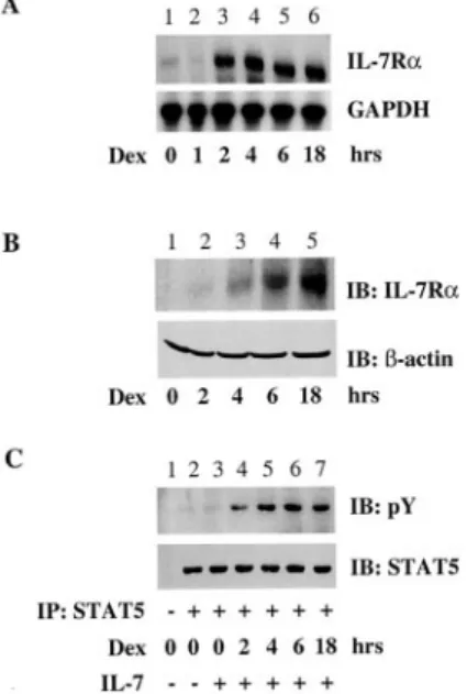
To confirm the induction of IL-7R␣ expression by glucocorticoids, the effect of Dex on an IL-7R␣-negative NK cell line (NK 3.3) was examined (Fig. 1). Dex induced IL-7R␣ mRNA expression in these cells within 2 h of treatment (Fig. 1A). A similar induction of protein was observed by immunoblotting after 4 h of Dex treat-ment (Fig. 1B). IL-7 exerts its effects by inducing tyrosine phos-phorylation of the transcription factor Stat5 (19). To assess whether the up-regulated IL-7R␣ was functional, we determined whether Dex treatment of NK cells permitted STAT5 phosphory-lation by IL-7. Consistent with the lack of detectable IL-7R␣ ex-pression, IL-7 alone had no effect on Stat5 activation in untreated
NK3.3 cells (Fig. 1C). However, preincubation with Dex permitted IL-7R␣ signaling and Stat5 phosphorylation. Thus, Dex induces the expression of functional IL-7R␣.
Glucocorticoids up-regulate IL-7R␣ expression on T cells
The physiological functions of IL-7 for T cells include promotion of survival and proliferation (20). To determine whether glucocor-ticoid up-regulation of IL-7R␣ expression is pertinent to T cells, we analyzed the effect of Dex on peripheral blood T lymphocytes. Dex induced an increase of ⬃3-fold in cell surface IL-7R␣ ex-pression (Fig. 2A, upper panel, thick line). Furthermore, both CD4 and CD8 subpopulations up-regulate IL-7R␣ (Fig. 2A), supporting the idea that glucocorticoids up-regulate IL-7R␣ on all T cells but not on monocytes or B cells (data not shown). Dex also up-regu-lated IL-7R␣ expression on naive CD4⫹human cord blood cells, indicating that the state of activation or differentiation of the T cells was not important for this effect (Fig. 2B). The Dex dose response was determined by FACS analysis, and as little as 10⫺10 M Dex induced IL-7R␣, with its expression being enhanced by increased Dex doses (Fig. 2C). These results were also confirmed at the mRNA level using RNase protection assays (data not
FIGURE 1. Glucocorticoids induce IL-7R␣ expression in NK cells. A, Time response of Dex. NK 3.3 cells were untreated (lane 1) or stimulated with Dex (10⫺7M) at the indicated times (lanes 2– 6). The cells were lysed, RNA was extracted, and IL-7R␣ expression was evaluated by RNase pro-tection assay. B, NK 3.3 cells were treated as described in A, and then subjected to immunoblotting with anti-IL-7R␣ or anti--actin Abs. C, IL-7R␣ signaling. NK 3.3 cells were untreated (lanes 1–3) or pretreated with Dex (10⫺7M) for 2–18 h as indicated (lanes 4 –7), left unstimulated (lanes
1 and 2), or stimulated with IL-7 (20 ng/ml; lanes 3–7). The cells were
lysed and immunoprecipitated with nonimmune rabbit polyclonal Ig (lane
1) or Stat5a (lanes 2–7) and subjected to immunoblotting with
anti-phosphotyrosine and anti-Stat5a as indicated. One representative experi-ment of at least three independent experiexperi-ments is shown.
FIGURE 2. Glucocorticoids induce IL-7R␣ expression in T cells. A, PBMC were analyzed for IL-7R␣ expression. Cells were untreated (thin line) or treated with Dex (10⫺7M) overnight (thick line), and stained with anti-IL-7R␣-PE, CD3-FITC, CD4-FITC, CD8-FITC, or isotype-matched IgG controls (dotted line), then analyzed by flow cytometry (n⫽ 16, from six donors). B, Naive cord blood purified CD4⫹T cells were untreated (thin line) or treated with Dex (10⫺7M) overnight (thick line) and analyzed for IL-7R␣ expression (n ⫽ 6, from three donors). C, Dex dose response. PBMC were activated with PHA (2g/ml) for 72 h and cultured for an additional day in the presence of IL-2 (40 IU/ml). Cells were rested and stimulated with the indicated concentration of Dex. CD3⫹cells (⬎95%) were gated and analyzed for IL-7R␣ expression. Values are the mean ⫾ SEM of three independent experiments. D, Dex time response. Peripheral T cells were untreated (lane 1) or were treated with Dex (10⫺7M) for 2–18 h as indicated (lanes 2– 4). This experiment was repeated three times on separate donors. E, Dex stimulation is reversed by RU-496. Peripheral T cells were treated for 18 h with Dex (10⫺7M; lanes 2 and 4) and with RU-496 (10⫺7M; lanes 3 and 4). D and E, The cells were lysed, RNA was extracted, and IL-7R␣ expression was evaluated by RNase protection assay using IL-7R␣- and GAPDH-specific probes.
shown). Furthermore, Dex caused up-regulation of IL-7R␣ mRNA in peripheral T cells as early as 2 h after initiation of treatment (Fig. 2D). The glucocorticoid antagonist RU-486 (Fig. 2E), but not the protein synthesis inhibitor cycloheximide (data not shown), inhibited the Dex-induced increase in IL-7R␣ mRNA, consistent with a direct effect of the liganded GR on IL-7R␣ transcription. The enhancement of IL-7R␣ expression occurred in a dose-depen-dent manner. Thus, glucocorticoids up-regulate IL-7R␣ mRNA and protein in T cells.
Antagonism between TCR- and glucocorticoid-mediated activation on IL-7R␣ expression
IL-7 has been shown to mediate homeostatic expansion of naive and memory T cells after viral infection (21). Therefore, we de-termined how IL-7R␣ expression was regulated by TCR stimula-tion. Purified human CD4⫹cord blood cells expressing high levels of IL-7R␣ were stimulated with anti-CD3/CD28 Abs. As ex-pected, with simultaneous anti-CD3/CD28 stimulation there was a large increase in IL-2R␣ expression (Fig. 3A, upper panel). The same treatment caused a dramatic decrease in the cell surface level of IL-7R␣ (Fig. 3A, lower panel). We determined whether the down-regulation of IL-7R␣ expression occurred at the transcrip-tional level by quantitating the effect of TCR signaling on IL-7R␣ mRNA. Human peripheral T cells stimulated for 18 h with anti-CD3/CD28 Abs showed a decrease in expression of IL-7R␣
mRNA (Fig. 3B). This effect was specific because levels of GAPDH and the common␥ cytokine subunit (␥c; the other chain
of the IL-7R) did not change. Thus, TCR-mediated activation down-regulates IL-7R␣ on mature T cells.
FIGURE 3. TCR activation down-regulates IL-7R␣ expression. A, Na-ive cord blood-purified CD4⫹T cells were analyzed for IL-2R␣ (upper
panel) and IL-7R␣ (lower panel) expression. Cells were untreated (0, thin
line) or were treated with anti-CD3 (1g/ml) and anti-CD28 (1 g/ml) Abs for 2 days (TCR, thick line). Cells were then incubated with anti-IL-7R␣-PE and anti-IL-2R␣-FITC mAbs or isotype-matched IgG-controls (dotted line) and then analyzed by flow cytometry (n⫽ 16, from six do-nors). B, Peripheral T cells were untreated or were stimulated with anti-CD3 and anti-CD28 Abs (5g/ml) for 18 h. Cells were lysed, RNA was extracted, and IL-7R␣ and ␥cexpression was evaluated by RNase
protec-tion assay using IL-7R␣-, ␥c-, and GAPDH-specific probes. This
experi-ment was performed three times on separate donors.
FIGURE 4. Antagonism between glucocorticoids and TCR activation on IL-2R␣ and IL-7R␣ expression. Cells were analyzed for IL-7R␣ (left
panels) and IL-2R␣ (right panels) expression. A, Peripheral T cells were
not activated (lanes 1 and 2) or activated with anti-CD3 (1g/ml) and anti-CD28 (1g/ml; lanes 3 and 4) and untreated (lanes 1 and 3) or treated with Dex (10⫺7M; lanes 2 and 4) for 18 h. Cells were lysed, RNA was extracted, and IL-7R␣ and IL-2R␣ expression was evaluated by RNase protection assay. This experiment was performed three times on separate donors. B, Naive cord blood purified CD4⫹T cells were activated with anti-CD3 (1g/ml) and anti-CD28 (1 g/ml) and were untreated (thin line) or treated with Dex (10⫺7M) for 2 days (thick line). Cells were then incubated with anti-IL-7R␣-PE and anti-IL-2R␣-FITC mAbs or isotype-matched IgG-controls (dotted line) and then analyzed by flow cytometry.
C, Naive cord blood purified CD4⫹T cells were not activated (lanes 1 and
2) or activated with anti-CD3 (1g/ml) and anti-CD28 (1 g/ml; lanes 3
and 4) and treated with Dex as described in B. Cells were analyzed for IL-7R␣ and IL-2R␣ expression, and mean fluorescence intensity is repre-sented. This experiment was repeated six times using separate donors. D, The cells were activated with graded concentrations of CD3 and anti-CD28 (0.1–10g/ml) and were untreated (E) or were treated with graded concentrations of Dex (10⫺11(Œ), 10⫺9(f), and 10⫺7M (F)) for 2 days, then analyzed for IL-7R␣ and IL-2R␣ expression.
Given that glucocorticoids and TCR-mediated activation have opposite effects on IL-7R␣ expression, we hypothesized that a bal-ance between TCR and GR signaling would determine IL-7R␣ expression levels and, hence, IL-7 responsiveness. RNase protec-tion assays were conducted on RNA from T cells treated with anti-CD3/CD28 Abs, Dex, or both (Fig. 4A, left panel). While Dex alone enhanced (Fig. 4A, lane 2) and TCR-mediated activation alone inhibited (Fig. 4A, lane 3) IL-7R␣ expression, the two stim-uli effectively antagonized each other (Fig. 4A, lane 4). By com-parison, Dex had no effect on IL-2R␣ mRNA expression, whereas TCR-mediated stimulation resulted in increased IL-2R␣ expres-sion, an effect that was inhibited by Dex (Fig. 4A, right panel). Analysis of cell surface expression of these receptors on CD3/ CD28-activated CD4⫹ T cells from cord blood yielded similar results (Fig. 4, B and C). A concomitant dose response of glu-cocorticoid and TCR signaling resulted in various levels of IL-7R␣
expression and showed that the two signaling pathways had op-posite effects on IL-7R␣ expression (Fig. 4D). Thus, a balance between GR and TCR signaling can regulate IL-7R␣ levels.
Glucocorticoid-induced IL-7R␣ enhances IL-7 responsiveness in T cells
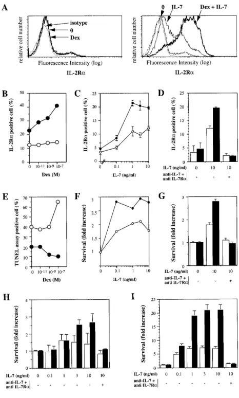
To determine whether the glucocorticoid-induced increase in IL-7R␣ in mature T cells has functional relevance, the expression of IL-2R␣, a cell surface molecule whose gene is up-regulated by IL-7 (22), was assessed in purified CD4⫹ cord blood T cells. Whereas Dex alone had little effect on IL-2R␣ expression (Fig. 5A,
left panel), IL-7 induced a moderate increase (Fig. 5A, right pan-el). Dex greatly enhanced IL-7-induced IL-2R␣ expression (Fig.
5A, right panel). Indeed, escalating concentrations of Dex gradu-ally enhanced IL-7-induced IL-2R␣ expression (Fig. 5B). More-over, we confirmed that the up-regulation of IL-2R␣ was IL-7 dose
FIGURE 5. Glucocorticoids enhance IL-7-medi-ated action on IL-2R␣ expression and survival. A, Naive cord blood purified CD4⫹T cells were un-treated or un-treated with Dex (10⫺7M), and untreated (left panel) or treated with IL-7 (10 ng/ml; right
panel) for 6 days and analyzed for IL-2R␣
expres-sion. The cells were stained with anti-IL-2R␣-FITC and isotype-matched FITC-IgG as a control and an-alyzed by flow cytometry. B, Naive cord blood pu-rified CD4⫹T cells were treated for 4 days with increasing concentration of Dex (10⫺11, 10⫺9, and 10⫺7M), without (E) or with IL-7 (10 ng/ml; F), and cells were analyzed for IL-2R␣ expression. One representative experiment of three is shown. C, Na-ive cord blood purified CD4⫹T cells were treated for 1 day with increasing concentrations of IL-7, without (E) or with Dex (10⫺7M; F), and cells were analyzed for IL-2R␣ expression. D, Naive cord blood purified CD4⫹T cells were treated for 1 day with IL-7 and with anti-IL-7 and anti-IL-7R␣ neu-tralizing Abs as indicated without (䡺) or with Dex (10⫺7M; f), and cells were analyzed for IL-2R␣ expression. E, Cells were treated as described in B and analyzed for apoptosis by bromodeoxyuridine staining using a modified TUNEL assay. F, Cells were treated as described in C and analyzed for ap-optosis using annexin V staining. G–I, Cells were treated as described in D and analyzed for apoptosis using annexin V staining after 1 (G), 3 (H), or 7 days (I). These experiments are representative of three donors, and the mean⫾ SEM of three independent experiments is represented.
dependent and was potentiated in the presence of glucocorticoids (Fig. 5C). Finally, we demonstrated that this enhanced IL-2R␣ expression was dependent upon IL-7 signaling, because it was completely inhibited by neutralizing Abs against the IL-7/IL-7R␣ complex (Fig. 5D). If the enhancement of IL-7 responsiveness by glucocorticoids has functional consequences, glucocorticoids might be expected to enhance the effect of IL-7 on T cell survival. As shown in Fig. 5E, IL-7 reduced by approximately half the spon-taneous apoptosis of CD4⫹cord blood cells. Whereas a high con-centration of Dex (10⫺7M) alone caused an increase in apoptosis, it inhibited apoptosis in the presence of IL-7. Using annexin V staining to measure apoptosis of purified CD4⫹T cells from cord blood, we analyzed the effect of IL-7 and neutralizing anti-IL-7/ IL-7R␣ Abs on the antiapoptotic effects of glucocorticoids and examined this effect in a dose-dependent manner (Fig. 5, F and G). Indeed, escalation of IL-7 doses favors survival, and this effect was further enhanced in the presence of glucocorticoids (Fig. 5F), whereas neutralizing Abs against the IL-7/IL-7R␣ complex com-pletely reversed the antiapoptotic effect of glucocorticoids and IL-7 (Fig. 5G). These results were confirmed with increasing doses of IL-7 in a time-dependent manner on days 3 (Fig. 5H) and 10 (Fig. 5I). Neutralizing Abs against the IL-7/IL-7R␣ complex com-pletely reversed the antiapoptotic effect of glucocorticoids and IL-7 on days 3 (Fig. 5H) and 10 (Fig. 5I). Thus, the IL-7-mediated inhibition of apoptosis increased with increasing concentrations of glucocorticoids and increasing doses of IL-7 in the presence of glucocorticoids, consistent with an enhanced sensitivity of Dex-treated cells to IL-7. These actions were specifically mediated by the IL-7/IL-7R␣ complex, as demonstrated with the blocking ex-periments. Therefore, glucocorticoids can paradoxically promote T cell survival in the presence of IL-7.
Discussion
IL-7 is a key cytokine for the development of T cells. Its impor-tance is illustrated by the fact that mice deficient for IL-7 (16) or IL-7R␣ (17) or humans with loss-of-function mutations in IL-7R␣ (18) have a marked deficiency in T cell development. The receptor for IL-7 is composed of the IL-7-specific subunit (IL-7R␣) (23) and␥c, the latter associating with the Janus kinase 3 (13, 20, 24,
25). Mutations in␥cand Janus kinase 3 also result in a reduction
in T cell number, and targeted mutations of other␥c-using
cyto-kines (IL-2, IL-4, and IL-15) clearly indicate that IL-7 has nonre-dundant functions in lymphopoiesis (26 –30). Several important functions have been ascribed to IL-7 in the thymus, including pro-motion of survival and proliferation of immature thymocytes (31– 33). IL-7 is also secreted by monocytes and dendritic and intestinal epithelial cells, participates in mature lymphoid survival and ex-pansion (34 –37), and inhibits T cell death in vitro (38, 39). Fur-thermore, IL-7 participates in regulation of the maintenance and the strength of the immune response and regulates the develop-ment and the cytolytic activity of intestinal␥␦ intraepithelial lym-phocytes (36, 40, 41). These intraepithelial lymlym-phocytes represent the frontline defense alimentary and bacterial gut lumen Ags and depend on IL-7R␣ signaling (42). Thus, IL-7R␣ signals IL-7-me-diated critical specific actions on T cell development and function, yet the control of its expression remains obscure.
Recognition of foreign Ags by mature T cells induces the ex-pression of many cytokine receptors that drive the immune re-sponse toward either a Th1 cellular or Th2 humoral type (43, 44). Two types of signals are needed to generate immune responses. The first is the recognition of Ag/MHC by the TCR. The second signal, termed costimulation, involves costimulatory molecule in-teractions and the activation of cytokine receptors with their cog-nate cytokines (1). Mounting evidence suggests the critical role of
the ligand-receptor complex IL-7/IL-7R␣ in the course of the T cell immune response (32, 34). For example, peripheral T cells from IL-7R␣⫺/⫺mice show impaired proliferation and increased apoptosis after TCR engagement. Therefore, IL-7R␣ expression appears to be important in the maintenance and strength of the immune response (34, 45, 46). In this regard it is noteworthy that TCR stimulation decreases IL-7R␣ expression. This down-regula-tion may represent a negative feedback loop to turn off lymphocyte activation and, hence, T cell-driven immune responses.
Adrenal-derived glucocorticoids suppress innate and specific immune responses through their negative actions on the expression of inflammatory mediators such as cytokines and cytokine recep-tors (5, 47, 48). Although widely used clinically, they clearly have protean effects and are far from optimal immunosuppressive agents. In this study we found that glucocorticoids increase IL-7R␣ expression in both naive and activated CD4⫹ T cells. By doing so, glucocorticoids were able to enhance the expression of IL-7-inducible genes and IL-7-mediated rescue from glucocorti-coid-induced apoptosis. The finding that glucocorticoids increase IL-7R␣ expression may provide a mechanism by which glucocor-ticoids can have a positive influence on the immune response. Based upon gene expression assessed with DNA microarrays, an-other top glucocorticoid-up-regulated gene is GILZ, with major antiapoptotic action. GILZ counteracts activation-induced T cell death and may contribute in part to the Dex-induced inhibition of TCR-activated apoptosis (19). Thus, glucocorticoids can have pro-survival activity during the T cell immune response by blocking TCR-induced clonal deletion and maintaining IL-7R␣ expression. During acute infections or flare-ups of inflammatory diseases, ad-renal glucocorticoid secretion is crucial for the restraint and con-trol of the adaptive immune response. In the neighborhood of IL-7-secreting epithelial or stromal cells, glucocorticoids may prevent T cell deletion by improving IL-7 signaling and help sustain the T cell immune response to overcome foreign Ags and pathogens.
New emigrants from generative lymphoid organs undergo pos-itive or negative selection in the periphery (2, 40, 49). The ho-meostasis of naive mature and memory T lymphocytes is depen-dent on ligand-TCR interactions (4, 50 –52). Also, T cell homeostasis is mediated by signals transmitted by growth factor receptors in secondary lymphoid and in nonlymphoid organs (3, 49, 53, 54). IL-7 potently enhances thymic-independent peripheral expansion and restores immunity in athymic T cell-depleted hosts (55). Furthermore, IL-7 is required for homeostatic expansion of naive CD8⫹and CD4⫹T cells in lymphopenic hosts and is par-tially required for the homeostatic proliferation of memory cells (22). These two studies point to IL-7 as an important cytokine in T cell homeostasis (22, 55). Therefore, improved IL-7R␣ signaling by glucocorticoids may contribute to IL-7-mediated expansion of memory T cells and maintenance of the peripheral T cell repertoire (56 –58).
References
1. Van Parijs, L., and A. K. Abbas. 1998. Homeostasis and self-tolerance in the immune system: turning lymphocytes off. Science 280:243.
2. Rocha, B., and H. von Boehmer. 1991. Peripheral selection of the T cell reper-toire. Science 251:1225.
3. Sprent, J., D. F. Tough, and S. Sun. 1997. Factors controlling the turnover of T memory cells. Immunol. Rev. 156:79.
4. Tanchot, C., F. A. Lemonnier, B. Perarnau, A. A. Freitas, and B. Rocha. 1997. Differential requirements for survival and proliferation of CD8 naive or memory T cells. Science 276:2057.
5. Wilckens, T., and R. De Rijk. 1997. Glucocorticoids and immune function: un-known dimensions and new frontiers. Immunol. Today 18:418.
6. Besedovsky, H. O., and A. del Rey. 1996. Immune-neuro-endocrine interactions: facts and hypotheses. Endocr. Rev. 17:64.
7. Ashwell, J. D., F. W. Lu, and M. S. Vacchio. 2000. Glucocorticoids in T cell development and function. Annu. Rev. Immunol. 18:309.
8. Boumpas, D. T., G. P. Chrousos, R. L. Wilder, T. R. Cupps, and J. E. Balow. 1993. Glucocorticoid therapy for immune-mediated diseases: basic and clinical correlates. Ann. Intern. Med. 119:1198.
9. Zacharchuk, C. M., M. Mercep, P. K. Chakraborti, S. S. Simons, Jr., and J. D. Ashwell. 1990. Programmed T lymphocyte death: cell activation- and ste-roid-induced pathways are mutually antagonistic. J. Immunol. 145:4037. 10. Baus, E., F. Andris, P. M. Dubois, J. Urbain, and O. Leo. 1996. Dexamethasone
inhibits the early steps of antigen receptor signaling in activated T lymphocytes. J. Immunol. 156:4555.
11. Iwata, M., S. Hanaoka, and K. Sato. 1991. Rescue of thymocytes and T cell hybridomas from glucocorticoid-induced apoptosis by stimulation via the T cell receptor/CD3 complex: a possible in vitro model for positive selection of the T cell repertoire. Eur. J. Immunol. 21:643.
12. Van Laethem, F., E. Baus, L. A. Smyth, F. Andris, F. Bex, J. Urbain, D. Kioussis, and O. Leo. 2001. Glucocorticoids attenuate T cell receptor signaling. J. Exp. Med. 193:803.
13. Johnston, J. A., M. Kawamura, R. A. Kirken, Y. Q. Chen, T. B. Blake, K. Shibuya, J. R. Ortaldo, D. W. McVicar, and J. J. O’Shea. 1994. Phosphory-lation and activation of the Jak-3 Janus kinase in response to interleukin-2. Na-ture 370:151.
14. Galon, J., C. Sudarshan, S. Ito, D. Finbloom, and J. J. O’Shea. 1999. IL-12 induces IFN regulating factor-1 gene expression in human NK and T cells. J. Im-munol. 162:7256.
15. Ascherman, D. P., T. S. Migone, M. C. Friedmann, and W. J. Leonard. 1997. Interleukin-2 (IL-2)-mediated induction of the IL-2 receptor␣ chain gene: critical role of two functionally redundant tyrosine residues in the IL-2 receptor chain cytoplasmic domain and suggestion that these residues mediate more than Stat5 activation. J. Biol. Chem. 272:8704.
16. von Freeden-Jeffry, U., P. Vieira, L. A. Lucian, T. McNeil, S. E. Burdach, and R. Murray. 1995. Lymphopenia in interleukin (IL)-7 gene-deleted mice identifies IL-7 as a nonredundant cytokine. J. Exp. Med. 181:1519.
17. Peschon, J. J., P. J. Morrissey, K. H. Grabstein, F. J. Ramsdell, E. Maraskovsky, B. C. Gliniak, L. S. Park, S. F. Ziegler, D. E. Williams, C. B. Ware, et al. 1994. Early lymphocyte expansion is severely impaired in interleukin 7 receptor-defi-cient mice. J. Exp. Med. 180:1955.
18. Puel, A., S. F. Ziegler, R. H. Buckley, and W. J. Leonard. 1998. Defective IL7R expression in T⫺B⫹NK⫹severe combined immunodeficiency. Nat. Genet. 20: 394.
19. D’Adamio, F., O. Zollo, R. Moraca, E. Ayroldi, S. Bruscoli, A. Bartoli, L. Cannarile, G. Migliorati, and C. Riccardi. 1997. A new dexamethasone-in-duced gene of the leucine zipper family protects T lymphocytes from TCR/CD3-activated cell death. Immunity 7:803.
20. Leonard, W. J., and J. J. O’Shea. 1998. Jaks and STATs: biological implications. Annu. Rev. Immunol. 16:293.
21. Akashi, K., M. Kondo, and I. L. Weissman. 1998. Role of interleukin-7 in T-cell development from hematopoietic stem cells. Immunol. Rev. 165:13. 22. Schluns, K., W. Kieper, S. Jameson, and L. Lefranc¸ois. 2000. Interleukin-7
me-diates the homeostasis of naive and memory CD8 T cells in vivo. Nat. Immunol. 1:426.
23. Goodwin, R. G., D. Friend, S. F. Ziegler, R. Jerzy, B. A. Falk, S. Gimpel, D. Cosman, S. K. Dower, C. J. March, A. E. Namen, et al. 1990. Cloning of the human and murine interleukin-7 receptors: demonstration of a soluble form and homology to a new receptor superfamily. Cell 60:941.
24. Miyazaki, T., A. Kawahara, H. Fujii, Y. Nakagawa, Y. Minami, Z. J. Liu, I. Oishi, O. Silvennoinen, B. A. Witthuhn, J. N. Ihle, et al. 1994. Functional activation of Jak1 and Jak3 by selective association with IL-2 receptor subunits. Science 266:1045.
25. Russell, S. M., J. A. Johnston, M. Noguchi, M. Kawamura, C. M. Bacon, M. Friedmann, M. Berg, D. W. McVicar, B. A. Witthuhn, O. Silvennoinen, et al. 1994. Interaction of IL-2R and ␥cchains with Jak1 and Jak3: implications for
XSCID and XCID. Science 266:1042.
26. Noguchi, M., Y. Nakamura, S. M. Russell, S. F. Ziegler, M. Tsang, X. Cao, and W. J. Leonard. 1993. Interleukin-2 receptor␥ chain: a functional component of the interleukin-7 receptor. Science 262:1877.
27. Macchi, P., A. Villa, S. Giliani, M. G. Sacco, A. Frattini, F. Porta, A. G. Ugazio, J. A. Johnston, F. Candotti, J. J. O’Shea, et al. 1995. Mutations of Jak-3 gene in patients with autosomal severe combined immune deficiency (SCID). Nature 377:65.
28. Nosaka, T., J. M. van Deursen, R. A. Tripp, W. E. Thierfelder, B. A. Witthuhn, A. P. McMickle, P. C. Doherty, G. C. Grosveld, and J. N. Ihle. 1995. Defective lymphoid development in mice lacking Jak3. [Published erratum appears in 1996 Science 271:17.] Science 270:800.
29. Russell, S. M., N. Tayebi, H. Nakajima, M. C. Riedy, J. L. Roberts, M. J. Aman, T. S. Migone, M. Noguchi, M. L. Markert, R. H. Buckley, et al. 1995. Mutation of Jak3 in a patient with SCID: essential role of Jak3 in lymphoid development. Science 270:797.
30. DiSanto, J. P., W. Muller, D. Guy-Grand, A. Fischer, and K. Rajewsky. 1995. Lymphoid development in mice with a targeted deletion of the interleukin 2 receptor␥ chain. Proc. Natl. Acad. Sci. USA 92:377.
31. Akashi, K., M. Kondo, U. von Freeden-Jeffry, R. Murray, and I. L. Weissman. 1997. Bcl-2 rescues T lymphopoiesis in interleukin-7 receptor-deficient mice. Cell 89:1033.
32. Maraskovsky, E., L. A. O’Reilly, M. Teepe, L. M. Corcoran, J. J. Peschon, and A. Strasser. 1997. Bcl-2 can rescue T lymphocyte development in interleukin-7 receptor-deficient mice but not in mutant rag-1⫺/⫺mice. Cell 89:1011. 33. von Freeden-Jeffry, U., N. Solvason, M. Howard, and R. Murray. 1997. The
earliest T lineage-committed cells depend on IL-7 for Bcl-2 expression and nor-mal cell cycle progression. Immunity 7:147.
34. Maraskovsky, E., M. Teepe, P. J. Morrissey, S. Braddy, R. E. Miller, D. H. Lynch, and J. J. Peschon. 1996. Impaired survival and proliferation in IL-7 receptor-deficient peripheral T cells. J. Immunol. 157:5315.
35. Reinecker, H. C., and D. K. Podolsky. 1995. Human intestinal epithelial cells express functional cytokine receptors sharing the common␥cchain of the
inter-leukin 2 receptor. Proc. Natl. Acad. Sci. USA 92:8353.
36. Watanabe, M., Y. Ueno, T. Yajima, Y. Iwao, M. Tsuchiya, H. Ishikawa, S. Aiso, T. Hibi, and H. Ishii. 1995. Interleukin 7 is produced by human intestinal epi-thelial cells and regulates the proliferation of intestinal mucosal lymphocytes. J. Clin. Invest. 95:2945.
37. Maeurer, M. J., and M. T. Lotze. 1998. Interleukin-7 (IL-7) knockout mice: implications for lymphopoiesis and organ-specific immunity. Int. Rev. Immunol. 16:309.
38. Vella, A., T. K. Teague, J. Ihle, J. Kappler, and P. Marrack. 1997. Interleukin 4 (IL-4) or IL-7 prevents the death of resting T cells: stat6 is probably not required for the effect of IL-4. J. Exp. Med. 186:325.
39. Vella, A. T., S. Dow, T. A. Potter, J. Kappler, and P. Marrack. 1998. Cytokine-induced survival of activated T cells in vitro and in vivo. Proc. Natl. Acad. Sci. USA 95:3810.
40. Rocha, B., P. Vassalli, and D. Guy-Grand. 1992. The extrathymic T-cell devel-opment pathway. Immunol. Today 13:449.
41. Rocha, B., P. Vassalli, and D. Guy-Grand. 1994. Thymic and extrathymic origins of gut intraepithelial lymphocyte populations in mice. J. Exp. Med. 180:681. 42. Maki, K., S. Sunaga, and K. Ikuta. 1996. The V-J recombination of T cell
re-ceptor-␥ genes is blocked in interleukin-7 receptor-deficient mice. J. Exp. Med. 184:2423.
43. Mosmann, T. R., and S. Sad. 1996. The expanding universe of T-cell subsets: Th1, Th2 and more. Immunol. Today 17:138.
44. Romagnani, S. 1997. The Th1/Th2 paradigm. Immunol. Today 18:263. 45. Hassan, J., and D. J. Reen. 1998. IL-7 promotes the survival and maturation but
not differentiation of human post-thymic CD4⫹T cells. Eur. J. Immunol. 28: 3057.
46. Thomis, D. C., and L. J. Berg. 1997. Peripheral expression of Jak3 is required to maintain T lymphocyte function. J. Exp. Med. 185:197.
47. Almawi, W. Y., H. N. Beyhum, A. A. Rahme, and M. J. Rieder. 1996. Regulation of cytokine and cytokine receptor expression by glucocorticoids. J. Leukocyte Biol. 60:563.
48. Wu, C. Y., K. Wang, J. F. McDyer, and R. A. Seder. 1998. Prostaglandin E2and
dexamethasone inhibit IL-12 receptor expression and IL-12 responsiveness. J. Immunol. 161:2723.
49. Tanchot, C., and B. Rocha. 1998. The organization of mature T-cell pools. Im-munol. Today 19:575.
50. Ernst, B., D. S. Lee, J. M. Chang, J. Sprent, and C. D. Surh. 1999. The peptide ligands mediating positive selection in the thymus control T cell survival and homeostatic proliferation in the periphery. Immunity 11:173.
51. Goldrath, A. W., and M. J. Bevan. 1999. Selecting and maintaining a diverse T-cell repertoire. Nature 402:255.
52. Viret, C., F. S. Wong, and C. A. Janeway, Jr. 1999. Designing and maintaining the mature TCR repertoire: the continuum of self-peptide:self-MHC complex recognition. Immunity 10:559.
53. Zhang, X., S. Sun, I. Hwang, D. F. Tough, and J. Sprent. 1998. Potent and selective stimulation of memory-phenotype CD8⫹T cells in vivo by IL-15. Im-munity 8:591.
54. Garcia, S., J. DiSanto, and B. Stockinger. 1999. Following the development of a CD4 T cell response in vivo: from activation to memory formation. Immunity 11:163.
55. Fry, T. J., B. L. Christensen, K. L. Komschlies, R. E. Gress, and C. L. Mackall. 2001. Interleukin-7 restores immunity in athymic T-cell-depleted hosts. Blood 97:1525.
56. Kos, F. J., and A. Mullbacher. 1993. IL-2-independent activity of IL-7 in the generation of secondary antigen-specific cytotoxic T cell responses in vitro. J. Im-munol. 150:387.
57. Dozmorov, I., and R. A. Miller. 1997. In vitro production of antigen-specific T cells from unprimed mice: role of dexamethasone and anti-IL-10 antibodies. Cell. Immunol. 178:187.
58. Dozmorov, I. M., and R. A. Miller. 1999. Age-associated decline in responses of naive T cells to in vitro immunization reflects shift in glucocorticoid sensitivity. Life Sci. 64:1849.