Thymosin
ββ4 has tumor suppressive effects and its decreased expression results
in poor prognosis and decreased survival in multiple myeloma
Jo Caers,1,2Dirk Hose,3,5Ine Kuipers,1Tomas Jan Bos,1Els Van Valckenborgh,1Eline Menu,1Elke De Bruyne,1
Hartmut Goldschmidt,3,5Ben Van Camp,1Bernard Klein,4and Karin Vanderkerken1
1Laboratory of Hematology and Immunology, Vrije Universiteit Brussel, Myeloma Center Brussels, Brussels, Belgium;2Department
of Hematology, CHU Université de Liège, Campus Sart-Tilman, Liège, Belgium;3Nationales Centrum für Tumorerkrankungen,
Heidelberg, Germany;4Institut National de la Santé et de la Recherche Médicale U475 and Unit for Cellular Therapy, CHU
Montpellier, Montpellier, France; 5Medizinische Klinik V, Universitätsklinikum Heidelberg, Heidelberg, Germany
Introduction
β-thymosins are a family of small peptides that were origi-nally proposed to be thymic hormones.1They were identified as actin monomer binding proteins, controlling the availabili-ty of actin for polymerization. They may, therefore, have a crucial role in regulating cellular functions involving actin polymerization/depolymerization cycles. Currently, 15 β-thymosins have been identified and characterized as highly conservative 5-kDa peptides containing 40 to 44 amino acid residues. In most mammalian tissues, thymosin-β 4 (Tβ4), the most abundant thymosin peptide, Tβ10 and Tβ15, have been studied as important members of the β-thymosin family.2 Several studies reported that these genes are over-expressed in solid tumors, which could be correlated to the angiogenic
and metastatic potential of the studied tumors.3
Multiple myeloma (MM) is a hematologic malignancy char-acterized by the accumulation of monoclonal plasma cells (PC) in the bone marrow (BM). MM cell biology can be dis-sected into the interactions of MM cells with their surround-ing stroma (matrix proteins, cytokines and BM cells) and in the acquisition of essential changes in cell behavior, such as self-sufficiency in growth signals, evasion of apoptosis and acquisition of invasive and spreading capacities.4 Earlier reports indicated that Tβ4 was down-regulated in RNA from primary human MM cells and cell lines.5
This observation is in contrast to the results obtained in most solid tumors where an upregulation is seen in malignant cells compared to their normal counterparts. Cha et al. showed that overexpression of Tβ4 resulted in an increased
JC and DH contributed equally to this manuscript. Acknowledgments: we thank Hae-Jae Cha (Research Center for Genetic Medicine, Children’s National Medical Center, Washington DC, USA) for providing the plasmid containing the murine thymosin β4 sequence. We also thank Angelo Willems, Carlo Heirman, Elsy Vaeremans, Erik Quartier, and Carine Seynaeve for excellent technical assistance.
Funding: this work was financially supported by the Fund for Scientific Research-Vlaanderen (FWO-Vlaanderen), the Association for International Cancer Research and by a grant from the European Commission FP6 to MSCNET. EM is a post-doctoral fellow of FWO-Vlaanderen.
Manuscript received on January 25, 2009. Revised version arrived on July 4, 2009. Manuscript accepted on July 7, 2009.
Correspondence: Karin Vanderkerken, Vrije Universiteit Brussel (VUB), Department of Hematology and Immunology, Laarbeeklaan 103, B-1090 Brussels, Belgium. E-mail: [email protected]
The online version of this article has a supplementary appendix.
Thymosin β4 (Tβ4) is a polypeptide involved in cellular proliferation, differentiation, and migration, over-expressed in several tumor entities. We evaluated its expression and function in 298 newly diagnosed multiple myeloma patients and the murine 5TMM model. Mean
Tβ4 expression was significantly lower in myeloma cells
compared to normal plasma cells (P<0.001). The same observation can be made in the 5TMM-mouse model by qRT-PCR and ELISA. Here, Tβ4 overexpression by lentivi-ral transduction of 5T33MMvt-cells led to significantly decreased proliferative and migratory capacities and increased sensitivity to apoptosis-induction. Mice injected with Tβ4 over-expressing myeloma cells showed a longer survival compared to mice injected with controls (88,9 vs. 65,9 days, P<0.05). In 209 MM patients treated with high-dose therapy and autologous stem cell transplantation,
expression of Tβ4 below the median was associated with a significantly shorter event free survival (37.6 vs. 26.2 months, P<0.05). In conclusion, our results indicate a pos-sible tumor suppressive function of Tβ4.
Key words: thymosin β4, cellular proliferation, multiple myeloma.
Citation: Caers J, Hose D, Kuipers I, Bos TJ, Van Valckenborgh E, Menu E, De Bruyne E, Goldschmidt H, Van Camp B, Klein B, and Vanderkerken K. Thymosin β4 has tumor suppressive effects and its decreased expression results in poor prognosis and decreased survival in multiple myeloma. Haematologica. 2010; 95:163-167. doi: 10.3324/haematol.2009.006411
©2010 Ferrata Storti Foundation. This is an open-access paper.
metastatic capacity of lung cancer cells and increased angiogenic response.6
Since migration, invasion and associated angiogenesis are key features in MM biology, we were interested in studying Tβ4 expression in a large panel of MM patients and its functionality in the murine 5TMM model.
Design and Methods
Gene expression analysis on human myeloma cells
Tβ4 expression was analyzed in purified PCs from BM samples obtained from 14 healthy donors, 11 patients with monoclonal gam-mopathy of unknown significance (MGUS) and 298 previously untreated multiple MM patients at the University Hospitals of Heidelberg or Montpellier.7Of these, 209 MM patients were treatedby high-dose therapy and autologous stem cell transplantation (ASCT). Biotinylated complementary RNA (cRNA) was amplified according to the Affymetrix labeling protocol (Affymetrix, Santa Clara, CA, USA). cRNA from a first group of patients (7 normal donors, 7 MGUS and 65 MM patients) was hybridized to the human U133 A and B. This group will be referred to as the HM1-group. A second independent validation group of patients (7 normal donors, 16 MGUS and 233 MM patients) was named the HM2 group. For these patients, the U133 2.0 GeneChip was used. These micro-array data had been previously used for several analyses, but thymosin β4 expression had never been analyzed before.8,9HM2 data were cor-rected for batch effect due to the usage of different labeling kits according to Johnson et al.10Expression data were gcrma-normalized and analyzed by the bioconductor packages for R. For patients’ char-acteristic see Online Supplementary Table S1.
The 5T2MM and 5T33MM murine models
of myeloma
The 5TMM models originated in elderly C57Bl/KaLwRij mice.11
The 5T33MMvivo (5T33MMvv) cells grow in vitro stroma-dependent-ly with a limited survival while the 5T33MMvitro (5T33MMvt) cell line is a clonally identical variant that originated from an in vitro cul-ture of 5T33MMvv cells, growing BM stroma-independently in RPMI-1640 supplemented with 10% bovine serum 1% natriumpyru-vate, 100 U/mL penicillin, 100 µg/mL streptomycin and 2 mM L-glu-tamine (all from Biowhittaker, Verviers, Belgium).12
Quantification of intracellular protein levels of T
β4 and
F-Actin G-Actin
Enzyme-Linked Immunosorbent Assays (ELISA) for measuring Tβ4 concentrations were performed according to the manufacturer’s instructions (Immundiagnostik, Bensheim, Germany). Cells (107)
were lyzed in a phosphate buffer containing 0.14 M NaCl, 2.6 M KCl, 8 mM Na2HPO4, 1.4 M KH2PO4 and 1% Triton X100 and sonicat-ed with an ultrasound finger. Protein levels and ratios between F-Actin and G-F-Actin were determined using the G-actin/F-actin in vivo assay kit (Cytoskeleton Inc, Denver, USA).
Quantitative real-time PCR
Quantitative real-time PCR (qRT-PCR) was performed using the ABI Prism 7700 Sequence Detection System. For the detection of both human and mouse Tβ4 mRNA and the endogenous reference gene GUS, Assays on Demand (Applied Biosystems) were used. To verify the results obtained with the microarrays studies, Tβ4 expres-sion was measured in 3 cell lines and in 3 patient samples and their correlations statistically verified using a Spearman correlation test.
Generation of 5T33MMvt cells over-expressing T
ββ4
A lentiviral transferplasmid encoding mouse Tβ4 (m Tβ4) was con-structed. The mTβ4 gene was obtained from HJ Cha (NIDCR, NIH, Bethesda, USA)6and inserted into the transferplasmidpHR'tripCMV-IRES-tNGFR-SIN.13mTβ4-encoding lentiviral vector particles were
produced in 293T cells, collected, ultracentrifugated and their viral titer determined.14After transduction, 5T33MMvt cells were surface stained using an in-house biotinylated anti-tNGFR antibody and puri-fied by FACS sorting into a 6-well plate (Becton Dickinson, FACSVantage). Next, they were analyzed for Tβ4 expression by RT-PCR. The 5T33MMvt cells over-expressing Tβ4 will be referred to as 5T33MMvtTβ4+.
In vitro and in vivo effects of T
ββ4 overexpression
In vitro proliferation was assessed by measuring DNA synthesis
using a 3H-thymidine incorporation assay, as described earlier.15
Apoptosis sensitivity of the MM cells was analyzed by staining with FITC labeled-annexin V and propidium iodide according to the man-ufacturers’ instructions (BD Biosciences, Erembodegem, Belgium). In
vitro migration studies were performed using Transwell chambers and
10% fetal calf serum as chemoattractant and were quantified through flow cytometry. To determine the effect of Tβ4 overexpression on survival, groups of 10 C57BLKaLwRij mice were intravenously inject-ed with either 5×1055T33MMvtTβ4+or wild-type 5T33MMvt cells.
Animals were sacrificed when they showed signs of morbidity, namely hind limb paralysis. Kaplan-Meier analysis was used to deter-mine a difference in the survival.
Results and Discussion
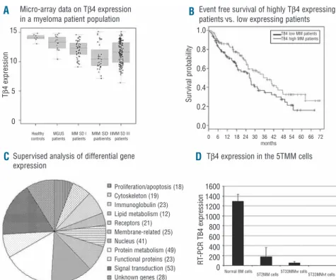
Different studies indicated a pivotal role of Tβ4 in the metastatic process of solid tumors.16,17 An adenoviral-based overexpression of Tβ4 was applied in a colon cancer and melanoma model showing increased growth, motility and invasive capacities in vitro and a larger tumor load in vivo.18,19Since proliferation, migration and invasion are part of the hallmarks of the biology of MM, we were interest-ed in investigating an involvement of Tβ4 in this disease. We first investigated the Tβ4 expression pattern in 298 pri-mary MM-cell samples and 14 normal plasma cell samples from healthy donors. Tβ4 expression is significantly lower in MM cells of the HM1 group (P<0.05) and HM2 group (P<0.001) compared to normal plasma cells. This holds true for a significantly lower Tβ4 expression in its pre-malignant stage (MGUS), its early (Durie Salmon stage I) or late stage (Durie Salmon II and III) in both HM1 and HM2 groups (P<0.001) (Figure 1A). No relevant correla-tion could be found between Tβ4 expression and percent-age of plasma cell infiltration in the bone marrow smear. Gene expression assessed by DNA-microarray correlates well with qRT-PCR performed on MM patient samples (coefficient of correlation r=0.993, P<0.001). These data are in agreement with results from Gondo et al. showing a decrease in Tβ4 expression in a small number of MM sam-ples by Northern blot analysis.5
Given the differential Tβ4 expression in MM patients, we subsequently investigated a possible prognostic value and influence on event free (EFS) and overall survival (OS) in our patient population. As Tβ4 was expressed in all MM cells, we examined the survival of 209 patients by compar-ing patients with Tβ4 expression above (Tβ4high) and below
(Tβ4low) the median (Figure 1B). Patients with Tβ4high com-pared with Tβ4lowshow a significantly longer median EFS (n=209, 37.6 months vs. 26,2, P<0.05), but only a trend regarding OS (P=0.1). Concerning EFS, multivariate analy-ses on Tβ4 expression with either ISS or β2-microglobulin indicated an independent (P=0.04) prognostic value of Tβ4 expression regarding ISS (P<0.001), but not (P=0.07) regarding β2-microglobulin (P=0.003) levels. In multi-vari-ate analyses for OS, ISS and β2-microglobulin appear as significant (P=<0.001) variables, whereas Tβ4 expression fails to reach independence (P=0.09 and P=0.01, respec-tively).
A supervized analysis of expression data comparing the Tβ4highto the Tβ4lowgroup identified over 300 significantly differentially expressed genes. These genes are listed in Online Supplementary Table S2. Analysis of their biological function allowed them to be divided into main functional categories and this distribution is illustrated in Figure 1C. Signal transduction, protein metabolism and nuclear func-tions were the largest categories, but 19 genes were impli-cated in cytoskeletal organization, and 32 genes in lym-phoid differentiation and immunoglobulin processing. In general, these gene clusters indicate a biological difference between MM cells of the two patient groups.
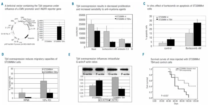
The data obtained in MM patients were also seen in the 5T33MM murine MM model by qRT-PCR demonstrating a decreased mRNA expression in 5TMM cells compared to normal BM cells (P<0.001, Figure 1D). Competitive ELISA confirmed these results on protein level (results not shown). To study functional effects of Tβ4, the gene was over-expressed using a lentiviral expression vector. The 5T33MMvt cell line was stably transduced and after sub-cloning, a 99% pure clone with strong t-NGFR expression was obtained. qRT-PCR confirmed the overexpression of Tβ4 compared to control cells (Figure 2A).
To assess the functional involvement of differential Tβ4 expression we used the 5T33MMvt and 5T33MMvtTβ4+ cell lines. In a 3H thymidine assay, 5T33MMvtTβ4+ cells showed a significant decrease in DNA synthesis compared to control cells (P<0.05). 5T33MMvtTβ4+ cells showed a sig-nificantly increased sensitivity to vinca-alkaloids (vin-blastin) and bortezomib (Figure 2B; P<0.001 for both bortezomib and vinblastin).
Likewise, bortezomib induced apoptosis was higher in 5T33MMvtTβ4+compared with 5T33MMvt cells (P<0.05; Figure 2C). In addition to affect survival pathways, Tβ4 overexpression reduced migratory capacities of 5T3MM cells; the percentages of cells that migrated in basal condi-tions and in 10%FCI was significantly lower in 5T33MMvtTβ4+compared to control cells (P<0.05; Figure 2D). The relative increase after stimulation (compared to basal conditions) was, however, similar in both popula-tions. We further examined the effects of Tβ4 expression on tumor development and survival of diseased mice by injecting mice intravenously with 5T33MMvtTβ4+or con-trol cells. In this study, the mean survival of mice injected with control cells was significantly shorter 65.9 days (SD 6.6 days), compared to 88.9 days (SD 9.3 days) for mice injected with 5T33MMvtTβ4+ cells (P<0.05; Figure 2F). These in vivo results confirm data obtained using the in vitro proliferation and apoptosis assays.
In solid tumors, Tβ4 expression is frequently upregulat-ed in malignant and metastatic cells. In these cancers, higher Tβ4 expression resulted in increased metastatic and invasive capacities of tumor cells, while proliferation remained unaffected.6In hematologic disorders, malignant plasma cell disorders, such as plasma cell leukemia and MM were the rare disorders that showed a decreased Tβ4 expression.5,20In contrast to solid tumors, publications on the function of Tβ4 in hematologic conditions are scanty
Figure 1. (A) The micro-array data
obtained for the Tβ4 expression in CD138+ sorted BM plasma
cells from healthy donors and MM patients. These results were validated by quantitative RT-PCR. MGUS: monoclonal gammopathy of undetermined significance, MM multiple MM, SD: Salmon and Durie Stage. (B) The event free survival of Tβ4high and Tβ4low
patients. Patients with Tβ4lowMM
had statistically significantly decreased event free survivals compared to patients with Tβ4high
MM (P<0.001), while also their overall survival tended to be shorter. (C) The differently expressed genes between Tβ4high
and Tβ4low patient groups. After
identification of the gene, these were grouped into similar biologi-cal function. A complete list of the genes can be found in Online
Supplementary Table S2. (D) A
similar gene expression pattern was observed in the murine 5TMM models where Tβ4 mRNA expression in 5T33MM and 5T2MM invaded BM was lowered compared to normal BM cells.
Micro-array data on Tβ4 expression in a myeloma patient population
Tβ4 expression in the 5TMM cells Supervised analysis of differential gene
expression Tβ 4 expression R T -PCR TB4 expression Sur vival probability Normal BM cells Proliferation/apoptosis (18) Cytoskeleton (19) Immunoglobulin (23) Lipid metabolism (12) Receptors (21) Membrane-related (25) Nucleus (41) Protein metabolism (49) Functional proteins (23) Signal transduction (53) Unknown genes (28) Healthy controls MGUS patients MM SD I patients MM SD II patients MM SD III patients TB4 low MM patients TB4 high MM patients 0 6 12 18 24 30 36 42 48 54 60 6672 months 5T33MMw cells 5T2MM cells 5T33MMvt cells Event free survival of highly Tβ4 expressing patients vs. low expressing patients A C D B 15 10 5 0 1600 1400 1200 1000 800 600 400 200 0 1.0 0.8 0.6 0.4 0.2 0.0
but indicate some inhibitory activity. Tβ4 was initially iso-lated and purified from a thymic protein preparation, called thymosin fraction-5. Addition of this protein frac-tion to different leukemic cell lines resulted in a decrease in growth responses.21 Similar inhibitory effects were recently described for Tβ4 on hematopoietic stem cells,22 bone marrow derived mast cells23and human promyelo-cytic leukemia cells,24in agreement with the results pre-sented here. Whereas a mechanistic explanation of this discrepancy is beyond the scope of this paper, further investigations are clearly merited.
Since Tβ4 has been shown to bind G-actin in a 1:1 man-ner and thus affects the polymerization of G-Actin into F-Actin, we analyzed in a semi-quantitative way, intracellu-lar G-actin and F-Actin. This quantification showed a low-ered G-Actin-F-Actin ratio after Tβ4 overexpression (Figure 2E). F-Actin is of particular importance in cytoskeleton changes involved in cellular migration and in microtubuli organization controlling the mitotic spin-dle.25,26In line with these results, vinca-alkaloids (e.g. vin-blastine used here) with micro-tubulin (polymerization) inhibitory activity, had more affect on the proliferation capacities of 5T33MMvtTβ4+ cells than on control cells (Figure 2B). Since immunohistochemical studies also showed a nuclear staining of Tβ4 in 5TMM cells (results not
shown), involvement of other pathways might also be implicated. Supervized gene analysis comparing Tβ4high with Tβ4low found different groups of genes differently expressed, including genes involved in cytoskeleton organization, nuclear homeostasis, lymphocyte differenti-ation and protein metabolism, which might indicate that the role of Tβ4 is more complicated than initially sup-posed.
In conclusion, our results propose a tumor suppressive function of Tβ4 expression in MM with impact on survival. Tβ4 was down-regulated in MM cells of patients compared to the normal BM plasma cells and studies with the murine 5T33MM model show a decreased in vitro and in vivo tumor growth for cells over-expressing the Tβ4 gene.
Authorship and Disclosures
JC and DH were the principal investigators and took pri-mary responsibility for the paper. JC, DH, IK, TJB, EDB and EM participated in the laboratory work for this study. BVC, EVV, BK and KV coordinated the research. HG and BK were responsible for patient recruitment and patient data. JC, DH, TJB, BK and KV wrote the paper.
The authors reported no potential conflicts of interest.
Figure 2. (A) Schematic representation of the modified lentiviral transfer plasmid and results of RT-PCR and qRT-PCR indicating the
pres-ence of the inserted Tβ4 gene in cultured 5T33MMvtTβ4+ cells. (B)3H thymidine uptake revealed a decreased DNA synthesis rate in
5T33MMvtTβ4+cells compared to wild-type cells. Incubation with the anti-MM agent bortezomib (5 nM) or the micro-tubuli inhibitor
vinblas-tine (2,5 µM) had significantly (P<0.001) stronger effects on 5T33MMvtTβ4+cells than on control cells. A similar observation was made in
apoptosis studies (C), where 5nM of bortezomib resulted in a significantly (P<0.05) increased apoptotic cell population after 18h incuba-tion. (D) The effects of Tβ4 overexpression on migration of 5T33MMvt cells: using 10% fetal calf serum as chemo-attractant, only 10.8%
(SD 6.6%) of 5T33MMvtTβ4+cells migrated compared to 34.7% (SD 15.9%) of the control 5T33MMvt cells (P<0.05). (E) (Upper) The F-actin
and G-actin bands of 5T33MMvt and 5T33MMvtTβ4+cells. The graph illustrates the ratios of quantified F-actin and G-actin. In 5T33MMt cells
actin is present in its polymerized form, whereas Tβ4 overexpression results in decreased F-actin formation and a greater pool of G-actin. (F) C57Bl/KaLwRij mice were injected with 5T33MMvt wild-type and 5T33MMvtTβ4+cells. Kaplan-Meier analysis showed a significantly
dif-ferent survival between these 2 groups with a mean survival of mice injected with 5T33MMvt wild type of 65.9 days (SD 6.6 days), com-pared to 88.9 days (SD 9.3 days) for mice injected with 5T33MMvtTβ4+cells. (P<0.05). LTR: long terminal repeat; gag: frame-shifted gag
gene; RRE: rev-responsive element; CMV: cytomegalovirus promotor trip: central polypurine tract + termination sequence; Ires: internal ribo-somal entry site; tNGFR: truncated form of the nerve growth factor receptor.
A lentiviral vector containing the Tβ4 sequence under influence of a CMV promotor and t-NGFR reporter gene
Tβ4 overexpression reduces migratory capacities of 5T33MMvt cells
Tβ4 overexpression influences intracellular G-actin/F-actin ratios
Survival curves of mice injected with 5T33MMvt TB4+and control cells
0.00 20.00 40.00 60.00 80.00 100.00 120.00 Days
control Bortezomib nM Basal bortezomib 5 nM vinblastin 2,5 M
5T33MM vt 5T33MM vt TB4 5T33MM vt 5T33MM vt TB4+ 5T33MMvt 5T33MMvt TB4+ 5T33MMvt 5T33MMvt TB4+ CMV CMV Primer Thymos in β4 IRES Primer IRES tNGFR 3’ LTR Ampicilin SV 40 gpt SV 40 Poly A 5’ LTR psi RRE cPPT pHR’tripCMV-Thymosin β4-IRES-tNGFR-SIN 5T33MMvt T33MMvt TB4
RPMI 10% FCI G-ACTIN F-ACTIN
5T33MMvt disease mice TB4 overpressing 1.0 0.8 0.6 0.4 0.2 0.0 0.6 0.55 0.5 0.45 0.4 0.35 0.3 60 50 40 30 20 10 0 35 30 25 20 15 10 5 0 35000 30000 25000 20000 15000 10000 5000 0 9000 8000 7000 6000 5000 4000 3000 2000 1000 0 Cum sur vival % migrated cells % apoptotic cells counts/min fold increase in TB4 expression P=0.037
Tβ4 overexpression results in decreased proliferation and increased sensibility to anti-myeloma agents
In vitro effect of bortezomib on apoptosis of 5T33MMvt
cells
A
D E F
References
1. Goldstein AL, Badamchian M. Thymosins: chemistry and biological properties in health and disease. Expert Opin Biol Ther. 2004;4(4):559-73.
2. Hannappel E. beta-Thymosins. Ann NY Acad Sci. 2007;1112:21-37.
3. Chen C, Li M, Yang H, Chai H, Fisher W, Yao Q. Roles of thymosins in cancers and other organ systems. World J Surg. 2005; 29(3):264-70.
4. Caers J, Van Valckenborgh E, Menu E, Van Camp B, Vanderkerken K. Unraveling the biology of multiple myeloma disease: can-cer stem cells, acquired intracellular changes and interactions with the sur-rounding micro-environment. Bull Cancer. 2008;95(3):301-13.
5. Gondo H, Kudo J, White JW, Barr C, Selvanayagam P, Saunders GF. Differential expression of the human thymosin-beta 4 gene in lymphocytes, macrophages, and granulocytes. J Immunol. 1987;139(11): 3840-8.
6. Cha HJ, Jeong MJ, Kleinman HK. Role of thymosin beta4 in tumor metastasis and angiogenesis. J Natl Cancer Inst. 2003;95 (22):1674-80.
7. Hose D, Moreaux J, Meissner T, Seckinger A, Goldschmidt H, Benner A, et al. Induction of angiogenesis by normal and malignant plasma cells. Blood. 2009;114(1): 128-43.
8. Sprynski AC, Hose D, Caillot L, Reme T, Shaughnessy JD Jr, Barlogie B, et al. The role of IGF-1 as a major growth factor for myeloma cell lines and the prognostic rele-vance of the expression of its receptor. Blood. 2009;113(19):4614-26.
9. Hose D, Reme T, Meissner T, Moreaux J, Seckinger A, Lewis J, et al. Inhibition of aurora kinases for tailored risk-adapted treatment of multiple myeloma. Blood.
2009;113(18):4331-40.
10. Johnson WE, Li C, Rabinovic A. Adjusting batch effects in microarray expression data using empirical Bayes methods. Biostatistics. 2007;8(1):118-27.
11. Radl J. Multiple myeloma and related disor-ders. Lessons from an animal model. Pathol Biol (Paris). 1999;47(2):109-14.
12. Manning LS, Berger JD, O’Donoghue HL, Sheridan GN, Claringbold PG, Turner JH. A model of multiple myeloma: culture of 5T33 murine myeloma cells and evaluation of tumorigenicity in the C57BL/KaLwRij mouse. Br J Cancer. 1992;66(6):1088-93. 13. Breckpot K, Dullaers M, Bonehill A, van
Meirvenne S, Heirman C, de Greef C, et al. Lentivirally transduced dendritic cells as a tool for cancer immunotherapy. J Gene Med. 2003;5(8):654-67.
14. Breckpot K, Heirman C, De Greef C, van der Bruggen P, Thielemans K. Identification of new antigenic peptide presented by HLA-Cw7 and encoded by several MAGE genes using dendritic cells transduced with lentiviruses. J Immunol. 2004;172(4):2232-7.
15. Caers J, Menu E, De Raeve H, Lepage D, Van Valckenborgh E, Van Camp B, et al. Antitumour and antiangiogenic effects of Aplidin in the 5TMM syngeneic models of multiple myeloma. Br J Cancer. 2008;98 (12):1966-74.
16. Nummela P, Yin M, Kielosto M, Leaner V, Birrer MJ, Holtta E. Thymosin beta4 is a determinant of the transformed phenotype and invasiveness of S-adenosylmethionine decarboxylase-transfected fibroblasts. Cancer Res. 2006;66(2):701-12.
17. Goldstein AL. Thymosin beta4: a new molecular target for antitumor strategies. J Natl Cancer Inst. 2003;95(22):1646-7. 18. Wang WS, Chen PM, Hsiao HL, Wang HS,
Liang WY, Su Y. Overexpression of the thy-mosin beta-4 gene is associated with increased invasion of SW480 colon
carcino-ma cells and the distant metastasis of human colorectal carcinoma. Oncogene. 2004;23(39):6666-71.
19. Wang WS, Chen PM, Hsiao HL, Ju SY, Su Y. Overexpression of the thymosin beta-4 gene is associated with malignant progres-sion of SW480 colon cancer cells. Oncogene. 2003;22(21):3297-306. 20. Shimamura R, Kudo J, Kondo H, Dohmen
K, Gondo H, Okamura S, et al. Expression of the thymosin beta 4 gene during differ-entiation of hematopoietic cells. Blood. 1990;76(5):977-84.
21. Spangelo BL, Pompilius M, Farrimond DD, Stevens N, Nieva R, Shroff S, et al. Presence of a peptide component of thymosin frac-tion-5 manifesting discrete cytostatic prop-erties in HL-60 human promyelocytic leukemia cells. Int Immunopharmacol. 2005;5(7-8):1317-29.
22. Bonnet D, Lemoine FM, Frobert Y, Bonnet ML, Baillou C, Najman A, et al. Thymosin beta4, inhibitor for normal hematopoietic progenitor cells. Exp Hematol. 1996;24(7): 776-82.
23. Leeanansaksiri W, DeSimone SK, Huff T, Hannappel E, Huff TF. Thymosin beta 4 and its N-terminal tetrapeptide, AcSDKP, inhibit proliferation, and induce dysplastic, non-apoptotic nuclei and degranulation of mast cells. Chem Biodivers. 2004;1(7):1091-100.
24. Huang WQ, Wang BH, Wang QR. Thymosin beta4 and AcSDKP inhibit the proliferation of HL-60 cells and induce their differentiation and apoptosis. Cell Biol Int. 2006;30(6):514-20.
25. Menu E, Braet F, Timmers M, Van Riet I, Van Camp B, Vanderkerken K. The F-actin content of multiple myeloma cells as a measure of their migration. Ann NY Acad Sci. 2002;973:124-36.
26. Xu FL, Saunders WS. Actin and micro-tubules: working together to control spin-dle polarity. Cancer Cell. 2008;14(3):197-9.