HAL Id: hal-02625983
https://hal.inrae.fr/hal-02625983
Submitted on 26 May 2020
HAL is a multi-disciplinary open access
archive for the deposit and dissemination of
sci-entific research documents, whether they are
pub-lished or not. The documents may come from
teaching and research institutions in France or
abroad, or from public or private research centers.
L’archive ouverte pluridisciplinaire HAL, est
destinée au dépôt et à la diffusion de documents
scientifiques de niveau recherche, publiés ou non,
émanant des établissements d’enseignement et de
recherche français ou étrangers, des laboratoires
publics ou privés.
Distributed under a Creative Commons Attribution| 4.0 International License
of Aspergillus section Flavi in spices marketed in
Lebanon
Joya Makhlouf, Amaranta Carvajal-Campos, Arlette Querin, Souria Tadrist,
Olivier Puel, Sophie Lorber, Isabelle P. Oswald, Monzer Hamze, Jean-Denis
Bailly, Sylviane Bally
To cite this version:
Joya Makhlouf, Amaranta Carvajal-Campos, Arlette Querin, Souria Tadrist, Olivier Puel, et al..
Morphologic, molecular and metabolic characterization of Aspergillus section Flavi in spices marketed
in Lebanon. Scientific Reports, Nature Publishing Group, 2019, 9 (1), �10.1038/s41598-019-41704-1�.
�hal-02625983�
Morphologic, molecular and
metabolic characterization of
Aspergillus section Flavi in spices
marketed in Lebanon
Joya Makhlouf
1,2, Amaranta Carvajal-Campos
1, Arlette Querin
1, soraya tadrist
1, olivier puel
1,
sophie Lorber
1, Isabelle p. oswald
1, Monzer Hamze
2, Jean-Denis Bailly
1& sylviane Bailly
1Spices are used extensively in Lebanon not only to flavour foods but also for their medicinal
properties. to date, no data are available regarding the nature of the toxigenic fungal species that may contaminate these products at the marketing stage in this country. Eighty samples corresponding to 14 different types of spices were collected throughout Lebanon to characterize the Aspergillus section Flavi contaminating spices marketed in Lebanon and the toxigenic potential of these fungal species. Most fungal genera and species were identified as belonging to Aspergillus section Flavi. Aspergillus flavus was the most frequent species, representing almost 80% of the isolates. Although identified as A. flavus by molecular analysis, some strains displayed atypical morphological features. seven strains of
A. tamarii and one A. minisclerotigenes were also isolated. Analyses of toxigenic potential
demonstrated that almost 80% of strains were able to produce mycotoxins, 47% produced aflatoxins, and 72% produced cyclopiazonic acid, alone or in combination with aflatoxins.
In Lebanon, as in many Mediterranean countries, spices are used extensively not only to flavour foods but also for their medicinal properties. However, spices can be contaminated with various hazards1–3, among which toxigenic
fungi are probably the most important. Indeed, some fungal species produce toxic secondary metabolites named mycotoxins as they develop on human food and animal feed4. Among the hundreds of known mycotoxins,
afla-toxins are the major ones for public health because they are the most potent of the known natural carcinogens, and the International Agency for Research on Cancer classified aflatoxin B1 (AFB1) in the group of molecules that are carcinogenic for both humans and animals (group 1)5. Chronic exposure to AFB1 is a major cause of
hepatocarcinoma6 and this food contaminant has been associated with the highest number of DALYs (deaths and
disability adjusted life years)7. Aflatoxins may contaminate many foods including cereals, dry fruits, and
ground-nuts. They are also frequent contaminants of spices. Indeed, spices are mainly produced in areas where both temperature and humidity favour fungal development and subsequent toxinogenesis. Methods of post-harvest processing (sun drying, handling, storage) can also allow secondary contamination and the development of moulds8. Previous studies have demonstrated that spices can be contaminated by mycotoxins and thus represent
a direct source of exposure for consumers9–11, as recapitulated in a recent review12. That is why spices are
specifi-cally concerned by regulations on aflatoxins. For instance, the E.U. regulation restricts contamination to 10 µg/kg for total aflatoxins and 5 µg/kg for AFB113. However, contamination of spices may exceed regulations and justify
the withdrawal of contaminated products. As an illustration, in 2016, the European Rapid Alert System for Food and Feed recorded 79 notifications of mycotoxin contamination of spices and herbs, most of which corresponded to the presence of aflatoxin B1 at levels exceeding European limits12,14.
Aflatoxins are produced by different fungal species that belong to the genus Aspergillus and more specifically to the Flavi section. For years, three main aflatoxigenic species were commonly considered in the section Flavi:
A. flavus, A. parasiticus and A. nomius15. In the last decade, the use of molecular tools enabled the identification
of new species belonging to the section Flavi, comprising 34 different species, of which 17 are aflatoxigenic16–22.
1Toxalim (Research Center in Food Toxicology), Université de Toulouse, INRA, ENVT, INP-Purpan, UPS, 180 Chemin de
Tournefeuille, F-31027, Toulouse, France. 2Health and environment Microbiology Laboratory, Lebanese University,
Beirut, Lebanon. Correspondence and requests for materials should be addressed to J.-D.B. (email: jd.bailly@envt.fr) Received: 1 June 2018
Accepted: 14 March 2019 Published: xx xx xxxx
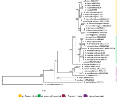
These species can be distinguished by subtle morphological specificities, molecular changes in some gene sequences22 (Fig. 1) and, most importantly, through their ability to produce different mycotoxins16,23. Indeed,
some species, including A. flavus, A. pseudotamarii and A. togoensis, produce aflatoxins of B type, whereas oth-ers, including A. parasiticus, A. minisclerotigenes, A. mottae, A. nomius, A. novoparasiticus, A. arachidicola and
A. korhogoensis produce both B and G type aflatoxins16,23. Some species may also produce other toxic secondary
metabolites such as cyclopiazonic acid (CPA)24.
To date, only few studies aimed to characterized the nature of fungi and more precisely of Aspergillus section
Flavi that may contaminate spices (Table 1). In most cases, fungal identification was done using a morphologic approach which may not have allowed the discrimination of recently identified species among the Flavi section. On the same way, the ability to produce aflatoxins was usually done with no distinction whether they belong to B or G type. With one exception9, the production of cyclopiazonic acid was not tested. No data are available
regard-ing the nature of the fungal species that may contaminate spices at the marketregard-ing stage in Lebanon despite such knowledge is extremely important for risk assessment.
Within this context, the aim of this study was to finely characterize the Aspergillus section Flavi that can con-taminate spices marketed in Lebanon and to determine the toxigenic potential of the isolated strains.
Figure 1. Phylogenetic tree of Aspergillus section Flavi. The phylogenic tree is based on concatenated sequences
from 4 genomic loci (benA, cmdA, PreB and rpb1). Bayesian tree was calculated from 38 strains and includes the type strains for most species. Strong bootstraps are shown at branch nodes. The sequences of primers and protocols used to generate gene fragments are available in Carvajal-Campos et al.19. Petromyces alliaceus NRRL
4181 was used as an outgroup taxon. (Adapted from Carvajal-Campos51).
Country Type of spices Number of samples
Method used for fungal identification Incidence of Aspergillus flavus (%) Toxigenic potential (nature of toxin tested) Incidence of toxigenic strains Reference
Brazil Rosemary, cinnamon, clove, fennel, pepper, pepperoni, oregano 200 Morphology 15 Aflatoxinsa 38% 26
India Red chili, black pepper, turmeric, coriander, cumin, fennel, caraway, fenugreek, ginger 311 Morphology 19.3 Aflatoxins 20–56%b 27
Morocco Paprika, cumin, black pepper, white pepper 80 Morphology and gene sequence 78 Aflatoxin B1, B2, G1, G2 and CPA 57% 9
Saudi Arabia Cinnamon, cumin, sumac, ginger, saffron, fenugreek, pepper, fennel, thyme,
cardamom, caraway, aniseed, clove 138 Morphology 17.4 ND ND
28
Table 1. Fungal contamination of spices and toxigenic potential of isolates. ano distinction between B and G
Results
Fungal counts and identification.
The total fungal counts of the spice samples are presented in Fig. 2. The overall mean fungal contamination was 7.6 × 10³ CFU/g, with marked variations among the samples. Most spicesdisplayed a moderate mean fungal load that ranged from 103 to 4 × 10³ CFU/g. These loads were associated with
spices harvested as seeds or dried grains. Some samples were even less contaminated. For instance, three out of five white pepper samples displayed no detectable fungal contamination. Four out of five cinnamon samples were found to be uncontaminated. Some ginger and turmeric samples were also found to be uncontaminated, but in smaller percentages (2/5 and 1/5 respectively).
Other spices were heavily contaminated with fungi. This was the case for spices resulting from a mixture of several ingredients such as Baar El Kaak, curry and Lebanese pepper. Heavy contamination was also the case for chili samples, which displayed a mean level of contamination of 2 × 10⁴ CFU/g.
Packaging did not limit fungal contamination of the spices. Indeed, for six out of the eight spices for which conditioned samples were analysed (caraway, chili, curry, Lebanese pepper, ginger, and Bhar Al Kaak), the most contaminated samples were the conditioned ones.
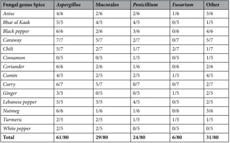
Identification of the fungal genera present in spices showed that Aspergillus was the most frequent genus, with Aspergillus isolates observed in 76% of samples (Table 2). Aspergillus was followed, in decreasing order, by Mucorales (36%), Penicillium (30%) and Fusarium (7.5%). The phytopathogenic genus Fusarium was only found in spices prepared from rhizomes (turmeric and ginger) or fruits (chili). Other fungal genera such as
Cladosporium, Alternaria, Acremonium, and Verticillium, which mostly are derived from field flora, as well as Paecilomyces, Spicaria and Hemispora, were also present at lower levels. The complete analysis of fungal
contam-ination of each sample is reported in Supplementary Table 1 (Table S1).
Aspergillus isolates belonged to 11 sections (Fig. 3), among which three predominated. Indeed, Aspergillus sec-tion Aspergillus isolates were found in almost 60% of the samples. They were present in all kinds of spices with the exception of cinnamon. Isolates of the Nigri section were also very common and were found in almost 50% of the analysed samples but not in the cinnamon or ginger. The section Flavi was observed in 40% of the samples. The distribution of these isolates was similar to that observed for those from the Nigri section. Isolates belonging to five other sections (Fumigati, Terrei, Versicolores, Candidi and Nidulantes) were also identified in 10 to 12% of the samples. Isolates belonging to the Restricti section were found in one sample of curry and one sample of nutmeg. In the same line, three isolates belonging to section Circumdati (2) and Wentii (1) were found in Lebanese pepper (2/3) and Bhar Al Kaak samples (1/3), respectively. Ten out of 11 sections were observed in Lebanese pepper.
Characterization of Aspergillus section Flavi.
Strains of Aspergillus section Flavi were heterogeneously distributed in 40% of the samples. They were present in all Bhar Al Kaak (5/5) samples and in most chili (5/7) and curry (4/7) samples. By contrast, they were only isolated in one sample of caraway, cumin, white pepper, and anise and in two samples of curcuma and coriander. No isolates of Aspergillus section Flavi were found in either cinna-mon or ginger samples. A total of 53 strains were isolated, with some samples displaying several morphologically distinct isolates that were purified and further identified. For instance, 12 strains were obtained from five out of the seven samples of chili analysed.Identification of Aspergillus species. Both morphological and molecular identification were performed to
iden-tify isolates at the species level. To this end, strains were cultivated on Malt Extract Agar (MEA), CREatinin sucrose Agar (CREA) and Aspergillus flavus and parasiticus Agar (AFPA) media, and their macroscopic and microscopic features were analysed. In parallel, ITS, benA and cmdA genes were sequenced. Complete results are given in Supplementary Table 2 (Table S2).
Figure 2. Total fungal load of spices samples (CFU/g). Fungal CFU were counted after 3 and confirmed after 5
days of culture on both MEA and salted MEA at 25 °C. Dots represent total fungal load of each sample whereas short bold lines represent the mean fungal load of each kind of spice. Dots circled in black correspond to packaged samples.
Forty-five out of the 53 isolates (85%) were identified as A. flavus isolates by molecular analysis. Among them, 42 displayed typical A. flavus morphological features, with a yellowish green colony, colourless or yellowish on the reverse side and long conidiophores that are rough in the distal part, with mostly radiate and biseriate heads and with some columns in aerial mycelium. Conidia are oblong and mildly rough. Some variations occurred in the depth and colour of the colony and sclerotia formation (Table S2). For instance, the J33a, J34a, J42a and J62a isolates displayed dark olive-green colonies with numerous uniseriate columnar Aspergillus heads. The J117a and J86a isolates produced large dispersed sclerotia, whose diameter exceeded 500 nm, whereas only the J7 isolate produced numerous small sclerotia (approximately 500 nm in diameter). All typical A. flavus isolates produced aspergillic acid, as evidenced by the presence of a bright orange reverse on the AFPA medium. The production of organic acid that resulted in yellow discoloration of the CREA medium varied with the isolate (Table S2).
Three isolates displayed unusual morphological features but were nevertheless identified as A. flavus by molec-ular analysis; these findings are summarized in Fig. 4.
Seven strains (J4a, J6a, J31a, J37, J63c, J116b, and J118a) were identified as A. tamarii by morphological and molecular analyses. These strains were isolated from different spices including chili (2 isolates), black pepper (1), white pepper (1), Lebanese pepper (1), curry (1) and Bhar al Kaak (1). They displayed highly sporulated dark green-bronze colonies. The conidiophores were long, wide and finely rough. The vesicles were oblong, bearing divergent chains of big, round echinulate spores. All A. tamarii strains displayed a dark-brown reverse side on the AFPA medium.
One isolate (J117c) was identified as A. minisclerotigenes. This strain was obtained from a sample of Bhar al Kaak. It produced numerous small sclerotia with diameters <400 nm.
Fungal genus Spice Aspergillus Mucorales Penicillium Fusarium Other
Anise 4/6 2/6 2/6 1/6 3/6 Bhar al Kaak 5/5 4/5 4/5 0/5 1/5 Black pepper 6/6 2/6 3/6 0/6 4/6 Caraway 7/7 5/7 2/7 0/7 5/7 Chili 5/7 2/7 1/7 2/7 1/7 Cinnamon 0/5 0/5 1/5 0/5 1/5 Coriander 6/6 2/6 1/6 0/6 2/6 Cumin 4/5 2/5 2/5 1/5 4/5 Curry 6/7 5/7 0/7 0/7 2/7 Ginger 3/5 0/5 0/5 1/5 2/5 Lebanese pepper 5/5 3/5 4/5 0/5 2/5 Nutmeg 6/6 1/6 1/6 0/6 3/6 Turmeric 2/5 2/5 1/5 1/5 1/5 White pepper 2/5 2/5 0/5 0/5 0/5 Total 61/80 29/80 24/80 6/80 31/80
Table 2. Fungal genera present in spice samples. Results are expressed as the number of contaminated samples.
Figure 3. Relative proportion of Aspergillus sections in spice samples. Isolates belonging to Aspergillus genus
were identified at the section level by both macroscopic and microscopic examination after five and seven days of culture at 25 °C on MEA, according to Pitt and Hocking45 and Samson et al.46. The relative proportion of each
Toxigenic potential of Aspergillus section Flavi strains. Since the section Flavi contains species with differing
abilities to produce mycotoxins, we further characterized the ability of the strains to produce different toxins. All
Flavi section strains were analysed for the production of both aflatoxins of B and G type as well as CPA. Details on
the levels of production of the different compounds are listed in Table S2.
Nearly half of the isolates (25/53) produced AFB1. Many isolates produced CPA alone (19/53) or simultane-ously with aflatoxin (19/53). A total of 83% of the isolates were able to produce at least one toxin (Table 3).
Eighty percent of A. flavus strains were able to produce mycotoxins, of which 40% (18/45) produced both AFB and CPA (Chemotype I), whereas 27% (12/45) produced only CPA (Chemotype IV), and 13% (6/45) produced only AFB (Chemotype III). As expected, none of the A. flavus strains produced AFG (Chemotype II). All A.
tamarii strains produced only CPA, and the A. minisclerotigenes strain produced both B and G aflatoxins, as well
as CPA.
When the toxigenic potential of strains is considered as a function of the nature of the spice from which they were isolated, approximately 60% of the strains isolated from chili, Bhar al Kaak and curcuma produced AFB1. By contrast, none of the few isolates from anise, white pepper, caraway, cumin, and coriander were aflatoxigenic.
Some strains displayed a high toxigenic potential, producing more than 200 µg aflatoxins/Petri dish after 7 days at 25 °C. To better evaluate the risk associated with the presence of such strains in spices, the ratio of AF/ CFU was calculated. As shown in Table 4, if a strain with high toxigenic potential contaminated a spice sample, 2 to 6 × 10⁴ CFU/g could be sufficient to reach the E.U. regulation of 10 ng aflatoxins/g. Fortunately, in most cases,
the fungal contamination levels observed for these strains in analysed samples were much lower, ranging from a few tens to a few hundred CFU/g. However, for one sample, a contamination level of 105 CFU Aspergillus flavus/g
Figure 4. Main macro- and microscopic features of atypical A. flavus isolates compared to the referenced
A. flavus NRRL62477 after 7 days of culture on MEA at 25 °C. Some isolates of the Aspergillus section Flavi
displayed unusual morphologic features but were nevertheless identified as Aspergillus flavus by molecular identification (sequence of β-tubulin and calmodulin genes) (Table 6). Compared to a reference strain (NRRL 62477, previously isolated from paprika9) displaying typical Aspergillus flavus features, those atypical strains
mainly differ by colour and aspect of conidial heads. The presence of such isolates demonstrates the strong phenotypical diversity that may exist within Aspergillus flavus species.
was measured, demonstrating the possibility for reaching and exceeding the threshold, especially in cases of non-optimal storage.
Discussion
The aim of this study was to investigate the presence of Aspergillus section Flavi in spices marketed in Lebanon. To this end, 80 samples of commonly used spices were collected in markets, and the fungal contamination was analysed. The average fungal load of samples agreed with that found in other recent surveys conducted in other countries25,26. Some differences were observed between spices, which could be due to either the composition of
the condiments or to the preparation process. For instance, some spices, such as cinnamon or anise, are rich in phenolic compounds that may have antifungal properties, which may explain their lower fungal contamina-tion27,28. Similarly, the terpenes in ginger and turmeric and an unidentified protein in ginger were shown to have
antifungal activity, which may help explain the low level of fungal contamination of these spices29.
In the case of other spices, the preparation process may also help reduce the initial fungal load by removing the external parts of the berries/roots before marketing. For instance, white pepper, which is obtained by removing the pericarp from the berries, was less contaminated than black pepper. In the same way, spices obtained from roots that are peeled during their preparation (i.e., ginger and turmeric) were less contaminated than spices obtained from whole dried seeds or fruits. Spices prepared by mixing different plants/seeds were usually more contaminated than others, as observed for Bhar Al Kaak (a mixture of black pepper, fennel, cardamom and clove), curry (mixture of ginger, garlic, onion, coriander, cinnamon) and Lebanese pepper (mixture of black pepper, chili, coriander and cinnamon). Each ingredient used in these preparations represents a separate source of con-tamination; moreover, the mixture of these ingredients may be an additional source of contamination. Since each ingredient may also display variable water activity, the water activity of the final mixture is unstable, and finding the appropriate storage procedure is complex. As a demonstration, these complex samples were the ones most frequently contaminated with mucorales, which are hygroscopic fungi.
In this study, we also analysed the fungal load of samples that were packed in hermetically sealed plastic bags for sale. Surprisingly, this did not prevent fungal contamination; instead, for six out of the eight types of con-ditioned spices that we analysed, the most highly contaminated samples were the concon-ditioned ones. Therefore, it appears that the traditional spice storage procedure, using open plastic containers that allow ventilation and limiting water migration and condensation, should be preferred to hermetically sealed packaging, which should
Chemotype
Spice Number of isolates
Chemotype I (AFB+/ CPA+)
Chemotype II (AFB+/
AFG+/CPA+) Chemotype III (AFB+) Chemotype IV (CPA+)
Chemotype V (no toxin) Anise 2 — — — 2/2 — Bhar al Kaak 8 4/8 1/8 — 3/8 — Black pepper 7 2/7 — 1/7 2/7 2/7 Caraway 1 — — — — 1/1 Chili 12 5/12 — 2/12 4/12 1/12 Cinnamon 0 — — — — — Coriander 3 — — — 3/3 — Cumin 1 — — — — 1/1 Curry 6 3/6 — — 1/6 2/6 Ginger 0 — — — — — Lebanese pepper 3 1/3 — — 2/3 — Nutmeg 3 1/3 — 1/3 — 1/3 Turmeric 6 2/6 — 2/6 1/6 1/6 White pepper 1 — — — 1/1 — Total (%) 53 18/53 (34%) 1/53 (2%) 6/53 (11%) 19/53 (36%) 9/53 (17%)
Table 3. Chemotypes of Aspergillus section Flavi isolated from spices.
Strain Aflatoxin production (µg/Petri dish)a CPA production (µg/Petri dish)a CFU (/Petri dish ± SD)a
AF/CFU ratio (×107)
Theoretical number of CFU to reach
10 ng µg AFb (×104) Number of CFU/g observed in samples
J4b 213 376 1.13 × 10⁹ ± 9.4 × 10⁷ 1.83 5.46 50 J6b 318 199 1.2 × 10⁹ ± 2 × 10⁸ 2.65 3.77 100,000 J71a 251 333 1.49 × 10⁹ ± 2.2 × 10⁸ 1.69 5.91 1,000 J75c 351 127 1.16 × 10⁹ ± 4 × 10⁷ 3.03 3.30 200 J86a 578 127 1.2 × 10⁹ ± 2 × 10⁸ 4.28 2.07 2,000 J117b 336 4532 1.48 × 10⁹ ± 1.4 × 10⁸ 2.27 4.41 150
Table 4. Estimation of the risk associated with the presence of highly aflatoxigenic strains in spices. aafter 7 days
be reserved for completely dried or sterilized spices. The development of breathable materials, allowing air to pass and avoiding condensation but also protecting against external contaminations, would probably be of interest to improve the preservation of spices.
Aspergillus was the main genus found in the samples, and although a great variety of sections were present,
sections Nigri and Flavi were very frequent, which is also in agreement with data reported in Morocco, India or Brasil9,30–32. Aspergillus section Aspergillus, formerly named Eurotium33, which corresponds to xerophilic isolates,
was also frequently observed, as expected in dry commodities.
Fifty-three distinct strains of Aspergillus section Flavi were isolated from samples, among which 85% were identified as A. flavus by molecular analysis, demonstrating relatively low biodiversity within the section Flavi. Although the molecular identification agreed with the results of morphological examination of the isolates in most cases, a few isolates displayed atypical morphological features even though they were identified as A. flavus strains by molecular analysis. In the last decade, the use of molecular tools and gene sequencing has shown that the Flavi section is much more complicated than previously thought, and many new species have been intro-duced, increasing the number to 3421,22. From our results, it appears that, beyond this genetic diversity, isolates
identified as A. flavus may also present some morphological particularities, demonstrating the wide phenotypic variation within the species. Two of the three strains with unusual morphology were able to produce mycotoxins (AFB and CPA), demonstrating the importance of these isolates that could easily have been misidentified due to their atypical colouration. Since these particular isolates originated from different spices, this phenotypic variety does not appear to be correlated with substrate specificity but could rely on the presence of mutations on genes involved in pigment synthesis or on a distinct basal expression of these genes34,35.
Seven out of 53 isolates (13%) were identified as A. tamarii. This proportion agrees with the results of previous studies reporting the presence of A. tamarii in spices, dry fruits and cereals25,30,36–38. As expected, all these isolates
produced CPA24.
Finally, only one isolate was identified as A. minisclerotigenes. This species was first described in 200823. It has
been reported in several geographic areas and on different substrates such as groundnuts in Argentina23,
ground-nuts in Algeria39, maize and almonds in Portugal17 and in spices marketed in Morocco9. Aspergillus miniscleroti-genes is of interest due to its ability to produce both B and G aflatoxins together with CPA. However, this species
is also quite difficult to isolate due to weak sporulation when cultured on MEA.
Our results show that more than 80% of the isolates belonging to the section Flavi are toxigenic. If we consider aflatoxin production, 47% of the strains produced AFB1 (chemotypes I and III). This proportion of aflatoxigenic strains agrees with data available on the toxigenic potential of Aspergillus section Flavi in different foodstuffs such as spices, chestnuts and cereals9,31,38,40. Some strains were able to produce great amounts of aflatoxins when
cultured in vitro. The presence of such highly toxigenic strains raise the question of the sanitary impact of the con-tamination. From data obtained while characterizing toxigenic potential, we demonstrated that the presence of only a few thousands CFU of these strains could be sufficient to reach the regulatory limit on the aflatoxin content in spices. Even if most samples of our study displayed relatively low levels of contamination, such fungal counts can be obtained, as observed in one sample of this study. This underscores the need for proper storage procedures, especially for spices that result from a mixture of different ingredients. The use of conditioning materials that protect from contamination but that also enable ventilation could improve the preservation of spices and limit the risk of fungal development and subsequent contamination with aflatoxins.
Notably, a high proportion of the strains were able to produce CPA, alone or in combination with aflatoxins. Although this mycotoxin has already been reported as a possible contaminant of several foods and feeds41, recent
data are lacking on the contamination of foods and feeds with that CPA-producing species. Notably, the produc-tion of CPA by the A. flavus strains used as biocontrol agents appeared to be a strong limitaproduc-tion, which needs to be evaluated since CPA has been demonstrated to be cytotoxic and immunotoxic to human cells42,43. Moreover,
due to its frequent coproduction with aflatoxins, an investigation of the possible interaction between the two toxic compounds when ingested simultaneously would be interesting. Considering the origin of toxigenic strains, we observed that some spices are more often contaminated with toxigenic isolates than others. For example, 58, 62 and 67% of the strains from chili, Bhar al Kaak and turmeric, respectively, are toxigenic. By contrast, none of the few strains of anise, black pepper, caraway, coriander and cumin were shown to be toxigenic. This finding is also of interest regarding the risk assessment of contamination of these spices with mycotoxins.
Gene Gene name Length bp
Primers
Sequence (Nucleotides: 5′→3′) Forward Reverse
ITS (1–2) 18S ribosomal RNA gene
partial sequence Internal Transcribed Spacer
565–613 ITS1 5′-GGAAGTAAAGTCGTAACAAGG ITS 2 5′-TTGGTCCGTGTTTCAAGACG
ITS (4–5) 300–330 ITS5 5′-GGAAGTAAAAGTCGTAACAAGG ITS 4 5′-TCCTCCGCTTATTGATATGC
benA - β-tubulin 1125 β-tub 2a 5′-GGTAACCAAATCGGTGCTGCTTTC β-tub 2b 5′-ACCCTCAGTGTAGTGACCCTTGGC
cmdA Calmodulin 543 Cmd5 5′-CCGAGTACAAGGAGGCCTTC-3′ Cmd6 5′-CCGATAGAGGTCATAACGTGG-3′
In conclusion, this study reports, for the first time, the diversity of Aspergillus section Flavi in spices marketed in Lebanon. The results reveal that A. flavus is the most frequent species in the samples analysed and that the pro-portion of toxigenic strains is high. This study also demonstrated that some spices are frequently contaminated with toxigenic strains and should probably be submitted to specific safety controls.
Methods
solvents and reagents.
All solvents were of HPLC grade and purchased from VWR International (Fontenay-sous-Bois, France). Medium and agar were purchased from BIOKAR Diagnostics (Allonne, France), and Tween 80, from Merck (Darmstadt, Germany). Mycotoxin standards were purchased from Sigma (Saint-Quentin-Fallavier, France). Aflatoxins were dissolved in toluene-acetonitrile (98:2, v/v) and cyclopiazonic acid in methanol to obtain 200 µg/ml stock solutions that were stored at −20 °C. Standard solutions used for HPLC analysis were stored at 4 °C and changed monthly. Taq DNA polymerase used for molecular identification was purchased from Invitrogen (Carlsbad, Ca, USA).samples.
Eighty samples were randomly collected from several retailers across all Lebanese regions. Fourteen different types of spices were analysed: anise (n = 6), Bhar Al Kaak (n = 5), caraway (n = 7), chili (n = 7), cinna-mon (n = 5), coriander (n = 6), cumin (n = 5), curry (n = 7), ginger (n = 5), nutmeg (n = 6), black pepper (n = 6), white pepper (n = 5), Lebanese pepper (n = 5), turmeric (n = 5). All samples were of commercial grade, and to be representative of what can enter human food chain, no macroscopically mouldy sample was taken.Sixty-three out of the 80 spices samples were taken from batches stored in plastic containers with no particular protection. Some samples were hermetically sealed in plastic bags before marketing (17 samples including Bhar Al Kaak (n = 2), caraway (n = 3), chili (n = 2), curry (n = 2), ginger (n = 2), nutmeg (n = 2), Lebanese pepper (n = 2) and turmeric (n = 2).
Approximately 100 g of each sample were collected and carefully placed in 200 ml sterile plastic jars (Thermo-Fischer, Dardilly, France). To avoid any fungal development without altering spore viability, samples were stored at 4 °C until analysis9,44. In the laboratory, samples in the form of grains were finely ground in a
Waring blender (Waring Laboratory, Torrington, CT, USA).
Fungal count and identification.
Twenty grams of each sample were mixed with 180 ml of 0.05% Tween 80 for 2 min in a Waring blender and then placed on a horizontal shaking table at 220 rpm for one hour. Decimal dilutions were prepared in 0.05% Tween 80, and 100 µl of each dilution were plated on both MEA medium and Salted MEA (MEA + 6% NaCl)9. This last medium was used to identify xerophilic species and to limitMucorale development that may prevent a correct enumeration and identification of species with a low growth rate. Fungal colonies were counted after three days of culture at 25 °C and confirmed after five days. The limit of detection for the fungal count was 10 CFU/g of sample. The colonies were then identified according to Pitt and Hocking45 and Samson et al.46. Aspergillus section Flavi strains were isolated from plates by several platings on
MEA and Salted MEA.
Characterization of Aspergillus section Flavi strains.
Morphological identification. Aspergillussec-tion Flavi strains were identified at the species level by both macroscopic and microscopic examinasec-tion after five and seven days of culture at 25 °C on MEA, CREA (3.0 g creatine, 30 g sucrose, 0.5 g KCl, 0.5 g MgSO4.7H2O, 0.5 g
FeSO4.7H2O, 1.3 g K2HPO4.3H2O, 0.05 g Bromocresol purple, 15.0 g agar, distilled water to bring to 1 litre) and
AFPA (Thermo-Fisher Diagnostics, Dardilly, France) media according to Pitt et al.47 and Varga et al.16.
Molecular identification. The molecular identification of all Aspergillus section Flavi strains was first
per-formed by internal transcribed spacer (ITS) rRNA sequencing. For strains displaying atypical morphological features, this was completed by beta-tubulin (benA) and calmodulin (cmdA) gene amplification and sequencing. The strains were cultured in yeast extract sucrose (YES) broth and placed on a shaking incubator at 160 rpm at 27 °C for three days. Genomic DNA was isolated from mycelia, as previously described48. Primers used for the
Strain Species Beta-tubulin Calmodulin
J4a Aspergillus tamarii MG957155 MG957162 J6a Aspergillus tamarii MG957156 MG957163 J31a Aspergillus tamarii MG957157 MG957164 J37 Aspergillus tamarii MG957158 MG957165 J63c Aspergillus tamarii MG957159 MG957166 J116b Aspergillus tamarii MG957160 MG957167 J118a Aspergillus tamarii MG957161 MG957168 J117c Aspergillus minisclerotigenes MG957169 MG957170 J4c Aspergillus flavus MG957171 MG957174 J75e Aspergillus flavus MG957172 MG957175 J123a Aspergillus flavus MG957173 MG957176
Table 6. Nucleotide accession numbers for benA and cmdA of A. flavus strains with atypical morphology, A.
molecular identification are listed in Table 5. PCR reactions were carried out in a GeneAmp PCR 2700 thermo-cycler (Applied Biosystems, Foster City, USA). PCR products were purified with a GenElute PCR Clean-Up Kit (Sigma-Aldrich) and sequenced on an ABI3130XL sequencer (Applied Biosystems) using the dye terminator technology. PCR products were sequenced in both directions. Nucleotide sequence accession numbers for benA and cmdA from A. flavus with atypical morphology, A. tamarii and A. minisclerotigenes are listed in Table 6.
Mycotoxigenic potential of isolates. To assess the toxigenic potential of Aspergillus section Flavi strains, a spore
suspension was prepared from a 7-day culture, and 100 spores were centrally inoculated on MEA and cultured for seven days at 25 °C. After this incubation period, three sporulating cultures were analysed for toxin production. In parallel, at the end of the incubation period, three other cultures were used to perform a CFU count as previously described (see paragraph 4.3).
Toxins were then extracted from the whole culture medium by mechanical agitation in appropriate solvents. Extracts were filtered, and toxins were, as a first screening step, separated by thin layer chromatography (TLC) and quantified by fluorodensitometry using a Shimadzu CS930 fluorodensitometer (Shimadzu Corp., Kyoto, Japan), as previously described9. Results were further confirmed by HPLC analysis.
Evaporated extracts were diluted with 500 μL of water-acetonitrile-methanol (65:17.5:17.5, v/v/v) and filtered through a 0.45 μm disk (Thermo Scientific Fisher, Villebon-Sur-Yvette, France). Sample analysis was performed with a Dionex UltiMate 3000 UHPLC system (Thermo Scientific, Illkirch, France) using a liquid chromatography column Luna
®
C18 (125 × 2 mm, 5 μm, 100 Å) (Phenomenex, Torrance, CA, USA) at 30 °C. Separation condi-tions were adapted from Fu et al.49, with slight modifications. Briefly, a 20 min isocratic mode program wasdeliv-ered at 82.5% of eluent A: acidified water (0.2% of acetic acid) and acetonitrile (79:21, v/v) and 17.5% of eluent B: pure methanol. The flow rate was at 0.2 mL/min with a sample injection volume of 10 μL. Aflatoxins were detected using a fluorescent detector at 365/430 nm excitation/emission wavelengths. Peak identity was further confirmed by analysis of the absorption spectrum with a diode array detector (DAD) coupled to the system. Production levels of aflatoxins in media were calculated based on a standard calibration curve.
Based on their toxigenic potential (AFB+/−, AFG+/−, CPA+/−), isolates were classified into five chem-otypes as proposed by Vaamonde et al.50: Chemotype I for AFB and CPA producers; Chemotype II for AFB,
AFG and CPA producers; Chemotype III for AFB-only producers; Chemotype IV for CPA-only producers; and Chemotype V for non-toxigenic isolates.
References
1. Di Bella, G. et al. Organic contamination of Italian and Tunisian culinary herbs and spices. J. Environ. Sci. Health B. 17, 1–12, https:// doi.org/10.1080/03601234.2019.1571364 (2019).
2. Man, A., Mare, A., Toma, F., Curticapean, A. & Santacroce, L. Health threats from contamination of spcies commercialized in Romania: risks of fungal and bacterial infections. Endoc. Metab. Immune Disord. Drug Targets 16, 197–204, https://doi.org/10.2174 /1871530316666160823145817 (2016).
3. Hariram, U. & Labbé, R. Spore prevalence and toxigenicity of Bacillus cereus and Bacillus thuringiensis isolates from U.S. retail spices.
J. Food Prot 78, 590–596, https://doi.org/10.4315/0362-028X.JFP-14-380 (2015).
4. Omotaya, O. P., Omotayo, A. O., Mwanza, M. & Babalola, O. O. Prevalence of mycotoxins and their consequences on Human health.
Toxicol. Res. 35, 1–7, https://doi.org/10.5487/TR.2019.35.1.001 (2019).
5. I.A.R.C. Some naturally occurring substances, food items and constituents, heterocyclic aromatic amines and mycotoxins, in
Monographs on the evaluation of carcinogenic risks to humans, World Health Organization, Lyon, 56, 245 (1993).
6. Liu, Y. & Wu, F. Global burden of aflatoxin-induced hepatocellular carcinoma: a risk assessment. Environ. Health Perspect. 118, 818–824, https://doi.org/10.1289/ehp.0901388 (2010).
7. Gibb, H. et al. World Health Organization estimates of the global and regional disease burden of four foodborne chemical toxins, 2010: a data synthesis. F1000Research 4, 1–14, https://doi.org/10.12688/f1000research.7340.1 (2015).
8. Mandeel, Q. A. Fungal contamination of some imported spices. Mycopathologia 159, 291–298, https://doi.org/10.1007/s11046-004-5496-z
(2005).
9. El Mahgubi, A. et al. Distribution and toxigenicity of Aspergillus section Flavi in spices marketed in Morocco. Food Contr. 32, 143–148, https://doi.org/10.1016/j.foodcont.2012.11.013 (2013).
10. Valle Garcia, M., Mallman, C. A. & Venturini Copetti, M. Aflatoxigenic and ochratoxigenic fungi and their mycotoxins in spices marketed in Brazil. Food Res. Int., 106, 10.106/j.foodres.2017.12.06 136–140 (2018).
11. Asghar, M. A., Zahir, E., Rantilal, S., Ahmed, A. & Iqbal, J. Aflatoxin in composite spices collected from local markets of Karachi, Pakistan. Food Addit. Contam. Part B Surveillance 9, 113–9, https://doi.org/10.1080/19393210.2016.1150356 (2016).
12. Kabak, B. & Dobson, A. D. W. Mycotoxins in spices and herbs: an update. Crit. Rev. Food Sci. Nutr. 57, 18–34, https://doi.org/10.10 80/10408398.2013.772891 (2017).
13. European Union Commission regulation 1881/2006 setting maximum levels for certain contaminants in foodstuffs. Off. J. Europ.
Un. L364, https://doi.org/10.1080/10408398.2013.772891 5–24 (2006).
14. European Commission. RAFF – Food and Feed Safety Alerts. https://ec.europa.eu/food/safety/rasff_en.
15. Cary, J. W. & Ehrlich, K. C. Aflatoxigenicity in Aspergillus: molecular genetics, phylogenetic relationships and evolutionary implications.
Mycopathologia 162, 167–177, https://doi.org/10.1007/s11046-006-0051-8 (2006).
16. Varga, J., Frisvad, J. C. & Samson, R. A. Two new aflatoxin producing species and an overview of Aspergillus section. Flavi. Stud.
Mycol. 69, 57–80, https://doi.org/10.3114/sim.2011.69.05 (2011).
17. Soares, C., Rodrigues, P., Peterson, S. W., Lima, N. & Venancio, A. Three new species of Aspergillus section Flavi isolated from almonds and maize in Portugal. Mycologia 104, 682–697, https://doi.org/10.3852/11-088 (2012).
18. Taniwaki, M. H. et al. Aspergillus bertholletius sp. nov. from Brazil nuts. PLoS One 7, e42480, https://doi.org/10.1371/journal. pone.0042480 (2012).
19. Carvajal-Campos, A. et al. Aspergillus korhogoensis, a novel aflatoxin producing species from the Côte d’Ivoire. Toxins 9, 353, https:// doi.org/10.3390/toxins9110353 (2017).
20. Pitt, J. I. et al. Aspergillus hancockii sp. nov., a biosynthetically talented fungus endemic to southeastern Australian soils. PLoS One
12, e0170254, https://doi.org/10.1371/journal.pone.0170254 (2017).
21. Singh, P., Orbach, M. J. & Cotty, P. J. Aspergillus texensis: a novel aflatoxin producer with S morphology from the United States.
Toxins 10, https://doi.org/10.3390/toxins10120513 (2018).
22. Frisvad, J. C. et al. Taxonomy of Aspergillus section Flavi and their production of aflatoxins, ochratoxins and other mycotoxins. Stu.
23. Pildain, M. B. et al. Two novel Aflatoxin-producing Aspergillus species from Argentinean peanuts. Int. J Syst. Evol. Microbiol. 58, 725–35, https://doi.org/10.1099/ijs.0.65123-0 (2008).
24. Uka, V. et al. Unravelling the diversity of the cyclopiazonic acid family of mycotoxins in Aspergillus flavus by UHPLC triple-TOF HRMS. Toxins 9, https://doi.org/10.3390/toxins9010035 (2017).
25. Hashem, M. & Alamri, S. Contamination of common spices in Saudi Arabia markets with potential mycotoxin-producin fungi.
Saudi J. Biol. Sci. 17, 167–175, https://doi.org/10.1016/j.sjbs.2010.02.011 (2010).
26. Feroz, F., Shimizu, H., Nishioka, T., Mori, M. & Sakagami, Y. Bacterial and fungal counts of dried and semi-dried foods collected from Dhaka, Bangladesh and their reduction methods. Biocontrol Sci. 21, 243–251, https://doi.org/10.4265/bio.21.243 (2016). 27. Ozcan, M. M. & Chalchat, J. C. Chemical composition and antifungal effect of anise (Pimpinella anisum L.) fruit oil at ripening stage.
Ann. Microbiol. 56, 353–358, https://doi.org/10.1007/BF0317531 (2006).
28. Liu, Q., Meng, X., Zhao, C. N., Tang, G. Y. & Li, H. B. Antibacterial and antifungal activities of spices. Int. J. Mol. Sci. 18, 1283, https:// doi.org/10.3390/ijms18061283 (2017).
29. Zacchino, S. A. et al. Plant phenolic and terpenoids as adjuvants of antibacterial and antifungal drugs. Phytomedicine 37, 27–48,
https://doi.org/10.1016/j.phymed.2017.10.018 (2017).
30. Valle Garcia, M., Parussolo, G., Brombilla Moro, C., Olivier Bernardi, A. & Venturini Copetti, M. Fungi in spices and mycotoxinogenic potential of some Aspergilli isolated. Food Microbiol. 73, 93–98, https://doi.org/10.1016/j.fm.2018.01.013 (2018).
31. Jeswal, P. & Kumar, D. Mycobiota and natural incidence of aflatoxins, ochratoxin A, and citrinin in Indian spices confirmed by LC-MS/MS. Int. J. Microbiol. 2015, https://doi.org/10.1155/2015/242486 (2015).
32. Aiko, V. & Meht, A. Prevalence of toxigenic fungi in common medicinal herbs and spices in India. 3 biotech 159, https://doi. org/10.1007/s13205-016-0476-9 (2016).
33. Chen, A. J. et al. Polyphasic taxonomy of Aspergillus section Aspergillus (formerly Eurotium) and its occurrence in indoor environments and food. Stud. Mycol. 88, https://doi.org/10.1016%2Fj.simyco.2017.07.001 37–135 (2017).
34. Valiante, V. et al. The Aspergillus fumigatus conidial melanin production is regulated by the bifunctional bHLH DevR and MADS-box RlmA transcription factors. Mol. Microbiol. 102, 321–335, https://doi.org/10.1111/mmi.13462 (2016).
35. Sung, C. T. et al. Overexpression of a three-gene conidial pigment biosynthetic pathway in Aspergillus nidulans reveals the first known to acetylate tryptophan. Fung. Genet. Biol. 101, 1–6, https://doi.org/10.1016/j.micres.2016.12.013 (2017).
36. Da Chagas, O., Freire, F., Kozakiewicz, Z. & Paterson, R. M. Mycoflora and mycotoxins in Brazilian black pepper, white pepper and Brazil nuts. Mycopathologia 149, 13–19 (2000).
37. Perrone, G. et al. Population structure and aflatoxin production by Aspergillus section Flavi from maize in Nigeria and Ghana. Food
Microbiol. 41, 52–59, https://doi.org/10.1016/j.fm.2013.12.005 (2014).
38. Prencipe, S. et al. Characterization of Aspergillus section Flavi isolated from fresh chestnuts and along chestnuts flour process. Food
Microbiol. 69, 159–169, https://doi.org/10.1016/j.fm.2017.08.004 (2018).
39. Guezlane-Tebitel, N., Bouras, N., Mokrane, S., Benayad, T. & Mathieu, F. Aflatoxigenic strains of Aspergillus section Flavi isolated from marketed peanuts (Arachis hypogaea) in Algiers (Algeria). Ann. Microbiol. 62, 1–11, https://doi.org/10.1007/s13213-012-0473-0 (2012). 40. Riba, A. et al. Aspergillus section Flavi and aflatoxins in Algerian wheat and derived products. Food Chem. Toxicol. 48, 2772–2777,
https://doi.org/10.1016/J.FCT.2010.07.005 (2010).
41. Fernandez Pinto, V., Patriarca, A., Locani, O. & Vaamonde, G. Natural co-occurrence of aflatoxin and cyclopiazonic acid in peanuts grown in Argentina. Food Addit. Contam. 18, 1017–1020, https://doi.org/10.1080/02652030110057125 (2001).
42. King, E. D., Bassi, A. B., Ross, D. C. & Druebbisc, B. An industry perspective of the use of “atoxigenic” strains of Aspergillus flavus as biological control agent and the significance of cyclopiazonic acid. Toxin Rev. 30, 33–41, https://doi.org/10.3109/15569543.2011.588818
(2011).
43. Hymery, N., Masson, F., Barbier, G. & Coton, E. Cytotoxicity and immunotoxicity of cyclopiazonic acid on human cells. Toxicol. in
Vitro 28, 940–947, https://doi.org/10.1016/j.tiv.2014.04.003 (2014).
44. Kong, W. et al. Occurrence of toxigenic fungi and determination of mycotoxins by HPLC-FLD in functional foods and spices in China markets. Food Chem., 146, https://doi.org/10.106/j.foodchem.2013.09.005 3220–326 (2014).
45. Pitt, J. I. & Hocking, A. D. Fungi and Food spoilage. Third Ed., Springer, New-York (2009).
46. Samson, R. A., Houbraken, J., Thrane, U., Frisvad, J. C. & Andersen, B. Food and indoor fungi. CBS Knaw, Uthrecht, The Netherlands (2010).
47. Pitt, J. I., Hocking, A. D. & Glenn, D. R. An improved medium for detection of Aspergillus flavus and A. parasiticus. J. Appl. Bacteriol.
54, 109–114, https://doi.org/10.1111/j.1365-2672.1983.tb01307.x (1983).
48. Adjovi, Y. et al. Contrast between natural occurrence of toxigenic Aspergillii of the Flavi section and Aflatoxin B1 in cassava: possible explanation. Food Microbiol. 38, 151–159, https://doi.org/10.1016/j.fm.2013.08.005 (2014).
49. Fu, Z., Huang, X. & Min, S. Rapid determination of aflatoxins in corn and peanuts. J. Chromatogr. A 1209, 271–274, https://doi. org/10.1016/j.chroma.2008.09.054 (2008).
50. Vaamonde, G., Patriarca, A., Fernandez Pinto, V., Comerio, R. & Degrossi, C. Variability of aflatoxin and cyclopiazonic acid production by Aspergillus section Flavi from different substrates in Argentina. Int. J. Food Microbiol. 88, 79–84, https://doi. org/10.1016/S0168-1605(03)00101-6 (2003).
51. Carvajal-Campos A. Characterization of Aspergillus section Flavi: molecular markers as tool to unmask cryptic species. Ph.D dissertation. Université de Toulouse, 209 p. http://thesesups.ups-tlse.fr/3930/ (2018).
Acknowledgements
The authors thank Mrs. Daphnee Goodfellow for the English language editing and Mr Anwar El Mahgubi for technical assistance. This work was financed by the projects Aflared (joint project, 001–2012 STDF-AIRD) and ToxinFree (Campus France PHC cèdre 32763).
Author Contributions
Makhlouf J. collected the samples and performed the mycological analysis with technical support from Querin A. Carvajal-Campos A. performed molecular analysis with technical support from Tadrist S. Puel O. and Lorber S. designed the molecular analysis. Oswald I. and Hamze M. supervised the work. Bailly J.D. designed and supervised experiments. Bailly S. supervised the mycological analysis and performed morphological identification. All authors participated in the manuscript preparation.
Additional Information
Supplementary information accompanies this paper at https://doi.org/10.1038/s41598-019-41704-1.
Competing Interests: The authors declare no competing interests.
Publisher’s note: Springer Nature remains neutral with regard to jurisdictional claims in published maps and
Open Access This article is licensed under a Creative Commons Attribution 4.0 International
License, which permits use, sharing, adaptation, distribution and reproduction in any medium or format, as long as you give appropriate credit to the original author(s) and the source, provide a link to the Cre-ative Commons license, and indicate if changes were made. The images or other third party material in this article are included in the article’s Creative Commons license, unless indicated otherwise in a credit line to the material. If material is not included in the article’s Creative Commons license and your intended use is not per-mitted by statutory regulation or exceeds the perper-mitted use, you will need to obtain permission directly from the copyright holder. To view a copy of this license, visit http://creativecommons.org/licenses/by/4.0/.