HAL Id: tel-02494023
https://tel.archives-ouvertes.fr/tel-02494023
Submitted on 28 Feb 2020HAL is a multi-disciplinary open access archive for the deposit and dissemination of sci-entific research documents, whether they are pub-lished or not. The documents may come from
L’archive ouverte pluridisciplinaire HAL, est destinée au dépôt et à la diffusion de documents scientifiques de niveau recherche, publiés ou non, émanant des établissements d’enseignement et de
Evaluation of the role of sodium-glucose co-transporters
SGLT1 and 2 in the induction of endothelial senescence
and dysfunction using an in vitro and in vivo approach
Sin-Hee Park
To cite this version:
Sin-Hee Park. Evaluation of the role of sodium-glucose co-transporters SGLT1 and 2 in the induction of endothelial senescence and dysfunction using an in vitro and in vivo approach. Pharmacology. Université de Strasbourg, 2019. English. �NNT : 2019STRAJ041�. �tel-02494023�
UNIVERSITÉ DE STRASBOURG
ÉCOLE DOCTORALE DES SCIENCES DE LA VIE ET DE LA SANTÉ
Regenerative Nanomedicine – INSERM UMR 1260
THÈSE
présentée par :
Sin-Hee PARK
soutenue le : 17 Mai 2019
pour obtenir le grade de :
Docteur de l’université de Strasbourg
Discipline/ Spécialité
: Hématologie et physiopathologie vasculaire
Evaluation du rôle des co-transporteurs
sodium-glucose SGLT1 et 2 dans
l’induction de la sénescence et de la
dysfonction des cellules endothéliales
à l’aide d’une approche in vitro et in vivo
THÈSE dirigée par :
M. Olivier MOREL Pr, université de Strasbourg Mme Valérie B. SCHINI-KERTH Pr, université de Strasbourg
RAPPORTEURS :
M. François ROUBILLE Pr, Université de Montpellier Mme Céline DEMOUGEOT Pr, Université de Franche-Comté
ANIMUM FORTUNA SEQUITUR
Everyone has talent. What is rare is the courage to follow the talent
to the dark place where it leads.
ACKNOWLEDGEMENTS
This journey would not have been possible without the support of my family, professors, and friends.
I would like to express my deep gratitude to Professor Olivier Morel and Professor Valérie B. Schini-Kerth, my research supervisors, for their patient guidance, enthusiastic encouragement and useful critiques of this research work. I especially would like to express my infinite respect to Professor Valérie B. Schini-Kerth for the continuous support of my Ph.D study and research, for her motivation and immense knowledge. Her guidance helped me in all the time of research and writing of this thesis. It was a great honor for me to do Ph.D study under your supervision.
Besides my supervisors, I would like to thank the rest of my thesis committee: Professor François Roubille, Professor Céline Demougeot, Professor Bernard Geny and Dr. Angela Tesse for accepting to judge this work.
I would like to express my special thanks of gratitude to Professor Min-Ho Oak who gave me the golden opportunity to do this wonderful research in this lab. Your guidance is always like a lighthouse to me.
My sincere thanks also go to Dr. Cyril Auger, Professor Paola, Dr. Gilles for their valuable and constructive suggestions from every aspect.
I thank my fellow labmates Dr. Hira, Dr. Farooq, Wahid for the stimulating discussions, for all the fun we have had in the last three years. We started together and our efforts finally have come to fruition. (Wahid, you’ll be soon!)
Also I thank other members of the lab Brigitte (who was together), Christophe, Lamia, Midou, Ursula.. and all the colleagues …
Special thanks to my friends Dr. Hyunho Lee and Dr. Eugenia Belcastro. My friendship with you has been a gift for which there could truly be no adequate thanks. Your constant support and “positive mirroring” have given me more than I can say.
To my beloved parents and two sisters, thank you for encouraging me in all of my pursuits and inspiring me to follow my dreams. I am especially grateful to my parents, who supported me emotionally and financially. I always knew that you believed in me and you were just supportive in the best ways possible.
LIST OF PUBLICATIONS
Publications
1. Min-Ho Oak, Cyril Auger, Eugenia Belcastro, Sin-Hee Park, Hyun-Ho Lee, Valérie B. Schini-Kerth, “Potential mechanisms underlying cardiovascular protection by polyphenols: Role of the endothelium”, Free Radical Biology and Medicine, 122, 161-170 (2018).
2. Sonia Khemais-Benkhiat, Eugenia Belcastro, Noureddine Idris-Khodja, Sin-Hee
Park, Lamia Amoura, Malak Abbas, Cyril Auger, Laurence Kessler, Eric Mayoux,
Florence Toti, Valérie B. Schini-Kerth, “Angiotensin II-induced redox-sensitive SGLT2 expression promotes high glucose-induced endothelial cell senescence”, Journal of Cellular and Molecular Medicine, 109, 461 (2019).
3. Kushal Sharma, Hyun-Ho Lee, Dal-Seong Gong, Sin-Hee Park, EunyoungYi, Valérie Schini-Kerth, Min-Ho Oak, “Fine air pollution particles induce endothelial senescence via redox-sensitive activation of local angiotensin system”, Environmental Pollution, 252, 317-329 (2019)
4. Hira Hasan, Sin-Hee Park, Cyril Auger, Eugenia Belcastro, Kensuke Matsushita, Benjamin Marchandot, Hyun-Ho Lee, Abdul Wahid Qureshi, Gilles Kauffenstein, Patrick Ohlmann, Valérie B Schini-Kerth, Laurence Jesel, Olivier Morel, “Thrombin induces angiotensin II-mediated senescence in atrial endothelial cells: Impact on pro-remodeling patterns”, Journal of clinical medicine, 8(10), 1570 (2019)
5. Sin-Hee Park et al., “The AT1R/NADPH oxidase pro-oxidant pathway induces expression of SGLT1 and 2 to sustain glucose and Na+-dependent oxidative stress
promoting endothelial dysfunction in response to Ang II and circulating microparticles of coronary artery disease patients”, in preparation
6. Sin-Hee Park et al., “Empagliflozin, a sodium-glucose cotransporter 2 inhibitor, improved heart remodeling, endothelial and vascular dysfunction in the metabolic syndrome ZSF1 rats”, in preparation
7. HH. Lee, SH. Park et al., “An anthocyanin-rich blackcurrant extract induced NO-mediated relaxation in coronary artery rings and eNOS phosphorylation in cultured endothelial cells: Role of sodium-glucose cotransporter 1 and 2.”, in preparation
8. MA. Farooq, L. Amoura, S. Gaertner, Z. Niazi, SH. Park et al., “Oral intake of EPA:DHA 6:1 improves ageing-related blunted endothelium-dependent relaxations and increased contractile responses in the mesenteric artery: role of oxidative stress and cyclooxygenases”, in preparation
9. S. Gaertner, MA. Farooq, B. Pollet, L. Amoura, S. Khemais-Benkhiat, SH. Park et al., “Ageing-related endothelial dysfunction in the femoral vein is mediated by cyclooxygenases: Role of thromboxane prostanoid receptors”, in preparation
10. A. Qureshi, R. Altamimy, A. El Habhab, L. Amoura, M. Kassem, S. Khemais, M. Farooq, H. Hasan, SH. Park et al., “Treatment of rats with the omega fatty acid 3 formulation EPA:DHA 6:1 decreases the leukocyte microparticles-induced endothelial pro-inflammatory responses and senescence”, in preparation
Poster presentations
1. Sin-Hee Park, Eugenia Belcastro, Hira Hasan, Christophe Bruckert, Cyril Auger, Valerie B Schini-Kerth, “Angiotensin II induced oxidative stress-mediated upregulation of sodium-glucose cotransporters 1 and 2 (SGLTs) expression in cultured
coronary artery endothelial cells”, World Congress of Basic and Clinical Pharmacology, July 2018, Kyoto(Japan)
2. Sin-Hee Park, Muhammad Akmal Farooq, Sébastien Gaertner, Christophe Bruckert, Abdul Wahid Qureshi, Hyun-Ho Lee, Djamel Benrahla, Brigitte Pollet, Dominique Stephan, Patrick Ohlmann, Eric Mayoux, Cyril Auger, Olivier Morel, Valérie B. Schini-Kerth, “Empagliflozin, a sodium-glucose cotransporter 2 inhibitor, improved heart remodeling, endothelial and vascular dysfunction in the metabolic syndrome ZSF1 rats”, Printemps de la Cardiologie, April 2019, Lille
3. Sin-Hee Park, Sonia Khemais-Benkhiat, Noureddine Idris-Khodja, Lamia Amoura, Malak Abbas, Cyril Auger, Laurence Kessler, Eric Mayoux, Florence Toti, Valerie B Schini-Kerth, “Upregulation of sodium-glucose cotransporter 2 (SGLT2) expression in cultured senescent endothelial cells and in arterial sites at risk in vivo in rats”, Printemps de la Cardiologie, April 2018, Montpellier & Doctoral School Days, March 2018, Strasbourg & Journées du campus d'Illkirch, May 2018, Strasbourg
4. H. Hasan, M. Abbas, C. Auger, E. Belcastro, M.A. Farooq, SH. Park et al., “Atrial endothelial cells senescence promotes thrombogenicity, inflammation and extracellular matrix remodeling: role of the local Ang II / AT1 receptor pathway”, Printemps de la Cardiologie, April 2018, Montpellier & Doctoral School Days, March 2018, Strasbourg & Journées du campus d'Illkirch, May 2018, Strasbourg
5. A. Qureshi, R. Altamimy, A. El Habhab, L. Amoura, M. Kassem, S. Khemais, M. Farooq, H. Hasan, SH. Park et al., “Treatment of rats with the omega fatty acid 3 formulation EPA:DHA 6:1 decreases the leukocyte microparticles-induced endothelial pro-inflammatory responses and senescence”, International Meeting on Ischemia Reperfusion Injuries in Transplantation, April 2018, Poitiers
6. S. Gaertner, MA. Farooq, B. Pollet, L. Amoura, S. Khemais-Benkhiat, SH. Park et al., “Ageing-related endothelial dysfunction in the femoral vein is mediated by cyclooxygenases: Role of thromboxane prostanoid receptors”, European Society of Cardiology Congress, August 2018, Munich(Germany)
7. HH. Lee, S. Khemais-Benkhiat, P. Chabert, C. Auger, SH. Park et al., “An anthocyanin-rich blackcurrant extract induced NO-mediated relaxation in coronary artery rings and eNOS phosphorylation in cultured endothelial cells: Role of sodium-glucose cotransporters 1 and 2”, International Conference on the Mechanism of Action of Nutraceuticals and the International Union of Basic and Clinical Pharmacology; Natural Products Section, September 2017, Aberdeen(Scotland) & Doctoral School Days, March 2018, Strasbourg
8. MA. Farooq, L. Amoura, S. Gaertner, Z. Niazi, SH. Park et al., “Oral intake of EPA:DHA 6:1 improves ageing-related blunted endothelium-dependent relaxations and increased contractile responses in the mesenteric artery: role of oxidative stress and cyclooxygenases”, European Society of Cardiology Congress, August 2017, Barcelona(Spain) & International Conference on the Mechanism of Action of Nutraceuticals and the International Union of Basic and Clinical Pharmacology; Natural Products Section, September 2017, Aberdeen(Scotland) & Doctoral School Days, March 2018, Strasbourg
TABLE OF CONTENTS
ACKNOWLEDGEMENTS ………2
LIST OF PUBLICATION ………..3
TABLE OF CONTENTS ………..………..…………7
LIST OF FIGURES AND TABLES ………..………..10
ABREVIATIONS ………..………12
INTRODUCTION ………..………...………16
Chapter I The endothelium ………..……….………...………17
I.1 Anatomy and physiology of the blood vessels ……….….….….….….………18
I.2 Vascular endothelium ………..….….….….….….….….….….….….….………….20
I.3 Key role of the endothelium in vascular homeostasis ………..….….………...22
I.3.1 Endothelium-derived relaxing factors ………..….….….…...….….….………23
I.3.1.1 Nitric oxide ………..….….….….….….….….….….….…....….………23
I.3.1.2 Prostacyclin ………..….….….….….….….….….….….……….25
I.3.2 Endothelium-derived hyperpolarization ………..….….….….….………26
I.3.3 Endothelium-derived contracting factors ………..….….….….….…...………28
I.3.3.1 Endothelin-1 ………..….….….….….….….….….….….….…...………28
I.3.3.2 Thromboxane A2 & Prostaglandin H2 ………..….….….….………28
I.3.3.3 Angiotensin II ………..….….….….….….….….….….….….…………29
I.4 Endothelial dysfunction and cardiovascular disease ………...…..…..…..….………32
I.4.1 Atherosclerosis ………...…..…..…..…..…..…..…..…..…..…..…..….………33
I.4.2 Aging ………...…..…..…..…..…..…..…..…..…..…..………..…..…..………35
I.4.3 Hypertension ………...…..…..…..…..…..…..…..…....…..…..…..…..………36
I.4.6 Obesity ………...…..…..…..…..…..…..…..…..…..…..…..…..……..……….38 I.4.7 Underlying mechanisms of endothelial dysfunction ………...…..………39
I.4.7.1 Disturbed shear stress ………...…..…..…..…..…..…..…..…….………39 I.4.7.2 Local angiotensin system ………...…..…..…..…..…..…..……..………41 I.4.7.3 Reactive oxygen species ………...…..…..…..…..…..…..……...………42 I.4.7.4 Microparticles ………...…..…..…..…..…..…..…..…..…..…….………44
Chapter II Gliflozins in cardiovascular system ………...…..…..…..…..…….………46
II.1 Introduction ………...…..…..…..…..…..…..…..…..…..…..…..…..…..….………47 II.1.1 Gliflozins and sodium-glucose cotransporters ………...…..…..…….………47 II.1.2 Discovery and development of gliflozins ………...…..…..…..……...………50 II.1.3 Classification and characteristics of gliflozins ………...…..…..…….………51 II.2 Cardiovascular and metabolic protective effects of gliflozin ………...………53 II.2.1 ex vivo studies ………...…..…..…..…..…..…..…..…..…..…..…..….………54 II.2.2 In vivo studies ………...…..…..…..…..…..…..…..…..…..…..…..….………55 II.2.3 Clinical trials in humans ………...…..…..…..…..…..…...…..…..…..………57 II.3 Potential targets for the gliflozin-induced cardiovascular mechanisms …...………60 II.3.1 Sodium-glucose cotransporters ………...…..…..…..…..…..…..…….………60 II.3.2 Na+/H+ exchanger ………...…..…..…..…..…..…..…..…..…..…..….………61 II.3.3 Na+/Ca2+ exchanger ………...…..…..…..…..…..…..…..…..…..………64
II.3.4 Na+/K+ ATPase ………...…..…..…..…..…..…..…..…..…..…..…….………65
AIM OF THE STUDY ………...…..…..…..…..…..…..…..…..…..…..…..…..…...………67 RESULTS .…. .…. .…...…. .…. ………...…..…..…..…..…..…..…..…..…..…..….………74 Article I ………...…..…..…..…………...…..…..…..…………...…..…..…..………75 Article II ……...…..…..…..…………...…..…..…..…………...…..………..…..………120
LIST OF FIGURES AND TABLES
Figures
Figure 1. Structure of blood vessels ………19 Figure 2. The release of various endothelium-derived regulatory substances and their effects on the smooth muscle and circulating blood cells ………...………22 Figure 3. Endothelium-derived hyperpolarization (EDH)-mediated responses ……..………27 Figure 4. Schematic describing the circulating renin-angiotensin system (RAS) and the vascular effects induced by angiotensin II (Ang II) ………..…………..31 Figure 5. The differences between normal and dysfunctional endothelium ………...………32 Figure 6. Endothelial dysfunction as an early indicator in the initiation and progression of atherosclerosis ……….………34 Figure 7. A principle features of senescence cells ………..………36 Figure 8. A schematic of the major arterial tree presenting hemodynamic forces responsible for atherosclerosis susceptible sites and various phenotypic modulations in the aortic arch ………..………40 Figure 9. Various mechanisms of oxidative stress-mediated endothelial dysfunction by cardiovascular risk factors ………..44 Figure 10. Composition and molecular mechanisms of circulating microparticles (MPs) on atherosclerosis plaque progression ………..………45 Figure 11. Reabsorption of glucose in the kidney of healthy individuals ……….…..………48 Figure 12. The maladaptive glucose homeostasis mechanism leading to upregulation of SGLT2 which eventually contributes to development of cardiovascular complications in impaired fasting glucose (IFG) and diabetes mellitus (DM) ………..49
Figure 14. Chemical structures of SGLT2 inhibitors approved by the FDA and EMA (dapagliflozin, ertugliflozin, canagliflozin, and empagliflozin), approved by Japan’s PMDA (tofogliflozin, ipragliflozin, and luseogliflozin), and currently in phase III clinical trials (sotagliflozin and bexagliflozin) ……….52 Figure 15. Schematic indicating effect of gliflozins on metabolic and cardiovascular outcome
……….………..………...53 Figure 16. Cardiovascular Outcomes from EMPA-REG study ….……….………58 Figure 17. Potential pathways leading to elevated [Na+]i via the involved of various
transporters in cardiomyocytes ………..………..………62 Figure 18. Mechanisms of Na+/H+ exchanger (NHE) activation contribute to progression of both heart failure and diabetes ……….………...………63 Figure 19. Schematic indicating the role of SGLT1 and 2 in the Ang II-induced endothelial senescence and dysfunction ….………...………..186 Figure 20. Schematic indicating the role of SGLT1 and 2 in the CAD MPs-induced pro-atherosclerotic responses via the local Ang II/AT1R/NADPH oxidase pathway ……...187
Figure 21. Schematic indicating the effect of a selective SGLT2 inhibitor empagliflozin on heart remodeling, endothelial and vascular dysfunction in a well characterized animal model of metabolic syndrome with HFpEF, the ZSF1 ……..………..188
Tables
Table 1. Human SLC5A gene family ………..47 Table 2. Selectivity of gliflozins for SGLT2 and SGLT1 ………..……….………52
ABBREVIATIONS
AA: arachidonic acid
ACE: angiotensin-converting enzyme ACh: acetylcholine
ADP: adenosine di-phosphate
AGEs: advanced glycation end-products ARB: angiotensin receptor blocker
ASC: apoptosis-associated speck-like protein containing a C-terminal caspase recruitment domain
AT1R: angiotensin II type 1 receptor
AT2R: angiotensin II type 2 receptor
ATP: adenosine tri-phosphate Ang II: angiotensin II
BH2: dihydrobiopterin BH4: tetrahydrobiopterin BK: bradykinin
BKCa: calcium-sensitive potassium channels of large conductance
COX: cyclooxygenase CVD: cardiovascular disease DM: diabetes mellitus
ECEs: endothelin converting enzymes ECs: endothelial cells
ED: endothelial dysfunction
EDRF: endothelium-derived relaxing factor EETs: epoxyeicosatrienoic acids
EMA: European Medicines Agency EPCR: endothelial protein C receptor ET-1: endothelin-1
FAK: focal adhesion kinase
FDA: Food and Drug Administration GTP: guanosine triphosphate
H2O2: hydrogen peroxide
HAECs: human aortic endothelial cells HF: heart failure
HFpEF: heart failure with preserved ejection fraction HNF: hepatocyte nuclear factor
HR: hazard ratio
HUVECs: human umbilical vein endothelial cells Hsp90: heat shock protein
ICAM-1: intercellular adhesion molecule IFG: impaired fasting glucose
IRS-1: insulin receptor substrate-1 JNK: jun C-terminal kinase KLF2: krüppel-like factor 2 LDL: low density lipoprotein
LOX-1: lectin-type oxidized LDL receptor-1 MAPK: mitogen activated protein kinase
MMP: matrix metalloproteinase MPs: microparticles MitoNCX: mitochondrial NCX NCX: Na+/Ca2+ exchanger NF-κB: nuclear factor κB NHE: Na+/H+ exchanger NKA: Na+/K+-ATPase
NLRP3: nucleotide-binding domain leucin-rich repeat [LRR] and pyrin-containing receptor 3 NO: nitric oxide
O2-: superoxide anion
OH·: hydroxyl radical ONOO-: peroxynitrite PA: palmitic acid
PAI-1: plasminogen activator inhibitor-1 PARs: protease activated receptors PDGF: platelet derived growth factor
PECAM-1: platelet–endothelial cell adhesion molecule-1 PGG2: prostaglandin G2
PGH2: prostaglandin H2
PGI2: prostacyclin
PKA: protein kinase A PKC: protein kinase C
PYK2: proline-rich tyrosine kinase 2 RAS: renin-angiotensin system
SA-β-gal: senescence-associated β-galactosidase SASP: senescence-associated secretory phenotype SGLTs: sodium-glucose cotransporters
SHRSP: stroke-prone spontaneously hypertensive rats SMCs: smooth muscle cells
SOD: superoxide dismutase
STAT1: signal transducer and activator of transcription-1 STZ: streptozotocin
T2DM: type 2 diabetes mellitus TCA: tricarboxylic acid
TGF-β : transforming growth factor TNF-α: tumor necrosis factor-alpha TXA2: thromboxane A2
VCAM-1: vascular cell adhesion molecule-1
ZSF1: zucker diabetic fatty/Spontaneously hypertensive heart failure F1 hybrid cAMP: cyclic adenosine monophosphate
cGMP: cyclic 3′,5′-guanosine monophosphate eNOS: endothelial nitric oxide synthase ox-LDL: oxidized-low density lipoprotein sGC: soluble guanylyl cyclase
t-PA: tissue-type plasminogen activator vWF: von Willebrand factor
Chapter I
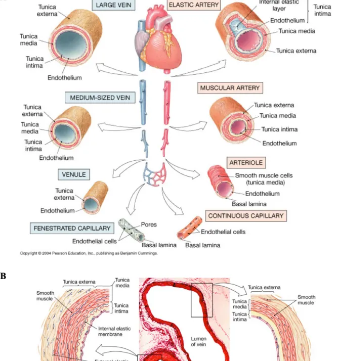
I.1 Anatomy and physiology of the blood vessels
The blood vessels which supply the organs with nutrients, gases and other substances by circulating blood throughout the body are the key connection between the heart and the tissues. Various types of blood vessels that differ in some structures have the same general characteristics. Arteries and arterioles which carry blood away from the heart have thicker walls and smaller lumens than veins and venules which return blood to the heart to resist surge of blood pressure. Between arterioles and venules, the capillaries exchange nutrients and wastes surrounding tissues through the thin walls.
The vascular wall of arteries and veins consists of three distinct layers, called tunics: the tunica intima, the tunica media and the tunica externa.
The tunica intima (also called the tunica interna) which is the most interior layer is comprised of a single layer of endothelial cells (ECs) in contact with blood and connective tissues. In case of the artery, there is also internal elastic membrane (also called the internal elastic lamina) which is a thick layer of elastic fibers at the frontier with the tunica media. The endothelium is continuous with endocardium lining the inner heart chambers and not only plays a role in separating the blood from the vessel wall as a physical barrier but also has physiologically critical functions contributing to regulate vascular tone, inflammation, and coagulation in vascular biology.
The tunica media is the middle layer composed of circularly organized strands of smooth muscle cells (SMCs) sustained by elastic fiber and connective tissue. This smooth muscle layer is responsible for controlling contraction and relaxation by neuronal and chemical mechanisms to regulate blood flow and blood pressure.
The tunica externa (also called the tunica adventitia) which is the outermost layer mostly made up of collagenous connective fibers functions to protect and hold the vessel in relative
Figure 1. Structure of blood vessels. (A) Differential types of blood vessels. (B) Comparison
I.2 Vascular endothelium
The surface of ECs which have various shape across the vascular tree, but typically thin and slightly elongated in the direction of blood flow, is constituted by approximately 1 to 6 X 10¹³ cells and weighs about 1 kg covering a surface area of more than 1,000 m2 in an adult human body (Jaffe 1987). The ECs serve as a central regulator in sustaining vascular homeostasis interacting with numerous released autocrine and paracrine substances in response to biological, physical and chemical stimuli. In fact, because of their morphology and location in direct contact with circulating blood, ECs have a very special localization to modulate diverse vascular biological responses, including thrombosis, leukocyte trafficking, vascular tone, platelet aggregation, inflammation and angiogenesis (Gori et al. 2007).
Under physiological conditions, the blood stream is in contact with the endothelium that controls the appropriate hemostatic balance between procoagulant and anticoagulant systems and helpsblood to remain fluid by providing anticoagulant and antiplatelet surface inhibiting clotting and platelet adhesion. ECs express anticoagulant factors such as endothelial protein C receptor (EPCR), thrombomodulin, tissue factor pathway inhibitor, heparan, ecto-ADPase, tissue-type plasminogen activator (t-PA), prostacyclin (PGI2), and nitric oxide (NO).
Whereas following vascular damage and under pathological conditions, the endothelium shifts the balance to a procoagulant/prothrombotic phenotype by synthesizing tissue factor, plasminogen activator inhibitor-1 (PAI-1), von Willebrand factor (vWF), thromboxane A2
(TXA2), and protease-activated receptors (PARs) (Bombeli et al. 1997; Rodgers 1988; Gross
& Aird 2000).
The passage of leukocytes from the circulatory system to the surrounding sites of vascular injury through the endothelium is a critical event in the inflammatory process of atherogenesis involving a multistep molecular cascade that includes capture, rolling, initial
injury is mediated by preformed components expressed on the endothelial surface, containing Weibel-Palade bodies and their major components, vWF, and P-selectin which mediate both leukocyte and platelet adhesion. It is concluded by firm adhesion between leukocyte integrins and immunoglobulin family, such as platelet–endothelial cell adhesion molecule-1 (PECAM-1), also known as CD31, vascular cell adhesion molecule-1 (VCAM-1), intercellular adhesion molecule-1 (ICAM-1) or junctional adhesion molecule and transmigration into inflamed site (Albelda & Buck 1990; Carlos & Harlan 1994).
Another crucial function of endothelium is to regulate the transport of solutes via a passively paracellular route and macromolecules using either transcellular or paracellular routes across the semi-permeable blood vessel endothelial barrier (Minshall & Malik 2006).
I.3 Key role of the endothelium in vascular homeostasis
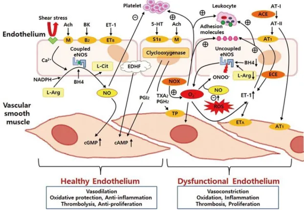
The endothelium contributes to the regulation of vascular tone and the function of smooth muscle and circulating blood cells via well controlled release of various regulatory substances in response to both mechanical and humoral stimuli (Figure 2).
Figure 2. The release of various endothelium-derived regulatory substances and their effects
on the smooth muscle and circulating blood cells (Park & Park 2015). ACE, angiotensin-converting enzyme; Ach, acetylcholine; AT-I, angiotensin I; AT-II, angiotensin II; AT1, angiotensin 1 receptor; BH4, tetrahydrobiopterin; BK, bradykinin; cAMP, cyclic
adenosine monophosphate; cGMP, cyclic guanosine monophosphate; ECE, endothelin converting enzyme; eNOS, endothelial nitric oxide synthase; EDHF, endothelium derived hyperpolarizing factor; ETA and ETB, endothelin A and B receptors; ET-1, endothelin-1;
oxide; NOX, nicotinamide adenine dinucleotide phosphate oxidase; PGH2, prostaglandin H2;
PGI2, prostacyclin; ROS, reactive oxygen species; S1B, serotonin receptor; TP, thromboxane
prostanoid receptor; TXA2, thromboxane; 5-HT, serotonin; Θ, inhibition; Å, stimulation.
I.3.1 Endothelium-derived relaxing factors
I.3.1.1 Nitric oxide
The presence of an endothelium-derived relaxing factor (EDRF) was recognized by Furchgott and Zawadzki in 1980. They observed that the relaxation of rabbit aortic rings by acetylcholine (ACh) is dependent on the intact endothelium (Furchgott & Zawadzki 1980). A few years later, it was revealed that EDRF was NO released from ECs (Palmer et al. 1987; Furchgott 1988). In the intact blood vessel, NO which is a free radical gas with a short half-life is produced by endothelial NO synthase (eNOS) in ECs through the oxidation of the amino acid L-arginine to L-citrulline (Palmer et al. 1988).
Under normal conditions, eNOS remains inactive by binding to the structural protein caveolin-1 located in caveolae, small invaginations of the cell membrane (Bucci et al. 2000). When agonists acting on membrane receptors such as ACh, bradykinin (BK), adenosine di-phosphate (ADP), adenosine tri-phosphate (ATP), thrombin and substance P increase the intracellular level of calcium, the displacement of eNOS from caveolin-1 to calcium/calmodulin occurs followed by the activation of eNOS (Topper et al. 1996). eNOS is the predominant enzyme in the regulation of vascular tone since eNOS knockout mice show increased systemic and pulmonary arterial pressures and endothelium-dependent relaxations in response to ACh of large arteries is markedly reduced (Huang et al. 1995; Chataigneau et al. 1999; Brandes et al. 2000). On the contrary, eNOS-overexpressing transgenic mice are
where it binds to the enzyme soluble guanylyl cyclase (sGC) by targeting the ferrous heme prosthetic group. The activated sGC enzyme catalyzes the increased conversion rate of guanosine triphosphate (GTP) to cyclic 3′,5′-guanosine monophosphate (cGMP) and subsequent relaxation of the vascular SMCs (Ignarro 1991).
In addition, shear stress evokes NO production by activating specialized calcium-activated potassium channels on the ECs surface, causing calcium entry into the cell (Cooke et al. 1991). In the first phase, the prompt separation to caveolin-1 and the following interaction with heat shock protein (Hsp90), which is a chaperon protein, are observed. Then, the protein kinase A (PKA)-dependent phosphorylation of eNOS at Ser1177 is immediately stimulated. In the second phase, the proline-rich tyrosine kinase 2 (PYK2)-dependent phosphorylation of the eNOS inhibitor site Tyr657 and the PKA-dependent phosphorylation of the eNOS activator site Ser633, contribute to control NO production (Fleming 2010; Boo & Jo 2003; Balligand et al. 2009).
Apart from vasorelaxation, endothelium-derived NO inhibits the adhesion and aggregation of platelets (Nong et al. 1997), the activation of leukocytes (Kubes et al. 1991) as well as the migration (Marks et al. 1995) and proliferation (Garg & Hassid 1989) of SMCs. Furthermore, inhibition of eNOS promotes advanced atherosclerosis in mice and rabbits (Kauser et al. 2000; Cayatte et al. 1994). The apoE-/- mice with the additional deletion of eNOS also showed enhanced progression of atherosclerosis and abdominal aortic aneurysm generation and ischemic heart disease, indicating that endogenous eNOS-derived NO plays a major role in anti-atherosclerotic process (Kuhlencordt et al. 2001; Chen et al. 2001).
I.3.1.2 Prostacyclin
Prostaglandin G2 (PGG2), Prostaglandin H2 (PGH2), and PGI2 derived from the
arachidonic acid (AA) are major products of vascular cyclooxygenase (COX). There are two isoforms of COX encoded by two distinct genes. COX-1 is constitutively expressed in many tissues including ECs, and can also be overexpressed, for example, in ECs, by shear stress (Doroudi et al. 2000). Both ECs and to a lesser extent SMCs of healthy blood vessels express COX-1 being the prominent isoform under steady states or under chronic shear stress (Tang & Vanhoutte 2008; Potter et al. 2011; DeWitt et al. 1983). COX-2 is not constitutively expressed, but can be induced rapidly and transiently when the endothelium is disturbed or exposed to inflamed states. PGI2 is produced from PGH2 by prostacyclin synthase (Coleman
et al. 1994). PGI2 acting as a paracrine factor binds to the IP receptor which is present on both
vascular SMCs and platelets to inhibit vasoconstriction and platelet aggregation, respectively, by activating adenylyl cyclase and the subsequent formation of the second messenger cyclic 3’,5’-adenosine monophosphate (cAMP) (Nicosia et al. 1987). In fact, the effect of PGI2 on
endothelium-dependent responses is enhanced in eNOS knockout mice (Sun et al. 1999). In coronary arteries of obese Zucker rats and in the mesenteric vascular bed of streptozotocin (STZ)-induced diabetic mice, endothelial dysfunction is improved by a compensatory up-regulation of the expression and activity of COX-2 (Sánchez et al. 2010; Nacci et al. 2009). Likewise, in coronary arterioles and forearm blood flow of humans with diabetes and hypertension, COX-2-derived PGI2 contributes as a compensatory factor for the diminished
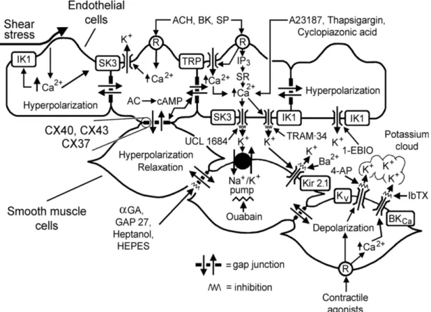
I.3.2 Endothelium-derived hyperpolarization
Endothelium-derived hyperpolarization (EDH), which causes hyperpolarization of the neighboring smooth muscle by inducing a more negative cell membrane potential, also plays significant role in the endothelium-dependent regulation of vascular tone (Feletou & Vanhoutte 1988). In general, EDH-mediated responses involve an increase in calcium levels resulting in potassium efflux via the stimulation of calcium-sensitive potassium channels of small and intermediate conductance in ECs. The SMCs respond to the transmission by electrical coupling through myo-endothelial gap junctions and/or discharge of potassium into the intercellular space, leading to hyperpolarizaion and relaxation (Edwards & Weston 2004; Sandow & Hill 2000) (Figure 3).
In addition, the endothelium releases substances that evoke hyperpolarization of the SMCs such as lipoxygenases derivatives (Faraci et al. 2001), epoxyeicosatrienoic acids (EET) (Campbell et al. 1996) and reactive oxygen species (Ellis & Triggle 2003). EETs, derived from cytochrome P450 epoxygenases, especially play a substantial role in endothelium-dependent hyperpolarization and relaxation of smooth muscle by opening of calcium-sensitive potassium channels of large conductance (BKCa) in several vascular beds
(Quilley & McGiff 2000) such as large coronary arteries from dogs (Widmann et al. 1998), cows and pigs as well as human (Miura et al. 1999). In isolated porcine and bovine coronary arteries and in cultured ECs including those of human origin, receptor-dependent and -independent agonists such as ACh, BK, AA and pulsatile stretch prompt EETs release from ECs, an effect blocked by cytochrome P450 inhibitors (Popp et al. 1998; Rosolowsky & Campbell 1996; Popp et al. 1996; Gebremedhin et al. 1998; Fisslthaler et al. 1999; Campbell et al. 1996).
Figure 3. Endothelium-derived hyperpolarization (EDH)-mediated responses (Feletou &
Vanhoutte 2006). R indicates receptor; Bk, bradykinin; SP, substance P; IP3, inositol trisphosphate; SR, sarcoplasmic reticulum; TRP, transient receptor potential channel; AC, adenylyl cyclase; cAMP, cyclic-AMP; αGA, glycyrrhetinic acid derivatives; CX, connexin; 4-AP, 4-aminopyridine; IbTX, iberiotoxin; SK3, small conductance calcium-activated potassium channel formed by SK3 α-subunits; IK1, intermediate conductance calcium-activated potassium channel formed by IK1 α-subunits; Kir2.1, inward rectifying potassium channel constituted of Kir2.1 α-subunits; KV, voltage-gated potassium channels;
I.3.3 Endothelium-derived contracting factors
I.3.3.1 Endothelin-1
Endothelin-1 (ET-1), which is generated from big ET-1 by endothelin converting enzyme (ECE), acts as a prominent endogenous vasoconstrictor peptide (Yanagisawa et al. 1988) and is also produced by vascular SMCs, macrophages and leukocytes (Ehrenreich et al. 1990; Sessa et al. 1991). The effects of ET-1 appear through the interaction with two G-protein-coupled receptors, ETA which has a high affinity for ET-1 and is predominately
expressed on vascular SMCs, and ETB which is also localized on the ECs as well as SMCs
(Arai et al. 1990; Sakurai et al. 1990). Binding to ETA receptor contributes to increase the
intracellular calcium concentration resulting in contraction of SMCs. By contrast, the activation of ETB receptor on the ECs counteracts the vasoconstricting action of ET-1 on the
SMCs by promoting the release of NO and PGI2, two potent vasodilators (de Nucci et al.
1988). In fact, an in vivo study using ECs-specific ET-1 knockout mice has shown that endothelium-derived ET-1 sustains basal vascular tone and blood pressure (Kisanuki et al. 2010). In a human study with normotensive subjects, it was observed that both an inhibitor of ECE and a selective ETA antagonist prevented forearm vasoconstriction by proET-1 (Haynes
& Webb 1994).
I.3.3.2 Thromboxane A
2& Prostaglandin H
2The effects of PGI2 counterbalance those of TxA2 that is converted from AA into
PGG2/PGH2 through COX-1 and the following synthesis by thromboxane synthase. TxA2
normotensive but show abnormal vascular responses to TxA2 and have a higher sensitivity to
bleeding (Thomas et al. 1998). The elimination of TP receptors alleviates vascular proliferation and activation of platelets in response to vascular injury in mice (Cheng et al. 2002), delays atherogenesis in apoE-/- mice (Kobayashi & Narumiya 2002), prevents
angiotensin II (Ang II)- and L-NAME-induced hypertension and the subsequent cardiac hypertrophy (Francois et al. 2008; Francois et al. 2004). Moreover, in apoE-/- mice and in patients with atherosclerosis, thromboxane synthase mRNA is expressed within the vascular wall of mice atherosclerotic aorta and in the core of human atherosclerotic lesions, and is associated with increased generation of TxA2, contributing to plaque instability and its
thrombotic complications (Gabrielsen et al. 2010).
I.3.3.3 Angiotensin II
Ang II, the active peptide generated from angiotensinogen by the circulating renin-angiotensin system (RAS), plays an essential role in controlling vascular homeostasis, and also in the initiation and development of hypertension, heart failure and atherosclerosis (Kim & Iwao 2000; Schmidt-Ott et al. 2000; Dzau 1986) (Figure 4). The action of both circulating and local Ang II, which is generated from angiotensin I by angiotensin-converting enzyme (ACE) expressed on the ECs, are mediated by activating Ang II type 1 receptor (AT1R) or type 2 receptor (AT2R) (de Gasparo et al. 2000). AT1R is mainly expressed in
different tissues and cells associated with cardiovascular function such as vascular smooth muscle contraction and proliferation. The activation of AT1R facilitates numerous
physiological and pathophysiological activities of Ang II via complicated intracellular signaling pathways and the generation of ROS through the activation of NAD(P)H oxidase.
by its stimulation remain under debate. However, it appears that the AT2R plays a role in
vasodilation, and may be increased as a counteracting mechanism in cardiac hypertrophy, hypertension and atherosclerosis (Lemarié & Schiffrin 2010).
The rise in oxidative stress stimulated by Ang II leads to diminished endothelial relaxation and endothelial dysfunction in experimental hypertensive rat model (Rajagopalan et al. 1996). In humans with elevated RAS activity, ROS-mediated endothelial dysfunction associated with vascular growth and inflammation has been involved in the formation of atheroma (Kolakovic et al. 2017). Ang II also acts as a proatherosclerotic factor causing vasoconstriction and stimulates the expression not only of cell adhesion molecules (CD31/PECAM-1, VCAM-1, ICAM-1) but also of growth factors, cytokines, and chemokines within the vascular wall (Touyz & Schiffrin 2000; Ushio-Fukai et al. 2002; Touyz & Schiffrin 1999).
Figure 4. Schematic describing the circulating renin-angiotensin system (RAS) and the
vascular effects induced by angiotensin II (Ang II). (A) The components of RAS and its receptors. (B) The role of Ang II on vascular cell functions (Montezano et al. 2014).
I.4 Endothelial dysfunction and cardiovascular diseases
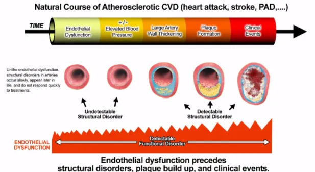
Endothelial dysfunction (ED) is a well-established early step associated with the accumulation of cardiovascular risk factors and leads to the development of atherosclerosis (Anderson et al. 1995; Kinlay & Ganz 1997) (Figure 5). The deleterious changes of endothelial function are characterized by an imbalanced contribution between the bioavailability of vasodilators and endothelium-derived contracting factors, causing an impaired endothelium-dependent vasodilation (Lerman & Burnett 1992). Furthermore, specific states of endothelial activation, which are proinflammatory, proliferative, and procoagulatory responses, favor the progressive development of atherogenesis (Anderson 1999). Therefore, the status of endothelial function has been suggested to be an important prognostic marker of future cardiovascular events in patients with cardiovascular risk factors (Gokce et al. 2003).
bradykinin; C-NP, C-type natriuretic peptide; ROS, reactive oxygen species; ET-1, endothelin-1; TXA2, thromboxane; Ang II, angiotensin II; PGH2, prostaglandin H2; t-PA,
tissue-type plasminogen activator; vWF, von Willebrand factor; PAI-1, plasminogen activator inhibitor-1; TF, tissue factor; CAMs, cell adhesion molecules; TGF-β, transforming growth factor; PDGF, platelet derived growth factor; bFGF, basic fibroblast growth factor; ILGF, insulin-like growth factor; LPL, lipoprotein lipase; NF-κB: nuclear factor κB.
I.4.1 Atherosclerosis
ED is an early indicator of the development of atherosclerosis, which is observed before structural alterations of the vascular wall are detectable by ultrasound or angiography (Luscher & Barton 1997). The early stages of atherosclerosis are characterized by a reduced NO bioavailability due to an increased level of ROS in response to cardiovascular risk factors (Warnholtz et al. 1999). The antiatherogenic role of NO is described in several experimental models of atherosclerosis such as apoE-/- mice as well as in humans. In these studies, the reduced endothelial NO formation facilitates the development of atherosclerotic lesion formation in the coronary artery and the aorta, and treatment of L-arginine retains vessel morphology. The increase in superoxide anion production is involved in the reduced bioavailability of NO in arteries predisposed to atherosclerosis (Cai & Harrison 2000; Landmesser & Harrison 2001). The enhanced ROS generation involves NADPH oxidase (Spiekermann et al. 2003), xanthine oxidase (White et al. 1996; Berry et al. 2000) and leads to the degradation of NO by O2- and the oxidation of tetrahydrobiopterin (BH4) into
dihydrobiopterin (BH2), which is not a cofactor of eNOS (Vásquez-Vivar et al. 2002; Tiefenbacher et al. 2000; Laursen et al. 2001; Landmesser et al. 2003). In addition, the
expression of adhesion molecules, cytokines and chemokines release, recruitment of leukocytes, enhanced low density lipoprotein (LDL) oxidation, activation of platelets, and VSMCs migration and proliferation. Furthermore, ED is not only the initial stage of the progression of atherosclerotic disease that occurs plaque formation, but it also can promote plaque growth leading to clinical cardiovascular complications, as one of the essential mechanisms in atherosclerotic diseases (Hegele 1996; Willerson 2002; Chiu & Chien 2011) (Figure 6).
Figure 6. Endothelial dysfunction as an early indicator in the initiation and progression of
I.4.2 Aging
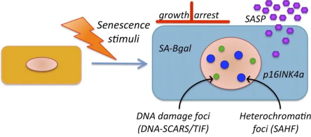
Aging is a physiological process causing a progressive worsening of structure and function of organs and is an independent cardiovascular risk factor for ED even in the absence of other risk factors such as diabetes, smoking, hypertension (Zeiher et al. 1993). Endothelial senescence, an irreversible phenomenon of cell cycle arrest, is known as both replicative senescence by aging over the time and premature senescence induced by pathological conditions with an increased expression of senescence-associated β-galactosidase (SA-β-gal) activity and p16INK4a (Dimri et al. 1995; Chen et al. 2006) (Figure 7). The senescent ECs are characterized by a prooxidative, and proinflammatory senescence-associated secretory phenotype (SASP) via the two major tumor suppressor pathways, known as the p53/p21 also involved in apoptosis and the p16/pRB pathways (Coppé et al. 2010). Several studies have shown that a variety of stimuli involved in cardiovascular disease promote endothelial senescence by increasing the intracellular level of oxidative stress and/or telomerase activity. Exposure of endothelial progenitor cells to Ang II promotes the onset of senescence through NADPH oxidase-dependent prooxidant response (Imanishi et al. 2005). Incubation of HUVECs with either high glucose or advanced glycation end-products (AGEs) increases SA-β-gal activity (Yokoi et al. 2006; Chen et al. 2002). Exposure of ECs to ox-LDL also promotes ECs senescence by impairing telomerase activity (Breitschopf et al. 2001). In addition, an increased SA-β-gal staining is observed in the thoracic aorta of Zucker diabetic rats (Chen et al. 2002) and mice model of oxidative stress (Ota et al. 2008). Furthermore, senescent ECs appear in human aortic arch and coronary arteries at sites overlapping atherosclerotic plaques (Vasile et al. 2001; Minamino et al. 2002).
Figure 7. A principle features of senescence cells (Rodier & Campisi 2011).
I.4.3 Hypertension
Elevated blood pressure is a crucial risk factor associated with increasing prevalence of myocardial infarction, heart failure, and stroke, resulting in higher cardiovascular mortality (Levy et al. 1996; Stamler et al. 1989). The hypertensive-associated ED is a common state characterized by an imbalance between endothelium-derived vasodilators and vasoconstrictor factors and increased oxidative stress which has an important role in the decreased bioavailability of NO, and antioxidant capacity. In fact, an enhanced oxidative stress in patients with essential hypertension causes lower NO availability leading to ED (Taddei et al. 1998). Antioxidants such as vitamins C and E ameliorate endothelium-dependent relaxation of mesenteric artery rings in stroke-prone spontaneously hypertensive rats (SHRSP) by decreasing NADPH oxidase activation and by increasing superoxide dismutase (SOD) activity (Chen et al. 2001). In addition, an elevation in Ang II levels influences an increase in blood pressure and sustenance of hypertension through the activation of NADPH oxidase to produce ROS and the inhibition of the angiotensin system improved ED in hypertensive state
upregulation of Ang II receptors in mesenteric arteries of DOCA-salt hypertensive rats (Schiffrin et al. 1983). Koh et al. identified that the treatment of hypertensive patients with the AT1R antagonist candesartan resulted in improved endothelial function, decreased PAI-1
and reduced level of vascular oxidative stress as well as decreased level of inflammatory cytokines including monocyte chemoattractant protein-1 (MCP-1), and tumor necrosis factor-alpha (TNF-α) (Koh et al. 2003).
I.4.4 Diabetes
Diabetes mellitus is a major chronic health problem affecting about 422 million adults worldwide. Its prevalence rate is increasing rapidly with higher than optimal blood glucose levels causing about 2.2 million deaths by raising the risk of cardiovascular diseases over the past few decades (World Health Organization 2016). The direct and indirect effects of hyperglycemia on the vascular tree are contributing to the induction of both macrovascular complications including stroke, coronary artery diseases, and peripheral vascular diseases, and microvascular complications including diabetic retinopathy, nephropathy, and neuropathy (Fowler 2008). ED is an early key event facilitating the initiation and development of diabetic vascular complications through multiple mechanisms including NADPH oxidase-derived oxidative stress, and eNOS uncoupling with the subsequent formation of ROS instead of NO (Hink et al. 2001). Studies using coronary arteries from animal models of type II diabetes indicate prominent impaired endothelium-dependent relaxations as well as worsened responses to vasoconstrictors (Elmi et al. 2008; Khazaei et al. 2008; Koltai et al. 1997; Bagi et al. 2003). Furthermore, peripheral arterioles (mesenteric, femoral, skeletal muscle and adipose tissue) of obese Zucker rats and insulin-resistant mice
hyperglycemia increases the formation of AGEs, which also contribute to ED by promoting oxidative stress as well as activation of inflammatory response including overexpression of VCAM-1 and tissue factor in HUVECs (Schmidt et al. 1995; Wautier et al. 2001).
I.4.5 Dyslipidemia
Hypercholesterolemia is one of the most well described risk factor triggering coronary artery disease (Multiple Risk Factor Intervention Trial Group 1982). Hypercholesterolemia as well as levels of LDL cholesterol lead to impaired endothelial function in both the coronary and the peripheral circulation in human subjects (Creager et al. 1990; Casino et al. 1993). Lowering cholesterol levels even within the normal range improved endothelium-dependent NO production and hence ameliorated endothelial function (Masumoto et al. 2001). It is worthy of note that reducing the average cholesterol level in patients with established coronary artery disease results in diminished myocardial infarction rates, and this protective effect may be partly because of improvement of the endothelial function (Gilligan et al. 1994; Casino et al. 1995).
I.4.6 Obesity
The progression of vascular disease in obesity may relate to the impact of the metabolic syndrome comprising insulin resistance, hypertension, dyslipidemia and excessive oxidative stress on the physiology of the endothelial function. Indeed, the effects of caloric restriction on endothelial function have been described to improve endothelium-dependent vasodilation in obese subjects with hypertension. Reduction of weight may improve endothelial function indirectly by ameliorating blood pressure and lipid profile (Sasaki et al. 2002). A lipase
placebo-controlled study involving 23 patients associated to an improved endothelial function (Bergholm et al. 2003). The aspect of aggravated oxidative stress in central obesity is based on an extension of the cytosolic triglyceride capacity in non-adipose tissue (Bakker et al. 2000). The deposit of long-chain fatty acyl-coenzyme A esters involved in fatty acids metabolism is postulated to prohibit mitochondrial adenosine translocation, with subsequent excess generation of oxygen radicals and accumulating evidence revealed that antioxidants may improve insulin resistance and ED (Paolisso & Giugliano 1996).
I.4.7 Underlying mechanisms of endothelial dysfunction
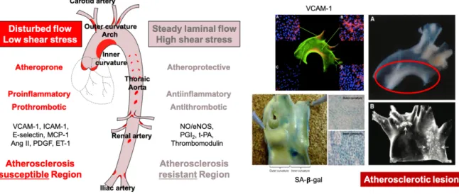
I.4.7.1 Disturbed shear stress
Even though the overall vascular tree is subjected to the cardiovascular risk factors of ED, phenotypic alterations to the endothelial function is expressed at specific regions of arteries such as bifurcations, branch points, and inner curvatures, where disturbed flow takes place and atherosclerosis is developing (Figure 8). Hemodynamic forces are not similar in the whole vascular system. While blood flow in the straight blood vessels of the arterial tree is commonly steady laminar with high shear stress, that in bifurcations is disturbed with low shear stress shifting endothelial function toward an atherogenic endothelial phenotype. Low shear stress promotes atherosclerosis by upregulating the expression of adhesion molecules, growth factors, cytokines, ET-1 and by impairing NO and PGI2 production, and lipid uptake
and catabolism, as well as inducing inflammation and prooxidative responses (VanderLaan et al. 2004; GimbroneJr 1999; Malek et al. 1999).
Figure 8. A schematic of the major arterial tree presenting hemodynamic forces responsible
for atherosclerosis susceptible sites and various phenotypic modulations in the aortic arch (Nakashima et al. 1994; Nakashima et al. 1998; Suo et al. 2007; Warboys et al. 2014).
A number of in vitro studies have characterized responses of ECs to shear stress as a function of time. Early responses of ECs to shear stress imply activation of specific channels or proteins at the plasma membrane including the induction of transmembrane K+ and Ca2+ channels opening (Naruse & Sokabe 1993; Olesen et al. 1988; Yoshikawa et al. 1997), stimulation of heterotrimeric G-proteins (Gudi et al. 1998), and PECAM-1 phosphorylation (Osawa et al. 1997). Moreover, within minutes, several intracellular signaling cascades are activated such as the calcium-dependent phosphorylation of eNOS at Ser1177 promoting its activation, and NO production (Gudi et al. 1998), PI3-kinase activation and signaling by integrins (Tzima et al. 2001). Within minutes to hours, shear stress causes activation of Rho family GTPases (Birukov et al. 2002; S. Li et al. 1999; Tzima et al. 2001; Tzima et al. 2002; Tzima et al. 2003; Tzima 2006; Wojciak-Stothard & Ridley 2003), as well as the adaptor protein Shc, c-Src, focal adhesion kinase (FAK) (Li et al. 1997), protein kinase C (PKC) (Traub et al. 1997), mitogen activated protein kinase (MAPK) (Tseng et al. 1995) and jun
within minutes (Hsieh et al. 1998) and facilitates long-term cellular effects via activation of diverse shear stress-responsive transcription factors including c-fos, c-myc, c-jun, and nuclear factor κB (NF-κB) (Khachigian et al. 1995). Subsequent to persistent shear stress for hours to days, ECs adapt by elevating expression of krüppel-like factor 2 (KLF2) (Dekker et al. 2002; SenBanerjee et al. 2004), E-selectin, platelet-derived growth factor (PDGF), ICAM-1, transforming growth factor-β (TGF-β), tissue factor and MCP-1 (Khachigian et al. 1995; Nagel et al. 1994; Resnick et al. 1993; Sampath et al. 1995).
I.4.7.2 Local angiotensin system
Although the primary targets of Ang II are VSMCs, Ang II has numerous effects on ECs, such as eliciting ROS generation, promoting apoptotic signaling pathways, and promoting thrombosis. The intracellular formation of ROS induced by Ang II stimulates the activation of the transcription factor of NF-κB subsequent to the degradation of its cytoplasmic inhibitor, IκB, resulting in raised levels of VCAM-1 (Pueyo et al. 2000). A similar observation was reported by Arenas et al., who showed that Ang II upregulates the secretion of inflammatory cytokine such as TNF-α, which critically contributes to vascular inflammation and promotes vascular disorders, and matrix metalloproteinase (MMP)-2 from ECs (Arenas et al. 2004). Ang II through AT1R triggers the production of TNF-α via a PKC-dependent pathway in
adult heart (Kalra et al. 2002). Ang II-induced NF-κB-mediated inhibition of fibrinolysis and induction of cell adhesion molecules including VCAM-1 and ICAM-1 contributes to the initiation and progression of atherosclerosis. Ang II-induced MCP-1 and IL-6 expression are dependent on the NAD(P)H oxidase-derived oxidative stress in VSMCs from rats as well as humans (Marui et al. 1993; Kranzhöfer et al. 1999; Chen et al. 1998). In cultured human
ApoE-/- mice, infusion of Ang II resulted in accelerated development of atherosclerosis and aneurysms (Weiss et al. 2001).
I.4.7.3 Reactive oxygen species
An increased vascular generation of ROS in diabetes, hypertension, obesity, aging is the most common contributor promoting the development of ED, which ultimately leads to cardiovascular disease (Figure 9). ROS which are mostly free radicals possess unpaired electrons or have oxidizing potential including molecular oxygen, have several sources in vascular cells. The reactive intermediate superoxide anion (O2-) is responsible for the
production of other reactive species under physiological responses, such as hydroxyl radical (OH·), hydrogen peroxide (H2O2) and peroxynitrite (ONOO-), and is generated by the
mitochondrial electron chain, NADPH oxidase, COXs, uncoupled eNOS and xanthine oxidase (Förstermann & Münzel 2006; Dröge 2002).
Numerous evidences suggest that ROS participate in vascular signaling and proatherogenic responses by modulating redox-sensitive transcription and transduction pathways (Kunsch & Medford 1999). In the surroundings of an elevated level of oxidative stress, ECs lose their protective phenotype and express proinflammatory molecules such as VCAM-1, ICAM-1 and MCP-1, all of which promote interactions between leukocyte and endothelium and contribute to the early stage of atherosclerosis. Appearance of inflammatory signals is predominantly regulated by NF-κB, which is a redox-sensitive transcription factor that also contributes to promote VSMCs growth, vascular remodeling, and atherogenesis (Valen et al. 2001). Cultured cells overexpressing free radical-scavenging enzymes such as catalase reveal restrained NF-κB activation in response to TNF-α, while those overexpressing
NF-κB activity is inhibited by antioxidants such as N-acetylcysteine and pyrrolidine dithiocarbamate (Kunsch & Medford 1999).
Oxidants also play a regulatory role in intracellular signaling. MAPK and tyrosine kinases are major regulatory proteins that modulate cellular responses to stress and growth stimuli (Wolin 2000; Kunsch & Medford 1999). In vascular cells, growth factors and Ang II are potent stimulators of extracellular signal-regulated kinase and p38 MAPK that stimulate proliferation and migration of SMCs fibroblasts via mechanisms involving hydrogen peroxide. These events cause neointimal growth contributing to atheroma progression and restenosis. Activation of VSMCs by the mitogen PDGF induces intracellular generation of hydrogen peroxide and phosphorylation of tyrosine (Sundaresan et al. 1995). This process is prohibited by increasing intracellular concentrations of catalase, and by N-acetylcysteine. ROS alter both Akt kinase and activity of caspase, which play an important role in proliferation of ECs and stimulation of apoptotic signals leading to loss of ECs, respectively (Irani 2000).
Moreover, ROS also modify metabolism of collagen matrix via activation of proteolytic MMPs that play a critical part in plaque characteristics and stability (Channon 2002). Expression of MMPs is raised in shoulder areas of atherosclerotic plaques where its enhanced activity may increase vulnerability possibly leading to plaque rupture (Galis et al. 1994). Atherectomy specimens from patients with unstable coronary syndromes show elevated levels of ROS compared with subjects with stable angina, suggesting a mechanistic role of ROS in composition of plaque and its activity (Azumi et al. 2002). N-acetylcysteine prevented the expression and stimulation of MMP-9 in hypercholesterolaemic rabbits, indicating a promising role for antioxidant treatment in the tuning of the plaque stability (Galis et al. 1998).
Figure 9. Various mechanisms of oxidative stress-mediated endothelial dysfunction by
cardiovascular risk factors (Förstermann & Münzel 2006).
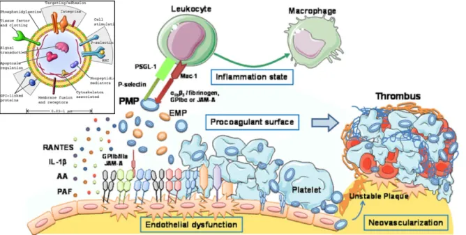
I.4.7.4 Microparticles
Circulating microparticles (MPs) are membrane vesicles with a diameter of 0.1 - 1 µm sheded from different cell types during cell activation by shear stress, cytokines, thrombin, and calcium ionophore A23187, apoptosis or senescence (Leeuwenberg et al. 1992; Sims et al. 1989; Miyazaki et al. 1996, Abbas 2017). The endothelium is one of the principal targets of circulating MPs acting as a biological transcellular signal delivery system by carrying membrane and cytoplasmic proteins and constituents, all of which represent their activated parent cells (Mause & Weber 2010). Under normal conditions, MPs which are mainly
released from platelets and to a lesser extent from ECs and leukocytes, participate in the regulation of ECs homeostasis (Tushuizen et al. 2011; Owens & Mackman 2011) (Figure 10). Under stress conditions, on the other hand, increased concentrations of circulating MPs contribute to ED especially by modulating the balance between NO and ROS generation in addition to stimulating procoagulant and proinflammatory responses (Mostefai et al. 2008; Brodsky et al. 2004). Moreover, a recent study by our lab has shown that the incubation of ECs with circulating MPs from acute coronary syndrome patients induced premature endothelial senescence involving the Ang II-induced NADPH oxidase pathway (Abbas et al. 2017).
Therefore, MPs isolated from blood have been considered as potential diagnostic biomarkers of vascular damage and inflammation in cardiovascular diseases including diabetes, acute myocardial infarction and atherothrombosis (Diamant et al. 2004; Feng et al. 2010; Boulanger et al. 2001).
Figure 10. Composition and molecular mechanisms of circulating microparticles (MPs) on
Chapter II
II.1 Introduction
II.1.1 Gliflozins and sodium-glucose cotransporters
Gliflozins are a class of blood glucose-lowering medications used in the management of type 2 diabetes by inhibiting sodium-glucose cotransporter 2 (SGLT2) mainly responsible for the reabsorption of glucose in the kidney.
Glucose transporters play a crucial role in the regulation of glucose homeostasis by carrying glucose across the plasma membranes. Among the glucose transport proteins in different tissues, sodium-glucose cotransporters (SGLTs), which are integral membrane proteins, transport glucose via a symport mechanism with the concomitant transfer of sodium and are independent of the presence of insulin. The driving force is due to the active sodium removal of the cell by the basolateral sodium/potassium-ATPase, thus promoting a low cytosolic [Na+] and hence, triggering glucose and sodium uptake. In human, six isoforms of SGLTs have been identified, but SGLT1 and SGLT2 are the best studied members of the SLC5A gene family (Table 1).
The average molecular weight of these transmembrane glycoproteins with 14 transmembrane helices is approximately 60 to 80 kDa and they contain 580 to 718 residues.
In the kidney of healthy individuals, approximately 160 - 180 g glucose per day filtered by the glomeruli is completely reabsorbed by the proximal tubules until the glucose concentration in the glomerular filtrate does not exceed the maximum glucose transport capacity. Nearly all of glucose reabsorption in the proximal tubules (S1/S2) occurs through both SGLT2 and SGLT1 proteins. SGLT2, a low-affinity, high-capacity transporter, is responsible for approximately 90% of this process that is independent of insulin and the remaining 10% is removed in the S3 segment by SGLT1, a related high-affinity, low-capacity transporter. SGLT1 is also substantially expressed in the small intestine (Wright et al. 2007; Wright 2001; Wright & Turk 2004; Hediger & Rhoads 1994; Brown 2000) (Figure 11).
Figure 11. Reabsorption of glucose in the kidney of healthy individuals.
The expression and activity of SGLT2 are higher in T2DM patients, and consequently, glucose reabsorption is more pronounced, which contributes to promote hyperglycemia in the blood circulation, ultimately leading to development of cardiovascular complications (Figure 12). Lowering hyperglycemia by inhibiting SGLT2 represents an insulin-independent
strategy that minimizes blood glucose levels by preventing excessive glucose reabsorption in the kidney and hence diminishing the renal glucose threshold.
Figure 12. The maladaptive glucose homeostasis mechanism leading to upregulation of
SGLT2 which eventually contributes to development of cardiovascular complications in impaired fasting glucose (IFG) and diabetes mellitus (DM).
Indeed, studies in primary cultures of human exfoliated proximal tubular epithelial cells from fresh urine of patients with T2DM showed that SGLT2 expression and renal glucose uptake were elevated compared with that from healthy individuals (Rahmoune et al. 2005). In addition, several studies in diabetic rodent models demonstrated increased renal SGLT2 and SGLT1 expression, including raised renal mRNA expression of SGLT1 and SGLT2 in diabetic obese Zucker rats compared with age-matched leans (Tabatabai et al. 2009) and also in alloxan-induced diabetic rats (Vestri et al. 2001). The mechanisms involved in upregulation of SGLT2 are still poorly understood. Recent studies suggested roles for insulin, PKC, and PKA activation and regulation of human SGLT2 activity (Ghezzi & Wright 2012). Ang II and AT1R (Osorio et al. 2009), and hepatocyte nuclear factor (HNF)–1α, a
transcription factor (Freitas et al. 2008) have been implicated in the increased SGLT2 expression in diabetic rats.
II.1.2 Discovery and development of gliflozins
The first SGLT inhibitor discovered was phlorizin, a natural phenolic O-glucoside consisting of a dihydrochalcone moiety, isolated from the bark of the apple tree in 1835 (Petersen 1835) (Figure 13A). It was firstly used in the treatment of infectious diseases such as malaria, because its bitter taste was similar to that of other tree extracts used as antipyretics (De Koninck 1836). In 1886, von Mering revealed that ingestion of phlorizin causes glycosuria and, a century later, investigations of its mechanism of action led to the characterization of SGLTs (Mering 1886). In 1987, in vivo evidence demonstrated the efficacy of phlorizin treatment to reduce glycemic level and normalize insulin sensitivity in diabetic rats (Rossetti et al. 1987). However, phlorizin acts as a nonspecific SGLT inhibitor by targeting both SGLT1 and SGLT2. The dual inhibition of SGLT1 and 2 restricts its applicability for human as a drug. Indeed, owing to poor selectivity, this drug displays important gastrointestinal side effects linked to the high expression level of SGLT1 in the intestine. In addition, limited oral bioavailability due to the hydrolysis of the O-glycoside by intestinal glycosidases (Betz et al. 1975) represents an important concern. Such limitations triggered important efforts, to develop new SGLT2 inhibitors appropriate for oral administration that led to the discovery of C-aryl glucoside-derived gliflozins, presenting nonhydrolyzable C−C bond (Larson 2015) (Figure 13B).