HAL Id: dumas-03124445
https://dumas.ccsd.cnrs.fr/dumas-03124445
Submitted on 28 Jan 2021
HAL is a multi-disciplinary open access archive for the deposit and dissemination of sci-entific research documents, whether they are pub-lished or not. The documents may come from teaching and research institutions in France or abroad, or from public or private research centers.
L’archive ouverte pluridisciplinaire HAL, est destinée au dépôt et à la diffusion de documents scientifiques de niveau recherche, publiés ou non, émanant des établissements d’enseignement et de recherche français ou étrangers, des laboratoires publics ou privés.
Quantification de la fixation myocardique à la
scintigraphie osseuse au 99mTc-bisphosphonate sur
caméra CZT dans l’amylose cardiaque à transthyrétine
David Dudoignon
To cite this version:
David Dudoignon. Quantification de la fixation myocardique à la scintigraphie osseuse au 99mTc-bisphosphonate sur caméra CZT dans l’amylose cardiaque à transthyrétine. Médecine humaine et pathologie. 2020. �dumas-03124445�
UNIVERSITÉ de CAEN NORMANDIE ---
UFR SANTÉ
FACULTÉ de MÉDECINE
Année 2019/2020
THÈSE POUR L’OBTENTION
DU GRADE DE DOCTEUR EN MÉDECINE
Présentée et soutenue publiquement le : 17 mars 2020
par
Monsieur Dudoignon David Alexandre Né le 12/02/1992 à Villiers-le-Bel (Val d’Oise)
:
Quantification de la fixation myocardique à la scintigraphie osseuse au 99mTc-bisphosphonate
sur caméra CZT dans l’amylose cardiaque à transthyrétine.
Président : Monsieur le Professeur AGOSTINI Denis
Membres : Monsieur le Professeur MANRIQUE Alain Monsieur le Professeur AIDE Nicolas
Monsieur le Docteur et maître de conférences LEGALLOIS Damien
U N I V E R S I T É D E C A E N · N O R M A N D I E
U F R S A N T É - F A C U L T E D E M E D E C I N E
Année Universitaire 2019/2020
Doyen
Professeur Emmanuel TOUZÉ Assesseurs
Professeur Paul MILLIEZ (pédagogie) Professeur Guy LAUNOY (recherche)
Professeur Sonia DOLLFUS & Professeur Evelyne EMERY (3ème cycle) Directrice administrative
Madame Sarah CHEMTOB PROFESSEURS DES UNIVERSITÉS - PRATICIENS HOSPITALIERS
M. AGOSTINI Denis Biophysique et médecine nucléaire
M. AIDE Nicolas Biophysique et médecine nucléaire
M. ALLOUCHE Stéphane Biochimie et biologie moléculaire
M. ALVES Arnaud Chirurgie digestive
M. AOUBA Achille Médecine interne
M. BABIN Emmanuel Oto-Rhino-Laryngologie
M. BÉNATEAU Hervé Chirurgie maxillo-faciale et stomatologie
M. BENOIST Guillaume Gynécologie - Obstétrique
M. BERGER Ludovic Chirurgie vasculaire
M. BERGOT Emmanuel Pneumologie
M. BIBEAU Frédéric Anatomie et cytologie pathologique
Mme BRAZO Perrine Psychiatrie d’adultes M. BROUARD Jacques Pédiatrie
M. BUSTANY Pierre Pharmacologie
Mme CHAPON Françoise Histologie, Embryologie
Mme CLIN-GODARD Bénédicte Médecine et santé au travail
M. DAMAJ Ghandi Laurent Hématologie
M. DAO Manh Thông Hépatologie-Gastro-Entérologie
M. DAMAJ Ghandi Laurent Hématologie
M. DEFER Gilles Neurologie
M. DELAMILLIEURE Pascal Psychiatrie d’adultes M. DENISE Pierre Physiologie
Mme DOLLFUS Sonia Psychiatrie d'adultes
M. DREYFUS Michel Gynécologie - Obstétrique
Mme ÉMERY Evelyne Neurochirurgie
M. ESMAIL-BEYGUI Farzin Cardiologie
Mme FAUVET Raffaèle Gynécologie – Obstétrique M. FISCHER Marc-Olivier Anesthésiologie et réanimation
M. GÉRARD Jean-Louis Anesthésiologie et réanimation
M. GUILLOIS Bernard Pédiatrie
Mme GUITTET-BAUD Lydia Epidémiologie, économie de la santé et prévention
M. HABRAND Jean-Louis Cancérologie option Radiothérapie
M. HAMON Martial Cardiologie
Mme HAMON Michèle Radiologie et imagerie médicale
M. HANOUZ Jean-Luc Anesthésie et réa. médecine péri-opératoire
M. HULET Christophe Chirurgie orthopédique et traumatologique
M. ICARD Philippe Chirurgie thoracique et cardio-vasculaire
M. JOIN-LAMBERT Olivier Bactériologie - Virologie
Mme JOLY-LOBBEDEZ Florence Cancérologie
M. JOUBERT Michael Endocrinologie
M. LAUNOY Guy Epidémiologie, économie de la santé et prévention
M. LE HELLO Simon Bactériologie-Virologie
Mme LE MAUFF Brigitte Immunologie
M. LOBBEDEZ Thierry Néphrologie
M. LUBRANO Jean Chirurgie viscérale et digestive
M. MAHE Marc-André Cancérologie
M. MANRIQUE Alain Biophysique et médecine nucléaire
M. MARCÉLLI Christian Rhumatologie
M. MARTINAUD Olivier Neurologie
M. MAUREL Jean Chirurgie générale
M. MILLIEZ Paul Cardiologie
M. MOREAU Sylvain Anatomie/Oto-Rhino-Laryngologie
M. MOUTEL Grégoire Médecine légale et droit de la santé
M. NORMAND Hervé Physiologie
M. PARIENTI Jean-Jacques Biostatistiques, info. médicale et tech. de communication M. PELAGE Jean-Pierre Radiologie et imagerie médicale
Mme PIQUET Marie-Astrid Nutrition
M. QUINTYN Jean-Claude Ophtalmologie
Mme RAT Anne-Christine Rhumatologie
M. RAVASSE Philippe Chirurgie infantile
M. REZNIK Yves Endocrinologie
M. ROD Julien Chirurgie infantile
M. ROUPIE Eric Médecine d’urgence Mme THARIAT Juliette Radiothérapie
M. TILLOU Xavier Urologie
M. TOUZÉ Emmanuel Neurologie
M. TROUSSARD Xavier Hématologie
Mme VABRET Astrid Bactériologie - Virologie
M. VERDON Renaud Maladies infectieuses
Mme VERNEUIL Laurence Dermatologie
M. VIVIEN Denis Biologie cellulaire PROFESSEURS ASSOCIÉS DES UNIVERSITÉS A MI-TEMPS
M. DE LA SAYETTE Vincent Neurologie
Mme DOMPMARTIN-BLANCHÈRE Anne Dermatologie
M. GUILLAUME Cyril Médecine palliative
M. LE BAS François Médecine Générale
M. SABATIER Rémi Cardiologie PRCE
Mme LELEU Solveig Anglais PROFESSEURS EMERITES
M. HURAULT de LIGNY Bruno Néphrologie
Mme KOTTLER Marie-Laure Biochimie et biologie moléculaire
M. LE COUTOUR Xavier Epidémiologie, économie de la santé et prévention
M. LEPORRIER Michel Hématologie
U N I V E R S I T É D E C A E N · N O R M A N D I E
U F R S A N T É - F A C U L T E D E M E D E C I N E
Année Universitaire 2019/2020
Doyen
Professeur Emmanuel TOUZÉ Assesseurs
Professeur Paul MILLIEZ (pédagogie) Professeur Guy LAUNOY (recherche)
Professeur Sonia DOLLFUS & Professeur Evelyne EMERY (3ème cycle) Directrice administrative
Madame Sarah CHEMTOB
MAITRES DE CONFERENCES DES UNIVERSITÉS - PRATICIENS HOSPITALIERS
M. ALEXANDRE Joachim Pharmacologie clinique
Mme BENHAÏM Annie Biologie cellulaire
M. BESNARD Stéphane Physiologie
Mme BONHOMME Julie Parasitologie et mycologie
M. BOUVIER Nicolas Néphrologie
M. COULBAULT Laurent Biochimie et Biologie moléculaire
M. CREVEUIL Christian Biostatistiques, info. médicale et tech. de communication M. DE BOYSSON Hubert Médecine interne
Mme DINA Julia Bactériologie - Virologie
Mme DUPONT Claire Pédiatrie
M. ÉTARD Olivier Physiologie
M. GABEREL Thomas Neurochirurgie
M. GRUCHY Nicolas Génétique
M. GUÉNOLÉ Fabian Pédopsychiatrie
M. HITIER Martin Anatomie - ORL Chirurgie Cervico-faciale
M. ISNARD Christophe Bactériologie Virologie
M. JUSTET Aurélien Pneumologie
Mme KRIEGER Sophie Pharmacie
M. LEGALLOIS Damien Cardiologie
Mme LELONG-BOULOUARD Véronique Pharmacologie fondamentale
Mme LEVALLET Guénaëlle Cytologie et Histologie
M. MITTRE Hervé Biologie cellulaire
M. SESBOÜÉ Bruno Physiologie
M. TOUTIRAIS Olivier Immunologie
MAITRES DE CONFERENCES ASSOCIÉS DES UNIVERSITÉS A MI-TEMPS
Mme ABBATE-LERAY Pascale Médecine générale
M. COUETTE Pierre-André Médecine générale
Mme NOEL DE JAEGHER Sophie Médecine générale
M. PITHON Anni Médecine générale
M. SAINMONT Nicolas Médecine générale
Mme SCHONBRODT Laure Médecine générale MAITRES DE CONFERENCES EMERITES
Mme DEBRUYNE Danièle Pharmacologie fondamentale
Mme DERLON-BOREL Annie Hématologie
Remerciements
Je tiens à remercier l’ensemble des professeurs et médecins qui m’ont tout d’abord appris mon métier et ensuite aidé dans l’élaboration de cette thèse.
Merci à mon directeur de thèse, le Professeur Manrique, de m’avoir guidé, enseigné et offert l’opportunité de publier à ses côtés.
Merci au Professeur Agostini de m’avoir accueilli au sein de son équipe avec le Professeur Aide, à Blandine Enilorac, à Catherine Nganoa et à Pia Tager, afin d’enseigner mon métier. Ma famille, ma fiancée et mes amis ont été des supports sans lesquels je ne pourrai avancer. Je remercie mes parents de me soutenir depuis toujours et ma fiancée, de m’épauler depuis notre rencontre.
Je remercie également les gestionnaires de scolarité, Yoann Villain pour ma thèse et Valérie Curto pour la constitution de mon dossier d’inter-CHU, pour leur gentillesse et leur réactivité.
Abréviations
ATTR: cardiac transthyretin amyloidosis
CZT myocardial SPECT: cadmium zinc telluride myocardial single photon emission computed tomography
HFpEF: heart failure with preserved ejection fraction H:B: heart-to-background
H:CL: heart to contralateral lung uptake ratio
Wb-H:B: whole-body scan heart-to-background ratio Ant-H:B: anterior reprojection ratio on CZT SPECT
LAO-H:B: anterior oblique reprojection ratio on CZT SPECT
3D-H:B: heart-to-background VOI in the contralateral lung on CZT SPECT ROI: region of interest
VG: ventricule gauche VOI: volume of interest
Tableaux et figures
Table 1. Patients characteristics
Table 2. Heart-to-background uptake ratio according to the Perugini grade
Table 3. Regression analysis and Pearson’s correlation between each individual method for
heart-to-background (H:B) uptake ratio.
Table 4. Heart-to-background uptake ratio according to the right ventricular (RV) uptake
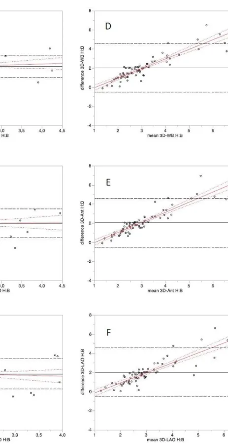
Figure 1: Bland Altman analysis showing (1) a high agreement between all planar methods
for the measurement of the heart-to-background (H:B) uptake ratio (A, B, and C), and (2) an underestimation of the high H:B uptake ratio using planar methods as shown by the increased difference between 3D-H:B and planar H:B ratios for high H:B ratio values (D, E, F). WB, whole-body H:B; Ant, anterior planogram H:B; LAO, left anterior oblique planogram H:B.
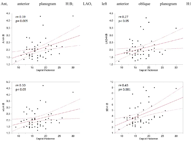
Figure 2: Correlation of the heart-to-background (H:B) uptake ratio to the septal wall
thickness. WB, whole-body H:B; Ant, anterior planogram H:B; LAO, left anterior oblique planogram H:B.
Sommaire
INTRODUCTION p 1 MATERIEL ET METHODES p 3 RESULTATS p 5 DISCUSSION p 6 CONCLUSION p 10 BIBLIOGRAPHIE p 11 ANNEXES p 121
INTRODUCTION
L’amylose cardiaque à transthyrétine (ATTR) représente une cause importante d’insuffisance cardiaque à fraction d’éjection préservée (HFpEF: Heart failure with preserved ejection fraction) (1-3), plus particulièrement chez les patients ayant une hypertrophie myocardiaque (4). L’utilisation de la scintigraphie planaire au bisphosphonate marqué au Technetium 99 peut permettre d’identifier les dépôts amyloïdes cardiaques à TTR à un stade précoce de la maladie et fait maintenant partie des critères diagnostiques non invasifs en l’absence de gammapathie monoclonale (5,6).
La médiane de survie après le diagnostic varie de 25 à 69 mois en fonction du génotype de la transthyrétine (variant héréditaire ou sauvage) et du stade de la maladie (7-9). Le Tafamidis meglumine se fixant sur la transthyrétine et empêchant l’amyloïdogenèse, a récemment obtenu une autorisation de mise sur le marché en raison d’une réduction de la mortalité globale et des hospitalisations pour cause cardiovasculaire. Ce nouveau médicament offre désormais de nouvelles perspectives de traitements dans l’amylose cardiaque à TTR, auparavant peu efficaces. De nombreuses études ont démontré une association entre l’importance de la fixation myocardique et la survie des patients avec amylose cardiaque à TTR (10,11). Cependant, il n’a pas été décrit de méthode standardisée d’évaluation semi-quantitative de la rétention cardiaque du bisphosphonate. En dehors de l’évaluation visuelle proposée par Perugini et al. (12) afin d’identifier les patients suspects d’amylose cardiaque à TTR, de nombreuses méthodes semi-quantitatives ont été proposées afin d’évaluer le pronostic des patients tels que des ratios coeur sur corps entier (H:WB), coeur sur poumon controlatéral (H:CL) ainsi que l’étude de la variation régionale de fixation au sein du ventricule gauche (10,13–15).
Dans une aire de nouvelles thérapies dans l’amylose cardiaque à TTR, une approche quantitative permettrait de réaliser le diagnostic, d’évaluer le pronostic du patient et de suivre la réponse thérapeutique. Avec la généralisation des caméras de tomoscintigraphie CZT, il est désormais possible de proposer une évaluation semi-quantitative de la fixation cardiaque du traceur en utilisant des reconstructions 3D et des images obtenues de rétroprojections planaires (16,17).
Le but de notre étude est donc de comparer différents ratios de fixation cardiaque en scintigraphie osseuse au 99mTc-bisphosphonate obtenus sur caméra CZT SPECT aux techniques d’imagerie planaire conventionnelle chez des patients adressés pour amylose cardiaque à transthyrétine.
2
INTRODUCTION
Transthyretin amyloidosis (ATTR) is an important cause of heart failure with preserved ejection fraction (HFpEF) (1–3), especially in patients with increased wall thickness (4). Cardiac planar radionuclide imaging using 99mTc-labeled bone seeking radiopharmaceuticals may identify cardiac ATTR amyloid deposits, even in the early course of the disease, and is now widely used as a noninvasive diagnostic criterion in patients without detectable monoclonal protein (5,6).
After the diagnosis, median survival ranges from 25 to 69 months depending on the transthyretin genotype (i.e. hereditary variant or wild-type ATTR) and the stage of the disease (7–9). Medical therapy using Tafamidis meglumine, that binds to transthyretin and prevents amyloidogenesis, recently demonstrated a reduction in all-cause mortality and cardiovascular-related hospitalizations, offering new therapeutic perspectives in patients with ATTR cardiac amyloidosis. Several studies demonstrated that a high level of cardiac retention of bone radiopharmaceuticals is associated with a decreased survival in patients with ATTR cardiac amyloidosis (10,11). However, there is no standardized method for semi-quantitative assessment of cardiac uptake of bone tracers. Beside the visual assessment proposed by Perugini and colleagues (12) to identify patients with cardiac ATTR, several semi-quantitative methods have been proposed to evaluate patients prognosis, including heart to whole-body (H:WB) retention, heart to contralateral lung (H:CL) uptake ratio and variation of regional left ventricular tracer uptake (10,13–15).
In the context of a new era for medical therapy of ATTR cardiac amyloidosis, there is a need for quantitative approaches that would be useful for diagnosis, assessment of patient prognosis, and also therapeutic response. With the widespread use of cadmium zinc telluride (CZT)-base single photon emission computed tomography (SPECT) camera, it is now feasible to propose a semi quantitative assessment of a cardiac tracer uptake using either 3D reconstructed acquisition or reprojected planar images (16,17).
The aim of this study was to compare several indexes of cardiac uptake of bone tracers obtained with CZT SPECT in comparison to conventional planar bone scan acquisition in patients with cardiac ATTR.
3
MATERIEL ET METHODES
Study population
Between October 2015 and March 2019, 97 consecutive patients with suspected cardiac ATTR were referred to the Nuclear Medicine department in two participating centers (Caen University Hospital and Toulouse University Hospital) for planar whole-body bone scintigraphy as part of routine diagnostic investigations. The need for supplemental tomographic acquisition was determined by the nuclear medicine physician on the basis of the results of whole-body scan. The final diagnosis of cardiac ATTR was based on the results of immunofixation electrophoresis of serum and urine, serum free light chain assay and bone scintigraphy. Patients characteristics and echocardiography results were obtained from hospital records. The investigation conforms with the principles outlined in the Declaration of Helsinki. This retrospective study was approved by our institutional review board.
Bone scintigraphy
Patients were scanned after intravenous injection of 10 MBq/kg of bisphosphonates (99m Tc-DPD in Caen and 99mTc-HMDP in Toulouse). Whole-body planar images were acquired 3 hours after injection, followed by a SPECT acquisition.
Whole-body images were acquired using a conventional Anger camera (Symbia, Siemens, Erlangen, Germany) with low energy, high-resolution collimators and a scan speed of 10 cm/min. Cardiac retention was assessed by the semi-quantitative visual score proposed by Perugini et al. (12) from 0 (no uptake) to 3 (uptake greater than bone, bone uptake attenuation and soft tissue uptake). Additional tomographic acquisition was not performed in case of Perugini score = 0.
The tomographic imaging was started with a 10-s prescan to help position the detectors on the cardiac area, followed by a 10-min list mode using a dedicated CZT cardiac SPECT camera (D-SPECT; Spectrum Dynamics, Biosensors, Caesarea, Israel) and a 10 % asymmetrical (−7 to +9 keV) energy windows centered on 140.5 keV. No scatter correction was performed, and reconstruction was performed using a dedicated workstation provided by the manufacturer. All acquisitions were gated at 16 intervals per cardiac cycle. Left ventricular volumes and ejection fraction were calculated using QGS software (Cedars-Sinai, Los Angeles, CA, USA). In addition to tomographic reconstruction, two planar equivalent images (planograms) were obtained as previously described (17) by projecting and summing all the elementary 2-D images that shared the same angle onto one large field of view virtual plane in anterior and 45° left anterior oblique views.
4 Heart-to-background ratio analysis
Different heart-to-background (H:B) uptake ratio were calculated using planar and SPECT imaging by drawing regions of interest (ROI) manually on the left ventricle and over a background region. Using planar conventional imaging, a ROI was manually draw over the heart in the anterior view, and then copied and pasted over the contralateral chest, including soft tissue, ribs, and blood pool. On planograms, a circular ROI was drawn over the heart while the size of the background ROI was determined automatically on the x and y dimensions and positioned manually over the contralateral chest (in anterior view) or the mediastinum (in LAO view). Finally, in transaxial reconstructed SPECT images, an elliptic volume of interest (VOI) was drawn manually to encompass the heart and a background VOI was placed over the contralateral lung. As a result, 4 different H:B uptake ratios were generated: wb-H:B (from planar whole-body image), ant-H:B (from anterior planogram), LAO-H:B (from left anterior oblique planogram), and 3D-H:B (from transaxial slices).
Statistical analysis
Paired data were compared using Student’s t-test for paired samples, and unpaired data were compared using Anova. Correlations and concordance between quantitative variables were tested using linear regression analysis with Pearson’s correlation and Bland-Altman analysis. Multivariate analysis was performed using a linear model. A two-tailed p value ≤0.05 was considered statistically significant. Statistical analysis was performed using JMP® version 11.0 (SAS Institute Inc., Cary, NC).
5
RESULTATS
Study population
Among the 97 consecutive patients screened in two university hospital (CHU de Caen, n= 59, and CHU de Toulouse, n= 38), 3 were excluded due to a final diagnosis of AL amyloidosis, and 27 were excluded due to incomplete SPECT or whole-body dataset. Finally, SPECT and planar whole-body radionuclide imaging collected from 67 patients were reviewed. Patients’ characteristics are summarized in Table 1. The cohort was predominantly male (57/67 patients, 85%) with NYHA class II or class III symptoms. Patients received either 99mTc-DPD (n=37) or 99mTc-HMDP (n=30). These patients presented with cardiac hypertrophy and relatively preserved ejection and 15 patients (25%) where in NYHA class 0-I at the time of investigation.
Cardiac retention of bone radiopharmaceuticals
Whole-body planar images demonstrated an abnormal cardiac retention of bone radiopharmaceuticals, predominantly graded Perugini 2 and 3 (Table 1). All planar methods (whole-body and planograms) yielded H:B uptake ratios that were not statistically different. However, 3D-H:B ratio was dramatically increased compared to planar results in patients with Perugini grade 2 and grade 3 (Table 2). Multivariate analysis using linear model confirmed that H:B uptake ratio was significantly influenced by both the Perugini grade (p<0.0001) and the calculation method (p<0.0001).
The correlation was high between all H:B ratios (table 3). Bland Altman analysis confirmed a high agreement between all planar methods (Figure 1).
However, compared to planar methods, 3D analyses consistently showed higher values and in addition showed increasing differences with higher uptake values.
Cardiac retention of bone radiopharmaceuticals was correlated to septal wall thickness. As depicted in Figure 2, the higher correlation between cardiac uptake and septal wall thickness was obtained when using 3D-H:B. Finally, patients with an abnormal right ventricular uptake of bone radiopharmaceutical on CZT SPECT imaging demonstrated higher H:B ratios compared to patients without right ventricular uptake (Table 4).
6
DISCUSSION
Le résultat de cette étude démontre la faisabilité d’une analyse semi-quantitative 3D CZT SPECT et que le ratio volumique “coeur sur bruit de fond” (3D-H:B) en scintigraphie osseuse au 99mTc-bisphosphonate est significativement plus élevé par rapport aux méthodes bidimensionnelles, avec une augmentation de la différence entre les deux méthodes se majorant avec les stades de la maladie.
L’amylose cardiaque à TTR est une maladie sous diagnostiquée, plus fréquente que précédemment estimée (2) avec une prévalence plus élevée chez les patients dans la septième et huitième décennie de vie (18).
L’échographie et l’imagerie par résonnance magnétique cardiaque disposent de critères diagnostiques non spécifiques parmi lesquelles une hypertrophie ventriculaire gauche fréquemment absente au stade initial de la maladie. Des techniques plus avancées d’analyse de la déformation longitudinale du tissu myocardique, “longitudinal strain”, permettent d’identifier un apex préservé de l’atteinte et d’augmenter les performances diagnostiques de l‘échographie, restant cependant peu utilisée (19). En dehors de l’hypertrophie du ventricule gauche, l’IRM cardiaque peut permettre d’identifier un rehaussement tardif du gadolinium, principalement sous endocardique, permettant également d’augmenter la sensibilité de l’examen (20). D’autre part, il a été démontré que l’analyse de la fixation myocardique en scintigraphie osseuse pouvait identifier précocement une amylose cardiaque à TTR et aider à écarter le diagnostic d’amylose à chaînes légères (5,11,12,21). Par conséquent, la scintigraphie osseuse au bisphosphonate (99mTc-DPD, 99mTc-PYP or 99mTc-HMDP) représente désormais un examen clé dans l’algorithme diagnostique chez les patients adressés pour amylose cardiaque à TTR (5,22).
Pourtant, l’évaluation visuelle de la fixation cardiaque par le grade de Perugini ne permet pas de prédire le pronostic des patients (23). Des études récentes ont montré que l’intensité de la fixation cardiaque est un puissant marqueur prédictif de l’évolution cardiaque défavorable (10,11). Rapezzi et al (11) ont démontré qu’un ratio planaire H:WB > 7,5, en scintigraphie osseuse au 99mTc-DPD, était prédictif d’un événement cardiovasculaire majeur. Dans cette étude, le calcul du ratio H:WB nécessitait une correction de la décroissance radioactive, du temps d’acquisition et une soustraction du tractus urinaire et de l’activité vésicale afin d’éliminer des sources potentielles d’erreurs. Dans une étude multicentrique, Castano et al. a proposé une alternative en évaluant la rétention cardiaque de 99mTc-PYP avec un ratio H:CL correspondant à un rapport du nombre total de coups dans la région d’intérêt planaire cardiaque sur le nombre total de coups sur le poumon controlatéral et a démontré une augmentation de la mortalité à 5 ans pour les patients avec un ratio H:CL ≥
7
1.6 (10). D’autres ratios ont été proposés pour quantifier la charge amyloïde en utilisant les gamma caméras Anger conventionnelles notamment via des rapports coeur/crâne, VG/bruit de fond vasculaire en SPECT, coeur/pelvis et coeur/médiastin (13,21,24), confortant le manque de standardisation des procédures en médecine nucléaire. Dans une autre étude récente, Gallini et al. ont évalué six indices différents obtenus sur des images planaires corps entier utilisant une gamma caméra Anger conventionnelle. En utilisant une courbe ROC, la sensibilité de ces indices variait de 89% à 100% avec un cut-off allant de 1.29 à 3.28. Dans cette étude, le ratio coeur/corps entier était le plus performant pour identifier une amylose cardiaque à TTR (13). Malgré la généralisation des caméras CZT cardiaques dédiées, notre étude est la première à comparer l’évaluation de la charge amyloïde utilisant ces caméras par rapport aux caméras conventionnelles planaires corps entier utilisées en scintigraphie.
Les images planaires issues des rétroprojections appelées planogrammes, sont obtenues à partir des acquisitions SPECT comme précédemment décrit (17). Le ratio H/B obtenu des planogrammes sont significativement plus faibles que les ratios volumiques 3D. Les statistiques de comptage des régions d’intérêt cardiaques et du bruit de fond sur les planogrammes prennent en compte la superposition des structures anatomiques fixant le traceur osseux, c’est à dire la cage thoracique, le sternum et les tissus mous (25). La fixation extracardiaque, notamment les tissus mous, n’est pas constante et varie en fonction du stade de la maladie (23). En revanche, le ratio volumique 3D-H:B, obtenu à partir des reconstructions 3D, traduit une charge amyloïde myocardique normalisée avec le bruit de fond pulmonaire et s’affranchit des fixations avoisinantes comme le sternum, la cage thoracique et les tissus mous qui ne sont pas inclus dans le volume d’intérêt.
D’autre part, Glaudemans et al. (21) a également retrouvé une augmentation du ratio VG sur bruit de fond vasculaire chez des patients atteints d’amylose cardiaque à TTR avec hypertrophie ventriculaire gauche comparativement au ratio H:WB (4.6 vs. 2.9), en tomoscintigraphie sur gamma caméra conventionnelle Anger.
En comparant aux précédentes études, nous avons retrouvé des ratios H:B relativement plus élevés. Une sensibilité plus élevée sur les caméras CZT cardiaques dédiées pourrait être une explication possible en raison des statistiques de comptage supérieures avec une relation proportionnelle et linéaire de la réponse contrairement au gamma caméras Anger conventionnelles qui présentent un phénomène de saturation pour un nombre élevé de coups (26).
D’après nos résultats, il existe une sous-estimation de la rétention cardiaque par les techniques d’imagerie planaire, qui se majore avec l’augmentation de la valeur moyenne des ratios évalués sur l’imagerie 3D CZT SPECT. De plus, le ratio volumique 3D-H:B fournit une
8
bonne corrélation à l’épaisseur septale et l’analyse volumique a permis de déceler une atteinte du ventricule droit chez 30% des patients, une population présentant une charge amyloïde plus élevée.
9
DISCUSSION
The results of this study demonstrated that a 3D semi-quantitative analysis of CZT SPECT is feasible and increases the heart-to-background ratio of 99mTc-labelled bone seeking tracer uptake compared to bidimensional methods, with a difference between the two methods increasing with the severity of the disease.
Cardiac ATTR is an underdiagnosed disease, more common than previously estimated (2), and that predominantly affects patients in their seventh and eighth decade of life (18). Echocardiographic and cardiac magnetic resonance imaging patterns are non-specific, demonstrating a left ventricular hypertrophy that is frequently absent in the early phase. Advanced techniques using speckle-tracking imaging may reveal an apical sparing of longitudinal strain that enhances diagnostic accuracy but remain poorly investigated (19). In addition to left ventricular hypertrophy, cardiac magnetic resonance imaging may show a late gadolinium enhancement, especially within the subendocardium, that increases diagnostic sensitivity (20). On the other hand, it has been demonstrated that cardiac uptake of bone radiopharmaceuticals can early identify cardiac ATTR and help distinguish transthyretin from light chain cardiac amyloidosis (5,11,12,21). Consequently, bisphosphonate (99mTc-TcDPD, 99mTc-PyP or 99mTc-TcHMDP) scintigraphy is now a key feature in the diagnostic algorithm in patients with suspected cardiac ATTR (5,22).
However, the visual assessment of cardiac uptake according to the widely used Perugini grading failed to predict patient prognosis (23). Recent data pointed out that the intensity of cardiac uptake is a powerful predictor of poor cardiac outcome (10,11). Rapezzi et al (11) demonstrated that a H:WB ratio of 99mTc-TcDPD uptake > 7.5 was predictive of major cardiac adverse event. In this study, the calculation of the H:WB ratio required a correction for decay and scan speed, and a subtraction of the urinary tract and bladder activity that are potential sources of errors. In a multicenter study, Castano et al. alternatively evaluated 99mTc-PYP cardiac retention by the H:CL ratio of total counts using planar cardiac imaging and demonstrated an increased 5-year mortality in patients with H:CL ≥ 1.6 (10). Several other indices have been proposed to quantify the amyloid burden using conventional Anger cameras, including heart/skull ratio, SPECT left ventricle/blood pool ratio, heart/pelvis and heart/mediastinum ratio (13,21,24), demonstrating the lack of standardization of nuclear imaging procedures. In a recent study, Gallini et al. evaluated six different indices obtained from planar whole body imaging using conventional Anger camera. Using ROC curve analysis, the sensitivity of these indices ranged from 89% to 100%, with cut-off values ranging from 1.29 to 3.28. In this study, the heart/whole-body ratios was the most accurate in identifying cardiac amyloidosis (13),
10
Despite the widespread use of dedicated cardiac CZT cameras, this is the first study comparing the assessment of amyloid load using these cameras in comparison with conventional whole-body bone scan. Planar equivalent images, i.e. planograms, were obtained from SPECT acquisition as previously described (17). The H:B ratio obtained from planograms were significantly lowers than those obtained from 3D images. The count statistics in heart and background regions in the planograms take into account the projection of every anatomic structure that binds the bone radiopharmaceutical, including the rib cage, the sternum and the soft tissues (25). Extra-cardiac uptake, including soft tissue involvement, is not constant and varies according to the stage of the disease (23). On the other hand, the H:B ratio obtained from 3D reconstructed images reflect the heart retention normalized to the lung uptake, a metric that is not influenced by sternum, rib cage or muscle uptake that are not included in VOIs. Interestingly, Glaudemans et al. (21) using a conventional Anger SPECT camera found an increased left ventricle-to-blood pool ratio in patients with ATTR and left ventricular hypertrophy that was further increased compared to H:WB ratio (4.6 vs. 2.9). Compared to these previous results, we found relatively higher H:B ratio. A possible explanation is the increased sensitivity of dedicated CZT cameras, with a linear count rate response compared to the non-linear response with a saturation at high count rate observed with conventional Anger cameras (26). Accordingly, our results demonstrated that the underestimation of cardiac retention by planar techniques increases with the mean value of this retention as assessed by 3D CZT SPECT imaging. In addition, 3D H:B provided a good correlation to myocardial thickness, and 3D analysis was able to demonstrate a right ventricular involvement in 30% of the patients, a condition associated with an increased cardiac retention.
CONCLUSION
L’analyse semi-quantitative 3D CZT SPECT optimise l’évaluation de la fixation cardiaque du bisphosphonate par rapport aux méthodes planaires chez les patients ayant une amylose cardiaque à TTR.
CONCLUSION
The semi-quantitative analysis of 3D CZT SPECT optimized the assessment of bisphosphonate myocardial uptake compared to 2D methods in patients with cardiac amyloidosis.
11
BIBLIOGRAPHIE
1. Bennani Smires Y, Victor G, Ribes D, et al. Pilot study for left ventricular imaging phenotype of patients over
65 years old with heart failure and preserved ejection fraction: the high prevalence of amyloid cardiomyopathy. Int J
Cardiovasc Imaging. 2016;32:1403-1413.
2. González-López E, Gallego-Delgado M, Guzzo-Merello G, et al. Wild-type transthyretin amyloidosis as a cause
of heart failure with preserved ejection fraction. Eur Heart J. 2015;36:2585-2594.
3. Mohammed SF, Mirzoyev SA, Edwards WD, et al. Left ventricular amyloid deposition in patients with heart
failure and preserved ejection fraction. JACC Heart Fail. 2014;2:113-122.
4. Damy T, Costes B, Hagège AA, et al. Prevalence and clinical phenotype of hereditary transthyretin amyloid
cardiomyopathy in patients with increased left ventricular wall thickness. Eur Heart J. 2016;37:1826-1834.
5. Gillmore JD, Maurer MS, Falk RH, et al. Nonbiopsy Diagnosis of Cardiac Transthyretin Amyloidosis.
Circulation. 2016;133:2404-2412.
6. Elliott PM, Anastasakis A, Borger MA, et al. 2014 ESC Guidelines on diagnosis and management of hypertrophic
cardiomyopathyThe Task Force for the Diagnosis and Management of Hypertrophic Cardiomyopathy of the European Society of Cardiology (ESC). Eur Heart J. 2014;35:2733-2779.
7. Ruberg FL, Maurer MS, Judge DP, et al. Prospective evaluation of the morbidity and mortality of wild-type and
V122I mutant transthyretin amyloid cardiomyopathy: the Transthyretin Amyloidosis Cardiac Study (TRACS). Am Heart
J. 2012;164:222-228.e1.
8. Grogan M, Scott CG, Kyle RA, et al. Natural History of Wild-Type Transthyretin Cardiac Amyloidosis and Risk
Stratification Using a Novel Staging System. J Am Coll Cardiol. 2016;68:1014-1020.
9. Gillmore JD, Damy T, Fontana M, et al. A new staging system for cardiac transthyretin amyloidosis. Eur Heart J.
2018;39:2799-2806.
10. Castano A, Haq M, Narotsky DL, et al. Multicenter Study of Planar Technetium 99m Pyrophosphate Cardiac
Imaging: Predicting Survival for Patients With ATTR Cardiac Amyloidosis. JAMA Cardiol. 2016;1:880-889.
11. Rapezzi C, Quarta CC, Guidalotti PL, et al. Role of 99mTc-DPD Scintigraphy in Diagnosis and Prognosis of
Hereditary Transthyretin-Related Cardiac Amyloidosis. JACC Cardiovasc Imaging. 2011;4:659-670.
12. Perugini E, Guidalotti PL, Salvi F, et al. Noninvasive etiologic diagnosis of cardiac amyloidosis using
99mTc-3,3-diphosphono-1,2-propanodicarboxylic acid scintigraphy. J Am Coll Cardiol. 2005;46:1076-1084.
13. Gallini C, Tutino F, Martone R, et al. Semi-quantitative indices of cardiac uptake in patients with suspected
cardiac amyloidosis undergoing 99mTc-HMDP scintigraphy. J Nucl Cardiol. February 2019.
14. Sperry BW, Vranian MN, Tower-Rader A, et al. Regional Variation in Technetium Pyrophosphate Uptake in
Transthyretin Cardiac Amyloidosis and Impact on Mortality. JACC Cardiovasc Imaging. 2018;11:234-242.
15. Van Der Gucht A, Cottereau A-S, Abulizi M, et al. Apical sparing pattern of left ventricular myocardial
99mTc-HMDP uptake in patients with transthyretin cardiac amyloidosis. J Nucl Cardiol Off Publ Am Soc Nucl Cardiol. 2018;25:2072-2079.
16. Blaire T, Bailliez A, Ben Bouallegue F, Bellevre D, Agostini D, Manrique A. Determination of the
Heart-to-Mediastinum Ratio of 123 I-MIBG Uptake Using Dual-Isotope ( 123 I-MIBG/ 99m Tc-Tetrofosmin) Multipinhole
Cadmium-Zinc-Telluride SPECT in Patients with Heart Failure. J Nucl Med. 2018;59:251-258.
17. Bellevre D, Manrique A, Legallois D, et al. First determination of the heart-to-mediastinum ratio using cardiac
dual isotope (123I-MIBG/99mTc-tetrofosmin) CZT imaging in patients with heart failure: the ADRECARD study. Eur J
Nucl Med Mol Imaging. 2015;42:1912-1919.
18. Dharmarajan K, Maurer MS. Transthyretin Cardiac Amyloidoses in Older North Americans. J Am Geriatr Soc.
2012;60:765-774.
19. Quarta CC, Solomon SD, Uraizee I, et al. Left ventricular structure and function in transthyretin-related versus
light-chain cardiac amyloidosis. Circulation. 2014;129:1840-1849.
20. Martinez-Naharro A, Treibel TA, Abdel-Gadir A, et al. Magnetic Resonance in Transthyretin Cardiac
Amyloidosis. J Am Coll Cardiol. 2017;70:466-477.
21. Glaudemans AWJM, van Rheenen RWJ, van den Berg MP, et al. Bone scintigraphy with
99mtechnetium-hydroxymethylene diphosphonate allows early diagnosis of cardiac involvement in patients with transthyretin-derived systemic amyloidosis. Amyloid. 2014;21:35-44.
22. Seferović PM, Polovina M, Bauersachs J, et al. Heart failure in cardiomyopathies: a position paper from the Heart
Failure Association of the European Society of Cardiology. Eur J Heart Fail. April 2019.
23. Hutt DF, Fontana M, Burniston M, et al. Prognostic utility of the Perugini grading of 99mTc-DPD scintigraphy in
transthyretin (ATTR) amyloidosis and its relationship with skeletal muscle and soft tissue amyloid. Eur Heart J
Cardiovasc Imaging. 2017;18:1344-1350.
24. Galat A, Van der Gucht A, Guellich A, et al. Early Phase 99Tc-HMDP Scintigraphy for the Diagnosis and Typing
of Cardiac Amyloidosis. JACC Cardiovasc Imaging. 2017;10:601-603.
25. Hutt DF, Quigley A-M, Page J, et al. Utility and limitations of 3,3-diphosphono-1,2-propanodicarboxylic acid
scintigraphy in systemic amyloidosis. Eur Heart J - Cardiovasc Imaging. 2014;15:1289-1298.
26. Bocher M, Blevis IM, Tsukerman L, Shrem Y, Kovalski G, Volokh L. A fast cardiac gamma camera with
dynamic SPECT capabilities: design, system validation and future potential. Eur J Nucl Med Mol Imaging. 2010;37:1887-1902.
12
ANNEXES
Table 1. Patients characteristics
Age 81±6 Gender, male, n (%) 57 (85) NYHA class, n (%) 0-I II III IV 17 (25) 18 (27) 20 (30) 12 (18) Echocardiography Septal thickness, mm LVEF, % 17±4 47±14 Perugini classification, n (%) grade 1 grade 2 grade 3 3 (5) 28 (42) 36 (53) Heart-to-background ratio Whole-body
SPECT anterior projection SPECT LAO projection SPECT 3D 2.05±0.64 1.97±0.61 2.06±0.64 4.06±1.77 CZT SPECT LV EDV, ml LV ESV, ml LV EF, %
Right ventricular uptake, n (%)
148±55 93±50 41±15 20 (30)
13
Table 2. Heart-to-background uptake ratio according to the Perugini grade
Heart-to-background ratio Perugini 1 Perugini 2 Perugini 3
Whole-body 1.45±0.04* 1.65±0.22†/‡ 2.42±0.67‡
SPECT Anterior projection 1.51±0.36†† 1.63±0.24†/‡ 2.27±0.67‡
SPECT LAO projection 1.24±0.06* 1.77±0.29†/‡ 2.35±0.70‡
SPECT 3D 1.96±0.88* 3.07±0.63† 5.01±1.85
*p<0.01. † p<0.0001 and ††p<0.05 vs. Perugini grade 3. ‡p<0.0001 vs. SPECT 3D.
Table 3. Regression analysis and Pearson’s correlation between each individual method for heart-to-background (H:B) uptake ratio. X Y Regression r p-value WB-H:B Ant-H:B Y = 0.2396689 + 0.8404054*X 0.89 <0.0001 WB-H:B LAO-H:B Y = 0.3885076 + 0.8136067*X 0.82 <0.0001 WB-H:B 3D-H:B Y = -0.536663 + 2.2380835*X 0.82 <0.0001 Ant-H:B LAO-H:B Y = 0.445274 + 0.8212357*X 0.78 <0.0001 Ant-H:B 3D-H:B Y = -0.521782 + 2.3309197*X 0.80 <0.0001 LAO-H:B 3D-H:B Y = -0.183172 + 2.0604316*X 0.74 <0.0001
Table 4. Heart-to-background uptake ratio according to the right ventricular (RV) uptake
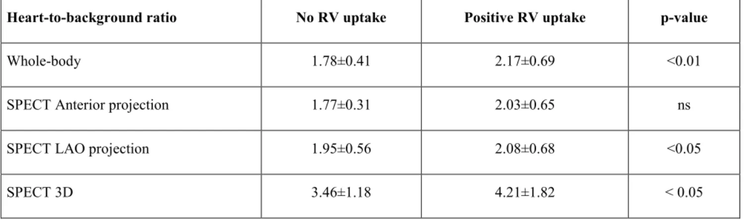
Heart-to-background ratio No RV uptake Positive RV uptake p-value
Whole-body 1.78±0.41 2.17±0.69 <0.01
SPECT Anterior projection 1.77±0.31 2.03±0.65 ns
SPECT LAO projection 1.95±0.56 2.08±0.68 <0.05
14
Figures.
Figure 1: Bland Altman analysis showing (1) a high agreement between all planar methods for the measurement of the heart-to-background (H:B) uptake ratio (A, B, and C), and (2) an underestimation of the high H:B uptake ratio using planar methods as shown by the increased difference between 3D-H:B and planar H:B ratios for high H:B ratio values (D, E, F). WB, whole-body H:B; Ant, anterior planogram H:B; LAO, left anterior oblique planogram H:B.
15
Figure 2: Correlation of the heart-to-background (H:B) uptake ratio to the septal wall thickness. WB, whole-body H:B;
« Par délibération de son Conseil en date du 10 Novembre 1972, l’Université n’entend donner aucune approbation ni improbation aux opinions émises dans les thèses ou mémoires. Ces opinions doivent être considérées comme propres à leurs auteurs ».
VU, le Président de Thèse
VU, le Doyen de la Faculté
VU et permis d’imprimer en référence à la délibération
du Conseil d’Université en date du 14 Décembre 1973
Pour le Président
de l’Université de CAEN et P.O
ANNEE DE SOUTENANCE : 2020
NOM ET PRENOM DE L’AUTEUR : DUDOIGNON David Alexandre
TITRE DE LA THESE : Quantification de la fixation myocardique à la
scintigraphie osseuse au 99mTc-bisphosphonate sur caméra CZT dans l’amylose cardiaque à transthyrétine.
RESUME DE LA THESE EN FRANÇAIS :
Propos. L’objectif de l’étude est de comparer différentes méthodes d’analyse semi-quantitative de la fixation myocardique en scintigraphie osseuse au 99mTc-bisphosphonate chez des patients adressés pour suspicion d'amylose cardiaque à transthyrétine.
Matériel. Des données sur 67 patients ont été recueillis et analysés rétrospectivement chez des patients ayant réalisé une scintigraphie osseuse et une SPECT myocardique sur caméra CZT 3 heures après injection de 99mTc-bisphosphonate. Une analyse visuelle de la fixation cardiaque a été réalisée sur les images planaires selon le score de Perugini. Des ratios ont été calculés sur les images planaires (wb-H:B) et tomographiques: rétroprojections antérieures (ant-H:B), obliques antérieures gauches (LAO-H:B) et volumiques (3D-H:B). L’épaisseur septale a été obtenue à partir des données de l’échographie cardiaque.
Résultats. Les ratios (H:B) wb-H:B, ant-H:B et LAO-H:B ne sont pas significativement différents (p=ns). Cependant, les ratios volumiques (3D-H:B) sont significativement plus élevés comparés aux ratios planaires (3D-H:B, 4.06±1.77, all P<0.0001 vs. wb-H:B, ant-H:B and LAO-H:B).
L’analyse de Bland-Altman montre une augmentation de la différence entre les ratios volumiques et planaires avec la valeur moyenne de la fixation cardiaque. Les ratios volumiques (3D-H:B) sont mieux corrélés à l’épaisseur septale (r= 0.45, p< 0.001).
Enfin, la fixation anormale du ventricule droit est associée à une fixation myocardique plus élevée. Conclusion. L’analyse semi-quantitative sur caméra CZT SPECT optimise l’évaluation de la fixation myocardique du 99mTc-bisphosphonate chez les patients ayant une amylose cardiaque à TTR. MOTS CLES : CZT SPECT, amylose cardiaque, scintigraphie osseuse, imagerie planaire
TITRE DE LA THESE EN ANGLAIS : Quantification of myocardial
99mTc-labeled bisphosphonate uptake with cadmium zinc telluride camera in patients with transthyretin-related cardiac amyloidosis.
RESUME DE LA THESE EN ANGLAIS :
Purpose. We aimed to compare different methods for semi-quantitative analysis of cardiac retention of bone tracers in patients with cardiac transthyretin amyloidosis (ATTR).
Methods. Data from 67 patients with ATTR who underwent both conventional wholebody scan and a CZT myocardial SPECT 3 hours after injection of 99mTc-labelled bone tracer were analyzed. Visual scoring of cardiac retention was performed on whole-body scan according to Perugini 4-point grading system. A planar heart-to-background (H:B) ratio was calculated using whole-body scan (wb-H:B). CZT SPECT was quantified using 3 methods: planar H:B ratio calculated from anterior reprojection (ant-H:B), left anterior oblique reprojection (LAO-H:B) and 3D-H:B ratio calculated from transaxial slices as mean counts in a VOI encompassing the heart divided by background VOI in the contralateral lung. Interventricular septal thickness was obtained using echocardiography.
Results. All H:Bs obtained from planar and reprojected data were not statistically different (wb-H:B, 2.05±0.64, ant-H:B, 1.97±0.61, LAO-H:B, 2.06±0.64, all P=ns).
However, 3D-H:B was increased compared to planar H:Bs (3D-H:B, 4.06±1.77, all P<0.0001 vs wb-H:B, ant-H:B and LAO-H:B). Bland-Altman plots demonstrated that the difference between 3D and planar H:Bs increased with the mean value of myocardial uptake. 3D-H:B was best correlated to septal thickness (r= 0.45, p<0.001). Finally, abnormal right ventricular uptake was associated with higher values of cardiac retention.
Conclusion. 3D semi-quantitative analysis of CZT SPECT optimized the assessment of 99mTc-labelled bone tracer myocardial uptake in patients with cardiac amyloidosis.