Developing VHH-based tools to study Ebolavirus Infection By
Jason V. M. H. Nguyen
B.S., Biochemistry and Molecular Biology University of California, Santa Cruz, 2012 Submitted to the Microbiology Graduate Program in partial fulfillment of the requirements for the degree of
Doctor of Philosophy At the
Massachusetts Institute of Technology
June 2019
2019 Massachusetts Institute of Technology. All rights reserved.
Signature redacted
Signature of Author...V Y'
... Jason V. M. H. Nguyen Microbiology Graduate Program May 13, 2019
Signature redacted
Certified by...It
Signature redacted
Accepted by... MASSACHUSETTS INSTITUTE OF TECHNOLOGYJUN
2
12019
LIBRARIES
ARCHIVES
...
Hidde L. Ploegh Senior Investigator Boston Children's Hospital...
Jacquin C. Niles Associate Professor of Biological Engineering Chair of Microbiology ProgramDeveloping VHH-based tools to study Ebolavirus Infection By Jason V. M. H. Nguyen
Submitted to the Microbiology Graduate Program on May 13th, 2019
in partial fulfillment of the requirements for the degree of Doctor of Philosophy
ABSTRACT
Variable domains of camelid-derived heavy chain-only antibodies, or VHHs, have
emerged as a unique antigen binding moiety that holds promise in its versatility and utilization as a tool to study biological questions.
This thesis focuses on two aspects on developing tools to study infectious disease, specifically Ebolavirus entry. In Chapter 1, I provide an overview about antibodies and how antibodies have transformed the biomedical field and how single domain antibody fragments, or VHHs, have entered this arena. I will also touch upon how VHHs have been used in various fields and certain aspects that remain underexplored. Chapter 2 focuses on the utilization of VHHs to study Ebolavirus entry using VHHs that were isolated from alpacas. Two VHHs were found to neutralize Ebolavirus in both Biosafety Level 2 and 4 laboratory conditions. Ongoing experiments to address mechanism focuses on two aspects of neutralization: Cathepsin inhibition or NPC1-mediated inhibition. Finally, Chapter 3 discusses the overall landscape for Ebolavirus therapeutics and will discuss future directions of this work.
Thesis Supervisor: Hidde L. Ploegh
Title: Senior Investigator, PCMM at Boston Children's Hospital 3
Acknowledgements
There are many people that I need to thank for supporting me during my graduate studies. I would like to first thank my advisor, Hidde Ploegh, for his guidance throughout my graduate school career. As a leader and professor, he has attracted some of the best people to
his lab, creating a fruitful environment for research. Your wisdom and insight have led to many unique projects that challenged my scientific curiosity.
I would also like to thank my committee, Michael Laub and Lee Gehrke for being
supportive over the many years. Our conversations have helped me both scientifically as well as keeping me grounded. Thank you both for providing assistance during our lab's move to Boston Children's Hospital. I would also like to thank Sean Whelan for reading my thesis and agreeing to be on my committee as an examiner.
Next, I would also like to thank all members of the Ploegh lab from the Whitehead Institute and Boston Children's Hospital for being great colleagues and friends through the years. I would like to specially thank Dr. Thibault Harmand, Dr. Nick McCaul, Dr. Matthias
Truttmann, and Lotte Wijne for their friendship and willingness to take the much-needed coffee breaks to discuss politics, TV shows, and the occasional science. Nick: for being a great friend and colleague. Your willingness to always help and teach people really speaks volumes. But also, to introducing me into Dungeons and Dragons. Thibault: Your energy and craziness meant that there has never been a dull moment in the lab. It has always been great working with you both in the lab and out. Matthias: for your wisdom and leadership and your willingness to dispense advice. Lotte: thank you for being a good friend. Whether it be bonding over food, music, corgis, or open houses. I'll miss our time in lab. I would also like to thank Joy Xie and Nova Pishesha for being great bay mates at Whitehead: we had so much fun traveling, eating, and bonding over whatever food Boston sent our way. Djenet Bousbaine, whose bike chats I really value, but also for being a great friend and colleague. For reading my thesis and providing feedback, I would like to thank Djenet Bousbaine, Ross Cheloha, and Nick McCaul for their valuable comments.
I would like to thank past Ploegh members. I would like to the Leo Hanke and Florian Schmidt for really introducing me into the lab and helping me get started on my thesis work. Jasdave Chahal: your infectious energy really invigorated the Whitehead crew. Our continued conversations outside the lab were very valuable and helpful in navigating graduate school.
I would also like to thank my Microbiology Program cohort: Josh, Nate, and Joseph. Your friendship and support over the years has been incredibly helpful especially when our lab moved to Harvard Medical School.
Special thanks to our lab manager, Robert Miller. Thank you for keeping the lab afloat and taking care of equipment whenever the occasion arises. Thank you to Simona Stella for dealing with the day-to-day administration of our lab.
I
would finally like to thank my family for always being supportive during the at-times
difficult road of graduate school. I know how hard it was for them to see me leave California,
but their visits to Boston were always happy reminders of the West Coast.
Table of Contents
C h a p te r 1
...
1 1
In tro d u ctio n ... 1 1 An Introduction to Ebolavirus ... 13 Ebolavirus Genom e ... 13 E b o la v iru s In fe ctio n ... 14 Ebolavirus Glycoprotein ... 16M odes of virus neutralization ... 18
Ebolavirus Therapeutics ... 20
Single Dom ain Antibody Fragm ents ... 23
Chapter 2 Neutralizing Single Dom ain Antibody Fragments against Ebolavirus ... 45
In tro d u ctio n ... 4 6 R e s u lts ... 4 9 In P ro g re ss ... 5 4 D isc u ss io n ... 5 9 M aterials and M ethods ... 61
C h a p te r 3 ... 8 2 Future Directions and Conclusions ... 82
F u tu re D ire ctio n s ... 8 3
C o n c lu sio n ... 8 4
Table of Figures
Introduction
Figure 1: Ebolavirus G enom e ... 28
Figure 2: Crystal Structure of Ebolavirus Glycoprotein... 29
Figure 3: Functional domains of GP are hidden by the mucin-like domain (MLD) ... 30
Figure 4: EBOV G P processing in host cells... 31
Figure 5: M odes of antibody-mediated virus neutralization... 32
Figure 6: Interaction of EBOV GP with neutralizing human antibody KZ52 blocks fusion ... 34
Figure 7: Comparison of Conventional Antibodies and VHHs ... 35
Figure 8: Hydrophilic substitutions remove the need for cognate light chain interaction ... 36
Chapter 2: Neutralizing Single Domain Antibody Fragments against Ebolavirus
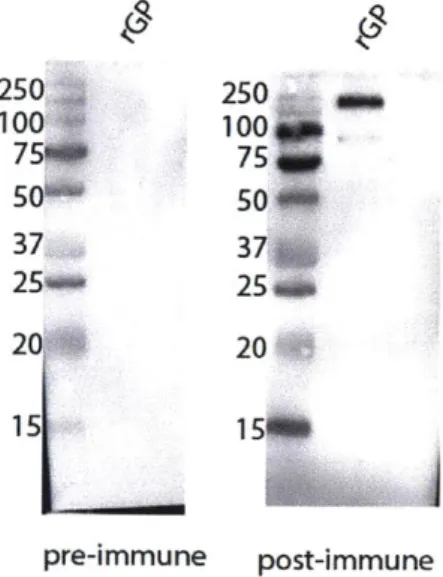
Figure 9 Ebolavirus Glycoprotein-M ediated Entry ... 67Figure 10: Immune response to GP is seen immunized alpaca serum ... 68
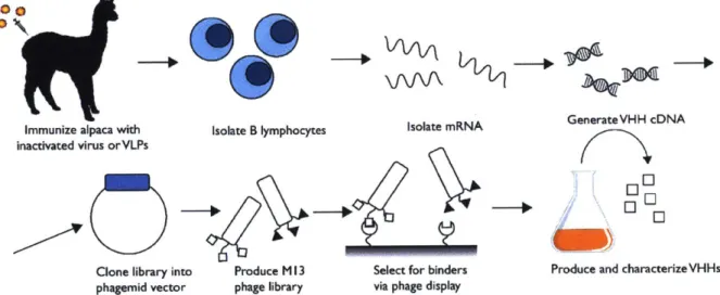
Figure 11: Schematic of VHH isolation from alpaca immunizations... 69
Figure 12: Sequences of anti-Ebolavirus Single domain antibody fragments ... 70
Figure 13: Neutralization of VSV-EBOV in cell culture... 71
Figure 14: EC50 of G10 and G84 against recombinant GP lacking the mucin domain (GPdM) ... 72
Figure 15: USA M RIID RIID Test- BL4 ... 73
Figure 16: Expression of Ebola G P ... 74
Figure 17: Cathepsin B Cleavage is altered by G84 ... 75
Supplemental
Supplem ental 1: V H H Sequences... 76Supplemental 2: Analysis of the germline-encoded V-region of selected VHHs... 77
Supplem ental 3: G10 and G84 pH sensitivity ... 78
Supplemental 4: G10 does not cross-react with Bundibugyo Ebolavirus ... 79
Supplemental 5: Removal of the glycan cap via thermolysin perturbs G10 binding ... 80
Supplemental 6: C-to-C VHH fusion or Ebolavirus neutralization ... 81
Chapter 1
Introduction
An Introduction to Ebolavirus
Ebolavirus (EBOV) is the causative agent of Ebola virus disease, a viral hemorrhagic fever
resulting from EBOV infection. EBOV was first discovered in 1976 in the Democratic Republic of
the Congo (formerly known as Zaire) and South Sudan. Between 1976 and 2014, there have
been over twenty documented epidemics caused by three species of EBOV: Zaire ebolavirus,
Sudan ebolavirus, and Bundibugyo ebolavirus (Malvy, McElroy, de Clerck, Gunther, & van
Griensven, 2019). These outbreaks accounted for approximately 2400 cases and 1600 deaths
(Table 1). In 2014, the largest Ebolavirus epidemic began, spanning multiple countries in
Western Africa. By its conclusion in 2016, there were 28,652 cases and 11,325 deaths
attributed to Zaire ebolavirus infection (Kaner & Schaack, 2016). As of spring 2019, the second
largest Ebolavirus outbreak is ongoing in the Democratic Republic of the Congo. Despite
extensive international intervention, the 2014-2016 outbreak highlighted the need for a better
understanding of Ebolavirus infection and considerations for therapeutics.
Ebolavirus Genome
Ebolaviruses belong to the genus Ebolavirus of the family Filoviridae in the order
Mononegavirales. This order consists of viruses that have a single-stranded RNA genome of
negative polarity. Within this genus, there are six known species, four of which cause disease in
humans: Zaire ebolavirus, Tar Forest ebolavirus, Bundibugyo ebolavirus, and Sudan ebolavirus.
The remaining two cause disease in nonhuman primates and pigs (Reston ebolavirus) and bats
(Bombali ebolavirus).
The approximately 19 kilobase genome of Ebolaviruses encodes seven genes:
nucleoprotein (NP), viral protein (VP) 30, polymerase cofactor (VP35), matrix (VP40), VP24,
polymerase (L) and glycoprotein (GP) (Figure 1). Each of the seven genes fulfill a specific role in
facilitating successful EBOV infection. Each gene is sequentially transcribed into mRNA by the
polymerase, L, an RNA-dependent RNA polymerase responsible for genome replication and
transcription of viral genes. NP encapsulates the genome, forming the viral ribonucleoprotein
complex. VP30 is a phosphoprotein responsible for transcription initiation (Xu et al., 2017).
VP35, an essential cofactor for L, also plays a role in interferon antagonism (Prins, Cardenas, &
Basler, 2009). VP40, the major matrix protein, facilitates viral budding at the host membrane
surface (Bornholdt et al., 2013). The function of VP24 is not well understood but it is thought to
play a role in immune evasion and virion formation (Banadyga et al., 2017). GP encodes the
glycoprotein that mediates entry into host cells. GP is the sole polycistronic gene in the EBOV
genome. GP mRNA is edited by the viral polymerase complex, producing three glycoprotein
isoforms with different functions: transmembrane GP, soluble GP (sGP), or second small GP
(ssGP) (Mehedi et al., 2011). Expression of viral genes facilitates the production of new viruses
and assists in mounting a successful infection.
Ebolavirus Infection
Ebolavirus is transmitted through contact with contaminated bodily fluids via open
wounds or mucus membranes. EBOV infects multiple cell types in humans and non-human
primates and results in a poor antibody response in non-survivors (Baize et al., 1999).
Initial targets of EBOV are antigen-presenting cells, such as dendritic cells or
macrophages. Infected cells travel to draining lymph nodes where viral replication continues, thereby allowing further dissemination (Geisbert et al., 2003; Baseler, Chertow, Johnson,
Feldmann, & Morens, 2017). Shortly thereafter, infection spreads to the liver and spleen (Malvy et al., 2019). Infection of endothelial cells causes cellular necrosis, leading to vascular
permeabilization, compromising the structural integrity of the endothelium (Wahl-Jensen et al., 2005). Eventually, tissue damage occurs in response to virus-associated cytopathic effects and massive release of inflammatory cytokines (Zarschler, Witecy, Kapplusch, Foerster, & Stephan, 2013).
Viruses have evolved sophisticated strategies to avoid recognition by the immune system. Likewise, EBOV encodes specific viral proteins to suppress the immune response. Immune suppression is mediated primarily by two viral proteins expressed during replication:
VP35 and VP24. VP35 was identified as an antagonist of type I interferon response by way of binding to double-stranded RNA, leading to suppression of RIG-1 mediated signaling (Cardenas et al., 2006). This, in turn, blocks IRF-3-dependent induction of interferon a and interferon /. Similarly, VP24 inhibits interferon production by prevent translocation of STAT1 into the nucleus, effectively blunting STATI-dependent interferon production (A. P. Zhang, Abelson, et
al., 2012; A. P. Zhang, Bornholdt, et al., 2012). It is unclear whether inhibition of STAT1 translocation is a consequence of direct VP24-STAT1 interaction or through binding of karyopherin al, a nuclear import protein (Reid et al., 2006). By means of this two-pronged approach, which perturbs the signaling pathways responsible for interferon production,
Ebolavirus effectively evades the immune response.
Ebolavirus Glycoprotein
Successful infection of EBOV depends on GP, an approximately 450 kDa trimeric
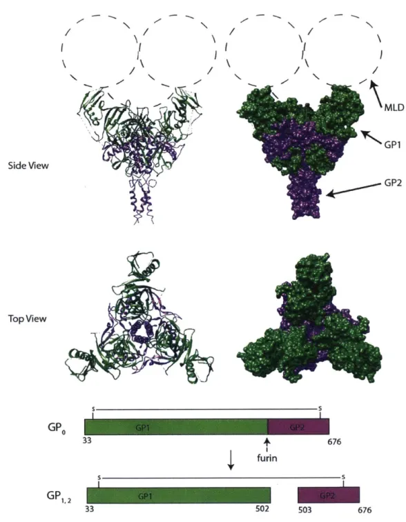
glycoprotein complex responsible for attachment, fusion, and entry into host cells. Prior to assembly of trimeric GP, the GP precursor (GPo) is cleaved by a furin-type protease in the Golgi into GP, and GP2 subunits, which remain linked by a disulfide bond (Figure 2). While many
receptors have been implicated in EBOV attachment to the plasma membrane, none have been shown to be solely necessary for entry thus far. Upon interaction with DC-SIGN/L-SIGN
(Simmons et al., 2003), LSECtin, hMGL (Takada et al., 2004), ft-integrins (Takada et al., 2000), or
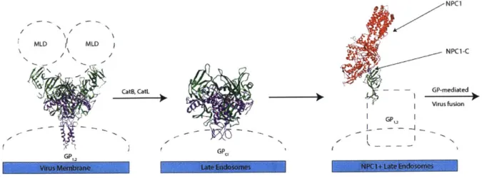
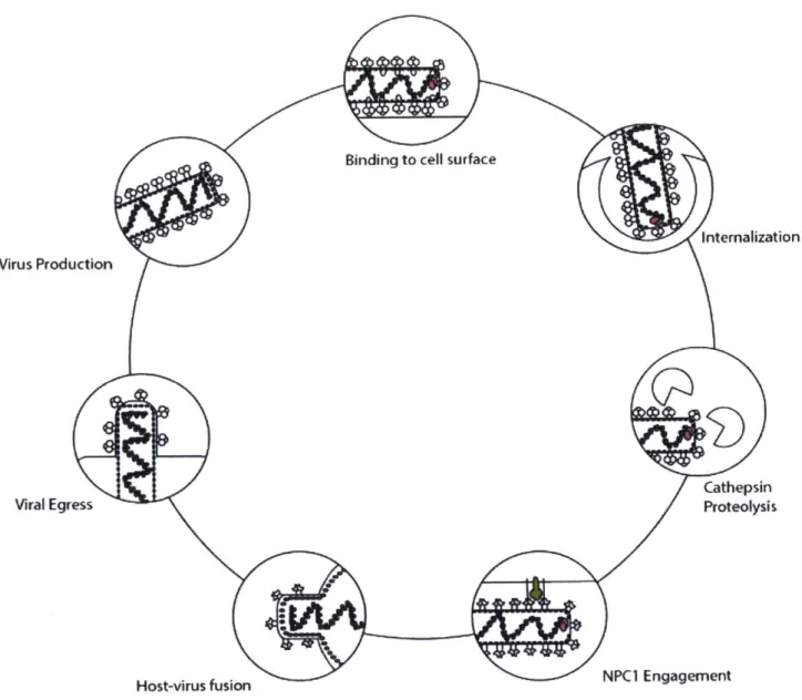
Tyro3 receptors (Shimojima et al., 2006), virions are internalized by macropinocytosis and trafficked to late endosomes. Proteolytic cleavage by cathepsins B and L activates the glycoprotein, priming it for fusion. It does so by removal of the mucin-like domain (MLD),
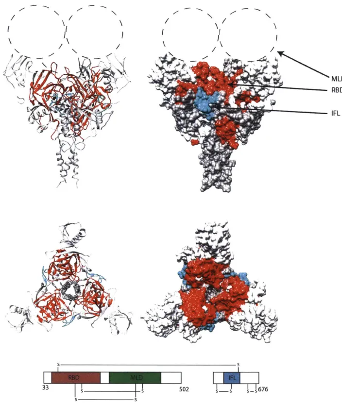
exposing the receptor binding domain (RBD) in GP1. The newly revealed RBD then interacts with
Niemann Pick C1 (NPC1), its entry receptor, by binding to the loop C domain of NPC1 (NPC1-C) (Figure 3, Figure 4). Proteolytic cleavage also potentiates GP2, liberating the internal fusion loop
(IFL), which projects into the host membrane (Cote et al., 2011; Brecher et al., 2012; Spence, Krause, Mittler, Jangra, & Chandran, 2016). Following GP-mediated membrane fusion between the host and viral membranes, the viral genome is released into the cytoplasm where
sequential transcription of viral genes by polymerase, L, begins.
Ebolavirus GP is a class I fusion protein. This class of fusion proteins, which includes the prototypical Influenza A virus (IAV) hemagglutinin (HA), are distinguished by proteolytic priming
and the fusion loop respectively, while GPo cleavage into GPI and GP2 by furin-like proteases
does not yield the same functional constituents (Figure 2). Instead, GP1, 2 requires further cleavage by endosomal cathepsins after internalization to render GP fusion-competent. While structural studies of GP-mediated fusion have not yet elucidated the specific conformational states of GP required for successful fusion, it occurs in NPC1-positive late endosomes through interaction with the NPC1-C loop C domain (Miller et al., 2012; Bornholdt et al., 2016; Spence et al., 2016; H. Wang et al., 2016) (Figure 4).
Aside from its central role in host entry, GP also influences viral replication and pathogenicity. The large structure of GP is believed to displace adhesion proteins needed for cellular attachment, as seen by cell rounding of infected and transfected cells (Takada et al., 2000). This is presumably responsible for the increased permeability of the endothelial barrier (Z. Y. Yang et al., 2000; Wolf, Beimforde, Falzarano, Feldmann, & Schnittler, 2011). Additionally,
mRNA editing of GP during transcription results in a soluble, dimeric GP that lacks a
transmembrane domain (sGP) (Figure 1) (Lee & Saphire, 2009). sGP is found in the circulation of infected individuals (Maruyama et al., 1999) and acts as an "antibody sink," thus blunting the humoral immune response (Pallesen et al., 2016). ssGP, another soluble form of GP, is expressed by EBOV-infected Vero E6 cells, but no function has been yet identified (Figure 1)
(Mehedi et al., 2011). Therefore, GP consists of multiple isoforms that subvert the immune response and potentially exacerbate infection.
Modes of virus neutralization
Viral neutralization can be achieved through neutralizing antibodies that block the
function of critical viral proteins. Neutralizing antibodies curtail infection by interfering with
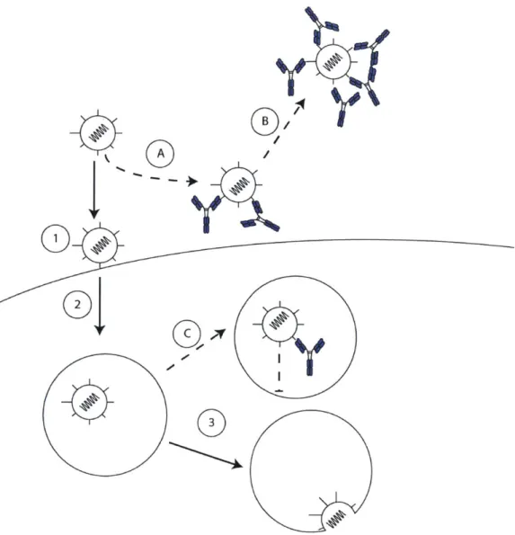
receptor interactions or by inhibiting fusion between the host and viral membranes (Figure 5).
Interference of virus-receptor interactions
Neutralizing antibodies can block attachment of virus to host cells by preventing
engagement of a viral protein to its cognate receptor. For example, some neutralizing
antibodies inhibit human immunodeficiency virus (HIV-1) infection by blocking or occluding the
CD4-binding site of gp120, one of the receptors required for HIV-1 entry (Dalgleish et al., 1984;
Kwong et al., 1998; Saphire et al., 2001; Raja, Venturi, Kwong, & Sodroski, 2003).
Inhibiting host-virus membrane fusion
Antibodies that bind the virus may not prevent receptor engagement but instead
prevent fusion between viral and host membranes. Overcoming the large kinetic barrier
presented by the fusion of two lipid bilayer membranes requires large conformational changes
of viral fusion proteins (reviewed in (Harrison, 2005, 2008)). By restricting these conformational
changes, antibodies that target conserved epitopes involved in the fusion transition should
block infection and prevent release of the viral genome into the host cytoplasm (Ekiert et al.,
2009). Palivizumab, an antibody against respiratory syncytial virus (RSV) F protein, is an
example of blocking cell-to-cell fusion through restriction of the conformational changes
required for fusion (Huang, Incognito, Cheng, Ulbrandt, & Wu, 2010; Swanson et al., 2011).
Vaccine-based virus protection
Vaccines can elicit adaptive immune responses via activation of B and T lymphocytes.
Protection against viral pathogens can be achieved through B and T cell activation. Activated B cells can secrete antibodies that recognize the vaccine immunogen. CD4*T cells assist in class switching and affinity maturation of these B cells. Finally, CD8' T cells can directly kill infected cells.
Vaccines fall into many categories: live, attenuated, subunit-based, inactivated, toxoid, conjugate, DNA, and recombinant vector vaccines. Each of these vaccine strategies carries its own advantages and disadvantages.
Most vaccines that confer protection are believed to induce the production of antigen-specific neutralizing antibodies. There is now increasing evidence that some vaccines induce antigen-specific T cells to provide long-lasting protection. In the field of HIV-1, T-cell based vaccines are attractive based on evidence that CD8' T cells predominate in controlling and eradicating HIV-1 infection (Borrow et al., 1997). DNA vaccines are used to induce CD8' T cell responses. Through administration of an DNA plasmid encoding the antigen of interest,
induction of CD81 T cells is observed. For example, following intramuscular injection of a DNA vaccine encoding Plasmodiumfalciparum PfCSP, antigen-specific CD8' T cells were elicited that targeted infected hepatocytes in both mice and humans, providing protection against infection (Sedegah, Hedstrom, Hobart, & Hoffman, 1994; R. Wang et al., 1998). Likewise, DNA
vaccination a plasmid encoding the IAV nucleoprotein also induced MHC-l restricted CD8+T cells and protected immunized mice against a lethal challenge of IAV (Fernando et al., 2016).
Antibody-dependent enhancement of infection
Although neutralizing antibodies provide protection from viruses, some antibodies may
also paradoxically promote infection. In the case of dengue virus (DENV) infection, there are
multiple serotypes: DENV-1, DENV-2, DENV-3, and DENV- 4. Serotype-specific neutralizing
antibodies of DENV are unable to neutralize different serotypes. Instead, these enhancing
antibodies bind to the envelope protein and are internalized along with the virus in an
FcR-dependent fashion (Goncalvez, Engle, St Claire, Purcell, & Lai, 2007). Hepatitis C infection can
be enhanced in a similar manner, in which neutralizing antibodies at a sub-neutralizing
concentration do not neutralize but instead enhance infection by internalization via FcRI, FcRII,
or FcRIII (Meyer, Ait-Goughoulte, Keck, Foung, & Ray, 2008).
Ebolavirus Therapeutics
The 2014-2016 Ebolavirus epidemic highlighted the need for novel Ebolavirus
therapeutics. Due to the rapid expansion of the outbreak, clinical trials were fast-tracked in
order to provide therapeutic options to combat this public health threat. Many candidate
therapeutics have emerged since, and these fall in two categories: small molecule inhibitors or
immunotherapeutics (G. Liu et al., 2017).
Vaccine Development against Ebolavirus
Vaccination plays an important role in providing protection to individuals during
outbreaks. For Ebolavirus, there are currently limited options for vaccinations strategies.
Recombinant vesicular stomatitis Indiana virus, pseudotyped with Ebola-GP (rVSV-EBOV), has emerged as a promising vaccine for use against Ebolavirus. rVSV-EBOV was reported to have efficacy in Phase III trials in Guinea, with no cases reported after vaccination (Henao-Restrepo et al., 2017). Vaccinations of at-risk populations were performed during the 2018 Ebolavirus outbreak to protect individuals who might have been in contact with infected individuals (Malvy et al., 2019). Other VSV or adenovirus-based vector vaccines (Table 2) are undergoing preclinical trials.
Small Molecule Inhibitors for Ebolavirus
Small-molecule inhibitors have primarily targeted the RNA-dependent RNA polymerase, L. One class of these inhibitors are nucleoside analogs that block RNA polymerase function and prevent further synthesis of viral genes. BCX4430, or Galidesivir, is an adenosine nucleoside analog that prevents RNA-polymerase termination once it is converted into its active
triphosphate form (Warren et al., 2014; Taylor et al., 2016). Incorporation of BCX4430 during RNA-dependent RNA synthesis leads to termination of RNA synthesis. The Phase I trial for
BCX4430 has been completed, but the results have not yet been made public. At this time, it is unclear whether there will be a Phase 11 trial for this drug.
Anti-sense RNA molecules are also under consideration for their potential as a therapeutic against EBOV. TKM-Ebola (Arbutus Biopharma), a lipid nanoparticle containing
three small-interfering RNAs (siRNAs) targeting VP24, VP35, and L, protected rhesus monkeys against infections with Ebolavirus (Thi et al., 2015). However, drug development has been suspended following development of flu-like symptoms during initial trials.
Phosphorodiamidate morpholino oligomers (PMOs) are synthetic antisense molecules
that can bind mRNAs and block their translation. AVI-7537, a PMO specific to VP24, was
administered to Rhesus monkeys infected with Ebolavirus. Six of the eight animals in the study
survived with no detectable viral RNA in the sera 8 days post-infection (Warren et al., 2015).
Despite promising Phase I outcomes, development of this drug has been halted by Sarepta
Therapeutics due to funding constraints and has yet to be revisited.
Ebolavirus Immune therapeutics
The use of antibodies as post-exposure prophylaxis has historically been successful for
treatment of several infectious diseases, such as rabies, RSV, cytomegalovirus, and vaccinia
(Keller & Stiehm, 2000). As GP is the only surface-exposed protein on EBOV, it is the preferred
target of immune therapeutics. Only recently have antibodies shown promising outcomes in
prophylactic treatment in non-human primate models, highlighting the challenging nature of
developing therapeutic antibodies for EBOV infection (Group et al., 2016).
While various monoclonal antibodies against EBOV have been generated and tested in
animal models, many of them fail to protect in non-human primate models (Gonzalez-Gonzalez
et al., 2017). For example, KZ52, one of the best characterized monoclonal antibodies against
GP, failed to protect against a lethal challenge in non-human primates, even at a high antibody
concentration of 50 mg/kg despite efficiently inhibiting viral fusion in cell culture (Oswald et al.,
2007; Davidson et al., 2015) (Figure 6). ZMapp is one of the few passive immunization
protection, its scalability limited deployment during the 2014 epidemic. Expanding the range of available antibody therapeutics would clearly be beneficial.
While vaccines against Ebolavirus are beginning to demonstrate promising field results,
therapeutics for post-exposure prophylaxis have not seen the same level of progress. Heavy-chain only antibodies provide a complementary antibody-based approach for studying Ebolavirus infection and neutralization.
Single Domain Antibody Fragments
Camelid-derived single domain antibody fragments, or the variable domain of the heavy chain of a heavy chain only antibody (VHH), can provide a complementary approach to the use of conventional antibodies, which consist of two identical heavy and light chains.
Heavy chain only antibodies are expressed by all Old World (Bactrian and dromedary camels) and New World (llamas, guanacos, alpacas, and vicuhas) camelids (Figure 7). Heavy-chain only antibodies do not associate with light Heavy-chain and lack the CH1 domain which normally
pairs with the light chain constant region (Hamers-Casterman et al., 1993; Ingram, Schmidt, & Ploegh, 2018)(Figure 8). VHHs do not require interaction with light chain variable regions due to the mutations from hydrophobic to more hydrophilic residues at positions 37, 44, 45, and 47
(Figure 8). This removes the hydrophobic interface necessary for variable heavy and light chain interaction (Achour et al., 2008). The variable regions of heavy chain only antibodies therefore do not rely on pairing of the heavy and light chain. Thus, a heavy-chain only antibody can be
truncated to its minimal unit, a 15 kDa VHH or nanobody (Figure 7). VHHs retain the same binding affinity as the full-length antibody from which they are derived. The minimal unit of
conventional antibodies that retains antigen binding requires expression of the variable heavy
(VH)
and variable light (VL) chains connected by a small linker, known as a single chain variable
fragment (scFv) (Ahmad et al., 2012). Unlike VHHs, scFvs are prone to aggregation and require
more extensive optimization to be practical (Gil & Schrum, 2013).
Compared with conventional antibodies, VHHs have many advantages that make them
attractive for biotechnological use. The antigen binding fragment is smaller in VHHs than in
scFvs, as they do not rely on the interaction with a variable light chain. Their small size improves
tissue penetration (Cortez-Retamozo et al., 2004), a property desirable for drug delivery in
cancer treatments and other applications where tissue penetration is important. VHHs can be
easily expressed in Escherichia coli at high yields of up to 200 mg/L ((Zarschler et al., 2013), as
they do not require disulfide bonds or N-glycans for either folding or antigen binding.
Furthermore, the longer CDR3s in VHHs have led to their use in identifying epitopes found in
enzyme active sites (De Genst et al., 2006) as well as conserved epitopes buried in trypanosome
proteins, which are typically inaccessible by conventional antibodies (Lauwereys et al., 1998;
Stijlemans et al., 2004). VHHs may be a valuable tool to probe the function of EBOV-GP. Only
few VHHs against this antigen have been reported (Liu, Shriver-Lake, Anderson, Zabetakis, &
Goldman, 2017).
VHHs as Antivirals
Given the widespread use of conventional antibodies as antivirals, it should be no
surprise that VHHs are also beginning to be explored in the context of treating infectious
diseases. Indeed, the ability of VHHs to recognize cryptic antigens coupled with their ease of
production makes them an attractive alternative to conventional immune therapeutics.
Antiviral VHHs are of great interest because of their propensity to recognize cryptic
epitopes in viral antigens that are inaccessible to conventional immunoglobulins. VHHs, such as
ALX-0171, a VHH against RSV, show promising therapeutic potential (Detalle et al., 2016). VHHs
have been described that block infection of many different pathogens:
IAV,
RSV, Rabies virus,
poliovirus, foot-and-mouth disease virus, rotavirus, HIV-1, hepatitis B virus, porcine retrovirus,
vaccinia virus, Marburg virus, tulip virus X, and bacteriophage p2 have all been targeted by
VHHs (Vanlandschoot et al., 2011). Other VHHs target intracellular viral antigens such as the IAV
and VSV nucleoprotein have been discovered and show antiviral effects (Ashour et al., 2015;
Hanke et al., 2016; F. I. Schmidt, L. Hanke, et al., 2016; Hanke et al., 2017). Although not useful
from a clinical perspective, such VHHs can provide insight into molecular aspects of virus-host
interactions. The growing list of antiviral VHHs illustrates a growing interest in antiviral VHHs
not only as therapeutics, but also as important tools to study different aspects of viral infection.
VHHs as Tools for Biological Research
VHHs are not only limited to their use as therapeutics or industrial purposes. Many
groups have explored their use as tools to study biological processes.
VHHs have been used extensively to characterize diverse cellular processes. Intracellular
expression of inhibitory VHHs helped characterize stages of IAV and VSV viral lifecycle (Hanke et
al., 2016; F.
1.
Schmidt, L. Hanke, et al., 2016; Hanke et al., 2017) Enzymatic activity can be
modulated by VHHs. For example, Huntingtin-associated yeast interacting protein E (HypE)
AMPylation activity was modulated using activating or inhibitory VHHs (Truttmann et al., 2015; Truttmann et al., 2016). Stabilization of an intermediate of inflammasome assembly was
confirmed by the use of a VHH specific for a component of the inflammasome. (F. 1. Schmidt, A. Lu, et al., 2016). Due to the small size of VHHs, microtubule organization has been also studied through the use of tubulin-specific VHHs in super-resolution microscopy, allowing for
visualization of individual microtubules (Mikhaylova et al., 2015). VHHs have great potential as tools in assisting biologists to better understand proteins of interests through the perturbation
of normal function or stabilization of intermediate states.
VHHs are of interest to crystallographers as well, because VHHs can be used as chaperones to aid in crystal formation. G-protein coupled receptors (GPCRs) are notoriously difficult to crystalize, especially when studying specific conformational states. The structure of the /32- adrenergic receptor was crystallized by using a VHH chaperone as an agonist to
stabilize the active state (S. G. Rasmussen et al., 2011). Many other structures for GPCRs and type IX secretion systems have been resolved by using this approach to elucidate the crystal
structure for a variety of proteins (Duhoo et al., 2017; Che et al., 2018). Thus, VHH interaction with enzymes can help stabilize particular conformational states and affect enzymatic activity.
VHHs provide a versatile toolkit in which to probe biological function by means of perturbation or stabilization of a protein of interest. My thesis focuses on developing tools to study infectious diseases through the use of VHHs and heavy chain only antibodies. To do this, I will use VHHs to study aspects of Ebolavirus infection.
South Sudan
284 151Sudan ebolavirus
1976Dem. Rep. of Congo
318
280
Zaire ebolavirus
1976
Dem. Rep. of Congo
1
1
Zaire ebolavirus
1977
South Sudan
34
22
Sudan ebolavirus
1979
Cote d'Ivoire (Ivory Coast)
1
0
Tal Forest ebolavirus
1994
Gabon
52
31
Zaire ebolavirus
1994
Dem. Rep. of Congo
315
250
Zaire ebolavirus
1995
South Africa
2
1
Zaire ebolavirus
1996
Gabon
60
45
Zaire ebolavirus
1996
Gabon
37
21
Zaire ebolavirus
1996
Uganda
425
224
Sudan ebolavirus
2000
Republic of Congo
57
43
Zaire ebolavirus
2001
Gabon
65
53
Zaire ebolavirus
2001
Republic of Congo
143
128
Zaire ebolavirus
2002
Republic of Congo
35
29
Zaire ebolavirus
2003
South Sudan
17
7
Sudan ebolavirus
2004
Uganda
149
37
Bundibugyo ebolavirus
2007
Dem. Rep. of Congo
264
187
Zaire ebolavirus
2007
Dem. Rep. of Congo
32
15
Zaire ebolavirus
2008
Uganda
1
1
Sudan ebolavirus
2011
Uganda
6
3
Sudan ebolavirus
2012
Dem. Rep. of Congo
36
13
Bundibugyo ebolavirus
2012
Uganda
11
4
Sudan ebolavirus
2012
Dem. Rep. of Congo
66
49
Zaire ebolavirus
2014
Multiple countries
28652
11325
Zaire ebolavirus
2014-2016
Dem. Rep. of Congo
8
4
Zaire ebolavirus
2017
Dem. Rep. of Congo
54
33
Zaire ebolavirus
2018
Dem. Rep. of Congo
ongoing ongoing
j
Zaire ebolavirus
2018
Table 1: List of Ebolavirus Outbreaks (1976 - present)
Adapted from "'Ebola Virus Disease Distribution Map: Cases of Ebola Virus Disease in Africa
Since 1976." By Center for Disease Control and Prevention. Retrieved May 14, 2019 from
https://www.cdc.gov/vhf/ebola/history/distribution-map.html.
27
Figure 1: Ebolavirus Genome
Ebolavirus is a negative-stranded, non-segmented RNA virus. Its genome encodes 7 genes: nucleoprotein (NP), viral protein (VP) 30, polymerase cofactor (VP35), matrix (VP40), VP24, polymerase (L) and glycoprotein (GP). Through mRNA editing, GP can be translated into 3 isoforms: GP, soluble GP (sGP), or second soluble GP (ssGP). Each GP isoform contributes to successful virus infection.
MLD GP1 Side View GP2 Top View
GP
1 33 + 676 furin S S GP1
2 Emi 33 502 503 676Figure 2: Crystal Structure of Ebolavirus Glycoprotein
Ebolavirus GP is expressed as a precursor, GPo. Cleavage by furin in the Golgi results in
disulfide-linked GP
1(green) and GP
2(purple). The large mucin-like domain (MLD) is a highly glycosylated
portion of GP, with 8 N-linked and 80 predicted
O-linked
glycosylation sites.
~>9
. he 3335 s s SS 4. I I76 02 s- 1 s 6 7 6Figure 3: Functional domains of GP are hidden by the mucin-like domain (MLD)
Prior to infection, the functional domains of EBOV are hidden by the MLD in its full-length form. The
receptor binding domain (RBD) in GP, is uncovered only upon cleavage by endosomal proteases,
I
ji
MLD RBD IFL'I-I I I
IINN
I
4kMLD
/
MWLD \ \N CatB, CatL S~GP. NPC1 NPC1-C - - - GP-mediated Virus fusion GP,Figure 4: EBOV GP processing in host cells
On the EBOV membrane, GP is expressed as GP
1,
2containing a mucin-like domain (MLD). Upon
internalization and trafficking to late endosomes, GP is proteolytically cleaved by host
cathepsins B and L, which removes the MLD. This results in cleaved GP (GPci). Upon proteolysis,
the receptor binding domain is revealed, allowing binding to NPC1 (via NPC1-C). This interaction
precedes fusion of the viral and host membranes. (PDB: 5JNX, 3CSY, 5JQ3)
OD/
0
Figure 5: Modes of antibody-mediated virus neutralization
In the course of infection of a cell, enveloped viruses first attach to host receptors via
viral entry proteins (1). Viruses are internalized (2) and trafficked through endosomal
compartments where they may interact with host factors. Eventually, the virions will fuse with
the host membrane and release the infectious genome (3).
Antibodies might interact with viral proteins to prevent infection (A) by interfering host
receptor engagement (B) or preventing fusion altogether (C).
Recombinant VSV-
Merck (USA)
VSV
Single dose
ZEBOV
ChAd3-EBO-Z with or without MVA-BN-Filo Ad26.ZEBOV with MVA-BN-Filo Ad5-ZEBOV GamEvac-CombiGlaxoSmithKline (UK) and, for
MVA-BN-Filo, Bavarian Nordic
(Denmark)
Johnson & Johnson (USA), and
MVA-BN-Filo from Bavarian Nordic
(Denmark)
Academy of Military Medical
Sciences and CanSino Biologics
(China)
Gamalei Scientific Research
Institute of Epidemiology and
Microbiology (Russia)
Chimpanzee
adenoviral
serotype 3 or
MVA
Human
adenoviral
serotype 26 or
MVA
Human adenoviral serotype 5VSV and
Ad5-vectored
vaccine
Single dose or
heterologous
prime-boost regimen
Heterologous
prime-boost regimen
Single dose or
homologous
prime-boost regimen
Heterologous
prime-boost regimen
Table 2: Summary of Promising Vaccines during the 2013-2016 Epidemic.
Adapted from "'Ebola Virus Disease" Prof Denis Malvy, MD; Anita K McElroy, PhD; Hilde de
Clerck, MD; Prof Stephan GOnther, MD; Prof Johan van Griensven, MD. By The Lancet. Retrieved
May 14, 2019 from httpIs://www.thelancet.com//*ournals//ancet/rticle/P/S0140-6736(18)33132-5/fulltext#seccestitlellO.
KZ52 GP,. / / / / / /
Figure 6: Interaction of EBOV GP with neutralizing human antibody KZ52 blocks fusion
The human monoclonal antibody KZ52 neutralizes EBOV by preventing fusion. KZ52 (red: KZ52
heavy chain, blue: KZ52 light chain) interacts with GP
2and is thought to prevent transition from
the pre-fusion to post-fusion conformations (Oswald et al., 2007; Lee et al., 2008; Davidson et
al., 2015). KZ52 binds to the internal fusion loop of GP
2(purple) and interacts with the interface
of GPI (green) and GP
2(purple). PDB: 5HJ3
34
%YK-Conventional
Immunoglobulin
150 kDa
Heavy-Chain Only
Immunoglobulin
100 kDa
Figure 7: Comparison of Conventional Antibodies and VHHs
Camelids express both conventional antibodies and heavy chain-only antibodies. Heavy
chain only antibodies lack a CH1 domain, resulting in a molecular mass of 100 kDa. Due to
the lack of cognate light chain interaction, the variable region of heavy-chain only
antibodies can be isolated as a 15 kDa VHH.
35
VP
VHH
15 kDa
FI
-Conventional Immunoglobulin 150 kDa Heavy-Chain Only Immunoglobulin 100 kDa I FR1 CDR1 FR2 CDR2 FR3 CDR3 | _ L, - I = FR1 CDR1 FR2 CDR2 FR3 CDR3 LA W
Figure 8: Hydrophilic substitutions remove the need for cognate light chain interaction
The germline encoded V-region in heavy-chain only antibodies contains non-synonymous
mutations in FR2 at positions 37, 44, 45, and 47. A change from hydrophobic to hydrophilic
residues removes the hydrophobic interface of variable heavy chain with the variable light
chain.
I1
/
I
I
/
I
I
I
/
I
I
References
Achour, I., Cavelier, P., Tichit, M., Bouchier, C., Lafaye, P., & Rougeon, F. (2008). Tetrameric and homodimeric camelid IgGs originate from the same IgH locus. J Immunol, 181(3), 2001-2009.
Ahmad, Z. A., Yeap, S. K., Ali, A. M., Ho, W. Y., Alitheen, N. B., & Hamid, M. (2012). scFv antibody: principles and clinical application. C/in Dev Immunol, 2012, 980250.
doi:10.1155/2012/980250
Ashour, J., Schmidt, F. I., Hanke, L., Cragnolini, J., Cavallari, M., Altenburg, A., Brewer, R., Ingram, J., Shoemaker, C., & Ploegh, H. L. (2015). Intracellular expression of camelid single-domain antibodies specific for influenza virus nucleoprotein uncovers distinct features of its nuclear localization. J Virol, 89(5), 2792-2800. doi:10.1128/JVI.02693-14
Baize, S., Leroy, E. M., Georges-Courbot, M. C., Capron, M., Lansoud-Soukate, J., Debre, P., Fisher-Hoch, S. P., McCormick, J. B., & Georges, A. J. (1999). Defective humoral responses and extensive intravascular apoptosis are associated with fatal outcome in Ebola virus-infected patients. Nat Med, 5(4), 423-426. doi:10.1038/7422
Banadyga, L., Hoenen, T., Ambroggio, X., Dunham, E., Groseth, A., & Ebihara, H. (2017). Ebola virus VP24 interacts with NP to facilitate nucleocapsid assembly and genome packaging.
Sci Rep, 7(1), 7698. doi:10.1038/s41598-017-08167-8
Baseler, L., Chertow, D. S., Johnson, K. M., Feldmann, H., & Morens, D. M. (2017). The
Pathogenesis of Ebola Virus Disease. Annu Rev Pathol, 12, 387-418. doi:10.1146/annurev-pathol-052016-100506
Bornholdt, Z. A., Ndungo, E., Fusco, M. L., Bale, S., Flyak, A. I., Crowe, J. E., Jr., Chandran, K., & Saphire, E. 0. (2016). Host-Primed Ebola Virus GP Exposes a Hydrophobic NPC1 Receptor-Binding Pocket, Revealing a Target for Broadly Neutralizing Antibodies. MBio, 7(1),
e02154-02115. doi:10.1128/mBio.02154-15
Bornholdt, Z. A., Noda, T., Abelson, D. M., Halfmann, P., Wood, M. R., Kawaoka, Y., & Saphire, E. 0. (2013). Structural rearrangement of ebola virus VP40 begets multiple functions in the virus life cycle. Cell, 154(4), 763-774. doi:10.1016/j.cell.2013.07.015
Borrow, P., Lewicki, H., Wei, X., Horwitz, M. S., Peffer, N., Meyers, H., Nelson, J. A., Gairin, J. E., Hahn, B. H., Oldstone, M. B., & Shaw, G. M. (1997). Antiviral pressure exerted by HIV-1-specific cytotoxic T lymphocytes (CTLs) during primary infection demonstrated by rapid selection of CTL escape virus. Nat Med, 3(2), 205-211.
Brecher, M., Schornberg, K. L., Delos, S. E., Fusco, M. L., Saphire, E. 0., & White, J. M. (2012). Cathepsin cleavage potentiates the Ebola virus glycoprotein to undergo a subsequent fusion-relevant conformational change. J Virol, 86(1), 364-372. doi:10.1128/JVI.05708-11 Cardenas, W. B., Loo, Y. M., Gale, M., Jr., Hartman, A. L., Kimberlin, C. R., Martinez-Sobrido, L.,
Saphire, E. 0., & Basler, C. F. (2006). Ebola virus VP35 protein binds double-stranded RNA and inhibits alpha/beta interferon production induced by RIG-I signaling. J Virol, 80(11), 5168-5178. doi:10.1128/JVI.02199-05
Che, T., Majumdar, S., Zaidi, S. A., Ondachi, P., McCorvy, J. D., Wang, S., Mosier, P. D., Uprety, R., Vardy, E., Krumm, B. E., Han, G. W., Lee, M. Y., Pardon, E., Steyaert, J., Huang, X. P., Strachan, R. T., Tribo, A. R., Pasternak, G. W., Carroll, F. I., Stevens, R. C., Cherezov, V.,
Katritch, V., Wacker, D., & Roth, B. L. (2018). Structure of the Nanobody-Stabilized Active
State of the Kappa Opioid Receptor. Cell, 172(1-2), 55-67 e15.
doi:10.1016/j.cell.2017.12.011
Cortez-Retamozo, V., Backmann, N., Senter, P. D., Wernery, U., De Baetselier, P., Muyldermans,
S., & Revets, H. (2004). Efficient cancer therapy with a nanobody-based conjugate.
Cancer Res, 64(8), 2853-2857.
Cote, M., Misasi, J., Ren, T., Bruchez, A., Lee, K., Filone, C. M., Hensley, L., Li, Q., Ory, D.,
Chandran, K., & Cunningham, J. (2011). Small molecule inhibitors reveal Niemann-Pick C1
is essential for Ebola virus infection. Nature, 477(7364), 344-348.
doi:10.1038/nature10380
Dalgleish, A. G., Beverley, P. C., Clapham, P. R., Crawford, D. H., Greaves, M. F., & Weiss, R. A.
(1984). The CD4 (T4) antigen is an essential component of the receptor for the AIDS
retrovirus. Nature, 312(5996), 763-767.
Davidson, E., Bryan, C., Fong, R. H., Barnes, T., Pfaff,
J.
M., Mabila, M., Rucker,
J.
B., & Doranz, B.
J. (2015). Mechanism of Binding to Ebola Virus Glycoprotein by the ZMapp, ZMAb, and
MB-003 Cocktail Antibodies. J Virol, 89(21), 10982-10992. doi:10.1128/JVI.01490-15
De Genst, E., Silence, K., Decanniere, K., Conrath, K., Loris, R., Kinne, J., Muyldermans, S., &
Wyns, L. (2006). Molecular basis for the preferential cleft recognition by dromedary
heavy-chain antibodies. Proc Natl Acad Sci US A, 103(12), 4586-4591.
doi:10.1073/pnas.0505379103
Detalle, L., Stohr, T., Palomo, C., Piedra, P. A., Gilbert, B. E., Mas, V., Millar, A., Power, U. F.,
Stortelers, C., Allosery, K., Melero, J. A., & Depla, E. (2016). Generation and
Characterization of ALX-0171, a Potent Novel Therapeutic Nanobody for the Treatment
of Respiratory Syncytial Virus Infection. Antimicrob Agents Chemother, 60(1), 6-13.
doi:10.1128/AAC.01802-15
Duhoo, Y., Roche, J., Trinh, T. T. N., Desmyter, A., Gaubert, A., Kellenberger, C., Cambillau, C.,
Roussel, A., & Leone, P. (2017). Camelid nanobodies used as crystallization chaperones
for different constructs of PorM, a component of the type IX secretion system from
Porphyromonas gingivalis. Acta Crystallogr
FStruct
Biol Commun, 73(Pt 5), 286-293.
doi:10.1107/S2053230X17005969
Ekiert, D. C., Bhabha, G., Elsliger, M. A., Friesen, R. H., Jongeneelen, M., Throsby, M., Goudsmit,
J., & Wilson, I. A. (2009). Antibody recognition of a highly conserved influenza virus
epitope. Science, 324(5924), 246-251. doi:10.1126/science.1171491
Fernando, G. J., Zhang, J., Ng, H. I., Haigh, 0. L., Yukiko, S. R., & Kendall, M. A. (2016). Influenza
nucleoprotein DNA vaccination by a skin targeted, dry coated, densely packed
microprojection array (Nanopatch) induces potent antibody and CD8(+) T cell responses.
J Control Release, 237, 35-41. doi:10.1016/j.jconrel.2016.06.045
Geisbert, T. W., Hensley, L. E., Larsen, T., Young, H. A., Reed, D. S., Geisbert, J. B., Scott, D. P.,
Kagan, E., Jahrling, P. B., & Davis, K. J. (2003). Pathogenesis of Ebola hemorrhagic fever in
cynomolgus macaques: evidence that dendritic cells are early and sustained targets of
infection. Am J Pathol, 163(6), 2347-2370. doi:10.1016/S0002-9440(10)63591-2
Goncalvez, A. P., Engle, R. E., St Claire, M., Purcell, R. H., & Lai, C. J. (2007). Monoclonal antibody-mediated enhancement of dengue virus infection in vitro and in vivo and strategies for prevention. Proc Nati Acad Sci US A, 104(22), 9422-9427. doi:10.1073/pnas.0703498104 Gonzalez-Gonzalez, E., Alvarez, M. M., Marquez-lpina, A. R., Trujillo-de Santiago, G.,
Rodriguez-Martinez, L. M., Annabi, N., & Khademhosseini, A. (2017). Anti-Ebola therapies based on monoclonal antibodies: current state and challenges ahead. Crit Rev Biotechnol, 37(1), 53-68. doi:10.3109/07388551.2015.1114465
Group, P. I. W., Multi-National, P. I. I. S. T., Davey, R. T., Jr., Dodd, L., Proschan, M. A., Neaton, J., Neuhaus Nordwall, J., Koopmeiners, J. S., Beigel, J., Tierney, J., Lane, H. C., Fauci, A. S., Massaquoi, M. B. F., Sahr, F., & Malvy, D. (2016). A Randomized, Controlled Trial of ZMapp for Ebola Virus Infection. N EnglJ Med, 375(15), 1448-1456.
doi:10.1056/NEJMoa16O4330
Hamers-Casterman, C., Atarhouch, T., Muyldermans, S., Robinson, G., Hamers, C., Songa, E. B., Bendahman, N., & Hamers, R. (1993). Naturally occurring antibodies devoid of light chains. Nature, 363(6428), 446-448. doi:10.1038/363446a0
Hanke, L., Knockenhauer, K. E., Brewer, R. C., van Diest, E., Schmidt, F. I., Schwartz, T. U., & Ploegh, H. L. (2016). The Antiviral Mechanism of an Influenza A Virus Nucleoprotein-Specific Single-Domain Antibody Fragment. MBio, 7(6). doi:10.1128/mBio.01569-16 Hanke, L., Schmidt, F. I., Knockenhauer, K. E., Morin, B., Whelan, S. P., Schwartz, T. U., & Ploegh,
H. L. (2017). Vesicular stomatitis virus N protein-specific single-domain antibody fragments inhibit replication. EMBO Rep, 18(6), 1027-1037.
doi:10.15252/embr.201643764
Harrison, S. C. (2005). Mechanism of membrane fusion by viral envelope proteins. Adv Virus Res,
64, 231-261. doi:10.1016/S0065-3527(05)64007-9
Harrison, S. C. (2008). Viral membrane fusion. Nat Struct Mol Bio/, 15(7), 690-698. doi:10.1038/nsmb.1456
Harrison, S. C. (2015). Viral membrane fusion. Virology, 479-480, 498-507. doi:10.1016/j.virol.2015.03.043
Henao-Restrepo, A. M., Camacho, A., Longini, I. M., Watson, C. H., Edmunds, W. J., Egger, M., Carroll, M. W., Dean, N. E., Diatta, I., Doumbia, M., Draguez, B., Duraffour, S., Enwere, G., Grais, R., Gunther, S., Gsell, P. S., Hossmann, S., Watle, S. V., Konde, M. K., Keita, S., Kone, S., Kuisma, E., Levine, M. M., Mandal, S., Mauget, T., Norheim, G., Riveros, X., Soumah, A., Trelle, S., Vicari, A. S., Rottingen, J. A., & Kieny, M. P. (2017). Efficacy and effectiveness of an rVSV-vectored vaccine in preventing Ebola virus disease: final results from the Guinea
ring vaccination, open-label, cluster-randomised trial (Ebola Ca Suffit!). Lancet,
389(10068), 505-518. doi:10.1016/S0140-6736(16)32621-6
Huang, K., Incognito, L., Cheng, X., Ulbrandt, N. D., & Wu, H. (2010). Respiratory syncytial virus-neutralizing monoclonal antibodies motavizumab and palivizumab inhibit fusion. J Virol,
84(16), 8132-8140. doi:10.1128/JVI.02699-09
Ingram, J. R., Schmidt, F. I., & Ploegh, H. L. (2018). Exploiting Nanobodies' Singular Traits. Annu Rev Immunoh, 36, 695-715. doi:10.1146/annurev-immunol-042617-053327
Kaner, J., & Schaack, S. (2016). Understanding Ebola: the 2014 epidemic. Global Health, 12(1), 53. doi:10.1186/s12992-016-0194-4
Keller, M. A., & Stiehm, E. R. (2000). Passive immunity in prevention and treatment of infectious diseases. Clin Microbiol Rev, 13(4), 602-614. doi:10.1128/cmr.13.4.602-614.2000
Kwong, P. D., Wyatt, R., Robinson, J., Sweet, R. W., Sodroski, J., & Hendrickson, W. A. (1998). Structure of an HIV gp120 envelope glycoprotein in complex with the CD4 receptor and a neutralizing human antibody. Nature, 393(6686), 648-659. doi:10.1038/31405
Lauwereys, M., Arbabi Ghahroudi, M., Desmyter, A., Kinne, J., Holzer, W., De Genst, E., Wyns, L., & Muyldermans, S. (1998). Potent enzyme inhibitors derived from dromedary heavy-chain antibodies. EMBO J, 17(13), 3512-3520. doi:10.1093/emboj/17.13.3512
Lee, J. E., Fusco, M. L., Hessell, A. J., Oswald, W. B., Burton, D. R., & Saphire, E. 0. (2008). Structure of the Ebola virus glycoprotein bound to an antibody from a human survivor.
Nature, 454(7201), 177-182. doi:10.1038/nature07082
Lee, J. E., & Saphire, E. 0. (2009). Ebolavirus glycoprotein structure and mechanism of entry.
Future Virol, 4(6), 621-635. doi:10.2217/fvl.09.56
Liu, G., Wong, G., Su, S., Bi, Y., Plummer, F., Gao, G. F., Kobinger, G., & Qiu, X. (2017). Clinical Evaluation of Ebola Virus Disease Therapeutics. Trends Mol Med, 23(9), 820-830. doi:10.1016/j.molmed.2017.07.002
Liu, J. L., Shriver-Lake, L. C., Anderson, G. P., Zabetakis, D., & Goldman, E. R. (2017). Selection, characterization, and thermal stabilization of llama single domain antibodies towards Ebola virus glycoprotein. Microb Cell Fact, 16(1), 223. doi:10.1186/s12934-017-0837-z Malvy, D., McElroy, A. K., de Clerck, H., Gunther, S., & van Griensven, J. (2019). Ebola virus
disease. Lancet, 393(10174), 936-948. doi:10.1016/S0140-6736(18)33132-5
Maruyama, T., Parren, P. W., Sanchez, A., Rensink, I., Rodriguez, L. L., Khan, A. S., Peters, C. J., & Burton, D. R. (1999). Recombinant human monoclonal antibodies to Ebola virus. J Infect
Dis, 179 Suppl 1, S235-239. doi:10.1086/514280
Mehedi, M., Falzarano, D., Seebach, J., Hu, X., Carpenter, M. S., Schnittler, H. J., & Feldmann, H. (2011). A new Ebola virus nonstructural glycoprotein expressed through RNA editing. J
Virol, 85(11), 5406-5414. doi:10.1128/JVI.02190-10
Meyer, K., Ait-Goughoulte, M., Keck, Z. Y., Foung, S., & Ray, R. (2008). Antibody-dependent enhancement of hepatitis C virus infection. J Virol, 82(5), 2140-2149.
doi:10.1128/JVI.01867-07
Mikhaylova, M., Cloin, B. M., Finan, K., van den Berg, R., Teeuw, J., Kijanka, M. M., Sokolowski, M., Katrukha, E. A., Maidorn, M., Opazo, F., Moutel, S., Vantard, M., Perez, F., van Bergen en Henegouwen, P. M., Hoogenraad, C. C., Ewers, H., & Kapitein, L. C. (2015). Resolving bundled microtubules using anti-tubulin nanobodies. Nat Commun, 6, 7933.
doi:10.1038/ncomms8933
Miller, E. H., Obernosterer, G., Raaben, M., Herbert, A. S., Deffieu, M. S., Krishnan, A., Ndungo, E., Sandesara, R. G., Carette, J. E., Kuehne, A. I., Ruthel, G., Pfeffer, S. R., Dye, J. M., Whelan, S. P., Brummelkamp, T. R., & Chandran, K. (2012). Ebola virus entry requires the host-programmed recognition of an intracellular receptor. EMBO J, 31(8), 1947-1960. doi:10.1038/emboj.2012.53
Pallesen, J., Murin, C. D., de Val, N., Cottrell, C. A., Hastie, K. M., Turner, H. L., Fusco, M. L., Flyak, A. I., Zeitlin, L., Crowe, J. E., Jr., Andersen, K. G., Saphire, E. 0., & Ward, A. B. (2016). Structures of Ebola virus GP and sGP in complex with therapeutic antibodies. Nat
Microbiol, 1(9), 16128. doi:10.1038/nmicrobiol.2016.128
Prins, K. C., Cardenas, W. B., & Basler, C. F. (2009). Ebola virus protein VP35 impairs the function of interferon regulatory factor-activating kinases IKKepsilon and TBK-1. J Virol, 83(7),
3069-3077. doi:10.1128/JVI.01875-08
Raja, A., Venturi, M., Kwong, P., & Sodroski, J. (2003). CD4 binding site antibodies inhibit human immunodeficiency virus gp120 envelope glycoprotein interaction with CCR5. J Virol,
77(1), 713-718. doi:10.1128/jvi.77.1.713-718.2003
Rasmussen, S. G., Choi, H. J., Fung, J. J., Pardon, E., Casarosa, P., Chae, P. S., Devree, B. T., Rosenbaum, D. M., Thian, F. S., Kobilka, T. S., Schnapp, A., Konetzki, I., Sunahara, R. K., Gellman, S. H., Pautsch, A., Steyaert, J., Weis, W. I., & Kobilka, B. K. (2011). Structure of a nanobody-stabilized active state of the beta(2) adrenoceptor. Nature, 469(7329), 175-180. doi:10.1038/nature09648
Reid, S. P., Leung, L. W., Hartman, A. L., Martinez, 0., Shaw, M. L., Carbonnelle, C., Volchkov, V. E., Nichol, S. T., & Basler, C. F. (2006). Ebola virus VP24 binds karyopherin alphal and blocks STAT1 nuclear accumulation. J Virol, 80(11), 5156-5167. doi:10.1128/JVI.02349-05 Saphire, E. 0., Parren, P. W., Pantophlet, R., Zwick, M. B., Morris, G. M., Rudd, P. M., Dwek, R. A.,
Stanfield, R. L., Burton, D. R., & Wilson, I. A. (2001). Crystal structure of a neutralizing human IGG against HIV-1: a template for vaccine design. Science, 293(5532), 1155-1159. doi:10.1126/science.1061692
Schmidt, F. I., Hanke, L., Morin, B., Brewer, R., Brusic, V., Whelan, S. P., & Ploegh, H. L. (2016). Phenotypic lentivirus screens to identify functional single domain antibodies. Nat
Microbiol, 1(8), 16080. doi:10.1038/nmicrobiol.2016.80
Schmidt, F. I., Lu, A., Chen, J. W., Ruan, J., Tang, C., Wu, H., & Ploegh, H. L. (2016). A single domain antibody fragment that recognizes the adaptor ASC defines the role of ASC domains in inflammasome assembly. J Exp Med, 213(5), 771-790.
doi:10.1084/jem.20151790
Sedegah, M., Hedstrom, R., Hobart, P., & Hoffman, S. L. (1994). Protection against malaria by immunization with plasmid DNA encoding circumsporozoite protein. Proc NatlAcadSci U
S A, 91(21), 9866-9870. doi:10.1073/pnas.91.21.9866
Shimojima, M., Takada, A., Ebihara, H., Neumann, G., Fujioka, K., Irimura, T., Jones, S., Feldmann, H., & Kawaoka, Y. (2006). Tyro3 family-mediated cell entry of Ebola and Marburg viruses.
J Virol, 80(20), 10109-10116. doi:10.1128/JVI.01157-06
Simmons, G., Reeves, J. D., Grogan, C. C., Vandenberghe, L. H., Baribaud, F., Whitbeck, J. C., Burke, E., Buchmeier, M. J., Soilleux, E. J., Riley, J. L., Doms, R. W., Bates, P., & Pohlmann,
S. (2003). DC-SIGN and DC-SIGNR bind ebola glycoproteins and enhance infection of macrophages and endothelial cells. Virology, 305(1), 115-123.
Spence, J. S., Krause, T. B., Mittler, E., Jangra, R. K., & Chandran, K. (2016). Direct Visualization of Ebola Virus Fusion Triggering in the Endocytic Pathway. MBio, 7(1), e01857-01815. doi:10.1128/mBio.01857-15
Stijlemans, B., Conrath, K., Cortez-Retamozo, V., Van Xong, H., Wyns, L., Senter, P., Revets, H., De Baetselier, P., Muyldermans, S., & Magez, S. (2004). Efficient targeting of conserved
