I
II UNIVERSITÉ DE SHERBROOKE
Role of Checkpoint Inhibitory Molecules in the development of Type
1 Diabetes
By
Pooja Sainarayan
Thesis presented to the Département de Immunologie as condition for the achievement of a Master’s degree in science (M. Sc.)
Programme d’immunologie Faculté de Médecine
Laboratory of Dr. Abdelaziz Amrani
III Evaluated by:
Professor Abdelaziz Amrani Research director
Université de Sherbrooke, Département d’Immunologie et Biologie Cellulaire
Professor Lee-Hwa Tai Internal member
Université de Sherbrooke, Département d’Immunologie et Biologie Cellulaire
Professor Viktor Steimle External member
IV
Table of materials
Table of materials ... IV List of figures ... VI List of abbreviations ... VII
Abstract ... 1
Résumé ... 2
1. Introduction ... 3
1.1 Prevalence ... 4
1.2 Mouse models of Type 1 diabetes ... 4
1.2.1 NOD mouse model ... 5
1.3 Contribution of genetic and environmental factors in T1D ... 6
1.3.1 Genetic Factors ... 6
1.3.2 Environmental Factors... 7
1.4 The immune system in Type 1 Diabetes ... 8
1.4.1 General Pathophysiology ... 9
1.4.2 Role of Antigen Presenting Cells (APCs) ... 9
1.4.2.1 Role of Dendritic Cells (DCs) ... 10
1.4.2.2 Role of Monocytes/Macrophages ... 11
1.4.2.3 Role of B-cells ... 12
1.4.3 Role of T-lymphocytes ... 13
1.4.3.1 T-cell origin and development ... 14
1.4.3.2 Implication of CD4+ T-cells in T1D ... 15
1.4.3.3 Implication of CD8+ T-cells in T1D ... 18
1.4.4 Role of Regulatory T-cells ... 19
1.4.4.1 Specificity of Regulatory T-cells ... 20
1.4.4.2 Mechanisms of Suppression ... 21
1.4.4.3 Checkpoint Inhibitory Molecules in Treg Suppression ... 24
1.4.4.4 Dysfunctional Treg Suppression in T1D ... 26
1.4.5 DC therapies in T1D ... 28
1.4.5.1 Harnessing tolerogenic DCs to improve Treg function ... 28
1.4.5.2 Evidence of protection from T1D of transgenic NOD.STAT5B+ mice ... 30
1.4.6 Project Rationale ... 34
V
3. Objectives... 35
4. Materials and Methods ... 36
4.1 Generation of NOD.STAT5B mice and monitoring diabetes ... 36
4.2 Antibodies and flow cytometry ... 36
4.3 Proliferation Assay ... 37
4.4 Statistical Analysis ... 37
5. Results ... 38
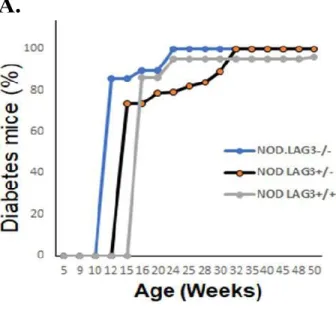
5.1 Loss of LAG-3 restores diabetes onset in NODSTAT5B+ mice ... 38
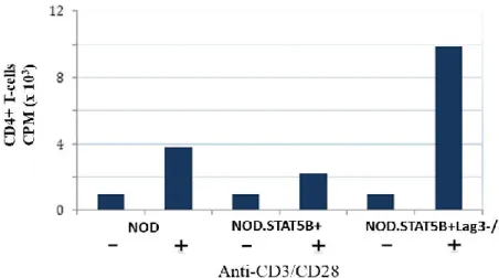
5.2 T-cells of NODSTAT5B+Lag-3-/- mice exhibit high proliferative response ... 40
5.3 Partial restoration of Treg pool in NODSTAT5B+Lag3-/-mice. ... 41
5.4 PD-1, CTLA-4 and LAG-3 expression levels in inactivated Tregs of NOD, NODSTAT5B+ and NODSTAT5B+Lag3-/- mice ... 43
5.5 CTLA-4 expression is substantially upregulated on Tregs upon activation in NODSTAT5B+ mice ... 45
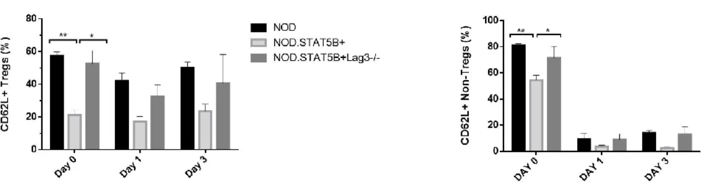
5.6 Lowered trend in expression of CD62L on Tregs and non-Tregs in NODSTAT5B+ mice 47 6. Discussion... 48
7. Conclusion ... 55
8. Acknowledgements ... 56
VI
List of figures
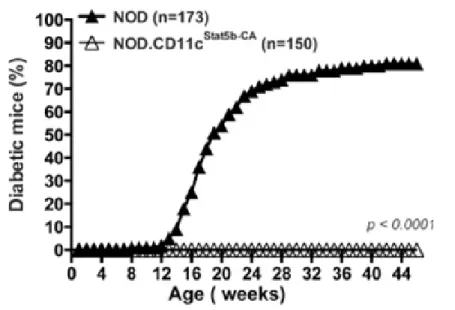
Figure 1. Increased expression of the active form of STAT5B in DCs of NODSTAT5B+ mice. ... 31 Figure 2. All NODSTAT5B+ are protected from T1D compared to NOD mice. ... 31 Figure 3. Increased percentage of Tregs in NODSTAT5B+ mice compared to NOD mice. ... 32 Figure 4. NODSTAT5B+ mice Tregs exhibit enhanced suppressive function compared to NOD mice Tregs. ... 33 Figure 5. NODSTAT5B+ and NOD mice monitored for diabetes development upon
being made heterozygotes or complete knockouts for LAG-3. ... 39 Figure 6. NODSTAT5B+ mice exhibit increased CD8+ T-cell and CD4+ T-cell
proliferation in the absence of LAG-3. ... 41 Figure 7. NODSTAT5B+ mice have increased Treg percentages compared to NOD and NODSTAT5B+Lag3-/- mice ... 42 Figure 8. No significant differences in basal PD-1, CTLA-4 and LAG-3 expression on Tregs in NODSTAT5B+ mice ... 44 Figure 9. CTLA-4 is significantly upregulated on Tregs following activation in
NODSTAT5B+ mice. ... 46 Figure 10. Lowered basal CD62L expression on Tregs and non-Tregs in NODSTAT5B+ mice. ... 47
VII
List of abbreviations
APC BB CA CCL CD CTL CTLA-4 CXCR DC DP EOMES ETP FITC GM-CSF HA HLA HSC ICOS IL IFN JAK KO LAG-3 LN MHC MLN mTEC NK NOD OVA PCR PD-1 PLN PTEN PTPN PKC RAG SHP SPAntigen presenting cell Biobreeding
Constitutively active CC chemokine ligand Cluster of differentiation Cytotoxic T-lymphocyte
Cytotoxic T-lymphocyte-associated protein CXC chemokine receptors
Dendritic cell Double positive Eomesodermin
Early thymic progenitors Fluorescein isothiocyanate
Granulocyte-macrophage colony-stimulating factor Haemagglutinin
Human leukocyte antigen Hematopoietic stem cell Inducible T cell costimulator Interleukin Interferon Janus kinase Knockout Lymphocyte-activation gene 3 Lymph node
Major histocompatibility complex Mesenteric lymph node
Medullary thymic epithelial cells Natural killer cell
Non-obese diabetic Ovalbumin
Polymerase chain reaction Programmed cell death protein 1 Pancreatic lymph node
Phosphatase and tensin homolog Protein tyrosine phosphatase Protein kinase C
Recombination-activating gene Small heterodimer partner Single positive
VIII STAT5 TCR TGF TIGIT TIM-3 TNF Tr1 TSLP WT ZAP70
Signal transducer and activator of transcription 5 T-cell receptor
Transforming growth factor beta
T cell immunoreceptor with Ig and ITIM domains T-cell immunoglobulin and mucin-domain containing-3 Tumor necrosis factor
Type 1 regulatory T cells Thymic stromal lymphopoietin Wild-type
1
Abstract
Type 1 diabetes (T1D) is an autoimmune disorder, where autoreactive cells are directed towards beta cells of the pancreas that produce insulin. This islet destruction and the chronic inflammation is facilitated by various immune cells specifically, macrophages, dendritic cells and T lymphocytes. We have previously shown that a dysfunction in the Treg pool and their suppressive function in NOD mice could be restored by GM-CSF or TSLP administration, which are recognized to activate the STAT5 signaling pathway. Since STAT5B dysfunction has emerged as a contributor to DC abnormalities that may contribute to Treg defect in T1D in NOD mice, we generated NOD.CD11c.STAT5B-CA transgenic mice expressing a constitutively active form of the STAT5B gene under the control of CD11c promoter. We observed that 100% of NOD.CD11c.STAT5B-CA transgenic mice were protected from disease, and protection was associated with an increased pool and suppressive function of Tregs. These data suggest that Treg development in STAT5B-CA.DCs environment leads to stable Tregs that are key cells in maintaining immune tolerance in NOD mice. Consistent with these findings, antigen-specific Tregs differentiated in vitro, in the presence of STAT5B-CA.DCs exhibited high suppressive function. Similarly, purified nTreg from NOD.CD11c.STAT5B-CA mice displayed high suppressive function compared nTregs purified from NOD mice. nTregs of NOD.CD11c.STAT5B-CA mice express high levels of suppressive inhibitory markers such as CTLA-4 when compared to nTregs of NOD mice. Checkpoint inhibitory molecules play a crucial role in optimal Treg suppression of autoreactive cells. Interestingly, NOD.CD11c.STAT5B-CA.LAG-3-/- mice become diabetic and have a drastic reduction of Tregs compared to NOD.CD11c.STAT5B-CA mice. These results suggest that Treg stability was restored in STAT5B-CA.DCs environment but lost in the absence of LAG-3. PD-1, CTLA-4 and LAG-3 checkpoint inhibitory molecules expressed on Tregs of NOD.CD11c.STAT5B-CA mice compared to pre-diabetic NOD and NOD.CD11c.STAT5B-CA.LAG-3-/- mice was characterized to provide an idea as to which may be more important in protection against disease. Interestingly, CTLA-4 was upregulated only on Tregs in NOD.CD11c.STAT5B-CA mice but not on Tregs of NOD and NOD.CD11c.STAT5B-CA.LAG-3-/- mice. Moreover, PD-1 and LAG-3 molecules were found to be upregulated only on non-Tregs (CD4+ T-cells) of NOD.CD11c.STAT5B-CA mice compared to NOD mice. These results suggest that CTLA-4 may be important for Treg mediated suppression of NOD.CD11c.STAT5B-CA mice, whereas PD-1 and LAG-3 expression may be important for certain CD4+ T-cell subsets in suppression of autoimmunity.
2
Résumé
Le diabète de type 1 (DT1) est une maladie auto-immune, dans laquelle les cellules autoréactives sont dirigées vers les cellules bêta du pancréas qui produisent de l'insuline. Cette inflammation chronique du pancréas et la destruction des îlots sont facilitées par diverses cellules immunitaires, notamment les macrophages, les cellules dendritiques et les lymphocytes T. Nous avions précédemment montré qu'un dysfonctionnement du pool et de la fonction suppressive des Tregs chez les souris NOD pourraient être rétablis par l'administration de GM-CSF ou de la TSLP, qui sont reconnues pour activer la voie de signalisation STAT5. Puisque le dysfonctionnement de STAT5B est devenu un facteur contributif des anomalies des cellules dendritiques (DC) pouvant contribuer à un défaut de Tregs chez les souris NOD, nous avons généré des souris transgéniques NOD.CD11cSTAT5B-CA exprimant une forme constitutivement active du gène STAT5B sous le contrôle du promoteur CD11c. Nous avons observé que 100% des souris transgéniques NOD.CD11cSTAT5B-CA étaient protégées contre la maladie et que cette protection était associée à une augmentation du pool et de la fonction suppressive des Tregs. Ces données suggèrent que le développement de Tregs en présence de STAT5B-CA.DC a conduit à des Treg stables qui jouent un rôle important dans le maintenance de la tolérance immunitaire chez les souris NOD. Conformément à ces découvertes, la différenciation in vitro des Tregs, spécifiques à un antigène du soi, en présence de STAT5B-CA.DCs, présentaient une fonction suppressive élevée. De façon similaire, les nTreg purifiés des souris NOD.CD11cSTAT5B-CA ont présenté une fonction suppressive élevée par rapport aux nTreg purifiés des souris NOD. Les nTregs des souris NOD.CD11cSTAT5B-CA expriment des niveaux élevés de marqueurs inhibiteurs tels que CTLA-4 par rapport aux nTregs des souris NOD. Les molécules inhibitrices du Tregs jouent un rôle crucial dans la suppression optimale des cellules autoréactives. Fait intéressant, 50% des souris NOD.CD11c.STAT5B-CA.LAG-3-/- deviennent diabétiques et présentent une réduction drastique des Tregs par rapport aux souris NOD.CD11cSTAT5B-CA. Ces résultats suggèrent que la stabilité des Tregs a été restaurée dans l'environnement STAT5B-CA.DCs mais perdue en l'absence de LAG-3. Les molécules inhibitrices PD-1, CTLA-4 et LAG-3 exprimées sur les Tregs et non Tregs des souris NOD.CD11cSTAT5B-CA ont été comparées aux souris pré-diabétiques NOD et NOD.CD11cSTAT5B-CA.LAG-3-/-. De façon intéressante, CTLA-4 est augmenté seulement chez les Tregs des souris NOD.CD11cSTAT5B-CA mais pas chez les Tregs des souris NOD.CD11cSTAT5B-CA.LAG-3-/-. De plus, PD-1 and LAG-3 sont régulés à la hausse seulement chez les cellules non-Tregs (CD4+ T cells) des souris NOD.CD11cSTAT5B-CA comparativement aux souris NOD. Nos résultats suggèrent que CTLA-4 serait important pour la fonction suppressive des Tregs des souris NOD.CD11cSTAT5B-CA alors que l’expression de PD-1 et LAG-3 par certaines populations de cellules T CD4+ seraient importants pour participer au contrôle des réactions autoimmunes.
3
1. Introduction
Type 1 diabetes (insulin-dependent) is a multifactorial autoimmune disorder characterized by pancreatic beta cell destruction, resulting in complete insulin deficiency. Type 2 diabetes (non-insulin dependent), on the other hand, is characterized by both resistance to insulin and failure in compensatory insulin secretion by the pancreas (Maahs et al. 2010). Although type 2 diabetic patients can go years without noticing any symptoms, type 1 diabetic patients may display symptoms suddenly and without warning. However, there is overlap between some of the symptoms of the two types of diabetes. Early common symptoms include hyperglycemia, polyuria, excessive thirst, weight gain or loss (more common in type 1 diabetes), blurry vision, fatigue and a tingling feeling of the extremities (Ramachandran 2014).
The most commonly used treatment regimen for type 1 diabetes includes combining strict diet and exogenous insulin administration (Pathak et al. 2019). Although various insulin delivery devices are in use, recurring episodes of hypoglycemia remains a challenge for patients and health care providers. Therefore, the use of continuous glucose sensors and infusion systems have become more popular. Additionally, this has paved way for the development of the artificial pancreas, also referred to as “closed-loop system”, that continuously screens blood glucose levels and injects only the needed amount of insulin (Pathak et al. 2019). This allows for improved blood glucose control resulting in reduced insulin spikes and hypoglycemia episodes. However, drawbacks of the closed loop system remain yet a challenge - including inconsistencies in outcome reporting, high costs, increased scar tissue caused by repeated microneedle injections and early sensor malfunction. Islet cell transplantation has also considerably improved in efficacy over the past 2 years, yet there are limitations including hazardous consequences of immunosuppressive drugs, islet cell apoptosis and ischemia that result in islet cell loss. Various immunotherapies have also been underway since the 1980s, yet repeatedly failed in clinical trials highlighting the urgency for improved immune therapies to treat patients successfully (Pathak et al. 2019).
4
1.1 Prevalence
Of the two major types of diabetes, type 1 accounts for 5-10% of the total cases. It accounts for the majority of cases seen in youths worldwide (Maahs et al. 2010). In a sample study of Canadian youth, the highest incidence age was observed in children aged 5 - 14 years where the most significant peak in incidence was between 10 - 14 years old (Fox et al. 2018). Epidemiological statistics reported Canadians diagnosed with T1D have been living with the disease for an average of 20.2 years, whereas type 2 averaged 12.2 years (Statistics Canada. Health Statistics Division 2017). In Canada, approximately 33,000 children between 5 - 18 years of age have type 1 diabetes, and several thousand younger than 5 years of age (The diabetic children's foundation 2019).
1.2 Mouse models of Type 1 diabetes
Mouse models of autoimmune diabetes have played a pivotal role in better understanding the pathogenesis and designing approaches to therapy. Although several rat models exist to study the disease, such as the Zucker Diabetic Fatty (ZDF) and BB rats, this section will mainly focus on mouse models in common use (Al-awar et al. 2016).
Mouse models used to study autoimmune diabetes can be broadly divided into two categories – humanized and spontaneous models. Humanized models are typically created by the introduction of genes such as those encoding for MHC, T-cell receptors and co-stimulatory molecules taken from humans. These models can aim to achieve the events initiating disease by transferring lymphocytes for example (von Herrath and Nepom 2009). A recent example of a humanized mouse model is the “YES” mice created by crossing mouse strains devoid of mouse MHC class I and II genes, and insulin genes (a known β-cell autoantigen). Instead, these mice express both human antigen presenting molecules and insulin genes as transgenes to illustrate autoantigen originating epitopes involved in disease initiation and heterogeneity in humans (Luce et al. 2018). Thereby, humanized models offer the advantage of a more detailed picture of in vivo immunoregulation of particular antigens presented in the framework of humanized antigen presenting modules, among other genes (von Herrath and Nepom 2009).
5 Spontaneous mouse models of autoimmune diabetes can ensue from pathway dysfunctions, insertion of transgenes or precisely known genetic mutations (von Herrath and Nepom 2009). For instance, double transgenic mouse models are made to carry transgenic expression of a specific antigen under the control of the insulin promoter (Rat Insulin promoter: RIP) on their pancreatic β-cells, along with TCR transgenes specific for that antigen. An example is the DO11.10 TCR-Tg×RIP-mOVA model, in which ovalbumin is expressed as the autoantigen within the pancreas, which along with the DO11.10 TCR transgene allows for the expansion of highly diabetogenic OVA-specific CD4+ T-cells. These mice become diabetic by 10 weeks of age and offer the advantage of focusing on a specific T-cell subset (van Belle, Taylor, and von Herrath 2009). Another popularly used model to study autoimmune diabetes is the NOD mice, due to its many similarities to the human form of disease. Diabetes initiation in NOD mice is rapid and disease progression is responsive to immune modulation (von Herrath and Nepom 2009). The similarities to the human form of disease are so striking that this model has been used for over 35 years by researchers. In addition, the model allows intrusive procedures and procurement of tissues that are seldom obtained from human patients making it a cornerstone for autoimmune diabetes research (Chen, Mathews, and Driver 2018).
1.2.1 NOD mouse model
The NOD mice serve as a preclinical model to autoimmune diabetes that outlines the immune pathogenesis closest to disease in humans. In addition to striking similarities in disease progression, the model provides a deeper understanding on genetic and environmental risk factors and therapies targeting various immune pathways. Yet, researchers noticed major differences in histopathology in patients and NOD mice, and challenges persist in translating therapies from NOD mice to humans (Zeng 2017).
NOD mice spontaneously develop T1D, unlike the majority of animal models that do not spontaneously develop autoimmunity. Both NOD mice and humans become diabetic early on, with NOD mice at around 10 weeks and humans around 6 months of age up to adolescence (Pearson, Wong, and Wen 2016). Genetic susceptibility genes, mainly
MHC-6 related genes are similar amongst both species. Autoantigens including GAD, insulin, ZnT8, IA-2 and IGRP are alike amongst NOD mice and humans. Both species do not exhibit lymphopenia, unlike the BB rat, highlighting one of the many reasons NOD mice have become more popularly used to study pathogenesis of T1D. Immune cell infiltration of pancreas leading to insulitis is characterized by DCs, macrophages, NK cells, B-cells, CD4 and CD8-T cells in both species (Pearson, Wong, and Wen 2016). Additionally, neither species show a TCR repertoire bias that can be implicated in pathogenesis. However, ketoacidosis, a serious side effect of diabetes, is more severe in humans compared to NOD mice. Moreover, there is a sex bias, where in NOD mice, a majority of females become diabetic, whereas in humans prior to puberty, there is no bias but after puberty, there is a slight skew towards males developing the disease. To address some of these differences, researchers are developing humanized NOD mouse models to better translate therapies to humans. Nevertheless, NOD mice in general and humanized NOD mice have and will continue to provide important discoveries into disease pathogenesis in humans (Pearson, Wong, and Wen 2016).
1.3
Contribution of genetic and environmental factors in T1D
Both genetics and environmental risk factors are known to play a role in autoimmune diabetes development. Recent investigations propose an increasing role for environmental factors (Kozhakhmetova and Gillespie 2015). Moreover, the considerable rise in T1D incidence over the past 30 years can only be explained by environmental or lifestyle changes (Rewers and Ludvigsson 2016a).
1.3.1 Genetic Factors
The MHC gene makes up the highest susceptibility factor contributing to T1D development, in both NOD mice and humans. MHC-II molecule I–Ag7, ortholog of
HLA-DQ in humans expressed by NOD mice plays the most significant role in diabetes development, compared to other MHC-related molecules (Pearson et al. 2016). A polymorphism in MHC-II gene, where serine at position 57 of I–Ag7 in NOD mice is
7 substituted by a charged aspartate residue, is also observed in human HLA-DQ, where alanine is substituted in the place of aspartate instead. When serine is replaced at position 57 with aspartate (as found in wild-type diabetes resistant mice), NOD mice are protected from T1D. Similarly, the DR3/4-DQ2/8 haplotypes are highly associated with susceptibility to diabetes in humans and protection ensues with other DR and DQ alleles (Pearson et al. 2016). Another important genetic susceptibility factor in humans includes the expression level of proinsulin in the thymus. Protection or susceptibility to disease is associated with the number of variable numbers of tandem repeats (VNTRs), upstream of the proinsulin gene (Pearson et al. 2016). For instance, 140-200+ repeats is correlated with increased thymic proinsulin and disease protection, whereas 26-63 repeats is correlated with decreased expression of proinsulin within the thymus and increased disease susceptibility (Pearson et al. 2016). When NOD mice were designed with insulin levels to mirror human proinsulin expression within the thymus, it was observed that increased thymic proinsulin expression led to greater negative selection and deletion of insulin autoreactive T-cells during their development associated with T1D protection (Pearson et al. 2016).
Additionally, variants in genes such as CTLA-4 and PTPN22 also contributes to increased disease risk (Pociot et al. 2010). CTLA-4, a checkpoint inhibitory molecule, that inhibits T-cell activation was characterized by association mapping using both NOD mice and human samples. Disease-linked CTLA-4 haplotypes likely inclines the individual towards a failure in peripheral tolerance in various organs or tissues leading to several known human autoimmune diseases (Pociot et al. 2010). Similarly, gene variants resulting in amino acid substitution of the gene encoding for lymphoid-specific protein tyrosine phosphatase (PTPN22) an inhibitor of T-cell activation, have been shown to affect its functionality in
vitro and in vivo, leading to increased susceptibility to T1D as well as to other autoimmune
diseases (Pociot et al. 2010).
1.3.2 Environmental Factors
Early epidemiological case studies have linked viruses as a probable trigger for T1D. Enteroviruses, among other viruses, have been linked as the strongest cause for pathogenesis
8 in both animal models and humans. Enteroviruses have been found in the pancreas of recently diagnosed T1D patients and possess tropism to islets both in vivo and in vitro (Rewers and Ludvigsson 2016b).
Dietary factors play a key role in pathogenesis of T1D induction. For instance, children breastfed around the time they were introduced to cereals had a decreased risk of developing disease, supporting a protective role of breastfeeding (Rewers and Ludvigsson 2016b). Studies investigating children drinking cows’ milk and its contribution to pathogenesis have yielded conflicting results, showing both increased and decreased risk for disease development and progression. In children at earlier stages of diabetes, increased cows’ milk consumption may promote disease progression. This is thought to be caused by specific fatty acids in cows’ milk and meat such as myristic, omega-7, 9, 16:1 and others (Virtanen et al. 2010). Vitamin D is a compound studied extensively as a major player in immune regulation and metabolic pathways involved in diabetes. However, birth cohort investigations have found very little evidence of vitamin D supplementation in protection against disease initiation or progression (Rewers and Ludvigsson 2016b).
Studying specific environmental triggers in autoimmune diseases presents a challenge as there are infinite candidate factors. Hence, the NOD mice model serves as a useful tool to study exactly how these environmental factors act in disease development. Some of the factors implicated in diabetes development in NOD mice include alterations in gut microbiota, exposure to wheat, gluten as well as to infectious agents (Pearson et al. 2016). Presently, a significant international study called TEDDY (The Environmental Determinants of Diabetes in the Young) is on-going, tracking young children over time to better understand the role of particular environmental risks in disease development (Anon 2007).
1.4 The immune system in Type 1 Diabetes
Destruction of pancreatic β-cells is a result of immune dysfunction, where both parts of the innate and adaptive immune system have been implicated. The past ten years have highlighted the role of cells in mediating β-cell damage. In healthy individuals, these T-cells are strictly controlled by various regulatory mechanisms, generally referred to as
9 “immunological tolerance.” Therefore, a break in immune tolerance plays a major role in disease development and progression (Hull, Peakman, and Tree 2017).
1.4.1 General Pathophysiology
Death of pancreatic β-cells due to a trigger such as viruses for instance, releases antigens and initiates the immune reaction towards other β-cells, resulting in ongoing inflammation of islets and eventually clinical manifestations of disease. Typically, APCs such as DCs and macrophages uptake these β-cell antigens and present them to CD4+ and CD8+ T-cells (Saberzadeh-Ardestani et al. 2018). In the periphery, autoreactive T-cells that have escaped negative selection in the thymus are activated and mediate β-cell killing by signals that include proinflammatory cytokine secretion and extrinsic apoptotic pathways involving Fas-FasL interactions of T-cells, increasing β-cell sensitivity to apoptosis (Paschou et al. 2018). In addition, cytokines secreted by APCs such as macrophages also harm β-cells, further sensitizing them to killing. Before T1D onset, persistent atrophic inflammation of the islets is detected, where T-cells, DCs, macrophages and B-cells are implicated. At this stage, patients are often asymptomatic and once this condition continues over several months or years, clinical manifestations of symptoms are observed. Several autoantibodies towards β-cell antigens can also be present prior to clinical symptoms in patients (Paschou et al. 2018). Overall, β-cell killing by effector mechanisms involves the combined co-operation of various APCs such as DCs, macrophages, B cells, as well as other members of the innate immune system such as NK cells and importantly, adaptive immune cells CD4+ and CD8+ T-cells (Saberzadeh-Ardestani et al. 2018). As gatekeepers of the immune system, Tregs function by various mechanisms to primarily suppress the inflammatory processes mediated by other immune cells (Hull et al. 2017). The specific roles of key immune cells involved in disease initiation and progression will be discussed in detail in the following sections.
1.4.2 Role of Antigen Presenting Cells (APCs)
It is well established that presentation of self-antigens by APCs to developing thymocytes or peripheral T-cells is necessary for all mechanisms leading to tolerance, either through deletion, anergy or Treg induction. T-cell responses are partly controlled by APCs
10 that upon interaction can inhibit inflammatory immune responses. Professional APCs such as DCs, macrophages and B cells are most widely investigated, and these APCs are a two-edged sword in the context of T1D as they can be primed towards either pro-inflammatory or anti-inflammatory immunogenic operations (Creusot, Postigo-Fernandez, and Teteloshvili 2018).
1.4.2.1 Role of Dendritic Cells (DCs)
DCs are professional antigen presenting cells that are characterized by phenotype heterogeneity endowed with different immune functions. Main functional DC subpopulations are conserved between mice and humans. Abnormal DC activation is enough to cause autoimmunity and cell-intrinsic mechanisms exist to negatively regulate DC activation to prevent autoimmunity. DCs can prime naïve CD4+ and CD8+ T-cells, which depending on the immune milieu, can promote or restrict autoimmunity. It has been shown that DCs can prime naïve CD4+ T-cells to become T-follicular helper (Tfh) cells that are implicated in systemic lupus erythematosus (SLE) (Ganguly et al. 2013). Similarly, DCs can prime naïve CD4+ T-cells to become Th17 cells implicated in psoriasis and experimental autoimmune encephalomyelitis (EAE) (Ganguly et al. 2013). In T1D, DCs can prime naïve CD4+ T-cells to become Th1 cells that are heavily implicated in disease development, or conversely, to Tregs that can result in disease restriction (Ganguly et al. 2013). DCs can also interact with autoreactive CD8+ T-cells to induce their proliferation and to increase their effector activity that contributes to disease initiation and progression. (Ganguly et al. 2013). DCs play central roles during all stages of T1D pathogenesis as key activators of naïve T-cells and in upkeeping self-tolerance. DCs within the pancreas uptake β-cell autoantigens and migrate to the pancreatic draining lymph nodes (LNs). In the LN they can prime naïve CD4+ and CD8+ T-cells, which, conditional on signals received by DCs, can differentiate into Th1 inflammatory effector subsets or Th2 anti-inflammatory or Tregs (Morel 2013). Activated T-cells then infiltrate the islets and perform their respective functions. There are two main subsets of DCs – plasmacytoid and classical. Both subsets have disease inducing and protective roles. Classical DCs (cDCs) contain certain subpopulations, such as merocytic DC, that can present autoantigens and home in inflamed
11 islets, propagating disease (Morel 2013). These DCs can release large amounts of IL-12 that promote Th1 responses and induce more pro-inflammatory cytokine secretion by T-cells and NK cells. TNF-α and IL-1β production by these subpopulations of cDCs can also induce DC maturation and result in a breakdown of immune tolerance (Morel 2013). Finally, they can also cross-present antigen to CD8+ T-cells resulting in increased effector T-cell activity. Protective subsets of cDCs such as the CD11b subset can be responsible for Treg induction, as well as interacting with thymic Tregs to influence their migration, and PD-1/PD-L1 interaction with T-cells resulting in T-cell anergy. Plasmacytoid DCs (pDCs) are high producers of type 1 interferons (IFN). In NOD mice, it has been shown that crosstalk between B-1a B cells, neutrophils and pDCs is involved in T1D induction through IFN production (Diana et al. 2013). However, it has been proposed that viral infection leading to T1D is dependent on virus tropism and the local milieu where IFN in produced. Thus, on one hand a pancreatic virus can stimulate IFN production protecting islets whereas, on another hand, IFN production by pDCs can activate autoreactive T-cells and result in T1D progression. Additionally, in agreement with pathogenic role pDCs in T1D, it was observed that pDC depletion decreased diabetes incidence in NOD mice (Morel 2013a).
DCs capacity to be manipulated, to re-establish T-cell tolerance and moderate autoantibody production has made them attractive therapeutic targets. A better understanding of DC key functions in autoimmunity can help enhance their tolerogenic functions. DCs of a semi-mature phenotype exhibiting decreased pro-inflammatory cytokine secretion but increased co-stimulatory and MHC molecules expression are considered efficiently tolerogenic in protection against autoimmunity. Additionally, tolerogenic DCs function to induce Treg cells or other regulatory mechanisms such as Th2 cells, including their expansion (Morel 2013a). Tolerogenic DCs have already began to be used in clinics, however effective translation of various other DC-based immunotherapies yet needs substantial research (Coutant and Miossec 2016).
1.4.2.2 Role of Monocytes/Macrophages
Macrophages are known to play important roles in T1D pathogenesis. They take up and present autoantigens to T-cells, regulate T-cell responses and CD4+ T-cell
12 differentiation into various T helper subsets by secreting cytokines, thereby influencing immune homeostasis (Espinoza-Jiménez, Peón, and Terrazas 2012).
Emerging studies suggest that macrophages are also implicated in the later stages of T1D. For instance, transferred diabetogenic T-cells (lymphocytes directly or indirectly responsible for β-cell death due to their antigen specificity, migratory capacity and ultimately cytotoxic function) were unable to cause diabetes post monocyte depletion, and it was shown that in vitro activated macrophages can directly destroy β-cells (Martin et al. 2008), (Roep and Peakman, 2011). Nevertheless, evidence for the latter is minimal and is still under investigation One study using several transgenic mouse models showed that monocytes can home to the islets when CCL2 chemokine is expressed in β-cells, killing β-cells and leading to diabetes, without mature T- and B- cell involvement (Martin et al. 2008). Despite the fact that diabetogenic T- and B-cells are key players in the pathogenesis of T1D in physiological environment, the study by Martin et al. (2008) and others have implicated monocytes/macrophages to play also a crucial role in pathogenesis. CCL2 is a key chemokine molecule responsible for recruiting monocytes and other immune cells to sites of inflammation. One important implication of the mentioned study is targeting CCL2 in APCs to decrease recruitment of autoreactive cells that home to sites of ongoing inflammation. In fact, it has been observed that anti-CCL2 therapy can oppose other autoimmune diseases such as lupus nephritis and murine demyelinating disease in animal models and several CCL2 inhibitors are currently under development (Yang 2008). Challenges are present in translating this therapy to humans, and investigations with respect to redundancy in chemokine pathways, in terms of the impact of modifying one chemokine pathway is yet to be determined (Yang 2008).
1.4.2.3 Role of B-cells
B-cells constitute the immune cell infiltrates in pancreatic islets in both mice and humans. In NOD mice, B-cells were found during the 4-7 weeks, age at which about 10% of islets are infiltrated. The role of B-cells in pathogenesis of T1D has been theorized to be most likely multifaceted, composing of faulty B-cell tolerance that permits autoreactive cells to escape and persist in the periphery, and remain activated well before the effector phases of
13 T1D (Hinman and Cambier 2014). B-cells, as APCs play a primary role in presenting β-cell autoantigens to diabetogenic T-cells. It was reported that in NOD mice, inhibition of antigen presentation by MHC-I or MHC-II on B-cells averts T1D development, highlighting the importance of B-cells as antigen presenters to CD4+ T-cells and cross-presentation to CD8+ T-cells (Noorchashm et al. 1999). They also exert effector mechanisms via the secretion of cytokines and autoantibodies towards islet autoantigens.
Although T1D is considered to be mainly a T-cell mediated disease, there has been considerable evidence highlighting a crucial role of B-cells in pathogenesis. In NOD mice, depletion of B-cells prevents diabetes development and additionally, depletion of B-cells in recently diagnosed T1D patients using the monoclonal antibody rituximab (chimeric mouse/human monoclonal IgG1 kappa antibody targeted against the CD20 antigen), conserved β-cell function and postponed the need for insulin administration 1 year post treatment (Smith, Simmons, and Cambier 2017). Overall these results suggest a considerable role of B-cells in disease development (Smith, Simmons, and Cambier 2017). As therapy, depleting B-cells will mostly not be distributed in clinical use and alternative minimal risk B-cell targeted therapy is still undergoing investigation (Hinman and Cambier 2014).
1.4.3 Role of T-lymphocytes
Autoantibodies characterize biomarkers used for T1D diagnoses and autoreactive T-lymphocytes are considered critical mediators of β-cell killing. Approximately 50-60% of genetic predisposition to T1D comes from HLA alleles that encode genes responsible for antigen presentation of peptides to T-cells (Pugliese 2017). Examination of T1D organ donor pancreas initially showed evidence of shared TCR sequences against target antigens that are shared amongst patients, as well as a wide repertoire of self antigens implicating the TCR in the insulitis lesions within the pancreas. Although both CD4+ and CD8+ T-cells are implicated in pathogenesis, CD8+ T-cells constitute the majority of T-cells that mediate inflammation seen in insulitis (Pugliese 2017).
14 1.4.3.1 T-cell origin and development
In mammals, T-cell development starts with haematopoietic stem cells (HSC) within the fetal liver and afterwards in the bone marrow where they differentiate into multipotent progenitors. Upon initiating transcription of recombination activating gene 1 and 2 (RAG 1 and 2), a subclass of multipotent progenitors develops into lymphoid-primed multipotent progenitors and then to common lymphoid progenitors (Cano and Lopera. 2013). Amongst all pluripotent cells, only a small portion will migrate to the thymus and differentiate into early thymic progenitor (ETP) cells. These ETP cells are multipotent, and can become T-cells, B-T-cells, NK T-cells, myeloid cells and DCs. Long-term thymopoiesis is solely dependent on the recruitment of thymus-inhabiting progenitors since the thymus itself does not encompass self-renewing progenitors. T-cell development to maturity is therefore conditional on progenitors entering the thymus within specific niches that allow compulsory stage-specific events needed for differentiation of progenitors to be reprogrammed into functional T-cells (Cano and Lopera. 2013).
ETP cells that are attracted by CCL9 and CCL21 chemokines come into the thymus by the corticomedullary junction (Cano and Lopera. 2013). Within the thymus stroma, the ETP come across several ligands for the notch receptors and growth factors such as Kit-ligand and IL-7, that aid in differentiation of these cells in the early stages of T-cell development. In fact, expression of notch-1 receptors and their contact with delta-like ligands is necessary for the expansion of T-cells within the thymus and for inhibiting non-T cell lineage maturation. ETP differentiate into double negative (DN) cells in the thymic cortex, i.e. they are both CD4- and CD8-. During this stage, ETP lose the ability to become B-cells and initiate expression of proteins necessary for the TCR gene arrangement like RAG1 and 2 (Cano and Lopera. 2013). Subsequently, proteins critical for TCR construction and signaling are also expressed, such as the CD3 chains, kinases, and phosphatases like LCK, ZAP70 and LAT. During the DN3 stage, there is cell-cycle arrest which allows for the rearrangement of the TCR β genes encoding for the variable region of the antigen receptors. TCR β genes that have been productively rearranged through V(D)J recombination, are then coupled to a surrogate chain Tα, which forms the TCR. Signaling through the pre-TCR allows cells to survive, proliferate and differentiate, in a process termed β selection.
15 Following the β selection checkpoint, thymocytes go on to become double positive (CD4+CD8+) cells, that undergo TCRα rearrangement, leading to an entirely assembled TCR (Cano and Lopera. 2013). Following the DP stage, cells undergo positive selection, where cells with functional TCRs are put to the test by their ability to bind self-peptides presented by epithelial cells via MHC-1 or MHC-II with intermediate affinity. Positive selection permits differentiation of DP thymocytes to single positive (SP) cells, restricted to their respective MHC classes. DP thymocytes unable to bind MHC-I or MHC-II go through apoptosis and only a subset of the DP cells expressing TCR with intermediate affinity towards the self-antigens live via survival signals. Afterwards, SP cells undergo negative selection upon entering the thymic medulla where medullary thymic epithelial cells (mTEC) and DCs present a variety of self-antigens to developing thymocytes. Genes expressed by mTECs include many tissue-restricted self-antigens. SP cells reacting with high affinity for binding self-antigens presented in the context of MHC-I or MHC-II die by apoptosis, thereby securing the death of possible autoreactive cells. Upon surviving negative selection, cells mature and complete their development into naïve T-cells. Naïve T-cells can then exit the thymus and migrate to the secondary lymphoid organs where they can encounter antigens, be primed and differentiate into effector T-cells with unique functionalities (Cano and Lopera. 2013).
1.4.3.2 Implication of CD4+ T-cells in T1D
Although CD8+ T-cells are primarily implicated in islet infiltration and inflammation, CD4+ T-cells are involved in ‘licencing’ CD8+ T-cells to be optimally activated to perform effector function (Katz, Benoist, and Mathis 1995). Additionally, CD4+ T-cells can differentiate into various T helper subsets after activation, and this choice has critical effects on their ensuing cytokine production and migration. Differentiation into a specific subtype also determines the nature of interaction between T-cells and other immune cells, thereby directly influencing effector functions. T-cell differentiation was mainly focused on T helper 1 (Th1) and T helper 2 (Th2) responses in terms of T1D, which was primarily seen as a Th1-mediated disease. However, over the years this view has expanded to include various other subsets such as Th17 and follicular T-helper cells (Tfh) (Katz, Benoist, and Mathis 1995).
16 Early evidence that T1D is a Th1 mediated disease came from a study showing that T-cells expressing a diabetogenic TCR differentiating towards Th1 and not Th2 phenotype, caused diabetes in neonatal NOD mice (Katz et al. 1995). Considering the Th1 phenotype includes IFN-γ dominated responses, rising levels of IFN-γ was observed to be correlated with T1D progression in NOD mice, and appeared to be a necessity in a virus-induced diabetes model (von Herrath and Oldstone 1997). Numerous studies have highlighted a role for IFN-γ in disease progression (Walker and von Herrath 2016). For instance, IFN-γ expression under control of human insulin promotor was enough to initiate diabetes development in mice, and IFN-γ inhibition in NOD mice could avert disease (Sarvetnick et al. 1988). Mechanisms of action of IFN-γ include up-regulation of MHC class I and II, increasing macrophage activation, stimulating adhesion molecule and chemokine expression to drive leucocyte extravasation, increasing the homing of autoreactive T-cells to pancreatic islets in NOD mice, and directly killing β-cells (Walker and von Herrath 2016). It is known that Th2 responses inhibit Th1, resulting in the ability of one T-cell subset reciprocally preventing the functionality of the other. As IL-4 is predominantly a Th2-type response, exogenous administration of IL-4 prevented diabetes in NOD mice, and transgenic expression of IL-4 under control of the insulin promoter in the pancreatic islets inhibited diabetes initiation (Rapoport 1993). Interestingly, helminth disease, known to promote Th2 response was shown to be protective against diabetes in NOD mice (Zaccone and Cooke 2013).
One important area of study of Th17 cells in T1D is the role of these cells to simultaneously produce IL-17 and IFN-γ. CD4+ T-cells secreting IL-17 has been implicated in many rodent disease models highlighting an importance for these cells in disease pathways in humans (Arif et al. 2011). For example, in multiple sclerosis, IL-17 gene expression is upregulated in plaques of the brain and IL-17 is expressed at increased levels by circulating T-cells. Similarly, Th17 cells that infiltrate synovial tissue is highly implicated in rheumatoid arthritis, where IL-17 promotes osteoclast-facilitated bone resorption (Arif et al. 2011). In human T1D, IL-17 has been directly linked to β-cell death. Pro-inflammatory cytokines such as IL-1β and IFN-γ cause upregulation of the IL-17 receptor which leaves β-cells greatly susceptible to apoptosis by IFN-γ, IL-17, IL-1β cytokines (Arif et al. 2011). T-cells positive for IL-17 can instigate IFN‐γ secretion leading to T-cells co-expressing both cytokines.
17 Several evidences exist to support a role of these cells in pathogenesis of T1D, including children exhibiting co-production of both cytokines, and mRNA levels of both cytokines being up-regulated within the pancreatic islets of a patient who died 5 days within disease diagnosis (Arif et al. 2011). Studies have demonstrated an increased tendency of IL-17+ T-cells to produce IFN‐γ in children diagnosed with T1D compared to healthy individuals (Walker and von Herrath 2016). Cumulatively, these studies warrant further investigation of T-cells co-producing IL-17 and IFN‐γ in relation to disease progression.
Tfh cells have also been implicated in diabetes in both humans and mouse models of T1D. Tfh cells secrete mainly IL-21, and this subset specifically helps B-cells in antibody production. As their name suggests, Tfh cells go into B-cell follicles of secondary lymphoid tissue where they begin the formation of specialized structures known as germinal centers (Walker and von Herrath 2016). Within these germinal centers, B-cells re-arrange their immunoglobulin molecules so that ones with the highest affinity for the antigen can be designated to become memory B-cells and long-lived plasma cells (Walker and von Herrath 2016). Microarray analysis of T-cells towards islet antigens in the pancreatic lymph nodes revealed an increase in Tfh cell specific genes including CXCR5 and IL-21 (Walker and von Herrath 2016). Adoptive transfer of CXCR5 enriched T-cells were shown to preferentially induce diabetes in mice (Walker and von Herrath 2016). In humans, increased Tfh phenotype, IL-21 production after ex-vivo activation of memory T-cells and in the blood circulation was reported in T1D patients compared to non-diabetic healthy controls (Walker and von Herrath 2016). The consequences of IL-21 production by CD4+ T-cells include facilitating their survival, increasing the ability of macrophages ability to stimulate CD4+ T-cell proliferation and by opposing Treg suppressive function. Additionally, IL-21 has also been critically implicated in T- B cell interactions which may be pertinent in the role of B-cells in disease (Walker and von Herrath 2016). Since IL-21 can be produced by other T-cell populations including Th17 cells and induced regulatory Tr1 type cells, the involvement of non-Tfh subsets in disease progression cannot be omitted. However, one study showed a strong significant correlation between Tfh frequency and IL-21+ T-cells, suggesting a crucial role of Tfh cells as major sources of IL-21 in T1D (Ferreira et al. 2015).
18 To elucidate the mechanisms by which CD4+ and CD8+ T-cells collaborate in islet β-cell killing, one study used two-photon microscopy in-vivo to follow hemagglutinin (HA) specific CD4+ and CD8+ T-cells within mice pancreatic β-cells expressing the HA antigen (Espinosa-Carrasco et al. 2018). They found that infiltrating CD4+ T-cells alone were enough to recruit APCs in the exocrine tissue that express increased MHC class II (mostly consisting of CD103+ DCs), leading to increased cell arrest in tissue. Effector CD8+ T-cells that lacked persistent help by APCs via anti-MHC II antibody treatment were unable to sustain optimal effector functions and exhibited decreased infiltration, uniquely in the pancreas thereby impeding disease progression. These results highlight a critical role of effector CD4+ T helper cells in continually helping effector CTL functions at target environments via APCs (Espinosa-Carrasco et al. 2018).
Protective subtypes of CD4+ T-cells such as Tregs and Tr1 cells with suppressive function have been shown to downregulate immune responses and protect against diabetes. Tregs are considered the gatekeepers of autoimmunity by keeping unwarranted responses at bay to inhibit disease development. Thorough characterization of Tregs including their generation in vitro and in vivo serves as a whole new venue to treating autoimmune diseases (Varela-Calvino et al. 2017). The mechanisms of action of these cells will be discussed in further detail in later sections.
1.4.3.3 Implication of CD8+ T-cells in T1D
CD8+ T-cells or cytotoxic T-lymphocytes (CTLs) specific towards insulin or other autoantigens are crucial for T1D development in both humans and mice. CTLs that recognize autoantigens presented via MHC-I kill β-cells using various mechanisms including via the release of perforin and granzyme (Burrack, Martinov, and Fife 2017). In diabetic patients, most of the infiltrating cells of the pancreas are CTLs, along with overexpression of MHC-I on β-cells. CTLs are also known to produce elevated amounts of IFN-γ that upregulate Fas expression on beta cells and therefore enabling CTLs to use Fas/FasL mechanisms to destroy β-cells. Naïve CD8+ T-cells recognize antigens presented by APCs and become activated. The proliferation, survival and development of naïve CD8+ T-cells as they differentiate into CTLs is promoted by various factors including IL-12, IFN-α, IFN-β, IFN-γ, and mTOR (Deng et al. 2019). Eomes and T-bet transcription factors enhance the expansion of memory
19 CD8+ T-cells and their effector function (Pearce et al. 2003). Studies suggest T-bet and Eomes function collaboratively to activate genes encoding for IFN-γ, perforin and granzyme in CTLs (Pearce et al. 2003). Eomes+/−Tbx21−/− (Tbx21 is a Th1-cell specific transcription
factor that mediates the expression of the key Th1 cytokine, IFN-γ) mice have dysfunctional IFN-γ induction and effector CD8+ T-cell activity (Pearce et al. 2003). Upon CTL killing of target cells, there is a release of several autoantigens which leads to increased production of autoantibodies that can result in self-apoptosis and increased local inflammation. Thus, CD8+ cell interaction with β-cells causes β-cell killing and an increase in autoreactive T-cells within islets (Deng et al. 2019).
Studies have demonstrated a role of micro-RNAs (miRNAs) in CD8+ T-cells with regards to gene silencing in T1D. Using adoptive transfer models of T1D, it has been shown that downregulating miR-29b reduced CD8+ T-cell cytolytic functions (Deng et al. 2019). Certain miRNAs such as miR-23b, miR-98 and miR-590-5p can decrease the expression of crucial apoptotic molecules, like Fas and TNF-related apoptosis-inducing ligand (TRAIL), suggesting they can act to modify T1D susceptibility in patients. Studies in epigenetic regulation of CD8+ T-cells in autoimmunity, and developing relevant therapeutic interventions are ongoing (Deng et al. 2019).
Recent investigations have reported a subset of CD8+ T-cells playing a suppressive role in autoimmunity, tumors and infections (Deng et al. 2019). These cells are characterized by the expression of the Treg specific master regulatory gene forkhead box p3 (Foxp3), the high affinity IL-2 receptor CD25, and inhibitory molecules such as PD-1, CTLA-4 and LAG- 3. These CD8+ Tregs can suppress effector T-cell responses in vitro in a CTLA-4 dependent manner. However, compared to CD4+ Tregs, CD8+ Tregs are less commonly studied and further investigation is required for their mechanisms of action (Deng et al. 2019).
1.4.4 Role of Regulatory T-cells
Regulatory T-cells (Tregs) are key immune suppressor cells that maintain peripheral tolerance. Decreased Tregs and/or their impaired suppression can lead to autoimmunity. Growing evidence in mouse models of various autoimmune disorders, including T1D, have
20 shown impairment in Treg suppressive function. Although in human diabetic patients, the frequency of Tregs in peripheral blood is normal, studies have shown these Tregs to have decreased suppressive function compared to Tregs of healthy controls (Visperas and Vignali 2016a).
There are two main classes of Tregs– natural or thymic-derived Tregs (nTregs) and inducible Tregs (iTregs) that can be differentiated in vitro via cytokines such as 10, IL-35 and TGF-β (Vignali 2012). This section will discuss various mechanisms by which Tregs suppress immune responses, highlighting the role of checkpoint inhibitory molecules, and how overall Treg suppression is impaired in T1D.
1.4.4.1 Specificity of Regulatory T-cells
Tregs are hallmark cells required to maintain self-tolerance, as well as towards unharmful, regular environmental antigens encountered at mucosal surfaces day by day (Bacher et al. 2016). Human target Treg antigens that promote mucosal tolerance are currently undergoing investigation. In mice, Treg specificity is largely influenced by self-antigens, and Tregs require continuous stimulation through the TCR by self-antigens to inhibit autoimmunity. Yet, exogenous antigen specific Tregs have been identified in naïve and pathogen-infected mice, as well as humans (Bacher et al. 2016). Murine Tregs specific for microbiota antigens were detected in intestinal tissues, and additionally, murine Tregs maintain tolerance towards fetal antigens during pregnancy. This provides evidence that Tregs are not solely a self-responsive population and they exist to also promote tolerance towards innocuous exogenous antigens. However, studies of the function of Treg tolerance towards aeroantigens is lacking and requires investigation (Bacher et al. 2016). One study by Spence et. al (2018) showed an accumulation of Tregs in inflamed islets, experienced in self-antigens, using NOD mice. Although progress has been made in mapping the specificity of autoreactive T-cells in T1D, Treg specificities that influence disease progression have not been established. Difficulties in mapping Treg specificities is partially due to the naturally extensive Treg repertoire and the comparative infrequency of islet antigen-specific Tregs. The challenge in measuring Treg suppressive function in diabetes protection in vivo also compounds the complexity of determining Treg specificity.
21 Interestingly, the group of Spence et. al (2018) performed MHC tetramer staining and single-cell TCR analysis of Tregs in islets of NOD mice to determine their specificity for insulin and various islet-derived antigens. NOD mice were found to be enriched in insulin-specific antigens in inflamed islets, an unexpected finding regarding the increased localization and prominence. It was shown that diabetes-susceptible recipients of Tregs from inflamed islets were protected from disease progression, although NOD mice eventually succumb to disease most likely due to IL-2 scarcity within inflamed islets and exhaustion of Tregs specific for islet-specific antigens. Overall, engineering Treg specificities based on insulin specific TCRs can be used as treatment for protection of β-cells in T1D, highlighting an important field of study regarding the specificities of Tregs in the context of autoimmunity (Spence et. al 2018).
1.4.4.2 Mechanisms of Suppression
CD4+CD25+Foxp3+ Tregs are key mediators of peripheral tolerance as they can suppress various immune cells such as B cells, NK cells, NKT cells, monocytes, DCs, CD4+ and CD8+ T-cells.
In order for Tregs to optimally suppress immune cells, Tregs have to be activated through their TCR in the presence of IL-2, although co-stimulation by CD28 is less stringent of a requirement (Schmidt, Oberle, and Krammer 2012). The majority of nTregs constitutively expresses the high affinity IL-2 receptor chain CD25 during their development in the thymus and their functionality is dependent on the expression of the transcription factor Foxp3. Primarily, Tregs were shown to have suppressive function when their depletion resulted in CD4+ T-cell mediated autoimmunity (Miyara and Sakaguchi 2007). Treg suppressive mechanisms that are best studied include the secretion of inhibitory cytokines, expression of checkpoint inhibitory receptors such as CTLA-4, LAG-3, cytotoxicity such as the granzyme/perforin pathway and metabolic interference such as depletion of IL-2 from the local environment (Vignali 2012).
Tregs can secrete large amounts of soluble and membrane-bound TGF-β. Inhibiting TGF-β partly blocked suppression of T-cell proliferation of both murine and human T-cells
22
in vitro (Schmidt et al. 2012). Additionally, TGF-β1 deficient mice develop T-cell mediated
autoimmune disease after a few weeks post-birth. Although this is controversial as other groups did not find any relation to TGF-β in Treg suppression of T-cells (Schmidt et al. 2012). Studies have shown that even though TGF-β deficient Tregs can inhibit proliferation of T-cells in vitro, in a colitis model, TGF-β1 secretion by Tregs was essential in preventing disease (Schmidt et al. 2012). Moreover, TGF-βRII deletion on conventional T cells did not render T-cells to be suppressed in vivo in colitis models, however TGF-β secreted by other cells than Tregs was probably implicated in these models of colitis instead (Schmidt et al. 2012). Tregs production of IL-10 was shown to be essential in regulating IFN-γ production by T-cells in the inflamed skin (Schmidt et al. 2012), although studies show it to be less necessary in controlling IFN-γ and T-cell proliferation in the lymph node (Schmidt et al. 2012). IL-10 mediated suppression of several immune cells including DCs has been well established, however direct action of IL-10 on memory/effector T-cells is essential in preventing T-cell mediated colitis. Studies have shown that IL-10R signaling is necessary for Tregs and in Th17 cells to optimally suppress colonic Th17 responses (Schmidt et al. 2012). In vitro experiments have demonstrated that through the action of IL-10, Tregs can induce the expression of V-set domain-containing T-cell activation inhibitor 1 (VTCN-1), the immune checkpoint molecule on DCs (Miyara and Sakaguchi 2007). IL-35 is a recently studied cytokine shown to play a role in Treg suppression by directly preventing conventional T-cell proliferation (Collison et al. 2010). IL-4 has been studied to be one of the tolerogenic cytokines in several autoimmune animal models and in clinical backgrounds. One study used IL-4 knockout (KO) mice to investigate how IL-4 acts on Treg-mediated suppression. Even though IL-4 KO and control mice showed similar Treg frequencies, Tregs from IL-4 KO mice had reduced suppression of conventional T-cells (Yang et al. 2017).
Tregs can also kill effector cells, as a way of inhibiting their action and leading to immune suppression (Miyara and Sakaguchi 2007). Perforin and granzyme A secretion can result in death of T-cells, monocytes and DCs. Secretion of granzyme B can lead to T-cell and B-cell death in a perforin-independent and perforin-dependent manner, respectively (Miyara and Sakaguchi 2007). Although inducing cell death is one mechanism of Treg-mediated suppression, it cannot account for all aspects, as nTregs can cause anergy and utilize other suppressive mechanisms to inhibit T-cell and APC effector function.
23 Another mechanisms by which Tregs can induce apoptosis of effector T-cells is by 2 deprivation. The majority of Tregs constitutively expresses CD25, the high-affinity IL-2 receptor, and Tregs have been shown to be significantly decreased in IL-IL-2 deficient mice (Miyara and Sakaguchi 2007). Administration of anti-IL-2 antibody leading to neutralization of circulating IL-2 decreases the frequency of Tregs, causing organ-specific autoimmunity, mirroring the effects of Treg depletion (Miyara and Sakaguchi 2007). These studies show a crucial role of IL-2 in Treg survival in the periphery and furthermore, IL-2 is necessary for Treg activation. IL-2 can upregulate Foxp3 expression by the signal transducer and activation of transcription 3 (STAT-3) and STAT-5 -dependent pathways (Miyara and Sakaguchi 2007). Mechanism of Treg-mediated IL-2 suppression mainly involves leaving less IL-2 for conventional T-cells, as Tregs express the high affinity IL-2 receptor (CD25). This lack of IL-2 in the environment leaves conventional T-cells with reduced activation, proliferation and ultimately their effector functions (de Sousa Linhares et al. 2018).
Tregs can also suppress responder T-cells by cell-to-cell contact mechanisms. Checkpoint inhibitory molecules such as PD-1, CTLA-4 and LAG-3 are constitutively expressed on Tregs, and their expression is upregulated following Treg activation. Additionally, these molecules are also expressed by other T-cells. The expression of checkpoint inhibitory molecules on Tregs and T-cells play key roles in sustaining peripheral tolerance (Paluch et al. 2018). Signaling via checkpoint inhibitory receptors leads to inhibition of various immune cells and stops excessive or unwarranted activation. Studies have shown that deletion or blockade of these molecules causes autoimmune disease in mice (Paluch et al. 2018). On the other hand, stimulation of signaling via these checkpoint inhibitory pathways can lead to abrogation of unwarranted immune activation and responses, such as in autoimmune diseases (Gianchecchi and Fierabracci 2018a). The mechanisms by which these molecules function to inhibit immune cells will be further discussed in the following subsection.
24 1.4.4.3 Checkpoint Inhibitory Molecules in Treg Suppression
Key co-stimulatory molecules include CD28, ICOS, TNFR-II, OX40 etc. and these molecules play a role in T-cell antigen priming, proliferation, survival and effector functions. In contrast, checkpoint inhibitory molecules such as PD-1, CTLA-4 and LAG-3 play a crucial role in downregulating the immune response by providing feedback inhibitory signals (Kumar, Bhattacharya, and Prabhakar 2018).
PD-1 recognizes two ligands, PD-L1 and PD-L2. PD-L1 is expressed widely on cells, such as T and B-cells, DCs, macrophages, mesenchymal stem cells and bone marrow-derived mast cells. On the other hand, PD-L2 expression is limited to mostly macrophages and DCs. Upon interaction with its ligand, PD-1 acts to inhibit T-cell proliferation, cytokine secretion and cytotoxic activity (Gianchecchi and Fierabracci 2018a) . Binding of PD-1 to its ligands during an ongoing T-cell effector response causes phosphorylation of tyrosine-based switch motif of PD-1, leading to recruitment of tyrosine phosphatase SHP-2. SHP-2 recruitment leads to dephosphorylation of Zeta chain Associated Protein Kinase (ZAP70) in T-cells, which along with subsequent inhibition of downstream PKC-θ signaling, results in inhibition of T-cell activation. Additionally, PD-1 signaling suppresses cell cycle progression that is induced via TCR signaling by suppressing casein kinase-2 (CK2), a suppressor of PTEN activation. Therefore, increased PTEN function inhibits downstream events of TCR signaling leading to decreased T-cell expansion and survival. PD-1 signaling has also been shown to decrease TCR expression by causing its internalization by augmenting the expression of E3-ubiquitin ligases (Riella et al. 2012). Additional consequences of PD-1 signaling include reduced expression of transcription factors such as GATA-3, T-bet and Eomes, which are linked to effector functions in T-cells, and reduced cytokine production. PD-1 signaling is also involved in reversing the dwell time of T-cells and their interaction with APCs, which can result in reduced T-cell activation and may induce generation of Tregs (Riella et al. 2012).
CLTA-4 recognizes CD80 and CD86 on APCs, the same ligands that are also recognized by CD28. CD28 signaling facilitates T-cell activation whereas CTLA-4 opposes T-cell activation via inhibitory signals and even binds ligands with stronger affinity than
25 CD28 (Walker 2013). Mechanisms of CTLA-4-mediated inhibition include 1) competition between ligands shared with costimulatory molecule CD28, 2) disturbance of localization of CD28 in the immunological synapse, 3) suppression of TCR mediated signaling pathways via recruitment of PP2A and SHP-2 phosphatases and 4) decreasing interaction time between naïve T-cells and APCs. One study investigated the protein structure versus function of CTLA-4 in suppression and found that Treg mediated inhibition and anergy were carried out by the external domain of CTLA-4 by binding APC costimulatory ligands, whereas the internal domain of CTLA-4 mediated TCR hypo-signaling (Tai et. al 2012). The externalization of CTLA-4 was required for suppression by either sequestering or eliminating costimulatory ligands from the surface of APCs. CTLA-4 externalization has also been shown to induce APCs to release indoleamine 2,3 -dioxygenase, which inhibits the activation of naïve T-cells metabolically (Tai et. al 2012).
LAG-3 recognizes MHC class II, a CD4 binding homolog, with higher affinity than CD4. Majority of the studies examining LAG-3 mediated inhibition has shown LAG-3 to associate with CD3, and its crosslinking with CD3 suppresses T-cell expansion, cytokine secretion and calcium flux (Anderson, Joller, and Kuchroo 2016). LAG-3 is expressed on activated CD4+ and CD8+ T-cells, as well as on certain NK cell subsets. As LAG-3 affects the function of CD8+ T-cells and NK cells that typically do not interact with MHC-II molecules, investigations regarding an alternative ligand for LAG-3 has been proposed. In this regard, a member of the DC-SIGN family of molecules LSECtin, has been found to be another ligand for LAG-3. LSECtin is expressed on several tumors as well as in the liver, thereby offering a potential mechanism by which CD8+ T-cells and NK cells expressing LAG-3 can be modulated in these tissues (Anderson, Joller, and Kuchroo 2016). Although the downstream signaling pathway is still not understood entirely, the unique cytoplasmic tail of LAG-3 has three regions that have been shown to be conserved between mice and humans. The primary region has a serine phosphorylation portion, the second region has a unique KIEELE motif and the third region has glutamic acid-proline (EP) repeats. Out of these three, the KIEELE motif has been found to be necessary for the inhibitory function of LAG-3 (Anderson, Joller, and Kuchroo 2016). In autoimmunity, LAG-3 functions to suppress T-helper cell responses via direct engagement of MHC-II (Anderson, Joller, and Kuchroo 2016). Inhibition via the KIEELE motif of LAG-3 acts by mediating cell growth,
26 that is by transmitting anti-proliferative signals to effector cells. The mechanisms by which LAG-3 controls extrinsic T-cell regulation is still under investigation. Studies have shown LAG-3 signaling to be necessary for effector cell suppression proposing that it may stimulate expression of regulatory cytokines or cell surface molecules (Huang et al. 2004). Although LAG-3 does not hinder with MHC:CD4 interaction (despite its stronger affinity), studies suggest LAG-3 to interfere with TCR signaling and suppress optimal T-cell activation (Huang et al. 2004). LAG-3 binding to MHC-II molecules stimulates an immunoreceptor tyrosine-based activation motif-mediated inhibitory signaling pathway (Zhao, Liao, and Kang 2017). Huang et. al (2004) have shown that ectopic expression of LAG-3 (but not a dysfunctional mutant) on CD4+ T-cells is enough to provide regulatory activity that is contact-dependent as determined by transwell experiments. Ectopic LAG-3 expression on T-cells has been found to remarkably decrease proliferative capacity and interestingly, the suppressive ability of LAG-3 was similar compared to parallel experiments of ectopically expressed Foxp3 (Huang et al. 2004). However, there has been speculation by some immunologists that LAG-3 is important for Treg-mediated suppressive function at only increased effector T-cell/Treg rations, but unessential at decreased ratios, an idea that requires further investigation. Currently, LAG-3 has been identified as a key immune regulator in chronic viral infection, parasitic infection, cancer and autoimmunity (Zhao, Liao, and Kang 2017).
1.4.4.4 Dysfunctional Treg Suppression in T1D
Studies of Tregs in T1D mouse models demonstrate a two-step hypothesis in the progression of disease from insulitis to overt diabetes. At the first step, autoreactive T-cells start homing to the islets but are still kept in check by Tregs. At the second step however, islet inflammation (insulitis) progresses to destructive insulitis leading to diabetes development as Tregs become dysfunctional and lose their ability to suppress effector T cells (Visperas and Vignali 2016b).
In human T1D patients, the frequency of Tregs in peripheral blood has been reported to be normal. However, a dysfunctional Treg phenoytope and suppressive function has been
27 observed (Visperas and Vignali 2016b). Due to the challenge present in not being able to obtain pancreas samples from T1D patients, the majority of studies done in humans have been done with peripheral blood cells. Thus, whether Tregs have a key role in controlling β-cell killing or present a modified phenotypic function within the islets during disease progression is unknown. For this reason, mouse models of T1D have been used widely to study disease progression (Visperas and Vignali 2016b).
In various autoimmune disorders including neuroinflammatory conditions, Tregs have defective expression of CTLA-4 and CD25 markers, while other studies have shown reduced expression of Foxp3 and IL-10 (Shevach 2018). A subset of Tregs that do not express Foxp3 but still have suppressive function via other mechanisms, known as Tr1 cells have been described (Zeng et al. 2015). Furthermore, studies have shown that specific deletions of CTLA-4 from Tregs accelerate development of autoimmune disease which coincided with several early observations showing Treg suppressive function can be reversed in vitro and in
vivo by administering anti-CTLA-4 (Shevach 2018). Similarly, PD-1 expression on Tregs
have also been shown to be important for their suppressive function, as PD-1+ Tregs have been shown to display stronger suppression during chronic viral infections (Francisco et. al 2010). A study suggested deletion of either PD-1 or its ligand can cause breakdown of immune tolerance and subsequently, autoimmunity (Francisco et al. 2010). LAG-3 has been shown to play a crucial role for maximal Treg suppression as well and blockade of LAG-3 on Tregs abolishes their suppressive function. However, investigations on contribution of LAG-3 in Treg suppression is controversial as some studies suggest perhaps LAG-3 is important for Treg suppressive function at high T-effector/Treg ratios but not so essential at reduced ratios (Zhao, Liao, and Kang 2017). Tr1 cells were found to express LAG-3, which also play a role in Tr1 induction (Zeng et al. 2015). In addition to LAG-3, Tr1 cells express other checkpoint inhibitory molecules following activation such as PD-1 and CTLA-4 (Zeng et al. 2015).