HAL Id: hal-01097774
https://hal.archives-ouvertes.fr/hal-01097774
Submitted on 22 Dec 2014HAL is a multi-disciplinary open access archive for the deposit and dissemination of sci-entific research documents, whether they are pub-lished or not. The documents may come from teaching and research institutions in France or abroad, or from public or private research centers.
L’archive ouverte pluridisciplinaire HAL, est destinée au dépôt et à la diffusion de documents scientifiques de niveau recherche, publiés ou non, émanant des établissements d’enseignement et de recherche français ou étrangers, des laboratoires publics ou privés.
PDMS MICRODEVICE ARRAYS FOR MEASURING
THE FORCES EXERTED BY GROWING
MULTICELLULAR TUMOR SPHEROIDS
Laurene Aoun, Stanislas Larnier, Pierre Weiss, Ariane Herbulot, Bernard
Ducommun, Valérie Lobjois, Christophe Vieu
To cite this version:
Laurene Aoun, Stanislas Larnier, Pierre Weiss, Ariane Herbulot, Bernard Ducommun, et al.. PDMS MICRODEVICE ARRAYS FOR MEASURING THE FORCES EXERTED BY GROWING MUL-TICELLULAR TUMOR SPHEROIDS. The 18th International Conference on Miniaturized Systems for Chemistry and Life Sciences (MicroTAS 2014), Oct 2014, San Antonio, Texas, United States. �hal-01097774�
PDMS MICRODEVICE ARRAYS FOR MEASURING THE FORCES
EXERTED BY GROWING MULTICELLULAR TUMOR SPHEROIDS
Laurene Aoun
1,2,3,4, Stanislas Larnier
1,2, Pierre Weiss
3,4, Ariane Herbulot
1,2,5,
Bernard Ducommun
3,4,6, Valérie Lobjois
3,4and Christophe Vieu
1,2,7 1CNRS, LAAS, 7 avenue du colonel Roche, F-31400 Toulouse, France,
2Univ de Toulouse,
LAAS, F-31400 Toulouse, France,
3CNRS, ITAV-USR3505, Toulouse, France,
4Univ de
Toulouse; ITAV-USR3505, Toulouse, France,
5Univ de Toulouse, Université Paul Sabatier,
LAAS, F31400, Toulouse, France,
6CHU de Toulouse, Toulouse, France, and
7Univ de
Toulouse, INSA, LAAS, F-31400 Toulouse, France
ABSTRACT
This paper presents a novel technique for measuring the mechanical forces exerted by a living MultiCellular Tumor Spehroid (MCTS) during growth. We used high aspect ratio microfabricated polydimethylsiloxane (PDMS) pillars as force sensors. During growth, spheroids induce a deformation of the micropillars, which is the parameter we used in order to measure the exerted forces. From 3 dimensional imaging of micropillars induced deflection and mechanical simulation, we were able to measure the force exerted by mammary cancer cell (MCF7) spheroids. Using this methodology we found a value of force of the order of 100-300 nanoNewtons.
KEYWORDS: Micro-tumors, Spheroids, Force, Volumetric imaging, Mechanical simulation.
INTRODUCTION
Tumor cells are known to be sensitive to external mechanical stimuli but little is known about the intrinsic growth-associated mechanical properties of micro-tumors [1]. In that context, the changing forces that a micro-tumor exerts on its microenvironment need to be better characterized. A MCTS is a 3D complex system that reproduces cell-cell and cell-matrix interactions and exhibits a cellular architecture that mimics micro-tumor organization. Many approaches have been developed to study the mechanical properties of spheroids. It includes cellular capsules to measure the pressure exerted by a spheroid [2], parallel plate tensiometry for rheological studies [3] or the micropipette technique to study the response of cellular aggregates to external stresses [4]. Our work differs from those mentioned before, firstly, by its high throughput and the ability to investigate a large number of spheroids within the array of microdevices at the same time. Secondly, our system enables to measure the force directly exerted by the spheroid during growth on its surrounding in the nanoNewton scale and for a large period of time without a drastic perturbation of the cell organization within the spheroid under investigation.
EXPERIMENTAL
The produced devices are arrays of high aspect ratio (1:10), round and flexible PDMS pillars, of 300 µm in height, identical in diameter and circularly distributed around a space where the spheroid will be placed. Each pillar is used as a force sensor by a simple recording of its bending. We fabricated devices of different pillar diameters in order to study the effect of the stiffness on the intensity of the generated forces. The technological challenge behind these devices was the optimization of a process capable to produce PDMS beams high enough compared to the MCTs (around 300 µm), soft enough (stiffness around 10 nN/µm) for measuring nano-newton scale forces, while preventing their mechanical collapse [5]. In order to measure forces exerted by growing spheroids, the deformations of the pillars and the contact zone of the spheroid with the microdevice are required. The growth of cancer cells spheroids was recorded by taking 3D confocal images for fluorescently stained microdevices and cells expressing a fluorescent marker (Figure 1).
Figure 1: (left) Scanning Electron microscopy images of an array of high aspect ratio PDMS microdevices and (right) a 3 dimensional confocal image of a fluorescently stained microdevice (green) surrounding a spheroid expressing a fluorescent marker (red).
RESULTS AND DISCUSSION
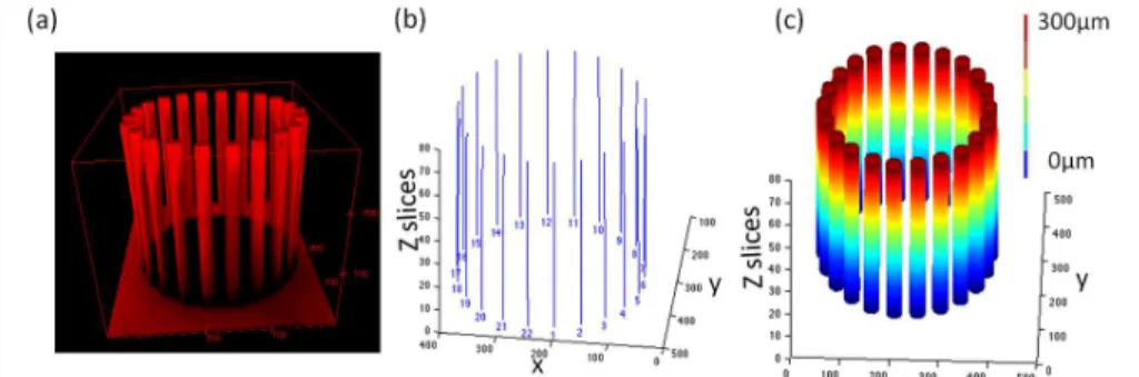
Spheroid growth induced bending of the micropillars, reflecting the forces exerted by the spheroid. On one hand, in order to characterize the 3 dimensional deformations of the pillars from the 3D confocal images, we developed a homemade MATLAB program that enables the quantification of a large number of devices at one time point (Figure 2).
Figure 2: Image analysis program for a microdevice with slightly displaced pillars induced by MCF7 spheroid (unlabelled), (a) a 3D confocal view of the whole microdevice to analyze the deformation of the pillars, (b) a representation of the detected centers of all pillars for each slice (80 slices with a z-step of 3,75µm) and (c) a 3 D reconstruction of the smoothed pillars with the microdevice’s parameters.
On the other hand, we used finite element method through the software COMSOL, to simulate the bending of the PDMS pillars. From various images, we were able to localize the contact region where the spheroid exerts its efforts on the pillars. Then by simulation, we could calculate the displacement field and the stress accumulated in the pillars for a large range of applied forces. The simulation results were then compared to the 3D experimental displacement in order to extract the value of the force responsible for this deformation (Figure 3).
Figure 3: (a) A simulation example of pillar deformation and Von Mises stress after the application of a distributed force in the region of contact with the spheroid, (b) bending profiles of a PDMS pillar showing the closest match between the simulated result (blue) and the experimental one (red).
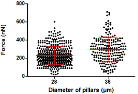
Using this procedure of force calculation, we recorded the forces exerted by MCF7 spheroids on the microdevices after a period of 4 days of contact between the spheroids and the microdevices. Our results showed that the forces exerted on a device with micropillars of 28 µm in diameter are in the order of 100-300 nN. Moreover we have found that spheroids confronted with stiffer pillars (36 µm in diameter), exerted slightly higher forces as shown in Figure 4.
Figure 4: A scattered plot with the standard deviation of the forces exerted by MCF7 spheroids after 4 days of growth within the microdevices for 2 pillars diameters: 28 and 36µm.
CONCLUSION
In this study, we offered a new methodology to investigate systematically the mechanical properties of spheroids with a high throughput. We gave for the first time a range of forces exerted by a living MCTS on its surroundings. In previous work we have shown the dynamic interaction between the spheroids and the microdevice [5], which opens a new perspective towards kinetics studies of the evolution of forces within a growing spheroid, in time. This method also opens original perspectives for pharmacological drug screening and for investigating the mechanisms of growth of micro-tumors.
AKNOWLEDGEMENTS
This work is funded by the foundation 2RITC (Toulouse).
REFERENCES
[1] F. Hirschhaeuser, H. Menne, C. Dittfeld, J. West, W. Mueller-Klieser and L. A. Kunz-Schughart, “Multicellular tumor spheroids: an underestimated tool is catching up again”, J Biotechnol, 2010, 148, 3-15.
[2] K. Alessandri, B. R. Sarangi, V. V. Gurchenkov, B. Sinha, T. R. Kiessling, L. Fetler, F. Rico, S. Scheuring, C. Lamaze, A. Simon, S. Geraldo, D. Vignjevic, H. Domejean, L. Rolland, A. Funfak, J. Bibette, N. Bremond and P. Nassoy, “Cellular capsules as a tool for multicellular spheroid production and for investigating the mechanics of tumor progression in vitro”, Proc Natl Acad Sci U S A, 110, 14843-14848.
[3] T. Vasilica Stirbat, S. Tlili, T. Houver, J. P. Rieu, C. Barentin and H. Delanoe-Ayari, “Multicellular aggregates: a model system for tissue rheology”, The European physical journal. E, Soft matter, 2013, 36, 84.
[4] K. Guevorkian, M. J. Colbert, M. Durth, S. Dufour and F. Brochard-Wyart, “Aspiration of biological viscoelastic drops”, Physical review letters, 2010, 104, 218101.
[5] L. Aoun, P. Weiss, A. Laborde, B. Ducommun, V. Lobjois, and C. Vieu, “Microdevice arrays of high aspect ratio poly(dimethylsiloxane) pillars for the investigation of multicellular tumour spheroid mechanical properties“, Lab on a Chip, 2014, 14(13), 2344-2353.
CONTACT